health occupations
DESCRIPTION
Health Occupations. Skeletal System – Unit 1. Skeletal System. Organs – BONES 206 in adult human Functions Framework Supports muscles, fat, skin Protection Surrounds vital organs (skull, ribs) Lever Muscles attach to bones to provide movement Production of blood cells - PowerPoint PPT PresentationTRANSCRIPT
Health Occupations
Skeletal System – Unit 1
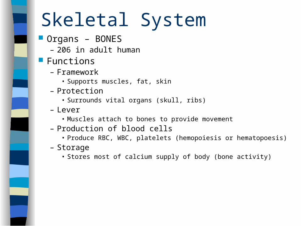
Skeletal System Organs – BONES
– 206 in adult human Functions
– Framework• Supports muscles, fat, skin
– Protection• Surrounds vital organs (skull, ribs)
– Lever• Muscles attach to bones to provide movement
– Production of blood cells• Produce RBC, WBC, platelets (hemopoiesis or hematopoesis)
– Storage• Stores most of calcium supply of body (bone activity)
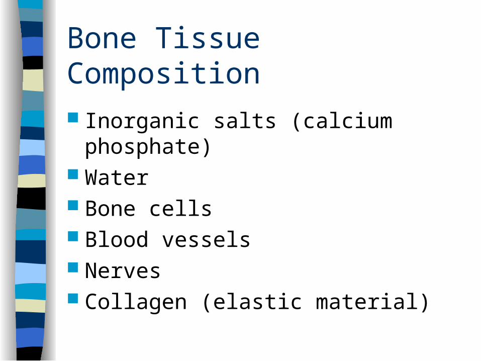
Bone Tissue Composition
Inorganic salts (calcium phosphate) Water Bone cells Blood vessels Nerves Collagen (elastic material)

Bone Tissue
Must continually receive food & oxygen Has fever nerves & blood vessels than other
tissues Grows for the first 18 – 20 years of life After growth stops, bone cells die & are
replaced by new cells– Osteoblasts – cells that MAKE bones– Osteoclasts – cells that BREAK DOWN bones &
reabsorb them
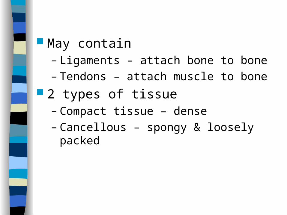
May contain– Ligaments – attach bone to bone– Tendons – attach muscle to bone
2 types of tissue– Compact tissue – dense – Cancellous – spongy & loosely packed
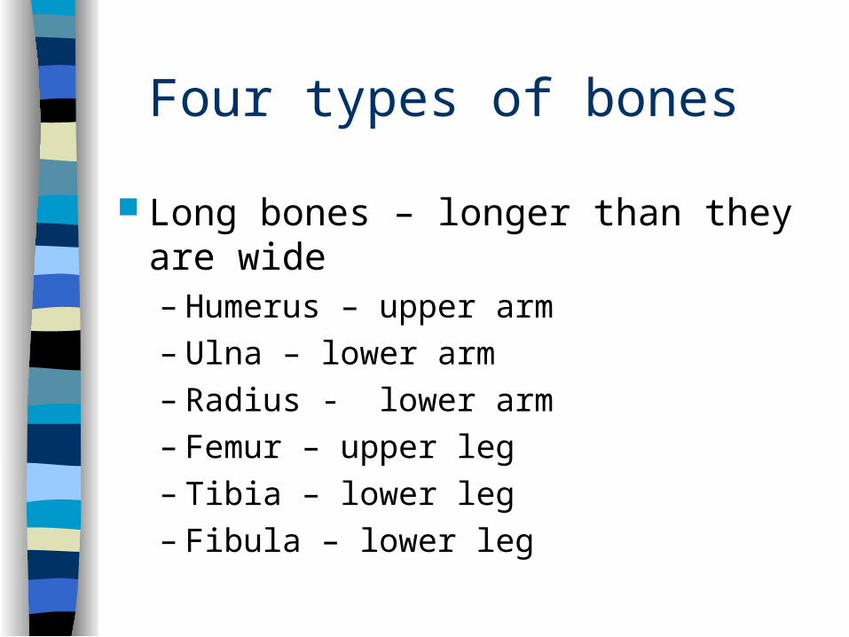
Four types of bones
Long bones – longer than they are wide– Humerus – upper arm– Ulna – lower arm– Radius - lower arm– Femur – upper leg– Tibia – lower leg– Fibula – lower leg
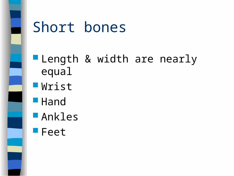
Short bones
Length & width are nearly equal Wrist Hand Ankles Feet
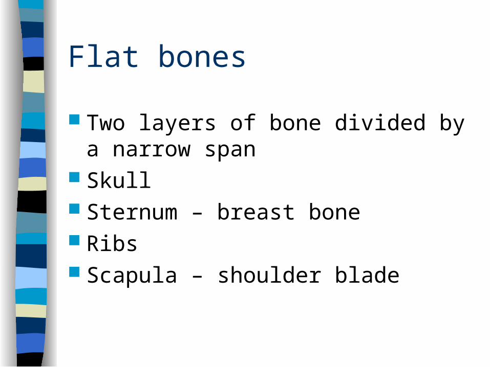
Flat bones
Two layers of bone divided by a narrow span
Skull Sternum – breast bone Ribs Scapula – shoulder blade
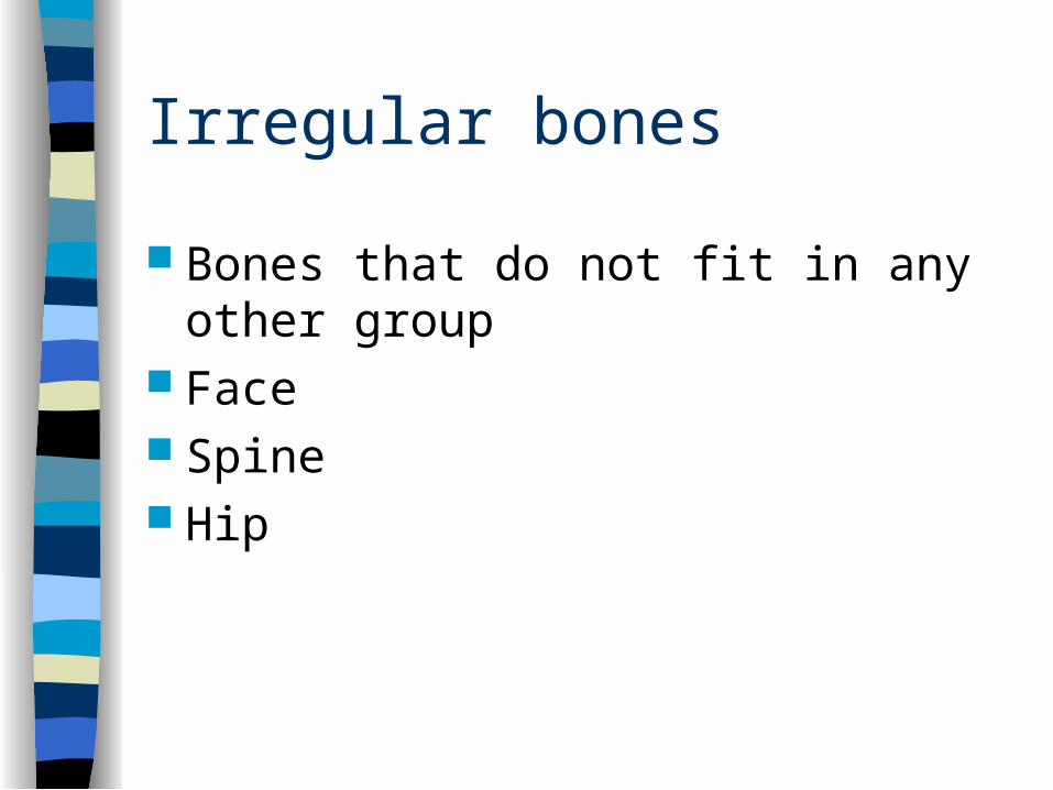
Irregular bones
Bones that do not fit in any other group Face Spine Hip
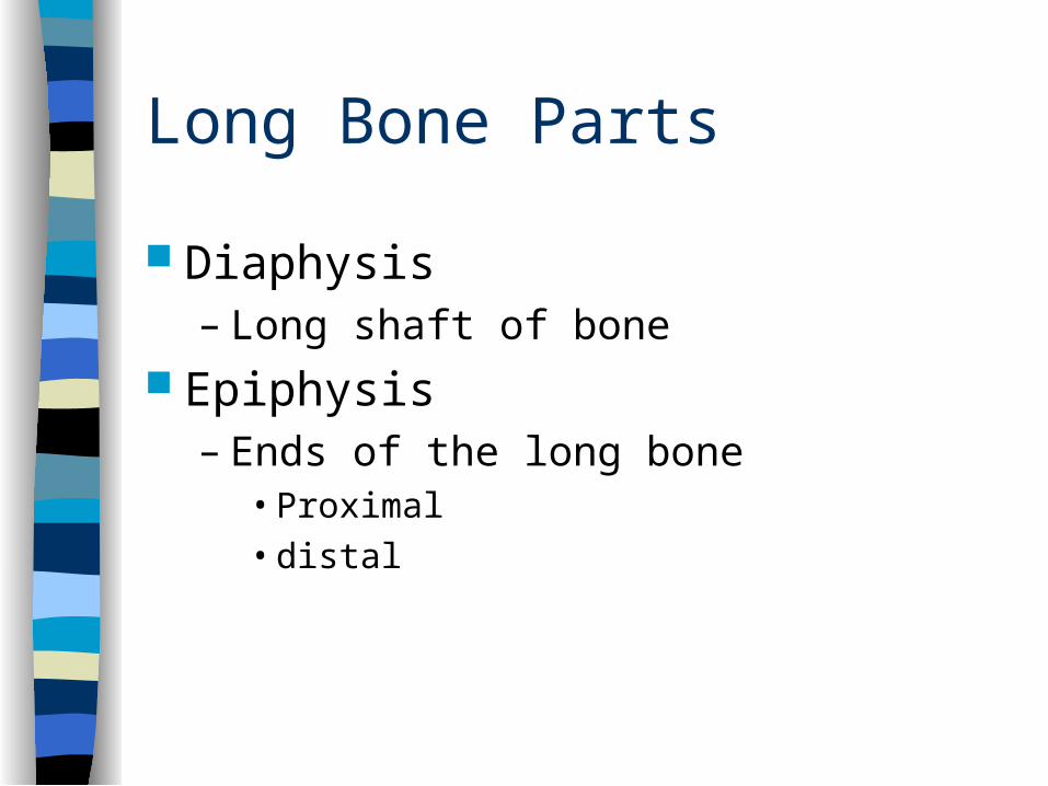
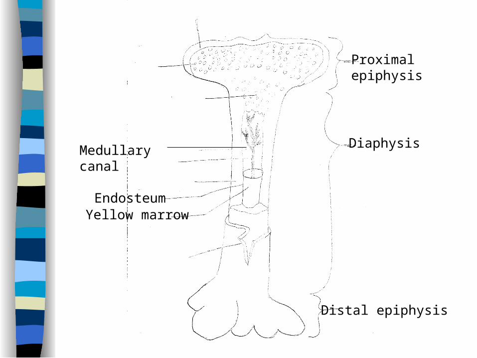
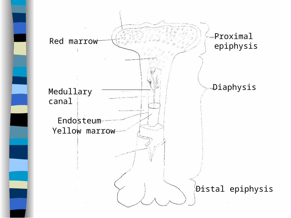
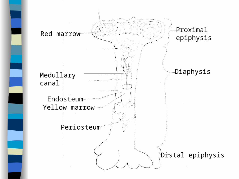
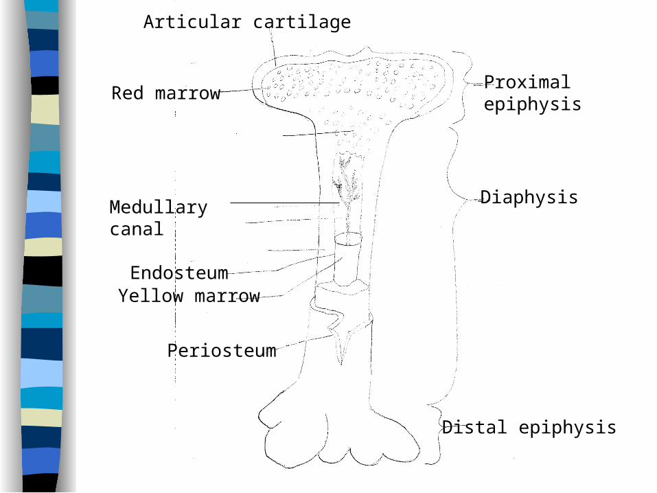
Long Bone Parts
Diaphysis – Long shaft of bone
Epiphysis– Ends of the long bone
• Proximal• distal
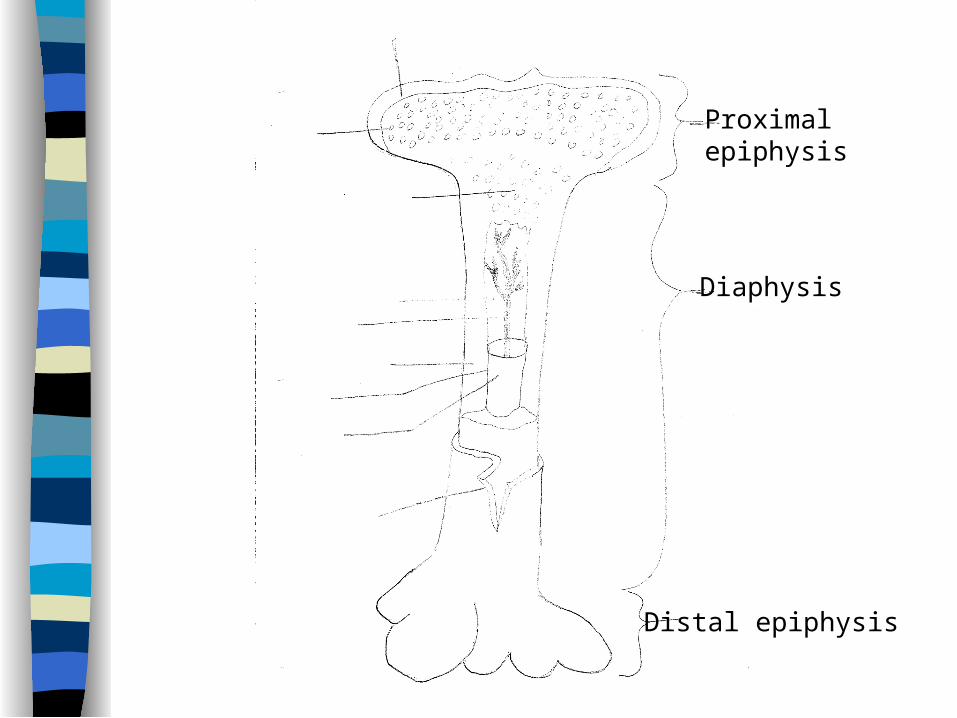
Diaphysis
Proximalepiphysis
Distal epiphysis
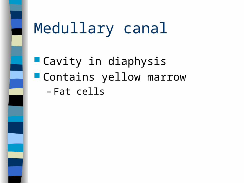
Medullary canal
Cavity in diaphysis Contains yellow marrow
– Fat cells
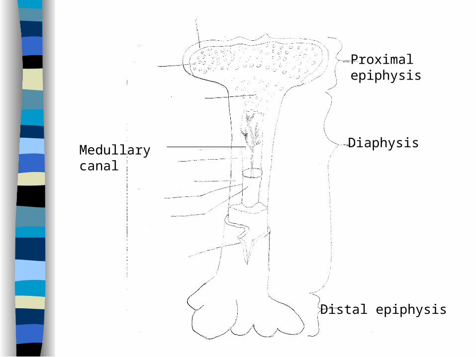
Diaphysis
Proximalepiphysis
Distal epiphysis
Medullary canal
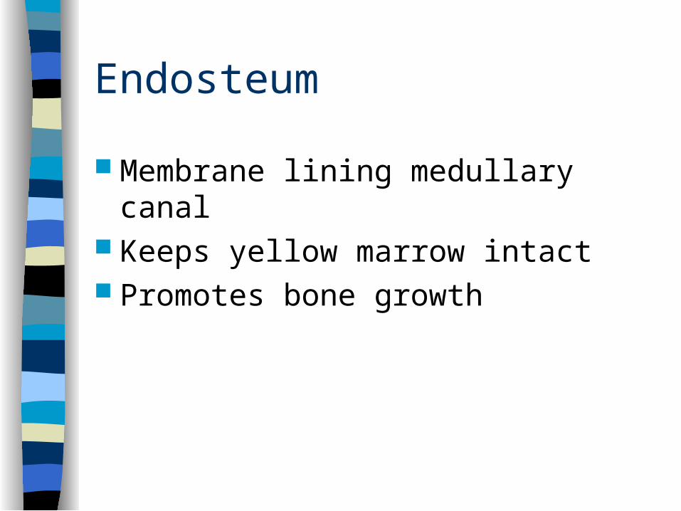
Endosteum
Membrane lining medullary canal Keeps yellow marrow intact Promotes bone growth
Diaphysis
Proximalepiphysis
Distal epiphysis
Medullary canal
EndosteumYellow marrow
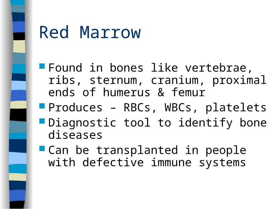
Red Marrow
Found in bones like vertebrae, ribs, sternum, cranium, proximal ends of humerus & femur
Produces – RBCs, WBCs, platelets Diagnostic tool to identify bone diseases Can be transplanted in people with
defective immune systems
Diaphysis
Proximalepiphysis
Distal epiphysis
Medullary canal
EndosteumYellow marrow
Red marrow
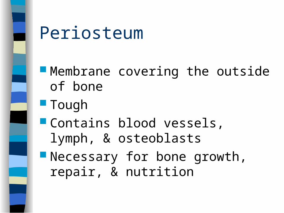
Periosteum
Membrane covering the outside of bone Tough Contains blood vessels, lymph, &
osteoblasts Necessary for bone growth, repair, &
nutrition
Diaphysis
Proximalepiphysis
Distal epiphysis
Medullary canal
EndosteumYellow marrow
Red marrow
Periosteum
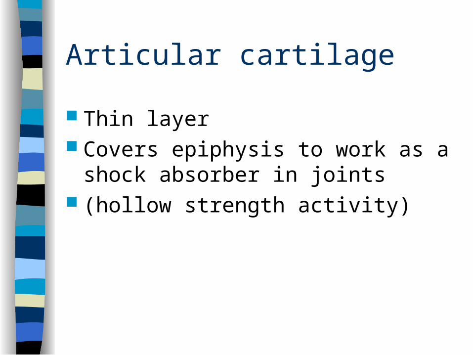
Articular cartilage
Thin layer Covers epiphysis to work as a shock
absorber in joints (hollow strength activity)
Diaphysis
Proximalepiphysis
Distal epiphysis
Medullary canal
EndosteumYellow marrow
Red marrow
Periosteum
Articular cartilage
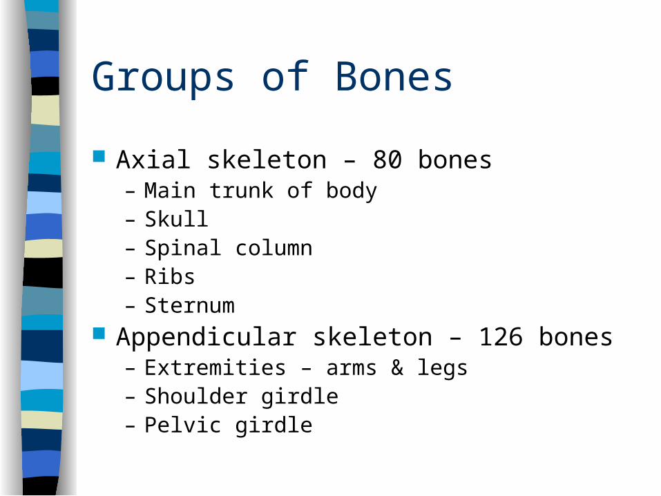
Groups of Bones
Axial skeleton – 80 bones– Main trunk of body– Skull– Spinal column– Ribs – Sternum
Appendicular skeleton – 126 bones– Extremities – arms & legs– Shoulder girdle– Pelvic girdle

Joints
Place where two bones meet Grouped by how much movement is allowed
– Synarthrosis joints – immoveable• Cranium, suture joints
– Amphiartrosis joints – slightly moveable• Vertebral discs, symphysis pubis, sacroiliac
– Diarthrosis joints – freely moveable• Shoulder, elbow, wrist, fingers, knees, ankles, toes
Ligaments – connect bone to bone– Hold bones together
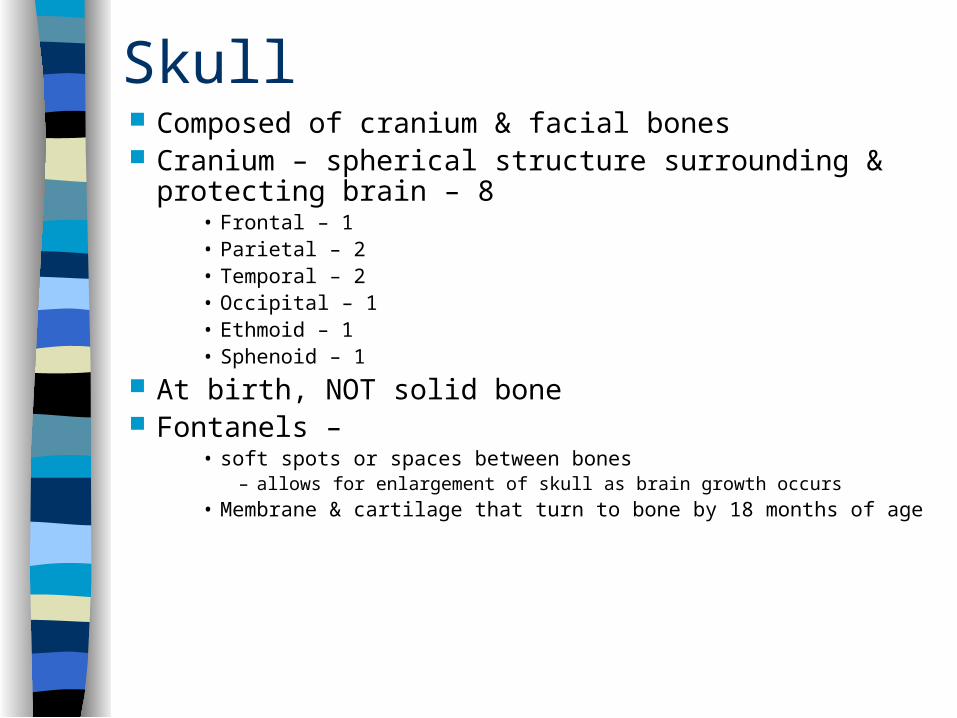
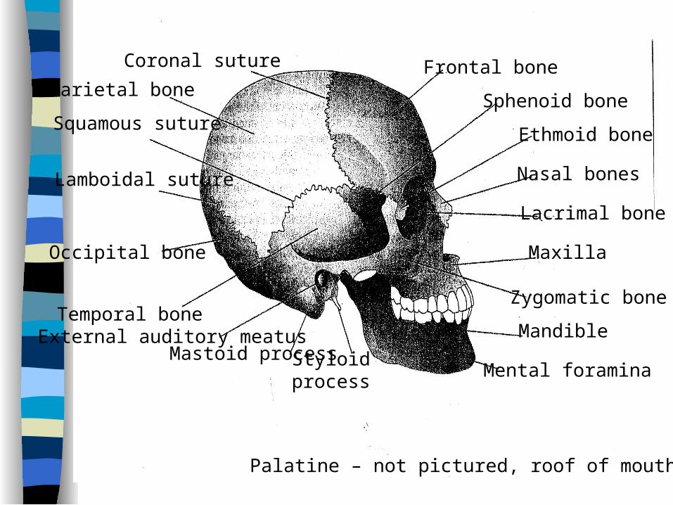
Skull Composed of cranium & facial bones Cranium – spherical structure surrounding & protecting
brain – 8 • Frontal – 1• Parietal – 2• Temporal – 2• Occipital – 1• Ethmoid – 1• Sphenoid – 1
At birth, NOT solid bone Fontanels –
• soft spots or spaces between bones– allows for enlargement of skull as brain growth occurs
• Membrane & cartilage that turn to bone by 18 months of age
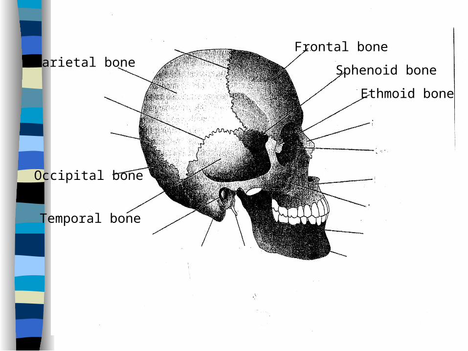
Frontal boneParietal bone
Temporal bone
Occipital bone
Sphenoid bone
Ethmoid bone
Skull
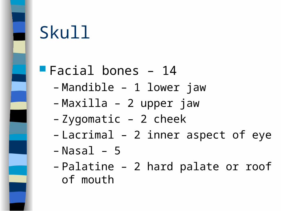
Facial bones – 14– Mandible – 1 lower jaw– Maxilla – 2 upper jaw– Zygomatic – 2 cheek– Lacrimal – 2 inner aspect of eye– Nasal – 5– Palatine – 2 hard palate or roof of mouth
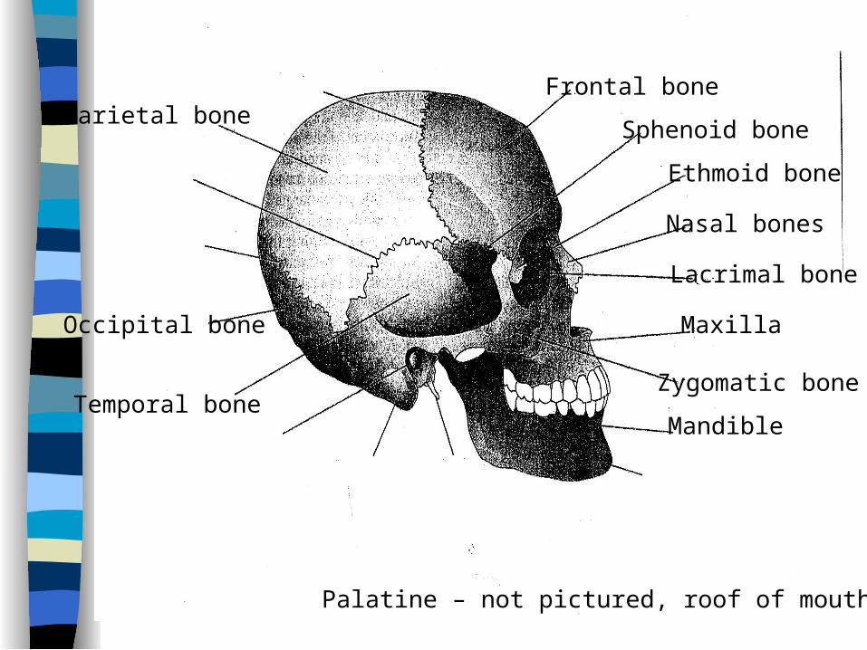
Frontal boneParietal bone
Temporal bone
Occipital bone
Sphenoid bone
Ethmoid bone
Mandible
Maxilla
Zygomatic bone
Lacrimal bone
Nasal bones
Palatine – not pictured, roof of mouth
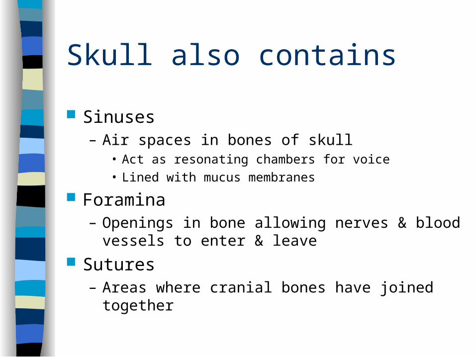
Skull also contains
Sinuses – Air spaces in bones of skull
• Act as resonating chambers for voice• Lined with mucus membranes
Foramina – Openings in bone allowing nerves & blood vessels
to enter & leave
Sutures – Areas where cranial bones have joined together
Frontal boneParietal bone
Temporal bone
Occipital bone
Sphenoid bone
Ethmoid bone
Mandible
Maxilla
Zygomatic bone
Lacrimal bone
Nasal bones
Palatine – not pictured, roof of mouth
Coronal suture
Squamous suture
Lamboidal suture
External auditory meatusMastoid process Styloid
processMental foramina