hedgehog signaling regulates sebaceous gland development
TRANSCRIPT

Short CommunicationHedgehog Signaling Regulates Sebaceous GlandDevelopment
Mary Allen,* Marina Grachtchouk,* Hong Sheng,*Vladimir Grachtchouk,* Anna Wang,* Lebing Wei,*Jianhong Liu,* Angel Ramirez,† Daniel Metzger,‡Pierre Chambon,‡ Jose Jorcano,† andAndrzej A. Dlugosz*From the Department of Dermatology and Comprehensive
Cancer Center,* University of Michigan, Ann Arbor, Michigan;
Project on Cell and Molecular Biology and Gene Therapy,†
Centro de Investigaciones Energeticas, Medioambientales y
Tecnologicas, Madrid, Spain; and Institut de Genetique et de
Biologie Moleculaire et Cellulaire,‡ College de France,
Communaute Urbaine de Strasbourg, Strasbourg, France
Epithelial progenitor cells in skin give rise to multiplelineages, comprising the hair follicle, an associatedsebaceous gland, and overlying epidermis; however,the signals that regulate sebocyte development arepoorly understood. We tested the potential involve-ment of the Hedgehog pathway in sebaceous glanddevelopment using transgenes designed to eitherblock or stimulate Hedgehog signaling in cutaneouskeratinocytes in vivo. Whereas inhibition of theHedgehog pathway selectively suppressed sebocytedevelopment, Hedgehog pathway activation led to astriking increase both in size and number of seba-ceous glands. Remarkably, ectopic Hedgehog signal-ing also triggered the formation of sebaceous glandsfrom footpad epidermis, in regions normally devoidof hair follicles and associated structures. These ec-topic sebaceous glands expressed molecular markersof sebocyte differentiation and were functional, se-creting their contents directly onto the skin’s surfaceinstead of into a hair canal. The Hedgehog pathwaythus plays a key role in sebocyte cell fate decisionsand is a potential target for treatment of skin disor-ders linked to abnormal sebaceous gland function,such as acne. (Am J Pathol 2003, 163:2173–2178)
Epithelial progenitor cells in skin give rise to epidermis aswell as the epithelial component of skin appendages,including hair follicles and associated sebaceousglands.1,2,3 Early-stage follicles consist of an epithelial
thickening, called a placode, and an adjacent mesenchy-mal condensate in the developing dermis.4 The follicleepithelium grows downward through the dermis and intothe subcutaneous fat, where it surrounds the conden-sate-derived hair follicle papilla to form the hair bulb.Rapidly proliferating matrix cells in the hair bulb give riseto the hair shaft and inner root sheath lineages, which aredriven upward toward the skin’s surface during hair mat-uration. Surrounding these cell layers is the follicle outerroot sheath, which is continuous with the interfollicularepidermis and contains epithelial stem cells in a regioncalled the bulge.5,6 The final differentiated cell type toappear in the developing follicle is the oil-rich sebocyte,which arises from cells within the superficial hair follicle.7
Over the course of several days, the expanding pool ofsebocytes forms a gland located outside of the hair fol-licle, with sebocytes releasing their contents into the haircanal not far below the skin’s surface.
Proper hair follicle development is dependent on aseries of inductive signals traveling between epithelialand mesenchymal follicle progenitors.8 Recent studieshave begun to identify some of the molecules regulatingfollicle morphogenesis and cell fate, and similar to manyother organ systems, hair follicles use both the Wnt andHedgehog pathways to guide their assembly. Wnt sig-naling regulates follicle initiation and reactivation of folli-cle growth during postnatal hair cycling.9–12 At laterstages of follicle maturation, the Wnt pathway also playsan important role in terminal differentiation of hair lineag-es.13,14 Hedgehog signaling plays a complementary role,as it is essential for the proliferative expansion of hairfollicle epithelium but is not required during follicle initia-tion or hair lineage differentiation.15–17
Given the involvement of Hedgehog proteins in regu-lating cell lineage specification in several other organs,18
we tested the potential involvement of this pathway in cellfate decisions in skin using both loss-of-function and
Supported by the National Institutes of Health through the University ofMichigan Comprehensive Cancer Center (CA46592) and the Center forOrganogenesis; and grants AR45973 and CA87837 (to A.A.D.).
Accepted for publication August 29, 2003.
Address reprint requests to Andrzej A. Dlugosz, M.D., U-M CancerCenter/Dermatology, 3310 CCGC, Box 0932, 1500E Medical CenterDrive, Ann Arbor, MI 48109-0932. E-mail: [email protected].
American Journal of Pathology, Vol. 163, No. 6, December 2003
Copyright © American Society for Investigative Pathology
2173

gain-of-function transgenic mouse models. We find thatinhibition of Hedgehog signaling in cutaneous keratino-cytes selectively blocks the formation of sebocytes. Incontrast, ectopic activation of the Hedgehog pathwaypromotes sebocyte development, even in regions thatnormally do not contain hair follicles or associated seba-ceous glands. Our findings strongly implicate Hedgehogsignaling in sebocyte cell fate decisions, and suggestthat modulation of the Hedgehog pathway may provide anovel means of regulating sebaceous gland function.
Materials and Methods
Construction and Breeding of Transgenic Mice
Because Hedgehog signaling modulates gene expres-sion via Gli proteins,19–21 we generated transgenic miceexpressing a skin-targeted dominant-negative Gli mutantdesigned to block Hedgehog responsiveness in cutane-ous epithelium. We produced a Gli2�C4 cDNA22 by di-gesting pcDNA3.1Flag-Gli2 (kindly provided by Drs. Hiro-shi Sasaki and Chi-chung Hui) with ApaI, adding afragment containing a stop codon flanked by ApaI sites,and re-ligating. Gli2�C4 cDNA was released frompcDNA3.1 using PmeI, and subcloned into the SnaBI siteof the bovine K5 cassette23 to generate K5-Gli2�C4. Allsubcloning was verified by sequencing. The insert waspurified and injected into C57BL/6 � SJL F2 mouse eggsby the University of Michigan Transgenic Core. Fivetransgenic founders were produced, and further charac-terization of these mice and their progeny will be describedin detail elsewhere. Studies reported in this manuscriptwere performed using F1 litters from crosses with C57BL/6Jbreeders (Jackson Laboratories, Bar Harbor, ME).
We modified the K5 transgenic cassette23 to enableCre-mediated expression of M2SMO, which encodes aconstitutively activated form of the hedgehog signalingeffector SMO,24 in skin. A fragment containing an EGFPcDNA with bovine growth hormone polyA signal, flankedby loxP sites in reverse orientation, was inserted into theNotI site of the K5 transgenic cassette. M2SMO cDNA24
was subcloned into the NheI site, yielding a constructwith the following elements: bovine K5 promoter, rabbit�-globin intron, loxP, EGFP, bovine growth hormone polyA,loxP, M2SMO, and 2xSV40 polyA, which we designatedK5-flxGFP-M2SMO. Eight transgenic founders were pro-duced and several mouse lines established, and thesewill be characterized in a subsequent publication. Toactivate M2SMO expression, K5-flxGFP-M2SMO micewere crossed with either K5-Cre or K5-CreERT225 mice togenerate double-transgenic progeny. Whereas recombi-nase activity in mice harboring the K5-CreERT2 trans-gene was previously shown to be dependent on treat-ment with 4-hydroxytamoxifen,25 low-level recombinationtook place in untreated double-transgenic mice de-scribed in this report (see Figure 2I). This most likelyreflects the variable sensitivity of different floxed alleles toCre-mediated recombination.26 All mice were housedand maintained according to University of Michigan in-stitutional guidelines.
Tissue Harvesting and Oil Red O Staining
For hematoxylin and eosin (H&E) staining, we fixed skinovernight in neutral-buffered formalin, transferred to 70%EtOH, processed, and embedded in paraffin. Skin wasalso embedded in Optimum Cutting Temperature com-pound for frozen sections. Oil Red O staining was per-formed using a modification of a protocol kindly providedby Dr. Karin Muller-Decker (Deutsches Krebsforschungs-zentrum, Heidelberg, Germany). Frozen sections werefixed in 1% neutral-buffered formalin for 5 minutes,washed in deionized water, and incubated in 60% iso-propanol for 5 minutes. Sections were stained with fil-tered Oil Red O working solution, prepared immediatelybefore use by making a 6:4 mixture of stock (0.5% OilRed O in 99% isopropanol) and deionized water. Sec-tions were transferred to 60% isopropanol, washed indeionized water, counterstained using hematoxylin, andmounted using 50% glycerol in PBS. A similar protocolwas used for Oil Red O staining of whole-mounts, exceptsamples were subsequently washed and stored in deion-ized water.
Semiquantitative RT-PCR
We isolated RNA from skin lysates prepared using Trizol(Invitrogen, Carlsbad, CA), according to the manufactur-er’s instructions. Semiquantitative reverse-transcription-polymerase chain reaction (RT-PCR) was performed us-ing 1-�g samples of total RNA for first-strand DNAsynthesis (Superscript II RT kit, Invitrogen). Primers wereselected to span an intron whenever possible, or PCRwas performed in the absence of reverse transcriptase torule out the possibility that amplification products werederived from contaminating DNA. PCR conditions areavailable on request; the following primers were used:actin (421 bp) forward 5�-TACCACAGGCATTGTGAT-GGA-3�, reverse 5�-CAACGTCACACTTCATGATGG-3�27;c-Myc (548 bp) forward 5�-AGTGCATTGATCCCTCAGT-GGTCTTTCCCTA-3�, reverse 5�-CAGCTCGTTCCTCC-TCTGACGTTCCAAGACGTT-3�; Gli1 (364 bp) forward5�-GTCGGAAGTCCTATTCACGC-3�, reverse 5�-CAGT-CTGCTCTCTTCCCTGC-3�; M2SMO (435 bp) forward5�-AAGCGGATCAAGAAGAGCA-3�, reverse 5�-GAG-GCAGTCGAGGAATGGTA-3�; Mc5r (490 bp) forward5�-AAATCCGATGCCAAGAAGTG-3�, reverse 5�-GG-TAGCGCAAGGCATAGAAG-3�; Scd3 (809 bp) forward5�-CTTGGATAACCACCCTGGGTG-3�, reverse 5�-CTC-CTCTGGAACATCACCAGCTTC-3�;28 Shh (241 bp)forward 5�-TCTGTGATGAACCAGTGGCC-3�, reverse5�-GCCACGGAGTTCTCTGCTTT-3�.29
ResultsSonic hedgehog (Shh) is produced and secreted bydeveloping hair follicle keratinocytes and activates sig-naling both in the follicular epithelium and mesen-chyme.30,31 Gli proteins mediate transcriptional re-sponses to Hedgehog family members,19,20 and theGli2�C4 transactivation-domain mutant blocks Gli func-
2174 Allen et alAJP December 2003, Vol. 163, No. 6
tion in a dominant-negative manner.22 To inhibit Hedge-hog signaling selectively in cutaneous epithelium, weused the bovine K5 promoter23 to generate K5-Gli2�C4transgenic mice. The K5 promoter is active in the epider-mal basal layer, embryonic hair follicle progenitor cells,and follicle outer root sheath, including the bulge regionwhich harbors multipotent stem cells.5,6
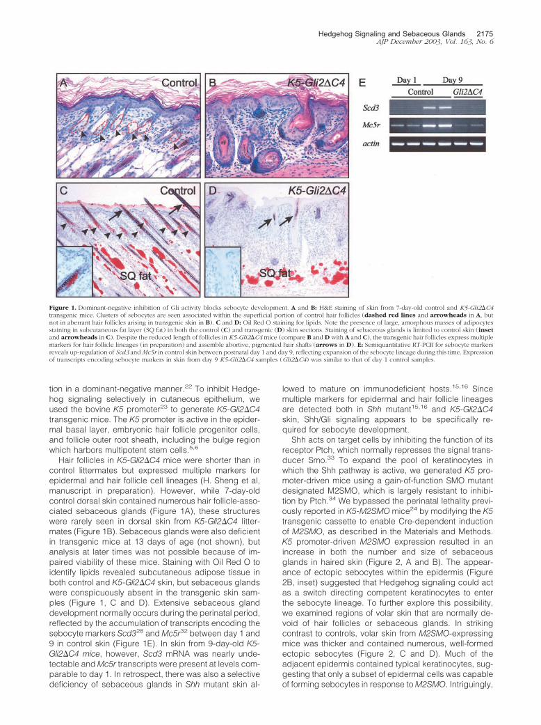
Hair follicles in K5-Gli2�C4 mice were shorter than incontrol littermates but expressed multiple markers forepidermal and hair follicle cell lineages (H. Sheng et al,manuscript in preparation). However, while 7-day-oldcontrol dorsal skin contained numerous hair follicle-asso-ciated sebaceous glands (Figure 1A), these structureswere rarely seen in dorsal skin from K5-Gli2�C4 litter-mates (Figure 1B). Sebaceous glands were also deficientin transgenic mice at 13 days of age (not shown), butanalysis at later times was not possible because of im-paired viability of these mice. Staining with Oil Red O toidentify lipids revealed subcutaneous adipose tissue inboth control and K5-Gli2�C4 skin, but sebaceous glandswere conspicuously absent in the transgenic skin sam-ples (Figure 1, C and D). Extensive sebaceous glanddevelopment normally occurs during the perinatal period,reflected by the accumulation of transcripts encoding thesebocyte markers Scd328 and Mc5r32 between day 1 and9 in control skin (Figure 1E). In skin from 9-day-old K5-Gli2�C4 mice, however, Scd3 mRNA was nearly unde-tectable and Mc5r transcripts were present at levels com-parable to day 1. In retrospect, there was also a selectivedeficiency of sebaceous glands in Shh mutant skin al-
lowed to mature on immunodeficient hosts.15,16 Sincemultiple markers for epidermal and hair follicle lineagesare detected both in Shh mutant15,16 and K5-Gli2�C4skin, Shh/Gli signaling appears to be specifically re-quired for sebocyte development.
Shh acts on target cells by inhibiting the function of itsreceptor Ptch, which normally represses the signal trans-ducer Smo.33 To expand the pool of keratinocytes inwhich the Shh pathway is active, we generated K5 pro-moter-driven mice using a gain-of-function SMO mutantdesignated M2SMO, which is largely resistant to inhibi-tion by Ptch.34 We bypassed the perinatal lethality previ-ously reported in K5-M2SMO mice24 by modifying the K5transgenic cassette to enable Cre-dependent inductionof M2SMO, as described in the Materials and Methods.K5 promoter-driven M2SMO expression resulted in anincrease in both the number and size of sebaceousglands in haired skin (Figure 2, A and B). The appear-ance of ectopic sebocytes within the epidermis (Figure2B, inset) suggested that Hedgehog signaling could actas a switch directing competent keratinocytes to enterthe sebocyte lineage. To further explore this possibility,we examined regions of volar skin that are normally de-void of hair follicles or sebaceous glands. In strikingcontrast to controls, volar skin from M2SMO-expressingmice was thicker and contained numerous, well-formedectopic sebocytes (Figure 2, C and D). Much of theadjacent epidermis contained typical keratinocytes, sug-gesting that only a subset of epidermal cells was capableof forming sebocytes in response to M2SMO. Intriguingly,
Figure 1. Dominant-negative inhibition of Gli activity blocks sebocyte development. A and B: H&E staining of skin from 7-day-old control and K5-Gli2�C4transgenic mice. Clusters of sebocytes are seen associated within the superficial portion of control hair follicles (dashed red lines and arrowheads in A, butnot in aberrant hair follicles arising in transgenic skin in B). C and D: Oil Red O staining for lipids. Note the presence of large, amorphous masses of adipocytesstaining in subcutaneous fat layer (SQ fat) in both the control (C) and transgenic (D) skin sections. Staining of sebaceous glands is limited to control skin (insetand arrowheads in C). Despite the reduced length of follicles in K5-Gli2�C4mice (compare B and D with A and C), the transgenic hair follicles express multiplemarkers for hair follicle lineages (in preparation) and assemble abortive, pigmented hair shafts (arrows in D). E: Semiquantitative RT-PCR for sebocyte markersreveals up-regulation of Scd3 andMc5r in control skin between postnatal day 1 and day 9, reflecting expansion of the sebocyte lineage during this time. Expressionof transcripts encoding sebocyte markers in skin from day 9 K5-Gli2�C4 samples (Gli2�C4) was similar to that of day 1 control samples.
Hedgehog Signaling and Sebaceous Glands 2175AJP December 2003, Vol. 163, No. 6

during early stages of development, ectopic sebocytesarose from cells at the base of epidermal invaginations(Figure 2D, inset), one of the sites where epidermal stemcells have been proposed to reside in non-hairy skin.35
The ectopic sebocytes in M2SMO-expressing volarskin stained with Oil Red O and were frequently orga-nized into glands (Figure 2, E and F). In the absence ofhair follicles, many of these glands released their con-tents into ducts leading directly to the skin’s surface(Figure 2F, inset), instead of into hair canals. Thus, theectopic sebocytes exhibited a remarkable degree of au-tonomy with regard to assembly into glandular structureswith rudimentary ducts. To better assess the extent ofectopic sebaceous gland development, footpads wereremoved from control and M2SMO transgenic mice,stained with Oil Red O, and examined as whole mounts.Large numbers of ectopic, Oil Red O-positive sebaceousglands were detected in footpads from M2SMO trans-genic mice, but not controls (Figure 2, G and H). Semi-quantitative RT-PCR analysis confirmed expression ofsebocyte markers in volar skin samples from M2SMO-expressing mice but not controls (Figure 2I). As ex-pected, the transgenic samples contained M2SMOmRNA and elevated levels of the Shh target gene Gli1,
confirming enhanced Hedgehog pathway activity.M2SMO samples did not contain detectable levels of ShhmRNA, arguing that the appearance of sebocytes intransgenic skin was not the result of up-regulated Shhexpression. In light of recent studies documenting en-hanced sebocyte development in mice expressing MYCin skin,36,37 it is noteworthy that c-Myc expression ap-peared to be up-regulated in volar skin of M2SMO mice,compared to controls.
DiscussionThe results of our studies strongly implicate Hedgehogsignaling in the development of sebocytes from compe-tent progenitor cells. Although sebaceous glands arenormally derived from hair follicles, ectopic Hedgehogsignaling leads to sebocyte formation within the epider-mis of hairy skin, as well as hairless volar skin. Previouswork using hairless skin38,39 or even corneal epithelium40
has demonstrated the presence of multipotent progenitorcells capable of giving rise to hair follicle and sebocytecell lineages. However, these studies entailed the use oftissue or cell-suspension recombinants, and did not iden-
Figure 2. Enhanced activation of Shh signaling in keratinocytes leads to formation of hyperplastic and ectopic sebaceous glands. A and B: H&E staining of controland M2SMO-expressing dorsal mouse skin at 6.5 months of age. Note small size of normal sebaceous glands in control skin (arrowheads in A) compared withincreased number and size of sebaceous glands in transgenic skin (arrowheads in B). In addition to glandular structures within the dermis, individual sebocytesor small aggregates are also found ectopically within the epidermis (inset in B). C and D: H&E staining of volar paw skin from control and transgenic mice at13 months of age. Note thickened epidermis and cornified cell layers typically seen in skin from this region, with a conspicuous absence of hair follicles andsebaceous glands (C). Ectopic sebaceous glands (arrowheads in D) develop from volar epidermis of mice expressing M2SMO. Sebaceous gland developmentis initiated at the base of epidermal downgrowths (inset in D). E and F: Oil Red O staining for lipids in control and transgenic volar skin. As expected, no stainingis detected in control skin in E, whereas multiple sebocytes are stained in transgenic mouse skin in F. Many of the ectopic sebaceous glands secrete Oil RedO-positive material directly onto the skin’s surface (see inset). G and H: Whole-mount analysis of footpads from control and transgenic mice. A large numberof Oil Red O-positive sebaceous glands are detected in transgenic (H) but not control (G) footpad. I: Semiquantitative RT-PCR analysis showing expression ofsebocyte markers (Scd3, Mc5r) in volar skin from M2SMO-expressing transgenic mice (M2SMO) and control skin containing hair follicles (hf), but not in controlvolar skin. Transgene-specific primers confirm expression of M2SMO in transgenic samples, which also contain elevated levels of the Shh target gene Gli1.
2176 Allen et alAJP December 2003, Vol. 163, No. 6

tify molecular signals that determine the fates of progen-itor cells. The appearance of sebaceous glands in re-sponse to ectopic M2SMO expression in keratinocytespoints to the Hedgehog pathway as a critical determinantof sebocyte cell fate. This concept is further strengthenedby the finding that formation of sebocytes, but not othercutaneous epithelial cell types, is inhibited by blockingGli protein function in K5-Gli2�C4 mice. Normal seba-ceous gland development is thus likely to be controlled,at least in part, by expression of Shh and downstreamtargets in developing hair germs but not epidermis.30,31
M2SMO can bypass this requirement, even in hairlessskin, by activating Hedgehog signaling in a ligand-inde-pendent manner.41
Sebaceous gland hyperplasia is also occasionallyseen in other mouse models with deregulated Hedgehogsignaling in skin, including K5-Gli142 and K5-Gli2 mice43
(data not shown). However, additional studies areneeded to define the precise conditions required forHedgehog-pathway-driven formation of this specializedcell type. Interestingly, ectopic expression of Shh in em-bryonic mouse skin is sufficient to drive the formation ofbasal cell carcinoma-like proliferations but not seba-ceous glands,44 in keeping with studies suggesting thatboth the timing45 and level46 of Hedgehog signaling ac-tivity are important determinants of skin phenotype. Inaddition to regulating normal sebaceous gland develop-ment, it will be interesting to ascertain whether Hedgehogsignaling is involved in the formation of sebaceous glandtumors and hyperplasias.
In contrast to its role in sebocyte development, Shh isnot required for terminal differentiation of hair lineag-es,15,16 which is controlled by members of the Wnt sig-naling pathway.1,2 Moreover, inhibition of Wnt targetgenes using a dominant-negative Lef-1 promotes sebo-cyte development while inhibiting differentiation of hairlineages.13,14 Taken together with these findings, ourdata raise the interesting possibility that sebocyte cellfate is governed by the relative levels of stimulatory(Hedgehog) and inhibitory (Wnt) signals acting on multi-potent progenitors. Further studies will be required to testthis hypothesis, and to ascertain whether the effects ofmesenchymal cell types on sebaceous gland develop-ment47,48 can be attributed to modulation of either ofthese developmental signaling pathways. The potentialinfluence of cross-talk involving Hedgehog and Wnt49 orBMP50 pathways will also need to be examined. In addi-tion, it will be important to explore the relationship be-tween the Hedgehog pathway and other molecules im-plicated in sebocyte development, including c-Myc,36,37
PPAR�,51,52 and COX-2.53
Hair follicles and sebaceous glands both undergo cy-clic changes after birth,54 and Shh plays a major role inregulating proliferation of follicle epithelium during timesof active growth.17 Thus, in addition to its involvement inthe initiation of sebaceous gland formation, the Hedge-hog pathway is also likely to play a role in postnatalfunction of sebaceous glands. Acne is the most commonskin disease in humans, and in its most severe forms itcan lead to significant disfigurement and emotional dis-tress.55 Although a number of factors contribute to the
development of acne, there is compelling evidence thatoil-producing sebaceous glands play a central role in thepathogenesis of this disease.56 Agents aimed at inhibit-ing Hedgehog signaling may thus provide a novel meansof reducing sebaceous gland activity and thereby im-proving acne. In keeping with this possibility, retinoidsused in the treatment of acne can inhibit sebocyte differ-entiation,57 and have also been shown to reduce Glitranscriptional activity in cultured keratinocytes.58
Note Added in ProofWhile this manuscript was under review, a report waspublished implicating Indian hedgehog in the proliferationof sebocyte progenitors (Niemann C, Unden AB, Lyle S,Zouboulis CC, Toftgard R, Watt FM: Indian hedgehogand b-catenin signaling: role in the sebaceous lineage ofnormal and neoplastic mammalian epidermis. Proc NatlAcad Sci USA 2003, 100(Suppl 1):11873–11880).
AcknowledgmentsWe thank Drs. Hiroshi Sasaki, Chi-chung Hui, and Fredde Sauvage for reagents; Drs. Bruce Morgan, Sarah Mil-lar, Deb Gumucio, Sean Morrison, Steve Prouty, andUlrike Lichti for helpful discussions. Transgenic micewere generated by the Transgenic Animal Model Core(Mark Berard and Thomas Saunders) of the University ofMichigan’s Biomedical Research Core Facilities.
References
1. Fuchs E, Merrill BJ, Jamora C, DasGupta R: At the roots of a never-ending cycle. Dev Cell 2001, 1:13–25
2. Niemann C, Watt FM: Designer skin: lineage commitment in postnatalepidermis. Trends Cell Biol 2002, 12:185–192
3. Byrne C, Hardman M, Nield K: Covering the limb: formation of theintegument. J Anat 2003, 202:113–123
4. Hardy MH: The secret life of the hair follicle. Trends Genet 1992,8:55–61
5. Cotsarelis G, Sun TT, Lavker RM: Label-retaining cells reside in thebulge area of pilosebaceous unit: implications for follicular stem cells,hair cycle, and skin carcinogenesis. Cell 1990, 61:1329–1337
6. Oshima H, Rochat A, Kedzia C, Kobayashi K, Barrandon Y: Morpho-genesis and renewal of hair follicles from adult multipotent stem cells.Cell 2001, 104:233–245
7. Paus R, Muller-Rover S, van der Veen C, Maurer M, Eichmuller S, LingG, Hofmann U, Foitzik K, Mecklenburg L, Handjiski B: A comprehen-sive guide for the recognition and classification of distinct stages ofhair follicle morphogenesis. J Invest Dermatol 1999, 113:523–532
8. Millar SE: Molecular mechanisms regulating hair follicle development.J Invest Dermatol 2002, 118:216–225
9. Gat U, DasGupta R, Degenstein L, Fuchs E: De novo hair folliclemorphogenesis and hair tumors in mice expressing a truncated�-catenin in skin. Cell 1998, 95:605–614
10. Huelsken J, Vogel R, Erdmann B, Cotsarelis G, Birchmeier W: �-Cate-nin controls hair follicle morphogenesis and stem cell differentiation inthe skin. Cell 2001, 105:533–545
11. Andl T, Reddy ST, Gaddapara T, Millar SE: WNT signals are requiredfor the initiation of hair follicle development. Dev Cell 2002, 2:643–653
12. Van Mater D, Kolligs FT, Dlugosz AA, Fearon ER: Transient activationof �-catenin signaling in cutaneous keratinocytes is sufficient to trig-ger the active growth phase of the hair cycle in mice. Genes Dev2003, 17:1219–1224
Hedgehog Signaling and Sebaceous Glands 2177AJP December 2003, Vol. 163, No. 6

13. Merrill BJ, Gat U, DasGupta R, Fuchs E: Tcf3 and Lef1 regulatelineage differentiation of multipotent stem cells in skin. Genes Dev2001, 15:1688–1705
14. Niemann C, Owens DM, Hulsken J, Birchmeier W, Watt FM: Expres-sion of DeltaNLef1 in mouse epidermis results in differentiation of hairfollicles into squamous epidermal cysts and formation of skin tu-mours. Development 2002, 129:95–109
15. St Jacques B, Dassule HR, Karavanova I, Botchkarev VA, Li J, Danie-lian PS, McMahon JA, Lewis PM, Paus R, McMahon AP: Sonic hedge-hog signaling is essential for hair development. Curr Biol 1998,8:1058–1068
16. Chiang C, Swan RZ, Grachtchouk M, Bolinger M, Litingtung Y, Rob-ertson EK, Cooper MK, Gaffield W, Westphal H, Beachy PA, DlugoszAA: Essential role for sonic hedgehog during hair follicle morphogen-esis. Dev Biol 1999, 205:1–9
17. Wang LC, Liu ZY, Gambardella L, Delacour A, Shapiro R, Yang J,Sizing I, Rayhorn P, Garber EA, Benjamin CD, Williams KP, Taylor FR,Barrandon Y, Ling L, Burkly LC: Regular articles: conditional disrup-tion of hedgehog signaling pathway defines its critical role in hairdevelopment and regeneration. J Invest Dermatol 2000, 114:901–908
18. Ingham PW, McMahon AP: Hedgehog signaling in animaldevelopment: paradigms and principles. Genes Dev 2001, 15:3059–3087
19. Matise MP, Joyner AL: Gli genes in development and cancer. Onco-gene 1999, 18:7852–7859
20. Ruiz I, Altaba A, Sanchez P, Dahmane N: Gli and hedgehog incancer: tumours, embryos and stem cells. Nat Rev Cancer 2002,2:361–372
21. Mill P, Mo R, Fu H, Grachtchouk M, Kim PC, Dlugosz AA, Hui CC:Sonic hedgehog-dependent activation of Gli2 is essential for embry-onic hair follicle development. Genes Dev 2003, 17:282–294
22. Sasaki H, Nishizaki Y, Hui C, Nakafuku M, Kondoh H: Regulation ofGli2 and Gli3 activities by an amino-terminal repression domain:implication of Gli2 and Gli3 as primary mediators of Shh signaling.Development 1999, 126:3915–3924
23. Ramirez A, Bravo A, Jorcano JL, Vidal M: Sequences 5� of the bovinekeratin 5 gene direct tissue- and cell-type-specific expression of alacZ gene in the adult and during development. Differentiation 1994,58:53–64
24. Xie J, Murone M, Luoh SM, Ryan A, Gu Q, Zhang C, Bonifas JM, LamCW, Hynes M, Goddard A, Rosenthal A, Epstein EH Jr, de SauvageF: Activating smoothened mutations in sporadic basal-cell carci-noma. Nature 1998, 391:90–92
25. Indra AK, Warot X, Brocard J, Bornert JM, Xiao JH, Chambon P,Metzger D: Temporally controlled site-specific mutagenesis in thebasal layer of the epidermis: comparison of the recombinase activityof the tamoxifen-inducible Cre-ER(T) and Cre-ER(T2) recombinases.Nucleic Acids Res 1999, 27:4324–4327
26. Vooijs M, Jonkers J, Berns A: A highly efficient ligand-regulated Crerecombinase mouse line shows that LoxP recombination is positiondependent. EMBO Rep 2001, 2:292–297
27. Walterhouse D, Ahmed M, Slusarski D, Kalamaras J, Boucher D,Holmgren R, Iannaccone P: Gli, a zinc finger transcription factor andoncogene, is expressed during normal mouse development. Dev Dyn1993, 196:91–102
28. Zheng Y, Prouty SM, Harmon A, Sundberg JP, Stenn KS, Parimoo S:Scd3: a novel gene of the stearoyl-CoA desaturase family with re-stricted expression in skin. Genomics 2001, 71:182–191
29. Takabatake T, Ogawa M, Takahashi TC, Mizuno M, Okamoto M,Takeshima K: Hedgehog and patched gene expression in adultocular tissues. FEBS Lett 1997, 410:485–489
30. Bitgood MJ, McMahon AP: Hedgehog and Bmp genes are coex-pressed at many diverse sites of cell-cell interaction in the mouseembryo. Dev Biol 1995, 172:126–138
31. Iseki S, Araga A, Ohuchi H, Nohno T, Yoshioka H, Hayashi F, Noji, S:Sonic hedgehog is expressed in epithelial cells during developmentof whisker, hair, and tooth. Biochem Biophys Res Commun 1996,218:688–693
32. Chen W, Kelly MA, Opitz-Araya X, Thomas RE, Low MJ, Cone RD:Exocrine gland dysfunction in MC5-R-deficient mice: evidence forcoordinated regulation of exocrine gland function by melanocortinpeptides. Cell 1997, 91:789–798
33. Kalderon D: Transducing the hedgehog signal. Cell 2000, 103:371–37434. Murone M, Rosenthal A, de Sauvage FJ: Sonic hedgehog signaling
by the patched-smoothened receptor complex. Curr Biol 1999,9:76–84
35. Potten CS, Booth C: Keratinocyte stem cells: a commentary. J InvestDermatol 2002, 119:888–899
36. Waikel RL, Kawachi Y, Waikel PA, Wang XJ, Roop DR: Deregulatedexpression of c-Myc depletes epidermal stem cells. Nat Genet 2001,28:165–168
37. Arnold I, Watt FM: c-Myc activation in transgenic mouse epidermisresults in mobilization of stem cells and differentiation of their prog-eny. Curr Biol 2001, 11:558–568
38. Reynolds AJ, Jahoda CA: Cultured dermal papilla cells induce follicleformation and hair growth by transdifferentiation of an adult epider-mis. Development 1992, 115:587–593
39. Ferraris C, Bernard BA, Dhouailly D: Adult epidermal keratinocytesare endowed with pilosebaceous forming abilities. Int J Dev Biol1997, 41:491–498
40. Ferraris C, Chevalier G, Favier B, Jahoda CA, Dhouailly D: Adultcorneal epithelium basal cells possess the capacity to activate epi-dermal, pilosebaceous and sweat gland genetic programs in re-sponse to embryonic dermal stimuli. Development 2000, 127:5487–5495
41. Hynes M, Ye W, Wang K, Stone D, Murone M, Sauvage F, RosenthalA: The seven-transmembrane receptor smoothened cell autono-mously induces multiple ventral cell types. Nat Neurosci 2000,3:41–46
42. Oro AE, Higgins K: Hair cycle regulation of Hedgehog signal recep-tion. Dev Biol 2003, 255:238–248
43. Grachtchouk M, Mo R, Yu S, Zhang X, Sasaki H, Hui CC, Dlugosz AA:Basal cell carcinomas in mice overexpressing Gli2 in skin. Nat Genet2000, 24:216–217
44. Oro AE, Higgins KM, Hu ZL, Bonifas JM, Epstein EH Jr, Scott MP:Basal cell carcinomas in mice overexpressing sonic hedgehog. Sci-ence 1997, 276:817–821
45. Morgan BA, Orkin RW, Noramly S, Perez A: Stage-specific effects ofsonic hedgehog expression in the epidermis. Dev Biol 1998, 201:1–12
46. Grachtchouk V, Grachtchouk M, Lowe L, Johnson T, Wei L, Wang A,de Sauvage F, Dlugosz AA: The magnitude of hedgehog signalingactivity defines skin tumor phenotype. EMBO J 2003, 22:2741–2751
47. Kartasova T, Scandurro AB, Denning MF, Wirth PJ, Yuspa SH, LichtiU: Factors mediating the interactions between epidermal and dermalcells in skin grafts that might be important for hair follicle develop-ment. J Invest Dermatol 1995, 104:21S–22S
48. Prouty SM, Lawrence L, Stenn KS: Fibroblast-dependent induction ofa murine skin lesion similar to human nevus sebaceus of Jadassohn.Lab Invest 1997, 76:179–189
49. Reddy S, Andl T, Bagasra A, Lu MM, Epstein DJ, Morrisey EE, MillarSE: Characterization of Wnt gene expression in developing and post-natal hair follicles and identification of Wnt5a as a target of Sonichedgehog in hair follicle morphogenesis. Mech Dev 2001, 107:69–82
50. Botchkarev VA, Botchkareva NV, Nakamura M, Huber O, Funa K,Lauster R, Paus R, Gilchrest BA: Noggin is required for induction ofthe hair follicle growth phase in postnatal skin. EMBO J 2001, 15:2205–2214
51. Rosen ED, Sarraf P, Troy AE, Bradwin G, Moore K, Milstone DS,Spiegelman BM, Mortensen RM: PPAR� is required for the differen-tiation of adipose tissue in vivo and in vitro. Mol Cell 1999, 4:611–617
52. Rosenfield RL, Deplewski D, Greene ME: Peroxisome proliferator-acti-vated receptors and skin development. Horm Res 2000, 54:269–274
53. Neufang G, Furstenberger G, Heidt M, Marks F, Muller-Decker K:Abnormal differentiation of epidermis in transgenic mice constitutivelyexpressing cyclooxygenase-2 in skin. Proc Natl Acad Sci USA 2001,98:7629–7634
54. Stenn KS, Paus R: Controls of hair follicle cycling. Physiol Rev 2001,81:449–494
55. Webster GF: Acne vulgaris. BMJ 2002, 325:475–47956. Leyden JJ: New understandings of the pathogenesis of acne. J Am
Acad Dermatol 1995, 32:S15–S2557. Deplewski D, Rosenfield RL: Role of hormones in pilosebaceous unit
development. Endocr Rev 2000, 21:363–39258. Goyette P, Allan D, Peschard P, Chen CF, Wang W, Lohnes D:
Regulation of gli activity by all-trans retinoic acid in mouse keratino-cytes. Cancer Res 2000, 60:5386–5389
2178 Allen et alAJP December 2003, Vol. 163, No. 6