helicobacter pylori couples motility and diffusion to … pylori couples motility and diffusion to...
TRANSCRIPT
Helicobacter pylori Couples Motility and Diffusion to Actively Create a HeterogeneousComplex Medium in Gastric Mucus
Seyed Amir Mirbagheri and Henry Chien Fu*
Department of Mechanical Engineering, University of Nevada, Reno, Reno, Nevada 89557, USA(Received 8 January 2016; revised manuscript received 1 April 2016; published 10 May 2016)
Helicobacter pylori swims through mucus gel by generating ammonia that locally neutralizes theacidic gastric environment, turning nearby gel into a fluid pocket. The size of the fluid zone is importantfor determining the physics of the motility: in a large zone swimming occurs as in a fluid throughhydrodynamic principles, while in a very small zone the motility could be strongly influenced bynonhydrodynamic cell-mucus interactions including chemistry and adhesion. Here, we calculate the size ofthe fluid pocket. We model how swimming depends on the de-gelation range using a Taylor sheetswimming through a layer of Newtonian fluid bounded by a Brinkman fluid. Then, we model how thede-gelation range depends on the swimming speed by considering the advection-diffusion of ammoniaexuded from a translating sphere. Self-consistency between both models determines the values of theswimming speed and the de-gelation range. We find that H. pylori swims through mucus as if unconfined,in a large pocket of Newtonian fluid.
DOI: 10.1103/PhysRevLett.116.198101
Microorganisms often navigate complex media andgeometries, including during infection and mammalianfertilization [1]. The effect of non-Newtonian environments[2–24] and geometrical confinement [25–34] have bothbeen the subject of much research, including situationscombining the two [35–37]. Usually, the medium rheologyand geometrical configuration are considered a backgroundenvironment that microorganisms do not change duringswimming [38]. Here, we address the active creation ofheterogeneous geometries in complex environments byswimming microorganisms, during which the geometry,medium response, diffusion, and motility couple to mutu-ally influence each other. For example, E. coli canmechanically deplete the polymer concentration near theirfast-rotating flagella, decreasing the local viscosity [40].In this Letter we concentrate on another such example,the local chemical alteration of gastric mucus from gel tosol by Helicobacter pylori [41].An ∼200 μm gastric mucus layer forms a barrier
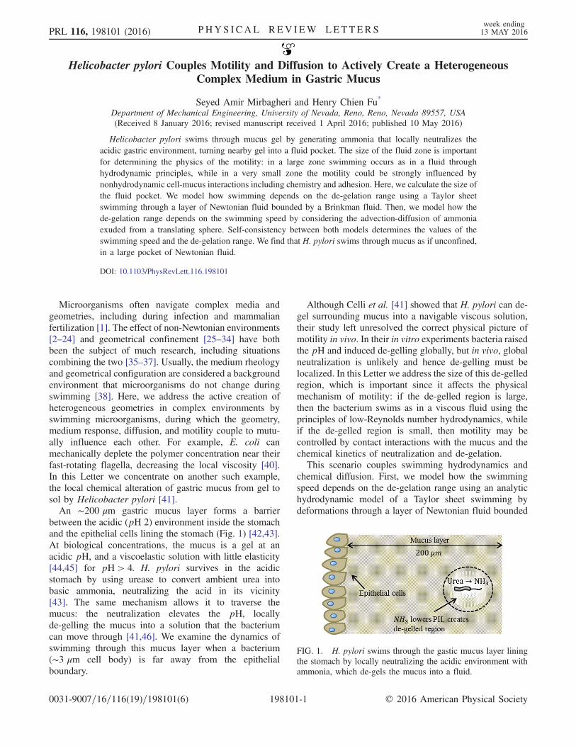
between the acidic (pH 2) environment inside the stomachand the epithelial cells lining the stomach (Fig. 1) [42,43].At biological concentrations, the mucus is a gel at anacidic pH, and a viscoelastic solution with little elasticity[44,45] for pH > 4. H. pylori survives in the acidicstomach by using urease to convert ambient urea intobasic ammonia, neutralizing the acid in its vicinity[43]. The same mechanism allows it to traverse themucus: the neutralization elevates the pH, locallyde-gelling the mucus into a solution that the bacteriumcan move through [41,46]. We examine the dynamics ofswimming through this mucus layer when a bacterium(∼3 μm cell body) is far away from the epithelialboundary.
Although Celli et al. [41] showed that H. pylori can de-gel surrounding mucus into a navigable viscous solution,their study left unresolved the correct physical picture ofmotility in vivo. In their in vitro experiments bacteria raisedthe pH and induced de-gelling globally, but in vivo, globalneutralization is unlikely and hence de-gelling must belocalized. In this Letter we address the size of this de-gelledregion, which is important since it affects the physicalmechanism of motility: if the de-gelled region is large,then the bacterium swims as in a viscous fluid using theprinciples of low-Reynolds number hydrodynamics, whileif the de-gelled region is small, then motility may becontrolled by contact interactions with the mucus and thechemical kinetics of neutralization and de-gelation.This scenario couples swimming hydrodynamics and
chemical diffusion. First, we model how the swimmingspeed depends on the de-gelation range using an analytichydrodynamic model of a Taylor sheet swimming bydeformations through a layer of Newtonian fluid bounded
FIG. 1. H. pylori swims through the gastic mucus layer liningthe stomach by locally neutralizing the acidic environment withammonia, which de-gels the mucus into a fluid.
PRL 116, 198101 (2016) P HY S I CA L R EV I EW LE T T ER Sweek ending13 MAY 2016
0031-9007=16=116(19)=198101(6) 198101-1 © 2016 American Physical Society

by a Brinkman fluid. Second, we model how the de-gelation range depends on swimming speed by using anadvection-diffusion model of ammonia exuded from atranslating sphere. The coupled problem demands thatboth the speed and neutralization range are in agreementfor both models. We show that swimming occurs in arelatively large zone of the Newtonian fluid, and that theassumptions within our approach are consistent with theresult. We discuss whether recent artificial swimmersmimicking H. pylori’s neutralization strategy [47] maybe in the same swimming regime as the bacteria.Effect of local confinement by mucus on swimming.—We
consider a waving two-dimensional sheet in the frame ofthe sheet, so the material points can be labeled by x (Fig. 2).The material points are displaced in the y direction fromy ¼ 0 by the deformation b sinðkx − ωtÞ. The half-spaceabove the sheet is a Newtonian fluid for y < h, and aBrinkman medium for y > h. Brinkman media are appro-priate representations of dilute gels [5] (gastric mucus is3%–5% w=v [46]) when the swimmer does not directlycontact the gel, as in our case, and the gel network is notdeformed by the swimmer [7].The velocity field satisfies incompressibility (∇ · v ¼ 0)
everywhere, the Stokes equations in the Newtonian fluid,and
−∇pþ μ
ϵ∇2v −
μα2
ϵðv þ VsÞ ¼ 0 ð1Þ
in the Brinkman fluid, where α ¼ ffiffiffiffiffiffiffiffiffi
ϵ=Kp
is the resistance,K is the permeability, and ϵ is the porosity (volume fractionof liquid) of the gel. We work in the frame of the sheetswimming with velocity Vs, so ðv þ VsÞ is the velocity ofthe fluid relative to the gel network, which is stationary inthe lab frame.At the sheet surface we use the no-slip boundary
conditions v(x;bsinðkx−ωtÞ)¼−bωcosðkx−ωtÞ. At theinterface between the fluid and the Brinkman medium, weuse boundary conditions maintaining a continuous velocity
v−ðxÞ ¼ vþðxÞ, where � corresponds to the limit y → hfrom below (−) or above (þ), and continuous traction½−Iðpþ − p−Þ þ μðϵ−1∇vþ −∇v−Þ� · y ¼ 0, where I is theidentity [48]. The full velocity field can be obtained from aboundary perturbation expansion in bk as in Taylor [54].The swimming velocity is obtained by imposing the force-free condition on the swimmer [55].In Fig. 2(b), the swimming speed normalized by the
unconfined (Newtonian) swimming speed VN is plotted asa function of layer height h for various values of resistanceα, constant values of porosity ϵ ¼ 0.95, and constantswimming stroke (ω, b, k constant). The effect of confine-ment by the gel is only large when hk < 1, and is very smallfor hk > 3. We examine various limits to check the result.As α → 0, the Newtonian swimming speed is recovered. Asα → ∞, the swimming speed confined by a solid boundaryat distance h [56] is recovered (solid black line). Finally, ash → 0, we recover the swimming speed of a sheet in aBrinkman medium [5], Vs ¼ 1
2ωkb2
ffiffiffiffiffiffiffiffiffiffiffiffiffiffiffiffiffiffiffiffiffiffi
1þ ðα=kÞ2p
.It is also interesting to examine the results for constant
power. The expended power can be calculated by integrat-ing the power per unit area at the swimmer surface[R
v · τ · ndA with τ the stress tensor], or by the sum ofpower lost by viscous dissipation and the action of Darcyresistance on the fluid [−
R
τ ·∇vdV þ ðμα2=ϵÞ R ðv þ VsÞ·ðv þ VsÞdV]. Agreement between the two methods pro-vides an internal check on our results. The lowest ordercontribution to the power comes from the OðbkÞ velocityfield and is shown in Fig. 2(c). The power increases as thegap size h decreases. In the limit h → 0, we obtainthe power expended in a Brinkman medium, 1
2b2ω2k½1þ
ðα=kÞ2 þffiffiffiffiffiffiffiffiffiffiffiffiffiffiffiffiffiffiffiffiffiffi
1þ ðα=kÞ2p
�, which agrees with a directcalculation of the power expended by a Taylor sheet in aBrinkman medium with no Newtonian layer [57]. Theresulting swimming speed at constant power is plotted inFig. 2(d). In contrast to the constant stroke case, theswimming speed remains finite as h → 0. However, in
FIG. 2. (a) Taylor swimming sheet in a layer of Newtonian fluid of thickness h confined by a Brinkman medium representing mucusgel. (b) Swimming speed normalized by unconfined speed versus layer thickness h for constant stroke, porosity ϵ ¼ 0.95, and variousvalues of resistance α. Solid black line is the result for a solid no-slip boundary at distance h. (c) Power dissipated normalized by thepower dissipated by an unconfined swimmer for the cases plotted in (b). (d) Swimming speed normalized by the unconfined speedversus layer thickness h for constant power dissipation, porosity ϵ ¼ 0.95, and various values of resistance α.
PRL 116, 198101 (2016) P HY S I CA L R EV I EW LE T T ER Sweek ending13 MAY 2016
198101-2

both cases the effect of confinement by the gel is only largewhen hk < 1, and is very small for hk > 3.Effect of swimming on size of local confinement.—We
examine the range of neutralization and de-gelling using asimplified model that treats the bacterium as a sphericalbody. Neutralization is controlled by a reaction-diffusionprocess involving urease, urea, ammonia, (bi)carbonate,and Hþ. Urease may act within the cell or be bound to itssurface [43], and in any case urease and urea diffusemore slowly than protons or ammonia and hence have lesseffect on the neutralization range. Thus, we assume that thepH is controlled by the diffusion of ammonia throughthe aqueous de-gelled solution surrounding the cell, withthe same diffusion constant as in water. We consider thediffusion of ammonia rather than Hþ since Hþ is suppliedthrough the mucus and diffuses in mucus gel 4–10 timesslower than in water [42]; however, using reasonably fasteror slower diffusion constants does not change our con-clusions [58]. Since H. pylori regulates the pH near its cellwall [43] (we assume a near-neutral pH 6), and the criticalde-gelation pH is near 4 [41], we model the concentrationof ammonia at the cell surface as a constant and at theboundary of the de-gelled region as decreased by a factorof 100.The diffusion of ammonia is affected by the swimming
flow of H. pylori, which we approximate as advection-diffusion from a stationary sphere in the presence of auniform background flow at the swimming velocity.Although this flow captures the dominant effect of advec-tion due to swimming translation, it differs from that of aforce-free bacterium since it results in a net force on thesphere, but as discussed later, the difference does not affectour conclusions. Advection-diffusion is controlled by thePeclet number Pe ¼ 2aVs=D, which weighs the relativeimportance of advection to diffusion. We estimate atypical Peclet number of 0.006 from the thickness of H.pylori (a ¼ 0.5 μm), the Newtonian swimming speed(Vs ¼ 10 μm=s [59]), and the diffusion constant of
ammonia in water (D ¼ 1.64 × 10⁻⁹ m2=s [60]); hence,the concentration profile is dominated by diffusion. If thebacterium swims faster due to the effects of confinement,the Peclet number may increase. For small Peclet numbers(Pe < 1), the solution to this advection-diffusion problemwas found via singular perturbation theory by Acrivos andTaylor [61], and we use their solution here.In Fig. 3(a) we show contours of equal concentration
near the sphere (surface concentration c0) obtained fromthe Acrivos and Taylor solution for Pe ¼ 0.006. In Fig. 3(b)we show the concentration contour c0=100, which repre-sents the boundary of the de-gelled region, for various Pe.As Pe increases (i.e., swimming velocity increases) thede-gelled region is swept into a narrower shape. The gapsize h in our 2D swimming model is perpendicular tothe traveling wave, so corresponds to the vertical distancefrom the sphere to the de-gelled boundary. By varying thePeclet number, we deduce a de-gelation range hA−DðVsÞ asa function of Vs [Fig. 3(c)].Self-consistent estimate of range of de-gelation.—
Finally, we estimate the range of de-gelation for swimmingH. pylori by demanding that the swimming speed and de-gelation range are consistent with both the hydrodynamicalswimming calculation and the diffusion-advection calcu-lation. Graphically, the swimming speed and gap size aredetermined by finding the intersection of the plots of thehydrodynamic swimming speed VH
s ðhÞ and hA–DðVsÞ fromdiffusion-advection (Fig. 4). The unconfined speed of theswimming sheet is set to the observed swimming speed(10 μm=s [59]) of H. pylori in a buffer solution, and weassume the effect of confinement is the same as for a sheet.Since the pitch P of H. pylori flagella has not beenmeasured we obtain the wave number k ¼ 2π=P fromthe value P ¼ 1.58 μm for V. alginolyticus [59]. Theresulting de-gelation size is h� ≈ 175=k, or 44 μm, muchlarger than the pitch or cell body. Therefore, swimmingoccurs in the unconfined regime and is largely unaffectedby the mucus gel surrounding the de-gelled region.
FIG. 3. (a) Concentration profile due to diffusion near a sphere in a uniform background flow in theþx direction for Pe ¼ 0.006. C0 isthe concentration at the sphere surface. (b) Contours of concentration 0.01C0 corresponding to de-gelation boundary for various Pe. Wetake the layer thickness for the confined sheet model from the distance in the y direction (h) to the de-gelation boundary. (c) De-gelationrange h as a function of velocity, for the cell parameters specified in the text.
PRL 116, 198101 (2016) P HY S I CA L R EV I EW LE T T ER Sweek ending13 MAY 2016
198101-3
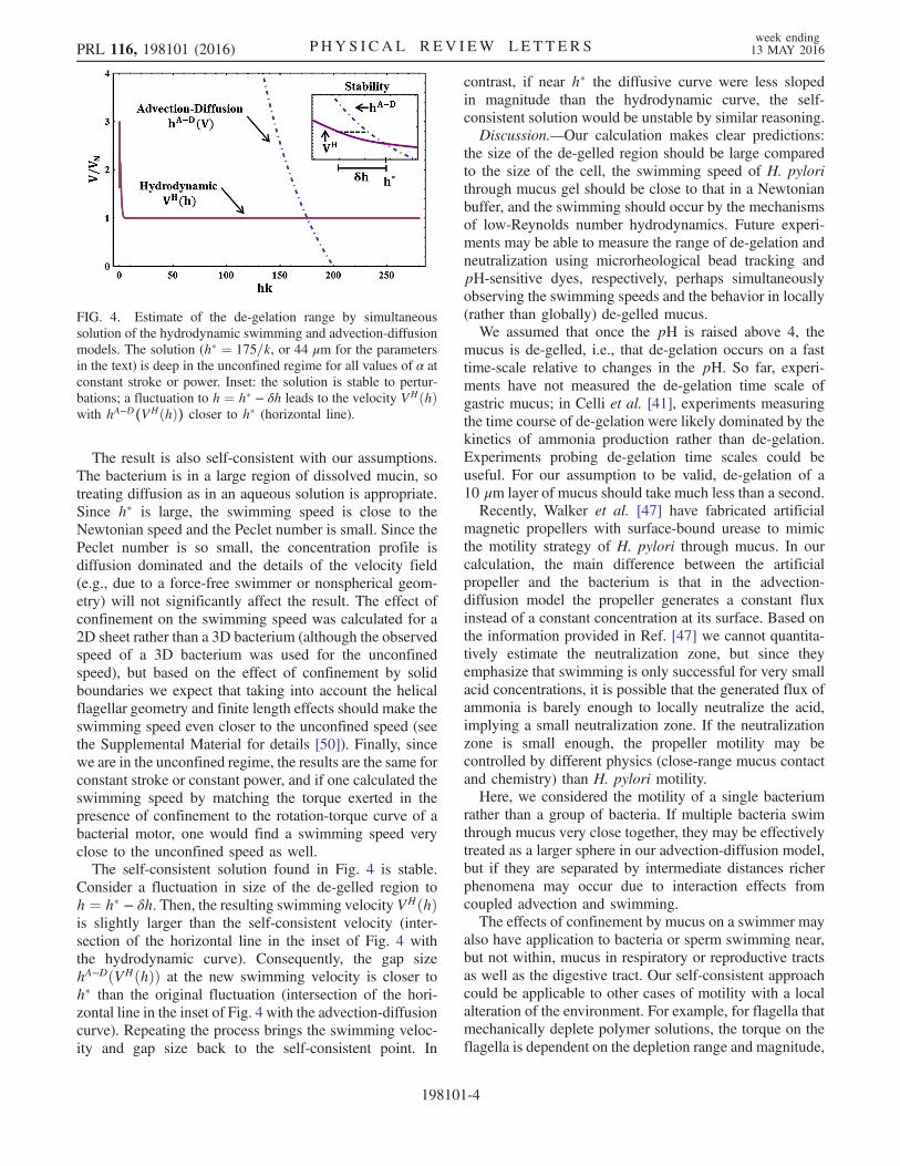
The result is also self-consistent with our assumptions.The bacterium is in a large region of dissolved mucin, sotreating diffusion as in an aqueous solution is appropriate.Since h� is large, the swimming speed is close to theNewtonian speed and the Peclet number is small. Since thePeclet number is so small, the concentration profile isdiffusion dominated and the details of the velocity field(e.g., due to a force-free swimmer or nonspherical geom-etry) will not significantly affect the result. The effect ofconfinement on the swimming speed was calculated for a2D sheet rather than a 3D bacterium (although the observedspeed of a 3D bacterium was used for the unconfinedspeed), but based on the effect of confinement by solidboundaries we expect that taking into account the helicalflagellar geometry and finite length effects should make theswimming speed even closer to the unconfined speed (seethe Supplemental Material for details [50]). Finally, sincewe are in the unconfined regime, the results are the same forconstant stroke or constant power, and if one calculated theswimming speed by matching the torque exerted in thepresence of confinement to the rotation-torque curve of abacterial motor, one would find a swimming speed veryclose to the unconfined speed as well.The self-consistent solution found in Fig. 4 is stable.
Consider a fluctuation in size of the de-gelled region toh ¼ h� − δh. Then, the resulting swimming velocity VHðhÞis slightly larger than the self-consistent velocity (inter-section of the horizontal line in the inset of Fig. 4 withthe hydrodynamic curve). Consequently, the gap sizehA−DðVHðhÞÞ at the new swimming velocity is closer toh� than the original fluctuation (intersection of the hori-zontal line in the inset of Fig. 4 with the advection-diffusioncurve). Repeating the process brings the swimming veloc-ity and gap size back to the self-consistent point. In
contrast, if near h� the diffusive curve were less slopedin magnitude than the hydrodynamic curve, the self-consistent solution would be unstable by similar reasoning.Discussion.—Our calculation makes clear predictions:
the size of the de-gelled region should be large comparedto the size of the cell, the swimming speed of H. pylorithrough mucus gel should be close to that in a Newtonianbuffer, and the swimming should occur by the mechanismsof low-Reynolds number hydrodynamics. Future experi-ments may be able to measure the range of de-gelation andneutralization using microrheological bead tracking andpH-sensitive dyes, respectively, perhaps simultaneouslyobserving the swimming speeds and the behavior in locally(rather than globally) de-gelled mucus.We assumed that once the pH is raised above 4, the
mucus is de-gelled, i.e., that de-gelation occurs on a fasttime-scale relative to changes in the pH. So far, experi-ments have not measured the de-gelation time scale ofgastric mucus; in Celli et al. [41], experiments measuringthe time course of de-gelation were likely dominated by thekinetics of ammonia production rather than de-gelation.Experiments probing de-gelation time scales could beuseful. For our assumption to be valid, de-gelation of a10 μm layer of mucus should take much less than a second.Recently, Walker et al. [47] have fabricated artificial
magnetic propellers with surface-bound urease to mimicthe motility strategy of H. pylori through mucus. In ourcalculation, the main difference between the artificialpropeller and the bacterium is that in the advection-diffusion model the propeller generates a constant fluxinstead of a constant concentration at its surface. Based onthe information provided in Ref. [47] we cannot quantita-tively estimate the neutralization zone, but since theyemphasize that swimming is only successful for very smallacid concentrations, it is possible that the generated flux ofammonia is barely enough to locally neutralize the acid,implying a small neutralization zone. If the neutralizationzone is small enough, the propeller motility may becontrolled by different physics (close-range mucus contactand chemistry) than H. pylori motility.Here, we considered the motility of a single bacterium
rather than a group of bacteria. If multiple bacteria swimthrough mucus very close together, they may be effectivelytreated as a larger sphere in our advection-diffusion model,but if they are separated by intermediate distances richerphenomena may occur due to interaction effects fromcoupled advection and swimming.The effects of confinement by mucus on a swimmer may
also have application to bacteria or sperm swimming near,but not within, mucus in respiratory or reproductive tractsas well as the digestive tract. Our self-consistent approachcould be applicable to other cases of motility with a localalteration of the environment. For example, for flagella thatmechanically deplete polymer solutions, the torque on theflagella is dependent on the depletion range and magnitude,
FIG. 4. Estimate of the de-gelation range by simultaneoussolution of the hydrodynamic swimming and advection-diffusionmodels. The solution (h� ¼ 175=k, or 44 μm for the parametersin the text) is deep in the unconfined regime for all values of α atconstant stroke or power. Inset: the solution is stable to pertur-bations; a fluctuation to h ¼ h� − δh leads to the velocity VHðhÞwith hA−D(VHðhÞ) closer to h� (horizontal line).
PRL 116, 198101 (2016) P HY S I CA L R EV I EW LE T T ER Sweek ending13 MAY 2016
198101-4
while the depletion is dependent on the torque via therotation rate and geometry.
This work was supported by National ScienceFoundation Grant No. CBET-1252182 to H. C. F.
*[email protected][1] S. Suarez and A. Pacey, Human Reprod. Update 12, 23
(2006).[2] E. Lauga, Phys. Fluids 19, 083104 (2007).[3] H. C. Fu, T. R. Powers, and C.W. Wolgemuth, Phys. Rev.
Lett. 99, 258101 (2007).[4] H. C. Fu, C. W. Wolgemuth, and T. R. Powers, Phys. Fluids
21, 033102 (2009).[5] A. M. Leshansky, Phys. Rev. E 80, 051911 (2009).[6] J. Teran, L. Fauci, and M. Shelley, Phys. Rev. Lett. 104,
038101 (2010).[7] H. C. Fu, V. B. Shenoy, and T. R. Powers, Europhys. Lett.
91, 24002 (2010).[8] G. Juarez, K. Lu, J. Sznitman, and P. E. Arratia, Europhys.
Lett. 92, 44002 (2010).[9] B. Liu, T. R. Powers, and K. S. Breuer, Proc. Natl. Acad.
Sci. U.S.A. 108, 19516 (2011).[10] X. N. Shen and P. E. Arratia, Phys. Rev. Lett. 106, 208101
(2011).[11] L. Zhu, E. Lauga, and L. Brandt, Phys. Fluids 24, 051902
(2012).[12] N. C. Keim, M. Garcia, and P. E. Arratia, Phys. Fluids 24,
081703 (2012).[13] M. Dasgupta, B. Liu, H. C. Fu, M. Berhanu, K. S. Breuer,
T. R. Powers, and A. Kudrolli, Phys. Rev. E 87, 013015(2013).
[14] J. Espinoza-Garcia, E. Lauga, and R. Zenit, Phys. Fluids 25,031701 (2013).
[15] S. E. Spagnolie, B. Liu, and T. R. Powers, Phys. Rev. Lett.111, 068101 (2013).
[16] Y. Gao, M. Neubauer, A. Yang, N. Johnson, M. Morse,G. Li, and J. X. Tang, BMC Microbiol. 14, 322 (2014).
[17] D. Gagnon, X. Shen, and P. Arratia, Europhys. Lett. 104,14004 (2013).
[18] T. D. Montenegro-Johnson, D. J. Smith, and D. Loghin,Phys. Fluids 25, 081903 (2013).
[19] D. Gagnon, N. Keim, and P. Arratia, J. Fluid Mech. 758, R3(2014).
[20] B. Thomases and R. D. Guy, Phys. Rev. Lett. 113, 098102(2014).
[21] L. Li and S. E. Spagnolie, Phys. Fluids 27, 021902 (2015).[22] C. Datt, L. Zhu, G. J. Elfring, and O. S. Pak, J. Fluid Mech.
784, R1 (2015).[23] S. D. Olson and K. Leiderman, J. Aero Aqua Bio-
mechanisms 4, 12 (2015).[24] B. Qin, A. Gopinath, J. Yang, J. P. Gollub, and P. Arratia,
Sci. Rep. 5, 9190 (2015).[25] E. Lauga, W. R. DiLuzio, G. M. Whitesides, and H. A.
Stone, Biophys. J. 90, 400 (2006).[26] A. P. Berke, L. Turner, H. C. Berg, and E. Lauga, Phys. Rev.
Lett. 101, 038102 (2008).[27] G. Li, L.-K. Tam, and J. X. Tang, Proc. Natl. Acad. Sci.
U.S.A. 105, 18355 (2008).
[28] G. Li and J. X. Tang, Phys. Rev. Lett. 103, 078101 (2009).[29] P. Denissenko, V. Kantsler, D. J. Smith, and J. Kirkman-
Brown, Proc. Natl. Acad. Sci. U.S.A. 109, 8007 (2012).[30] V. Kantsler, J. Dunkel, M. Polin, and R. E. Goldstein, Proc.
Natl. Acad. Sci. U.S.A. 110, 1187 (2013).[31] B. Liu, K. S. Breuer, and T. R. Powers, Phys. Fluids 26,
011701 (2014).[32] H. Shum and E. A. Gaffney, Phys. Rev. E 91, 033012
(2015).[33] F. Z. Temel and S. Yesilyurt, Bioinspiration Biomimetics 10,
016015 (2015).[34] M. Contino, E. Lushi, I. Tuval, V. Kantsler, and M. Polin,
Phys. Rev. Lett. 115, 258102 (2015).[35] M. Jabbarzadeh, Y. K. Hyon, and H. C. Fu, Phys. Rev. E 90,
043021 (2014).[36] S. Yazdi, A. Ardekani, and A. Borhan, J. Nonlinear Sci. 25,
1153 (2015).[37] G.-J. Li, A. Karimi, and A. Ardekani, Rheol. Acta 53, 911
(2014).[38] Except perhaps through deformation by mechanical com-
pliance, e.g., Ref. [39].[39] R. Ledesma-Aguilar and J. M. Yeomans, Phys. Rev. Lett.
111, 138101 (2013).[40] V. A. Martinez, J. Schwarz-Linek, M. Reufer, L. G. Wilson,
A. N. Morozov, and W. C. Poon, Proc. Natl. Acad. Sci.U.S.A. 111, 17771 (2014).
[41] J. P. Celli, B. S. Turner, N. H. Afdhal, S. Keates, I. Ghiran,C. P. Kelly, R. H. Ewoldt, G. H. McKinley, P. So, S.Erramilli, and R. Bansil, Proc. Natl. Acad. Sci. U.S.A.106, 14321 (2009).
[42] A. Allen and G. Flemström, Am. J. Physiol. 288, C1 (2005).[43] C. Montecucco and R. Rappuoli, Nat. Rev. Mol. Cell Biol.
2, 457 (2001).[44] X. Cao, R. Bansil, K. R. Bhaskar, B. S. Turner, J. T. LaMont,
N. Niu, and N. H. Afdhal, Biophys. J. 76, 1250 (1999).[45] J. P. Celli, B. S. Turner, N. H. Afdhal, R. H. Ewoldt, G. H.
McKinley, R. Bansil, and S. Erramilli, Biomacromolecules8, 1580 (2007).
[46] R. Bansil, J. P. Celli, J. M. Hardcastle, and B. S. Turner,Front. Immunol. 4, 310 (2013).
[47] D. Walker, B. T. Käsdorf, H.-H. Jeong, O. Lieleg, and P.Fischer, Sci. Adv., 1, e1500501 (2015).
[48] Ochoa-Tapia and Whitaker [49] have shown that thehomogenized boundary conditions can allow a jump intangential traction. The estimates we obtain for the de-gelation range are not changed by such a boundary con-dition. See the Supplemental Material [50] for details.
[49] J. A. Ochoa-Tapia and S. Whitaker, Int. J. Heat MassTransfer 38, 2635 (1995).
[50] See Supplemental Material at http://link.aps.org/supplemental/10.1103/PhysRevLett.116.198101, which in-cludes Refs. [51–53], for details of solution, includingresults when a jump in tangential traction is included,and comparison of effect of confinement by solid boundaryon 2D and 3D swimmer models.
[51] M. Sauzade, G. J. Elfring, and E. Lauga, Physica(Amsterdam) 240D, 1567 (2011).
[52] J. Happel and H. Brenner, Low Reynolds Number Hydro-dynamics: with Special Applications to Particulate Media,Vol. 1 (Springer Science&BusinessMedia, NewYork, 1983).
PRL 116, 198101 (2016) P HY S I CA L R EV I EW LE T T ER Sweek ending13 MAY 2016
198101-5
[53] M. Ramia, D. Tullock, and N. Phan-Thien, Biophys. J. 65,755 (1993).
[54] G. I. Taylor, Proc. R. Soc. A 209, 447 (1951).[55] Details are in the Supplemental Material [50].[56] A. Reynolds, J. Fluid Mech. 23, 241 (1965).[57] This result differs from that of Ref. [5], which ignored the
dissipation by the Darcy resistance in our second calculationmethod for the power.
[58] Reproducing the calculation in the next section withthe faster diffusion constant of Hþ in water(D ¼ 9.31 × 10⁻⁹ m2=s [60]) yields an estimate for the
size of the de-gelled region of hk ≈ 196 (h ¼ 49 μm).Approximating the effect of mucus on the diffusion constantof ammonia by slowing it by a factor of 4 yields anestimate for the size of the de-gelled region of hk ≈ 132(h ¼ 33 μm), instead of hk ≈ 175 for ammonia in water.
[59] L. E. Martínez, J. M. Hardcastle, J. Wang, Z. Pincus, J.Tsang, T. R. Hoover, R. Bansil, and N. R. Salama, Mol.Microbiol. 99, 88 (2016).
[60] E. Cussler, Diffusion, Mass Transfer in Fluid Systems,2nd ed. (Cambridge University Press, Cambridge, 1997).
[61] A. Acrivos and T. D. Taylor, Phys. Fluids 5, 387 (1962).
PRL 116, 198101 (2016) P HY S I CA L R EV I EW LE T T ER Sweek ending13 MAY 2016
198101-6