hematological disorders of newborn
TRANSCRIPT

7/28/2019 Hematological Disorders of Newborn
http://slidepdf.com/reader/full/hematological-disorders-of-newborn 1/98
Haematological Disorders of
Newborn
Hasmawati bt HassanConsultant Neonatologist
HRPZII
7/28/2019 Hematological Disorders of Newborn
http://slidepdf.com/reader/full/hematological-disorders-of-newborn 2/98
Common Hematological Disorders
• Anaemia
• Polycythaemia
• Bleeding and Coagulation Disorders

7/28/2019 Hematological Disorders of Newborn
http://slidepdf.com/reader/full/hematological-disorders-of-newborn 3/98
Introduction
• Blood volume and red cell mass at birth and in
neonatal period depend on
– Volume of placental transfusion and,
– Subsequent readjustments of blood volume

7/28/2019 Hematological Disorders of Newborn
http://slidepdf.com/reader/full/hematological-disorders-of-newborn 4/98
Placental Transfusion
• Occurs within 3 min. of delivery
• Contributes 25% of total neonatal bloodvolume.
• This amount will be increased in:
– Elevated maternal blood pressure
– Use of oxytocic drugs
– Late clamping or milking of the cord – Infant held in a low dependent position.
7/28/2019 Hematological Disorders of Newborn
http://slidepdf.com/reader/full/hematological-disorders-of-newborn 5/98
Placental Transfusion
• Average blood volume 85-90ml/kg.
• Ranges between 75-100 ml/kg.
• Readjustment of blood volume – Occur the first 3-4 hours after birth with
heamoconcentration to compensate for expansion
of intravascular volume.

7/28/2019 Hematological Disorders of Newborn
http://slidepdf.com/reader/full/hematological-disorders-of-newborn 6/98
Neonatal Anaemia

7/28/2019 Hematological Disorders of Newborn
http://slidepdf.com/reader/full/hematological-disorders-of-newborn 7/98
Neonatal Anaemia
• Defined as: Hb. Less than 13 g%

7/28/2019 Hematological Disorders of Newborn
http://slidepdf.com/reader/full/hematological-disorders-of-newborn 8/98
Causes of Neonatal Anaemia
• Physiological anaemia (1)
• Anaemia of prematurity (2)
• Haemorrhage (3)
– Antepartum haemorrhage – Fetomaternal transfusion
– Twin twin transfusion
– Neonatal internal haemorrhage –Hemorrhagic Disease of
Newborn• Haemolysis (4)
• Aplasia (5)
7/28/2019 Hematological Disorders of Newborn
http://slidepdf.com/reader/full/hematological-disorders-of-newborn 9/98
Physiological anaemia (1)
• Term born with Hb range 15-23.5g%
• Preterm slightly lower
• Slight increase due to hemoconcentration• Over 1 week Hb drops and remains low for
most of the first year.
• Also known as physiological anaemia
7/28/2019 Hematological Disorders of Newborn
http://slidepdf.com/reader/full/hematological-disorders-of-newborn 10/98
Anaemia of prematurity (2)
• Defined as: physiological anemia occurring inthe preterm infant which occur earlier, severeand prolonged.
• Causes: – Lack of erythropoietin
– Hemodilution
– Iron deficiency – Haemolysis

7/28/2019 Hematological Disorders of Newborn
http://slidepdf.com/reader/full/hematological-disorders-of-newborn 11/98
Anaemia of prematurity (2)
Lack of erythropoietin.
• Due to relatively hypoxic fetal state to
hyperoxic stage.
• This suppresses erythropoietin secretion for
the first 7-8 weeks.
•Bone marrow resistant to stimulation of
erythropoietin.

7/28/2019 Hematological Disorders of Newborn
http://slidepdf.com/reader/full/hematological-disorders-of-newborn 12/98
Anaemia of prematurity (2)
Haemodilution – increase of plasma volume
over the first months of life.
• Poor red cell production -the haemoglobin
falls.
• Also referred as ‘early anaemia.’

7/28/2019 Hematological Disorders of Newborn
http://slidepdf.com/reader/full/hematological-disorders-of-newborn 13/98
Anaemia of prematurity (3)
Iron deficiency:
• In preterm the iron stores are exhausted more
quickly due to rapid growth.
• Infant of 1.5 kg has half the iron stores at
birth compared with a 3.0 kg infant.
•This account for ‘late anemia’ seen at 4
months of age.

7/28/2019 Hematological Disorders of Newborn
http://slidepdf.com/reader/full/hematological-disorders-of-newborn 14/98
Anaemia of prematurity (2)
Haemolysis:
• Due to vitamin E deficiency.
• Treatment with vit. E may reduce the extent of
late anemia of prematurity.
• In term, lowest Hb occurs between 6 –10weeks when it falls to 10-11 g/dl.
• In preterm it occurs earlier and last longerwith a nadir 7-8 g.

7/28/2019 Hematological Disorders of Newborn
http://slidepdf.com/reader/full/hematological-disorders-of-newborn 15/98
Symptoms of Anaemia in Preterm
• Breathlessness with feeds
• Tachycardia
• Apnoea• Bradycardia
• Failure to gain weight

7/28/2019 Hematological Disorders of Newborn
http://slidepdf.com/reader/full/hematological-disorders-of-newborn 16/98
Treatment of anaemia of prematurity
• Iron given to preterm from 2 weeks of age.
• Elemental iron 1-2 mg/kg is recommended
until age 12 months of age.
• Blood transfusion 10ml/kg if Hb < 8 g or if
symptomatic.
•Recombinant human erythropoietin.

7/28/2019 Hematological Disorders of Newborn
http://slidepdf.com/reader/full/hematological-disorders-of-newborn 17/98
Anemia base on Hematocrit –
indication for blood transfusion
• Hct < 35 + ventilated
• Hct <30 + on Oxygen
• Hct <25 + symptomatic
• Hct <20 – all babies

7/28/2019 Hematological Disorders of Newborn
http://slidepdf.com/reader/full/hematological-disorders-of-newborn 18/98
Haemorrhage (3)
7/28/2019 Hematological Disorders of Newborn
http://slidepdf.com/reader/full/hematological-disorders-of-newborn 19/98
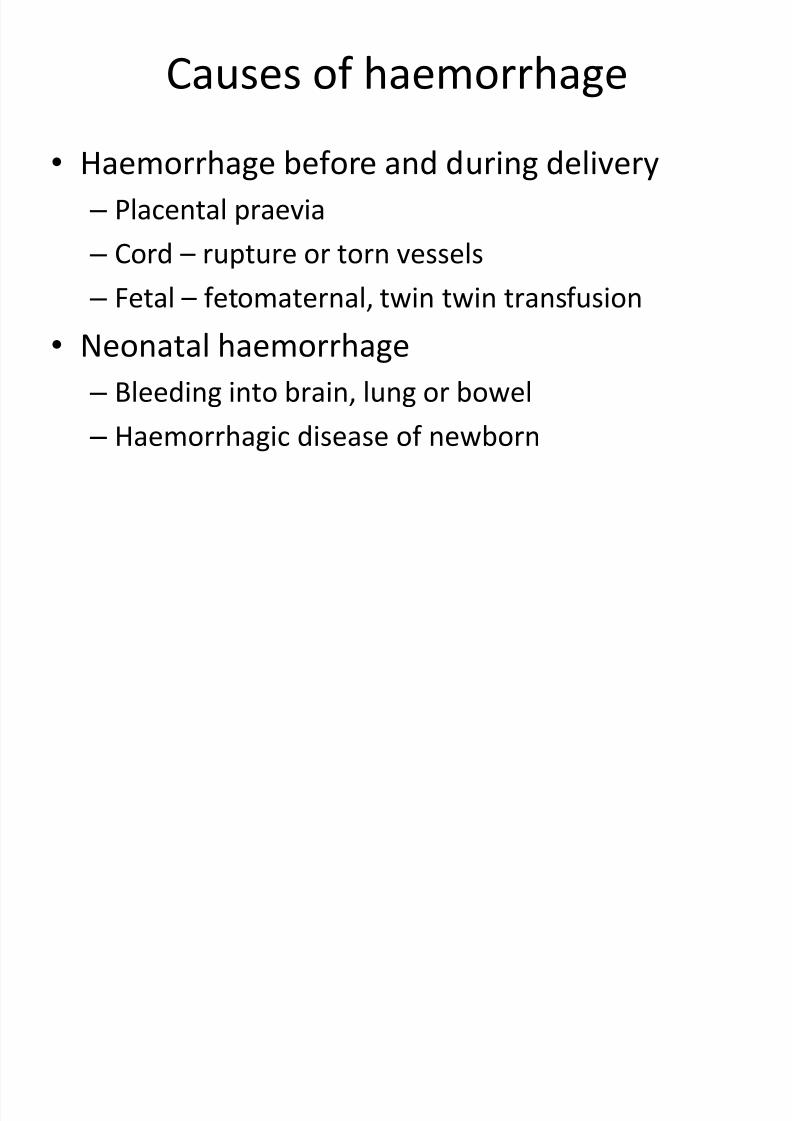
Causes of haemorrhage
• Haemorrhage before and during delivery
– Placental praevia
– Cord – rupture or torn vessels
– Fetal – fetomaternal, twin twin transfusion
• Neonatal haemorrhage
– Bleeding into brain, lung or bowel
– Haemorrhagic disease of newborn

7/28/2019 Hematological Disorders of Newborn
http://slidepdf.com/reader/full/hematological-disorders-of-newborn 20/98
Management of Haemorrhage
• Clinical assessment for shock and hypovolaemia.
• Investigations include:-
– Haemoglobin and haematocrit
– Blood group
– Cross match blood
– Kleihauer’s test
– Coagulation studies
– Ultrasound cranium, abdomen, stools for blood.

7/28/2019 Hematological Disorders of Newborn
http://slidepdf.com/reader/full/hematological-disorders-of-newborn 21/98
Haemorrhagic Disease of Newborn

7/28/2019 Hematological Disorders of Newborn
http://slidepdf.com/reader/full/hematological-disorders-of-newborn 22/98
History
• Townsend in Boston (1864) described 50 cases of “hemorrhagic disease of the newborn” during first 2
weeks of life
• In 1929, Vitamin K isolated from alfalfa by Dam and Doisy
(Nobel Prize, 1942), and conducted clinical trials showingVitamin K protects against HDN
• 1961, Am Acad Pediatrics and Am College Obstetrics andGynecology recommended routine prophylaxis with Vit K
for all newborns• Controversy in Britain in 1990s resolved to satisfaction of
AAP, ACOG, Canada, Australia, New Zealand and others

7/28/2019 Hematological Disorders of Newborn
http://slidepdf.com/reader/full/hematological-disorders-of-newborn 23/98
Haemorrhagic disease of newborn
• Early
• Classical – due to deficiency of vitamin K
dependent clotting factors
• Late onset

7/28/2019 Hematological Disorders of Newborn
http://slidepdf.com/reader/full/hematological-disorders-of-newborn 24/98
Primary HDN
• Often fatal condition• Diffuse hemorrhage in otherwise healthy infant
• During the first week of life
• Particularly in low birth weight babies• Results of low levels of prothrombin and other vitaminK dependent clotting factors, (Factors II, VII, IX and X)caused by vitamin K deficiency
• An exaggerated of physiologic deficiency of clottingfactors normal in the first few days of life
• Incidence between 2.5 to 17.0 per thousand newbornsnot given vitamin K prophylactically

7/28/2019 Hematological Disorders of Newborn
http://slidepdf.com/reader/full/hematological-disorders-of-newborn 25/98
Haemorrhagic disease of newborn
• Classical haemorrhagic disease of thenewborn is due to deficiency of vit.Kdependent clotting factors.
• Vitamin K is produced by bacterial flora of thegut which there is little production during firstweek of life.
• Decline due to routine IM vitamin K at birth.

7/28/2019 Hematological Disorders of Newborn
http://slidepdf.com/reader/full/hematological-disorders-of-newborn 26/98
Late HDN
• Between 2-12 weeks of life,
• Especially in breast-fed babies.
• Immaturity of liver affects production of clottingfactors
• Late HDN primarily in breast fed infants without
or inadequate vitamin K rates of 4.4-7.2/100,000
live births

7/28/2019 Hematological Disorders of Newborn
http://slidepdf.com/reader/full/hematological-disorders-of-newborn 27/98
Common Clinical Manifestations
• Bleeding in the
– gastrointestinal tract
– urinary tract
– umbilical stump – nose
– scalp
– intracranial hemorrhage – Shock
– death

7/28/2019 Hematological Disorders of Newborn
http://slidepdf.com/reader/full/hematological-disorders-of-newborn 28/98
Clinical features:
• Spontaneous bleeding usually GIT.
• Umbilical bleeding or postcircumcision.
• Occurs late in first week of life esp to breast-fed infant.
• DD from bleeding due to swallow blood.
–Differentiate by APT’s Test.

7/28/2019 Hematological Disorders of Newborn
http://slidepdf.com/reader/full/hematological-disorders-of-newborn 29/98
American Academy of Pediatrics 1961
• Prophylactic use of Vit K recommended by the
American Academy of Pediatrics, and by the American
College of Obstetricians and Gynecologists since 1961.• Up until 1987, administration of vit K at birth was
mandatory in only five states in the US
• AAP recommendation renewed in 1993 and remains
current

7/28/2019 Hematological Disorders of Newborn
http://slidepdf.com/reader/full/hematological-disorders-of-newborn 30/98
Investigations:
• The diagnosis is confirmed by a prolonged
prothrombin time (PT) but normal partial
thromboplastin time (PTT).
• Apt’s test – resistance of Fetal RBCs to
denaturation by sodium hydroxide.
7/28/2019 Hematological Disorders of Newborn
http://slidepdf.com/reader/full/hematological-disorders-of-newborn 31/98
Treatment
• Vitamin K1, 1 mg im or iv
• Blood transfusion if indicated.
7/28/2019 Hematological Disorders of Newborn
http://slidepdf.com/reader/full/hematological-disorders-of-newborn 32/98
Renewed Interest in Vit K
• Since the 1980s attention – UK, Europe, Japan, Canada,Australasia and Middle East
• HDN and vit K deficiency reported in both developedand developing countries where it is not routinely used,or where use may be waning
• Controversy re oral versus parenteral use of routine VitK largely resolved
• Intramuscular administration within the first 6 hoursafter birth more effective in preventing both early andlate HDN

7/28/2019 Hematological Disorders of Newborn
http://slidepdf.com/reader/full/hematological-disorders-of-newborn 33/98
Other Countries
• Still not routine in Japan, Germany, UK
• Routine prophylactic Vitamin K for newborns
adopted in – Canada
– Australia
– New Zealand
– Croatia, 1988

7/28/2019 Hematological Disorders of Newborn
http://slidepdf.com/reader/full/hematological-disorders-of-newborn 34/98
Public Health Importance
• Japanese incidence of HDN reported as 1/1,700 in
breast fed babies and 1:4,500 in all infants
• Of these, 82% were reported to have intracranial
hemorrhage (ICH)• NDN still significant; even more in developing countries
e.g. India, Thailand, Singapore and Taiwan
• Thailand reports incidence of 35-72/100,000 births
• ICH not always identified as HND related and may be
significant factor in birth-related cerebral palsies

7/28/2019 Hematological Disorders of Newborn
http://slidepdf.com/reader/full/hematological-disorders-of-newborn 35/98
Summary
• Deficiency of Vit K remains a significant worldwide causeof neonatal morbidity and mortality
• Routine prophylactic use of vitamin K should always beused to prevent HDN (“good public health practice”)
• Administration by intramuscular injection (0.5-1.0 mgm)within 6 hours of birth is preferable
• May be given orally as 3 doses spread over the first 4weeks of life
• Vit K showing up in literature on osteoporosis• A safe, inexpensive preventive procedure that should bemandatory component of newborn care.
7/28/2019 Hematological Disorders of Newborn
http://slidepdf.com/reader/full/hematological-disorders-of-newborn 36/98
Haemolysis (4)
(Hemolytic Disease of Newborn,
HDN)

7/28/2019 Hematological Disorders of Newborn
http://slidepdf.com/reader/full/hematological-disorders-of-newborn 37/98
Causes of neonatal haemolysis
Immune haemolysis (positive Coombs’ test)
Rhesus incompatibility
ABO incompatibility
Minor blood group
Maternal SLE
Non-Immune haemolysis
Congenital infectionDIC
G6PD def
Pyruvate kinase
Alpha Thal
7/28/2019 Hematological Disorders of Newborn
http://slidepdf.com/reader/full/hematological-disorders-of-newborn 38/98
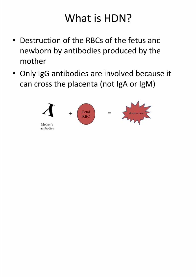
What is HDN?
• Destruction of the RBCs of the fetus and
newborn by antibodies produced by the
mother
• Only IgG antibodies are involved because it
can cross the placenta (not IgA or IgM)
+ Fetal
RBC= destruction
Mother’s
antibodies
7/28/2019 Hematological Disorders of Newborn
http://slidepdf.com/reader/full/hematological-disorders-of-newborn 39/98
7/28/2019 Hematological Disorders of Newborn
http://slidepdf.com/reader/full/hematological-disorders-of-newborn 40/98
Pathophysiology
• Although transfer of maternal antibodies isgood, transfer of antibodies involved in HDNare directed against antigens on fetal RBCs
inherited by the father• Most often involves antigens of the Rh and
ABO blood group system, but can result fromany blood group system
• Remember: The fetus is POSITIVE for anantigen and the mother is NEGATIVE for thesame antigen

7/28/2019 Hematological Disorders of Newborn
http://slidepdf.com/reader/full/hematological-disorders-of-newborn 41/98
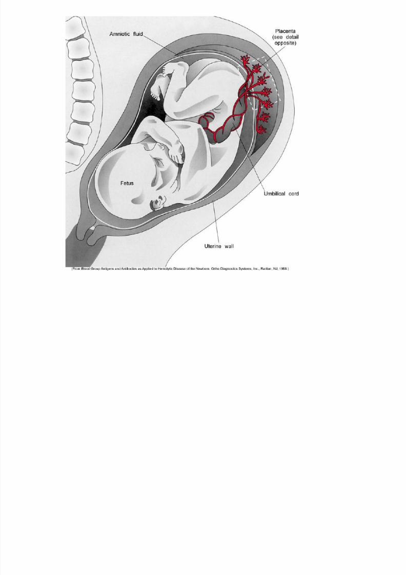
Pathophysiology
• HDN develops in utero
• The mother is sensitized to the foreign antigen
present on her child’s RBCs usually through
some seepage of fetal RBCs (fetomaternal
hemorrhage) or a previous transfusion
• HDN occurs when these antibodies cross the
placenta and react with the fetal RBCs

7/28/2019 Hematological Disorders of Newborn
http://slidepdf.com/reader/full/hematological-disorders-of-newborn 42/98
Rhesus Haemolytic Disease
• This occurs because the mother’s immune
system has been sensitized by rhesus-positive
cells from her fetus. Due to:-
– Fetomaternal transfusion.
– Rhesus incompatible transfusions.
• 95% due to antigen D.
• 83% of population are D positive.

7/28/2019 Hematological Disorders of Newborn
http://slidepdf.com/reader/full/hematological-disorders-of-newborn 43/98
Rh HDN
• Mother is D negative (d/d) and child is D positive (D/d)
• Most severe form of HDN
• 33% of HDN is caused by Rh incompatibility
• Sensitization usually occurs very late in pregnancy, so thefirst Rh-positive child is not affected
– Bleeds most often occur at delivery
– Mother is sensitized
– Subsequent offspring that are D-positive will be affected

7/28/2019 Hematological Disorders of Newborn
http://slidepdf.com/reader/full/hematological-disorders-of-newborn 44/98

7/28/2019 Hematological Disorders of Newborn
http://slidepdf.com/reader/full/hematological-disorders-of-newborn 45/98
FetoMaternal Hemorrhage
• Sensitization occurs as a result of seepage of
fetal cells into maternal circulation as a result
of a fetomaternal hemorrhage
– Placental membrane rupture (7%)
– Trauma to abdomen
– Delivery (>50%)
– Amniocentesis
– Abortion
7/28/2019 Hematological Disorders of Newborn
http://slidepdf.com/reader/full/hematological-disorders-of-newborn 46/98
Pathogenesis
• Maternal IgG attaches to antigens on fetalcells
– Sensitized cells are removed by macrophages in
spleen – Destruction depends on antibody titer and
number of antigen sites
– IgG has half-life of 25 days, so the condition can
range from days to weeks
• RBC destruction and anemia cause bonemarrow to release erythroblasts, hence the
name “erythroblastosis fetalis”)
7/28/2019 Hematological Disorders of Newborn
http://slidepdf.com/reader/full/hematological-disorders-of-newborn 47/98
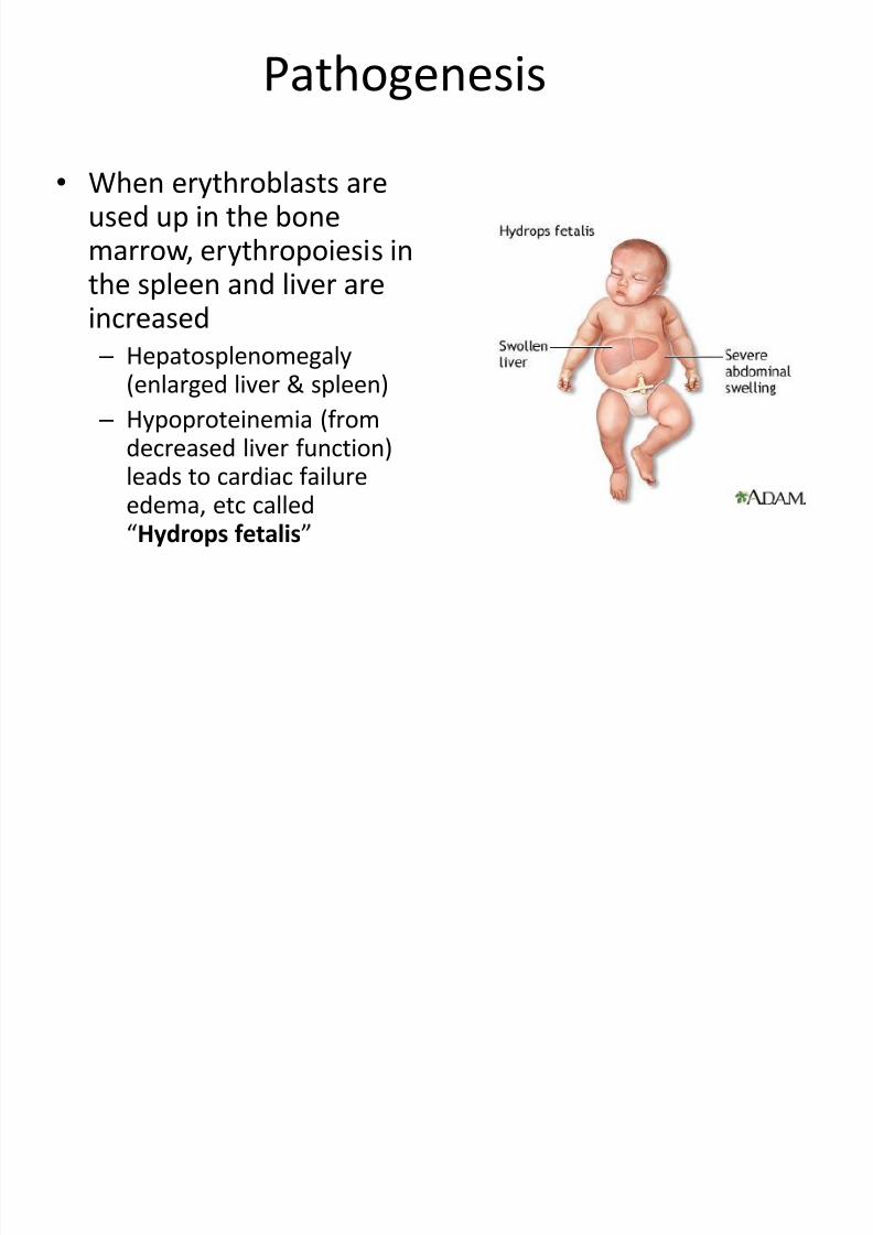
Pathogenesis
• When erythroblasts areused up in the bonemarrow, erythropoiesis inthe spleen and liver are
increased – Hepatosplenomegaly
(enlarged liver & spleen)
– Hypoproteinemia (fromdecreased liver function)leads to cardiac failureedema, etc called“Hydrops fetalis”

7/28/2019 Hematological Disorders of Newborn
http://slidepdf.com/reader/full/hematological-disorders-of-newborn 48/98
Bilirubin
• Hemoglobin is metabolized to bilirubin
– Before birth, “indirect” bilirubin is transported
across placenta and conjugated in maternal liver
(“direct”) where it is excreted – After birth, the newborn liver is unable to
conjugate the bilirubin
• Unconjugated (“indirect”) bilirubin can reach toxic
levels (18-20 mg/dL)
• This is called kernicterus and can lead to permanent
brain damage

7/28/2019 Hematological Disorders of Newborn
http://slidepdf.com/reader/full/hematological-disorders-of-newborn 49/98
Prevention of Rh Haemolytic Disease
• Anti-D gamma globulin 100-200 ug. Given to
all rhesus-negative mum who gave birth to
rhesus-positive infants within 72 hours.
• Also given to at-risk rhesus negative mum
after abortion or after amniocentesis esp if
Kleihauer test shows a fetomaternal
transfusion.

7/28/2019 Hematological Disorders of Newborn
http://slidepdf.com/reader/full/hematological-disorders-of-newborn 50/98
Dose
• Each vial of RhIg contains enough anti-D to
protect against a FMH of 30 mL
– One vial contains 300 μg of anti-D
– Given intramuscularly of intravenously
– Massive fetomaternal hemorrhage (>30 mL)
requires more than one vial
– To assess a FMH, a maternal sample is screenedwithin 1 hour of delivery (rosette test)

7/28/2019 Hematological Disorders of Newborn
http://slidepdf.com/reader/full/hematological-disorders-of-newborn 51/98
Diagnosis & Management
• Serologic Testing (mother & newborn)
• Amniocentesis and Cordocentesis
• Intrauterine Transfusion
• Early Delivery
• Phototherapy & Newborn Transfusions

7/28/2019 Hematological Disorders of Newborn
http://slidepdf.com/reader/full/hematological-disorders-of-newborn 52/98
Management during pregnancy
• Routine testing of rhesus antibodies.
• If present, an amniocentesis may be indicated.
• Also if there is a previous infant affected and
exchange transfusion was done, and previousstillbirth.
• Amniocentesis is commonly done at 30-32
weeks.

7/28/2019 Hematological Disorders of Newborn
http://slidepdf.com/reader/full/hematological-disorders-of-newborn 53/98
Management during pregnancy
• Assessment of bilirubin in liquor amnii.
• Using spectrophotometric technique at wavelengthof 450nm.
• This is the region of maximal bilirubin absorption.• The optical density difference between patient’samniotic fluid and normal amniotic fluid at specificgestational age is plotted on the Liley chart

7/28/2019 Hematological Disorders of Newborn
http://slidepdf.com/reader/full/hematological-disorders-of-newborn 54/98
Liley chart (Liley, 1961)
• Provides guidelines for severity of rhesusisoimmunization, treatment and expectedcord blood haemoglobin.
• Also it help obstetrician to plan the delivery.• If fetus severely affected and too immature to
deliver, then in utero transfusion may be lifesaving.

7/28/2019 Hematological Disorders of Newborn
http://slidepdf.com/reader/full/hematological-disorders-of-newborn 55/98
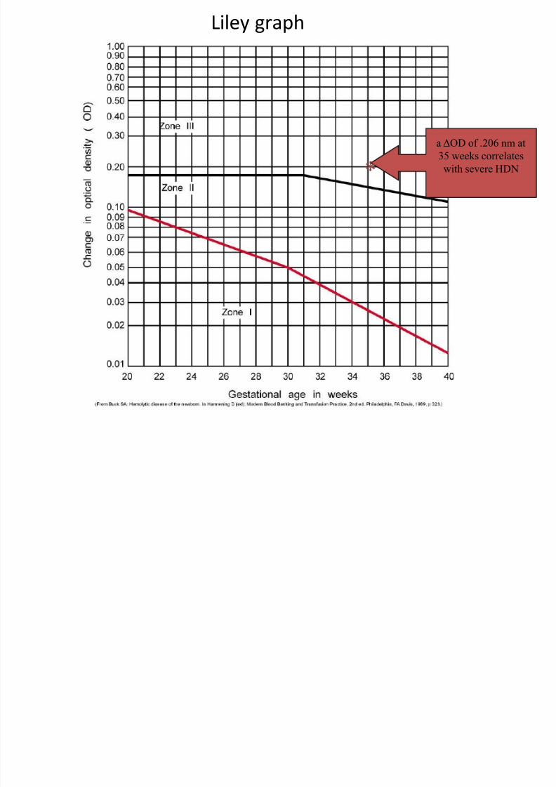
Liley graph
• The ΔOD is plotted on the Liley graph
according to gestational age
• Three zones estimate the severity of HDN
– Lower: mildly or unaffected fetus (Zone 1)
– Midzone: moderate HDN, repeat testing (Zone 2)
– Upper: severe HDN and fetal death (Zone 3)
Liley graph
7/28/2019 Hematological Disorders of Newborn
http://slidepdf.com/reader/full/hematological-disorders-of-newborn 56/98
Liley graph
a ΔOD of .206 nm at
35 weeks correlates
with severe HDN*

7/28/2019 Hematological Disorders of Newborn
http://slidepdf.com/reader/full/hematological-disorders-of-newborn 57/98
Management of the rhesus immunized infant
• The infant should be assessed for
maturity,pallor, jaundice,ascites…
• Placenta send for pathology examination.
• Cord blood taken for HB.,DCT, Hb. And platelet
and total Serum Bilirubin.

7/28/2019 Hematological Disorders of Newborn
http://slidepdf.com/reader/full/hematological-disorders-of-newborn 58/98
Indications for immediate/early exchange transfusion
• Cord haemoglobin < 8 g/dl.
• Hydrops fetalis
• Cord bilirubin > 85umol/L
• Rapidly rising bilirubin crossing the level for
exchange transfusion.
•Strong positive Coombs’ test.

7/28/2019 Hematological Disorders of Newborn
http://slidepdf.com/reader/full/hematological-disorders-of-newborn 59/98
Complications of rhesus incompatibility
• Kernicterus and bilirubin encephalopathy.
• Hyaline membrane disease.
• Beta cell hyperplasia leading to hypoglycaemia.
• Hypoalbuminaemia and lung oedema.
• Thrombocytopenia and DIC.
• Inspissated bile syndrome.
• Complications of exchange transfusion.• Anaemia: Folic acid, and tansfusions.

7/28/2019 Hematological Disorders of Newborn
http://slidepdf.com/reader/full/hematological-disorders-of-newborn 60/98
ABO HDN

7/28/2019 Hematological Disorders of Newborn
http://slidepdf.com/reader/full/hematological-disorders-of-newborn 61/98
ABO HDN
• ABO incompatibilities are the most common
cause of HDN but are less severe
– About 1 in 5 pregnancies are ABO-incompatible
– 65% of HDN are due to ABO incompatibility
• Usually, the mother is type O and the child has
the A or B antigen…Why?
– Group O individuals have a high titer of IgG anti-A,B in addition to having IgM anti-A and anti-B
7/28/2019 Hematological Disorders of Newborn
http://slidepdf.com/reader/full/hematological-disorders-of-newborn 62/98
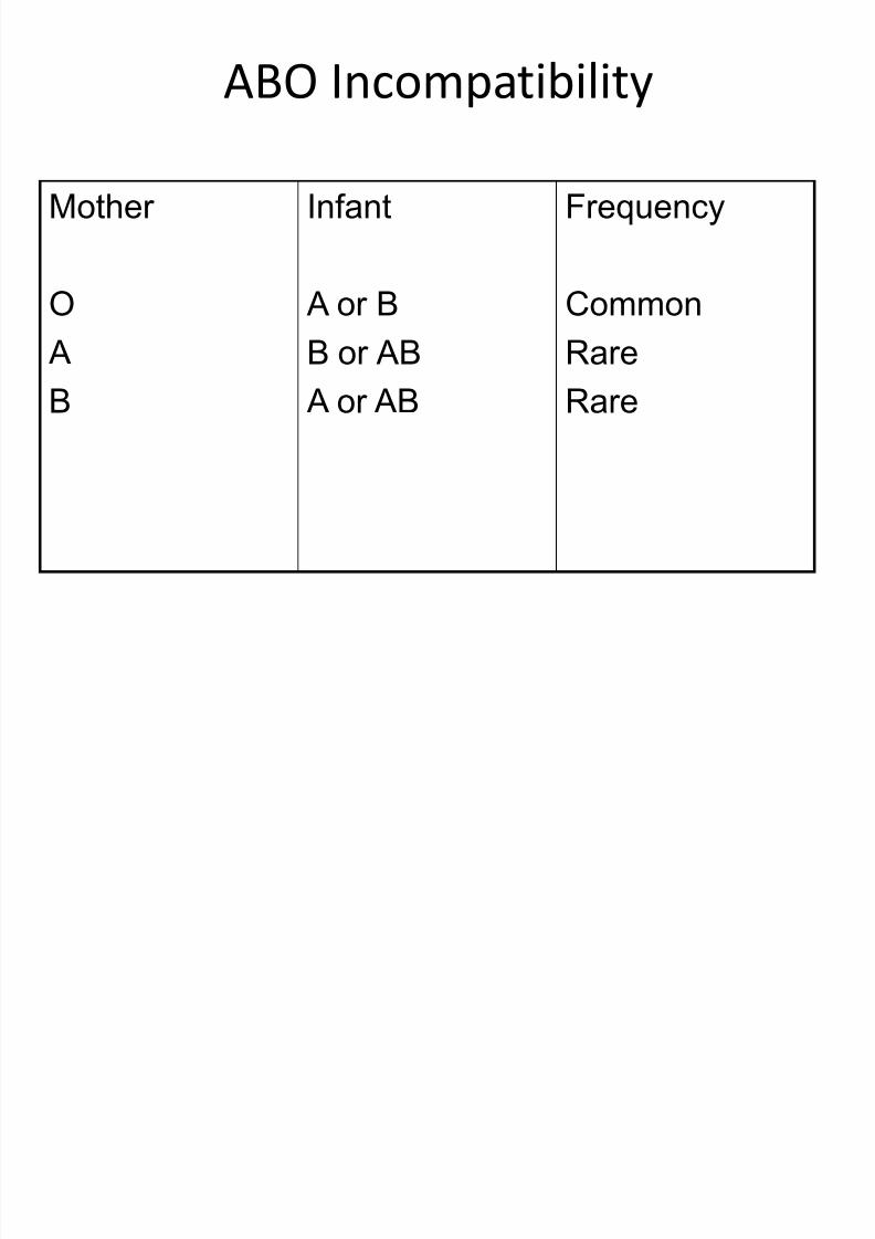
ABO Incompatibility
Mother
O A
B
Infant
A or BB or AB
A or AB
Frequency
CommonRare
Rare
7/28/2019 Hematological Disorders of Newborn
http://slidepdf.com/reader/full/hematological-disorders-of-newborn 63/98
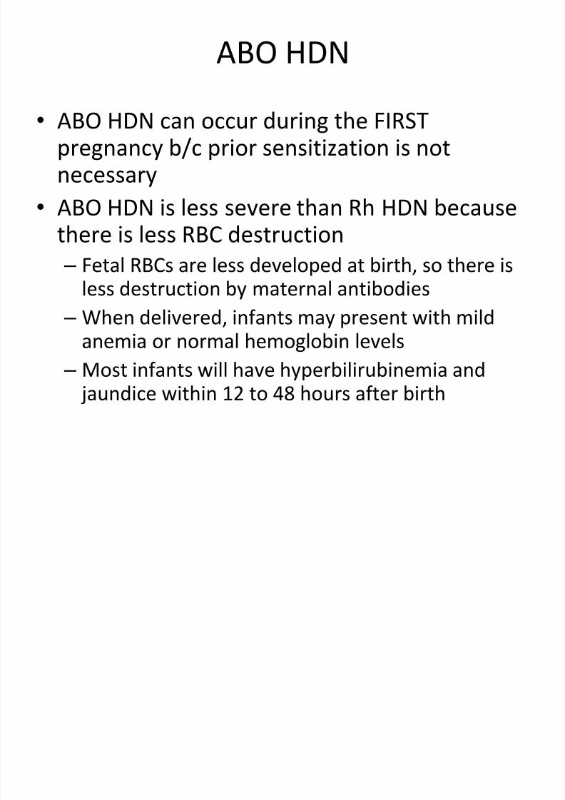
ABO HDN
• ABO HDN can occur during the FIRSTpregnancy b/c prior sensitization is notnecessary
• ABO HDN is less severe than Rh HDN becausethere is less RBC destruction
– Fetal RBCs are less developed at birth, so there isless destruction by maternal antibodies
– When delivered, infants may present with mildanemia or normal hemoglobin levels
– Most infants will have hyperbilirubinemia and
jaundice within 12 to 48 hours after birth
7/28/2019 Hematological Disorders of Newborn
http://slidepdf.com/reader/full/hematological-disorders-of-newborn 64/98
Clinical features
• Jaundice first 24 hours of life
• No hepatosplenomegaly
• Kernicterus unusual.
• Hydrops occasionally being reported.
• Late anemia is seldom a problem.

7/28/2019 Hematological Disorders of Newborn
http://slidepdf.com/reader/full/hematological-disorders-of-newborn 65/98
Diagnosis of ABO HDN
• Infant presents with jaundice 12-48 hrs after
birth
• Testing done after birth on cord blood
samples:
– Sample is washed 3x to remove Wharton’s jelly
– Anticoagulated EDTA tube (purple or pink)
– ABO, Rh and DAT performed
– Most cases will have a positive DAT
• If DAT positive, perform elution to ID antibody

7/28/2019 Hematological Disorders of Newborn
http://slidepdf.com/reader/full/hematological-disorders-of-newborn 66/98
Investigations:
• Suspected in mother who is blood group O and
infant either group A or less commonly group B.
• DCT usually negative but indirect coombs test may
be positive.• Blood smear may show features of haemolysis.
• Immune anti-A or anti-B may be elicated from fetal
RBCs or cord blood.

7/28/2019 Hematological Disorders of Newborn
http://slidepdf.com/reader/full/hematological-disorders-of-newborn 67/98
Treatment of ABO HDN
• Only about 10% require therapy
• Phototherapy is sufficient
• Rarely is exchange transfusion needed
• Phototherapy is exposure to artificial orsunlight to reduce jaundice
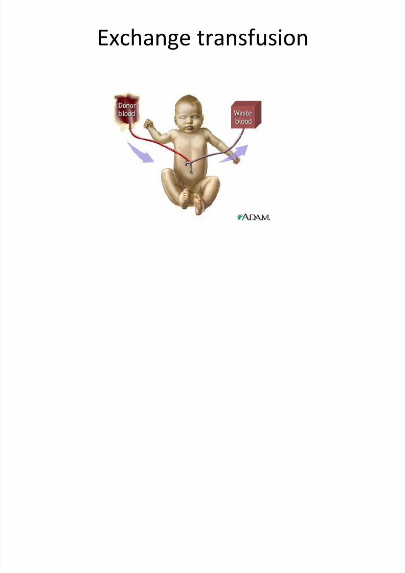
• Exchange transfusion involves removingnewborn’s RBCs and replacing them withnormal fresh donor cells
7/28/2019 Hematological Disorders of Newborn
http://slidepdf.com/reader/full/hematological-disorders-of-newborn 68/98
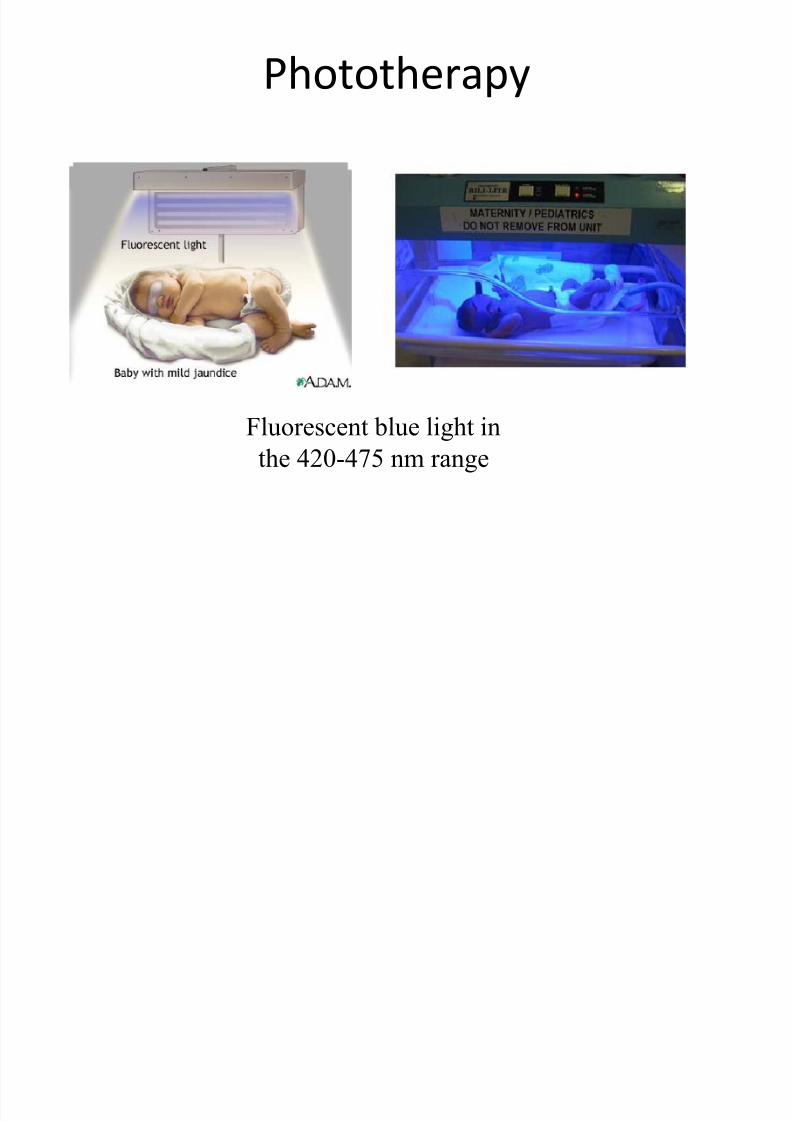
Phototherapy
Fluorescent blue light inthe 420-475 nm range
7/28/2019 Hematological Disorders of Newborn
http://slidepdf.com/reader/full/hematological-disorders-of-newborn 69/98
Exchange transfusion

7/28/2019 Hematological Disorders of Newborn
http://slidepdf.com/reader/full/hematological-disorders-of-newborn 70/98
Non-immune hemolysis – G6PD
Glucose 6 phosphate dehydrogenase
7/28/2019 Hematological Disorders of Newborn
http://slidepdf.com/reader/full/hematological-disorders-of-newborn 71/98
Glucose-6-phosphate dehydrogenase
deficiency
• X-linked recessive thus occur in male.
• Heterozygote female may manifest mild
disease.
• Due to deficiency of enzyme within RBC which
render cells more susceptible to haemolysis.
• Many variants to conditions.
Drugs that may cause haemolysis in infants

7/28/2019 Hematological Disorders of Newborn
http://slidepdf.com/reader/full/hematological-disorders-of-newborn 72/98
Drugs that may cause haemolysis in infants
with G6PD deficiency
• Antimalarials - primaquine, quinine
• Nitrofurantion
• Sulphonamides
• Nalidixic acid
• Naphthalene
• Chloramphanicol• Vitamin K (large doses)
• Fava beans (kachang parang/itek)
7/28/2019 Hematological Disorders of Newborn
http://slidepdf.com/reader/full/hematological-disorders-of-newborn 73/98
Others condition causing haemolysis
• Pyruvate kinase deficiency
• Hereditary spherocytosis
• Alpha Thallasaemia
7/28/2019 Hematological Disorders of Newborn
http://slidepdf.com/reader/full/hematological-disorders-of-newborn 74/98
Hydrops Fetalis
• This term is used to describe an infant who
shows severe and generalized oedema, ascites
and pleural effusions at birth.

7/28/2019 Hematological Disorders of Newborn
http://slidepdf.com/reader/full/hematological-disorders-of-newborn 75/98

7/28/2019 Hematological Disorders of Newborn
http://slidepdf.com/reader/full/hematological-disorders-of-newborn 76/98
Causes of Hydrops Fetalis
• Immune: severe haemolytic disease of
newborn.
• Non-immune

7/28/2019 Hematological Disorders of Newborn
http://slidepdf.com/reader/full/hematological-disorders-of-newborn 77/98
Causes of Hydrops Fetalis
• Non-immune:
– Fetal onset SVT
– Severe anemia in utero
– Chronic twin-twin transfusion
– Severe CHD
– Premature closure of foramen ovale and ductus
arteriosus – Congenital Nephrotic syndrome.
f

7/28/2019 Hematological Disorders of Newborn
http://slidepdf.com/reader/full/hematological-disorders-of-newborn 78/98
Causes of Hydrops Fetalis
• Non-immune:
– Congenital infections
– Congenital malformations; obstructive uropathy
– Parvovirus B19
– Idiopathic: 50% of cases.
f d l

7/28/2019 Hematological Disorders of Newborn
http://slidepdf.com/reader/full/hematological-disorders-of-newborn 79/98
Management of Hydrops Fetalis
• May include:
– Haemoglobin electrophoresis.
– Kleihauer’s test.
– Coombs’ test and full blood count.
– TORCH investigations.
– Total serum proteins and serum albumin

7/28/2019 Hematological Disorders of Newborn
http://slidepdf.com/reader/full/hematological-disorders-of-newborn 80/98
Polycythaemia
l h i

7/28/2019 Hematological Disorders of Newborn
http://slidepdf.com/reader/full/hematological-disorders-of-newborn 81/98
Polycythaemia
• Common and is defined as a venous haemotocrit of 65% or more (= Hb 22g/dl), during the first week.
• Not equal to hyperviscous.
• Blood viscosity depends largely on packed cellvolume.
• Viscosity much more greater in small vessels thanlarge vessels.
• Daignosed on free flowing venous specimen and notfrom heel prick sample.

7/28/2019 Hematological Disorders of Newborn
http://slidepdf.com/reader/full/hematological-disorders-of-newborn 82/98
Causes of neonatal polycythaemia
Chronic intrauterine hypoxia:
SGA infants
Postmaturity
Excessive tranfusion of blood
Delayed clamping
Twin twin transfusion
Infants of Diabetic mothersDown’s syndrome
Neonatal thyrotoxicosis
CAH
Cli i l f

7/28/2019 Hematological Disorders of Newborn
http://slidepdf.com/reader/full/hematological-disorders-of-newborn 83/98
Clinical features
• Infants look plethoric.• Neurological
– Jitteriness,apnoea,convulsions.
• Cardiovascular
– Congestive heart failure,pulmonary hypertension withRight to left shunting and cyanosis.
• Gastrointestinal - NEC
• Renal – renal vein thrombosis• Others: hypoglycaemia, thrombocytopenia,
jaundice,hypocalcemia.
M

7/28/2019 Hematological Disorders of Newborn
http://slidepdf.com/reader/full/hematological-disorders-of-newborn 84/98
Management
• Anticipation
• At risk infants haemotocrit should be measured.
• Infants without symptoms but haematocrit > 70%
should have a dilutional exchange transfusion.• Symptomatics infants with venous PCV of 65-70%
may require dilutional exchange transfusion.
Dil ti l h t f i

7/28/2019 Hematological Disorders of Newborn
http://slidepdf.com/reader/full/hematological-disorders-of-newborn 85/98
Dilutional exchange transfusion
• Performed using normal saline / plasma andaimed at reducing the haematocrit to about
50% using formula:
• Volume = actual Hct – desired Hct / actual Hctx weight (kg) x blood volume.

7/28/2019 Hematological Disorders of Newborn
http://slidepdf.com/reader/full/hematological-disorders-of-newborn 86/98
Bleeding and Coagulation disorders

7/28/2019 Hematological Disorders of Newborn
http://slidepdf.com/reader/full/hematological-disorders-of-newborn 87/98
Bleeding and coagulation disorders
• May be due:
– Thrombocytopenia
– Deficiency of clotting factors
– Abnormal capillaries
– Combination of all
Cli i l f t

7/28/2019 Hematological Disorders of Newborn
http://slidepdf.com/reader/full/hematological-disorders-of-newborn 88/98
Clinical features
• Bleeding from umbilical or venepuncture sites.
• Bruising, purpura or petechial haemorrhages.
C f th b t i
7/28/2019 Hematological Disorders of Newborn
http://slidepdf.com/reader/full/hematological-disorders-of-newborn 89/98
Causes of thrombocytopenia
• Infection – Bacterial infection
– TORCH infections
• Isoimmune
• Maternal disease – ITP
– SLE
– Drug induced
• Neonatal drug exposure
– Thiazide diuretics – Quinine
– Sulphonamides
I i th b t i
7/28/2019 Hematological Disorders of Newborn
http://slidepdf.com/reader/full/hematological-disorders-of-newborn 90/98
Isoimmune thrombocytopenia
• Rare condition. Analogous to Rh isoimmunization.• Transfusion of fetal A1 antigen-positive platelets into
maternal circulation may produce maternal IgGantibodies if the mother is platelet A1 antigen
negative.• Mother has normal platelet count.
• Newborn may have severe thrombocytopenia but istransient.
• Platelets transfusion or exchange transfusion may beneeded in severe cases.
M t l ITP
7/28/2019 Hematological Disorders of Newborn
http://slidepdf.com/reader/full/hematological-disorders-of-newborn 91/98
Maternal ITP
• Due to transplacental maternal antibodies.• Mother may have thrombocytopenia and the lower
mum platelets count the more severe affected theinfant is.
• Steroids and IVIG had been advocated for treatment.
• Blood transfusion or platelet may be required if PC <20,000.
• Intracerebral bleed may occur before onset of labour.
Causes of thrombocytopenia

7/28/2019 Hematological Disorders of Newborn
http://slidepdf.com/reader/full/hematological-disorders-of-newborn 92/98
Causes of thrombocytopenia
• Neonatal drug exposure – Thiazide diuretics
– Quinine
– Sulphonamides
• DIC• TAR syndrome
• Kasabach Merritt syndrome
• Fanconi’s anaemia
• Leukaemia• Pancytopenias
Di i t d I t l C l ti (DIC)
7/28/2019 Hematological Disorders of Newborn
http://slidepdf.com/reader/full/hematological-disorders-of-newborn 93/98
Disseminated Intravascular Coagulation (DIC)
• DIC an acquired coagulation disordercharaterized by intravascular consumption of
platelets and clotting factors II,V,VIII and
fibrinogen.• Intravascular coagulation results from
deposition of thrombi in small vessels and
consumption of clotting factors leading tohaemorrhage.
Neonatal conditions and DIC
7/28/2019 Hematological Disorders of Newborn
http://slidepdf.com/reader/full/hematological-disorders-of-newborn 94/98
Neonatal conditions and DIC
• Septicaemia
• Severe shock
• Severe perinatal asphyxia
• IRDS in VLBW infants• Severe Rhesus disease
• TORCH infections
• Hypothermia• Maternal DIC with transplacental effect.
Investigations for DIC

7/28/2019 Hematological Disorders of Newborn
http://slidepdf.com/reader/full/hematological-disorders-of-newborn 95/98
Investigations for DIC
• Blood film shows haemolysis with fragmentedand distorted red cells.
• Thrombocytopenia.
• Prolonged PT,PTT and thrombin time.
• Low fibrinogen.
• Increased FDPs.
Presence of three or more makes DIC likely.
Treatment

7/28/2019 Hematological Disorders of Newborn
http://slidepdf.com/reader/full/hematological-disorders-of-newborn 96/98
Treatment
• Very complex
• Treat underlying disease process
• Treatment of the haematological abnormality.
Others coagulation disorders
7/28/2019 Hematological Disorders of Newborn
http://slidepdf.com/reader/full/hematological-disorders-of-newborn 97/98
Others coagulation disorders
• Immaturity of clotting factors secondary toliver immaturity.
• Inherited disorders of coagulation
– Haemophilia A (Factor VIII def)
– Chrismas disease (Factor IX def)

7/28/2019 Hematological Disorders of Newborn
http://slidepdf.com/reader/full/hematological-disorders-of-newborn 98/98