hematology ii - nursing411.orgnursing411.org/army/md0857.pdf · hematology ii introduction this...
TRANSCRIPT

U.S. ARMY MEDICAL DEPARTMENT CENTER AND SCHOOL FORT SAM HOUSTON, TEXAS 78234-6100
HEMATOLOGY II
SUBCOURSE MD0857 EDITION 200
DEVELOPMENT
This subcourse is approved for resident and correspondence course instruction. It reflects the current thought of the Academy of Health Sciences and conforms to printed Department of the Army doctrine as closely as currently possible. Development and progress render such doctrine continuously subject to change.
ADMINISTRATION
For comments or questions regarding enrollment, student records, or shipments, contact the Nonresident Instruction Section at DSN 471-5877, commercial (210) 221-5877, toll-free 1-800-344-2380; fax: 210-221-4012 or DSN 471-4012, e-mail [email protected], or write to: COMMANDER AMEDDC&S ATTN MCCS HSN 2105 11TH STREET SUITE 4192 FORT SAM HOUSTON TX 78234-5064 Approved students whose enrollments remain in good standing may apply to the Nonresident Instruction Section for subsequent courses by telephone, letter, or e-mail. Be sure your social security number is on all correspondence sent to the Academy of Health Sciences.
CLARIFICATION OF TRAINING LITERATURE TERMINOLOGY When used in this publication, words such as "he," "him," "his," and "men" are intended to include both the masculine and feminine genders, unless specifically stated otherwise or when obvious in context. .
USE OF PROPRIETARY NAMES
The initial letters of the names of some products are capitalized in this subcourse. Such names are proprietary names, that is, brand names or trademarks. Proprietary names have been used in this subcourse only to make it a more effective learning aid. The use of any name, proprietary or otherwise, should not be interpreted as an endorsement, deprecation, or criticism of a product; nor should such use be considered to interpret the validity of proprietary rights in a name, whether it is registered or not.
MD0857 i
TABLE OF CONTENTS Lesson Paragraphs INTRODUCTION 1 DIFFERENTIAL LEUKOCYTE COUNT AND OTHER PROCEDURES Section I. Differential Leukocyte Count ..................................... 1-1--1-3 Section II. Erythrocyte Indices and Fragility Tests...................... 1-4--1-6 Section III. Demonstration of L.E. Cells....................................... 1-7--1-8 Section IV. Special Stains............................................................ 1-9--1-12 Exercises 2 BLOOD COAGULATION Section I. Introduction................................................................ 2-1--2-2 Section II. Coagulation System .................................................. 2-3--2-5 Section III. The Coagulation Mechanism..................................... 2-6--2-8 Section IV. Coagulation Studies .................................................. 2-9--2-30 Exercises 3 ANEMIA Section I. Diagnosing Anemia ................................................... 3-1--3-3 Section II. Laboratory Assessment of Anemia............................ 3-4--3-7 Section III. Anemia as Categorized by Morphology..................... 3-8--3-11 Exercises 4 LEUKEMIA Section I. Morphologic Disorders of the Leukocyte ................... 4-1--4-4 Section II. Classification of Leukemias ....................................... 4-5--4-16 Section III. Diagnostic Special Stains .......................................... 4-17 Exercises APPENDIX A, Glossary of Terms
MD0857 ii
CORRESPONDENCE COURSE OF THE U.S. ARMY MEDICAL DEPARTMENT CENTER AND SCHOOL
SUBCOURSE MD0857
HEMATOLOGY II
INTRODUCTION
This subcourse is concerned with the blood tests performed in the hematology section of the laboratory. The purpose of these tests is to aid the physician in diagnosis. Thus, these tests are important and often essential to the health and life of the patient. Thorough study of this subcourse should enable you to better fulfill your role in health care.
ACKNOWLEDGEMENT
Portions of this subcourse are extracted from TM 8-227-4, Clinical Laboratory Procedure--Hematology, dated 5 December 1973; from Brown, Barbara, Hematology Principles and Procedures, 4th ed., Lea and Febiger; and from Operator's Manual for QBC II Centrifugal Hematology System, Clay Division of Becton Dickinson Company. Written consent of the copyright owner has been obtained. Under no circumstances will this material be sold, commercially used, or copied.
Subcourse Components: The subcourse instructional material consists of four lessons and an appendix as follows:
Lesson 1, Differential Leukocytes Count and Other Procedures Lesson 2, Blood Coagulation. Lesson 3, Anemia. Lesson 4, Leukemia Appendix A, Glossary of terms Here are some suggestions that may be helpful to you in completing this subcourse: --Read and study each lesson carefully. --Complete the subcourse lesson by lesson. After completing each lesson, work the exercises at the end of the lesson
MD0857 iii
--After completing each set of lesson exercises, compare your answers with those on the solution sheet that follows the exercises. If you have answered an exercise incorrectly, check the reference cited after the answer on the solution sheet to determine why your response was not the correct one. Credit Awarded: Upon successful completion of the examination for this subcourse, you will be awarded 8 credit hours. To receive credit hours, you must be officially enrolled and complete an examination furnished by the Nonresident Instruction Section at Fort Sam Houston, Texas. You can enroll by going to the web site http://atrrs.army.mil and enrolling under "Self Development" (School Code 555).
MD0857 1-1
LESSON ASSIGNMENT LESSON 1 Differential Leukocyte Count and Other Procedures. TEXT ASSIGNMENT Paragraphs 1-1 through 1-12. LESSON OBJECTIVES After completing this lesson, you should be able to: 1-1. Select the statement that best describes the appropriate materials and procedures used to specially stain and examine blood smears. 1-2. Select the correct principles and steps used in the calculation of erythrocyte indices and in performing the various erythrocyte fragility tests.
1-3. Select the statement that best describes the principles used in the demonstration of LE cells.
1-4. Select the materials and procedures used in performing a screening test for G-6-PD deficiency. SUGGESTION After completing the assignment, complete the exercises of this lesson. These exercises will help you to achieve the lesson objectives.
MD0857 1-2
LESSON 1
DIFFERENTIAL LEUKOCYTE COUNT AND OTHER PROCEDURES
Section I. DIFFERENTIAL LEUKOCYTE COUNT 1-1. INTRODUCTION a. The critical examination of a blood smear includes the following: quantitative and qualitative study of platelets, differential count quantitating the three types of leukocytes (granulocytes, lymphocytes, monocytes), and morphological characteristics of erythrocytes and leukocytes. Staining the blood smears is a critical part of the examination. The procedure for staining is described in lesson 3 of Subcourse MD0853. To accurately perform the differential count it is necessary for a technician to recognize all the characteristics of normal blood cells. This includes normal biological variation. For instance, not every lymphocyte is exactly the same size, nor do all lymphocytes have exactly the same number of azurophilic granules. b. Certain morphological and histochemical characteristics are utilized to differentiate blood cells. A review of the significant features promotes a better understanding of blood differentials. Cellular characteristics such as relative size, shape, cytoplasmic granulation, nuclear- cytoplasmic ratio, nuclear configuration, chromatin or nucleoli are very important. These features are discussed in Subcourse MD0853, lesson 4. c. Experience is the foremost teacher in hematology. It is readily acquired in a busy hematology section where the opportunity for differential analysis occurs frequently. Experience can be diversified and interesting if proficiency slides and material from cases of confirmed diagnoses are maintained as study sets. This study material should be available to all technicians in the laboratory. d. All routine blood smears should be kept until the physicians have reviewed the differential reports. A 1-week period is usually adequate. Occasionally, a review of a specific problem slide results in findings that were not originally apparent and reinforces confidence in the laboratory by the medical staff. This practice also adds to the experience and proficiency of the technician. 1-2. EXAMINATION OF PERIPHERAL BLOOD SMEARS a. Principle. The stained blood smear permits the study of the appearance and the identification of the different kinds of leukocytes, and the appearance of erythrocytes and thrombocytes (blood platelets).
MD0857 1-3
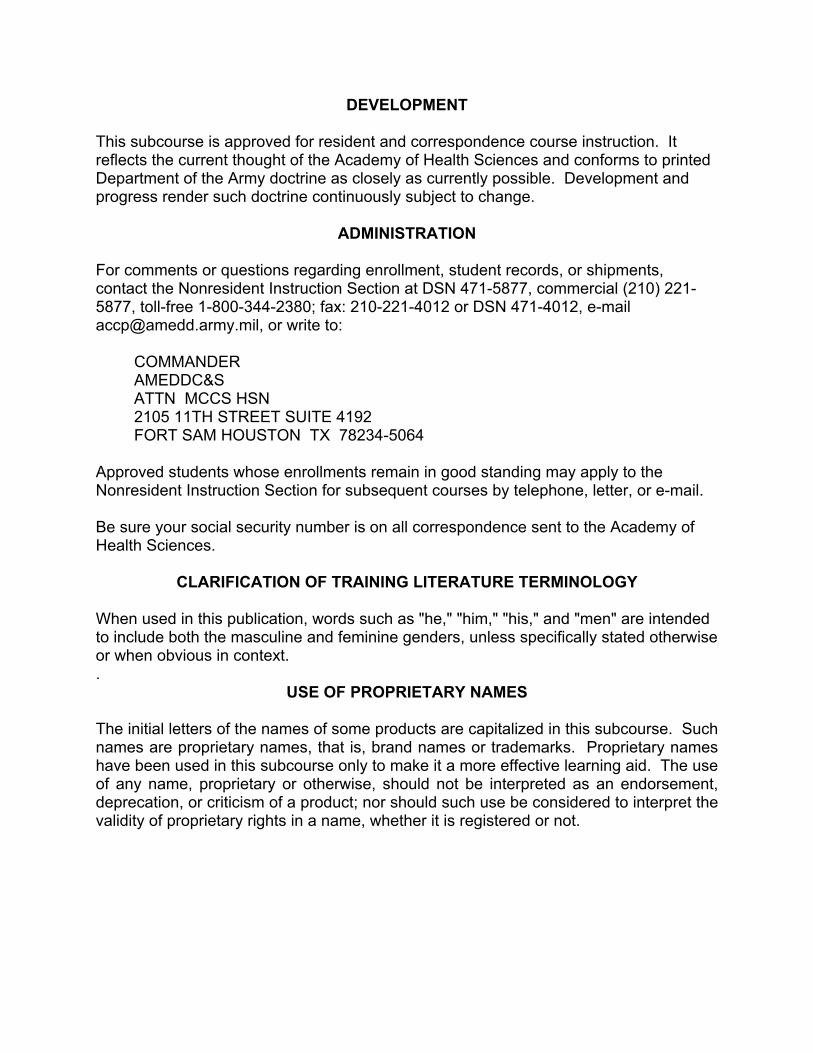
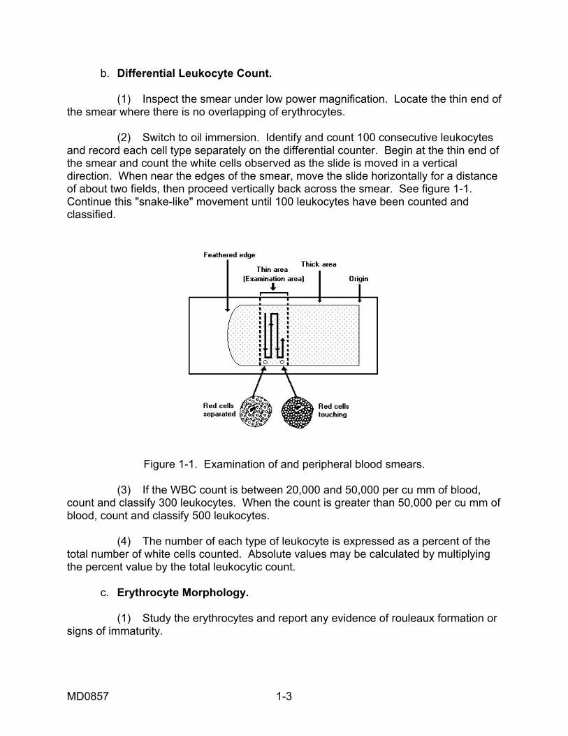
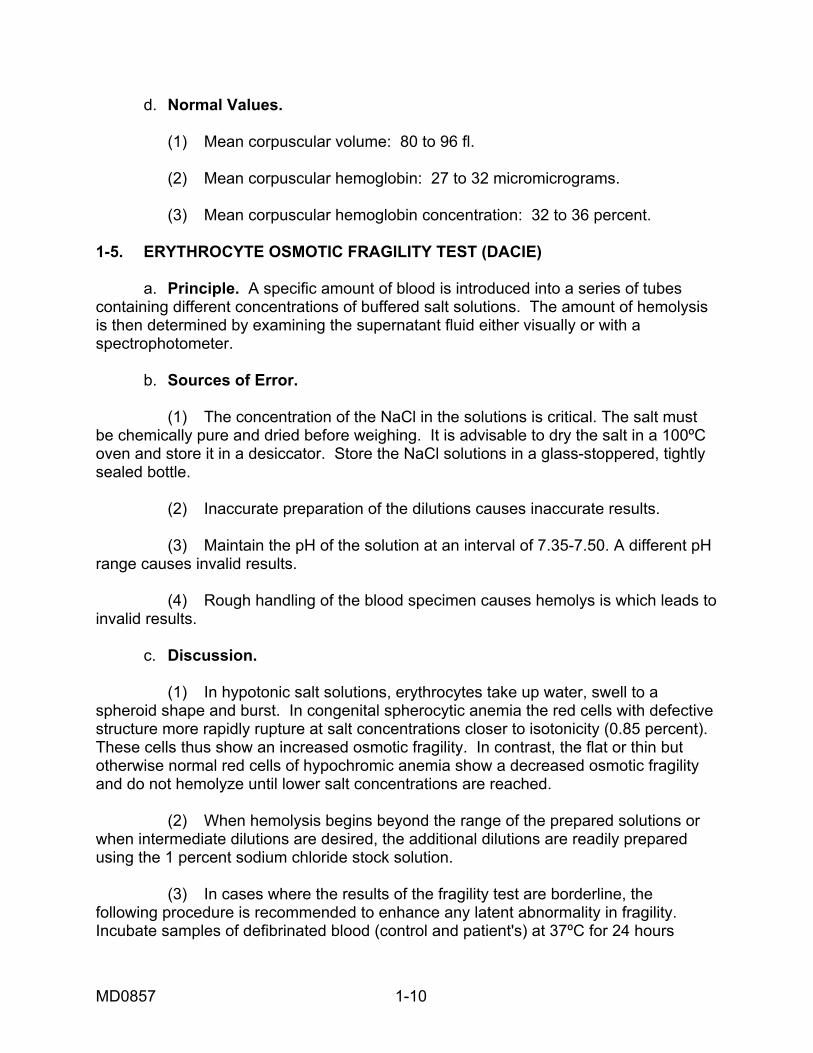
b. Differential Leukocyte Count. (1) Inspect the smear under low power magnification. Locate the thin end of the smear where there is no overlapping of erythrocytes. (2) Switch to oil immersion. Identify and count 100 consecutive leukocytes and record each cell type separately on the differential counter. Begin at the thin end of the smear and count the white cells observed as the slide is moved in a vertical direction. When near the edges of the smear, move the slide horizontally for a distance of about two fields, then proceed vertically back across the smear. See figure 1-1. Continue this "snake-like" movement until 100 leukocytes have been counted and classified.
Figure 1-1. Examination of and peripheral blood smears. (3) If the WBC count is between 20,000 and 50,000 per cu mm of blood, count and classify 300 leukocytes. When the count is greater than 50,000 per cu mm of blood, count and classify 500 leukocytes. (4) The number of each type of leukocyte is expressed as a percent of the total number of white cells counted. Absolute values may be calculated by multiplying the percent value by the total leukocytic count. c. Erythrocyte Morphology. (1) Study the erythrocytes and report any evidence of rouleaux formation or signs of immaturity.
MD0857 1-4
(2) Report the erythrocyte morphology with reference to size, shape, staining characteristics, and inclusions. Report the degree of the specific morphological characteristic (for example, moderate hypochromia). (3) If nucleated erythrocytes are found (usually these are metarubricytes), report the number per 100 leukocytes counted. d. Qualitative Platelet Evaluation. (1) Observe the thrombocytes in several oil immersion fields to obtain a rough estimation of their numbers (normal, increased, or decreased). Normal is an average of 8 to 10 per oil immersion field. (2) Note any abnormality in morphology (giant platelets, etc.) If the thrombocytes appear to be significantly decreased, a thrombocyte count and/or a clot retraction test may be indicated. e. Discussion. (1) All abnormal white cells (for example, immature, hypersegmented, toxic, atypical lymphocytes, etc.) should be classified or described and reported in percent, separately. Cells that are ruptured, fragmented, or degenerated are not included in the differential count, but should be noted separately and reported as the number seen per 100 leukocytes. (2) In view of the gradual transition from the metamyelocyte to the banded neutrophil and then to the segmented neutrophil, exact classification is sometimes difficult. In such cases, classify the cell according to the more mature form. (3) Size considerations in differentiating blood cells require a defined linear standard. The micron (.001 mm) is usually used in reference to microscopic dimensions. Ocular micrometers are available through Federal medical supply channels and are easily calibrated, using a hemacytometer that has standardized dimensions. In routine screening of blood smears, an experienced technician relates the size of a normocytic erythrocyte (seven to eight microns) to the size of the white cell to be differentiated, since erythrocytes are usually present throughout the microscopic field. Finally, it should be understood that personal visual discrimination is an inaccurate gauge of linear measure. Some reference measure should be employed. (4) The shape of blood cells often depends upon the smear and staining technique. Variations that have no clinical significance occur from physical and chemical distortions that result from technical error. These variations are avoided with careful technique. Each routine smear should be scanned initially to evaluate the smear and stain quality before differential analysis.
MD0857 1-5
(5) Cytoplasmic granulation--neutrophilic, basophilic, or eosinophilic--is an important morphological observation. Differences in granule color in Wright-stained preparations are caused by the variable dye affinity of specific granules. The intensity of colors and the relative blueness or redness of the erythrocytes is used to evaluate the quality of the stain. The familiar basophilic (blue), eosinophilic (red), and neutrophilic (pink) granules are quite obvious in routine blood smears. The presence, absence, type, and quantity of granules are characteristic attributes used to differentiate leukocytes. (6) The size ratio of nucleus to cytoplasm (N:C) is a differentiating characteristic. For instance, a cell with a nuclear mass equal to the cytoplasmic mass would have an N:C ratio of 1:1. The total cell mass is usually greater in the more immature cells and decreases as the cell matures. The nuclear mass usually decreases also as the cell matures. Of course, lymphocytes are the exception to this generality. (7) The nuclear configurations of leukocytes help distinguish these cells. Round, oval indented, band, or segmented are terms used to describe variations in shape. These normal configurations can be distorted by physical and chemical factors. Some of the leukocytes are so fragile that in thick blood smears their normal configuration may be distorted by the pressure of erythrocytes forced against them. These artifacts should be recognized as such in an intelligent evaluation of blood differentials. (8) In addition to nuclear shape and size, the internal nuclear morphology shows differential inclusions. The chromatin appears finely reticulated in some cells, or as a coarse network, or even clumped, in others. The parachromatin, a lighter staining material beside the chromatin, is scant or abundant. The appearance of the chromatin and the quality of parachromatin are utilized to differentiate blood cells. The presence, absence, and number of nucleoli in the nucleus are the most distinctive characteristics of immature nuclei in blood cells. (9) All abnormal blood smears should be examined by another trained person for confirmation of the results. f. Normal Differential Values. (1) Banded neutrophil: 0 to 6 percent. (2) Segmented neutrophil: 40 to 75 percent. (3) Eosinophils: 1 to 7 percent. (4) Basophils: 0 to 2 percent.
MD0857 1-6
(5) Lymphocytes: 22 to 40 percent. (6) Monocytes: 1 to 10 percent. 1-3. EXAMINATION OF BLOOD MARROW SMEARS a. Principle. Nucleated blood cells are counted and classified from a bone marrow smear stained with a Romanowsky stain containing both Wright and Giemsa stains. b. Procedure. (1) Using oil immersion magnification, count and classify 300 to 500 nucleated cells. (2) Classify all blood cells according to cell type and various stages of maturation. (3) Calculate myeloid-erythroid ratio by dividing the number of nucleated erythrocytes into the number of granulocytic (myeloid) cells. (4) A peripheral blood evaluation usually accompanies the bone marrow reports. This evaluation usually includes an erythrocyte count, leukocyte count, differential count, hemoglobin, hematocrit, and a reticulocyte count. c. Discussion. (1) The differential cell count on a bone marrow smear is carried out by a hematologist, pathologist, or trained technician. (2) Since interpretation of findings in bone marrow examinations is very difficult, it is of utmost importance that the smears and stains are carefully prepared using scrupulously clean equipment. (3) The laboratory technician is usually responsible for preparing bone marrow smears, staining the smears, checking the quality of the stained smear, and coverslipping the slides. d. Normal Values. (1) Leukocytes. (a) Myeloblast: 0 to 1 percent. (b) Promyelocytes: 2 to 5 percent.
MD0857 1-7
(c) Neutrophilic myelocytes: 5 to 19 percent. (d) Neutrophilic metamyelocytes: 13 to 22 percent. (e) Neutrophilic bands: 17 to 33 percent. (f) Neutrophilic segmented cells: 3 to 11 percent. (g) Eosinophilic cells: 0 to 3 percent. (h) Basophilic cells: 0 to 1 percent. (i) Lymphocytes: 5 to 15 percent. (j) Monocytes: 0 to 2 percent. (k) Plasmocytes: 0 to 1 percent. (2) Erythrocytes. (a) Rubriblasts: 0 to 1 percent. (b) Prorubricytes: 1 to 4 percent. (c) Rubricytes: 3 to 10 percent. (d) Metarubricytes: 5 to 25 percent. (3) Megakaryocytes. 0 to 3 percent. (4) Myeloid-Erythroid Ratio (M:E). 3-4:1.
Section II. ERYTHROCYTE INDICES AND FRAGILITY TESTS 1-4. ERYTHROCYTE INDICES a. Principle. By using accurately determined red blood cell counts, hematocrits, and hemoglobin values, the size and hemoglobin content of the average red cell in a given blood sample is calculated. The values obtained are the erythrocyte indices which aid in the classification and study of anemias. They consist of the mean corpuscular volume (MCV), mean corpuscular hemoglobin (MCH), and mean corpuscular hemoglobin concentration (MCHC).

MD0857 1-8
b. Calculation of Erythrocyte Indices. (1) Mean corpuscular volume (MCV)--The average volume of the individual red blood cell. Femtoliter (fl) or 10-15 liter = 1 fl. Hematocrit (percent) x 10 --------------------------------------- = fl Red cell count (x 10/L) Example: Hematocrit 45 percent Red count 5,000,000 per cu mm 45 X 10 ----------- = 90 fl 5.0 (2) Mean corpuscular hemoglobin (MCH)--The average weight of hemoglobin of the individual red cell. Hemoglobin (x 10 gm/ dL) ------------------------------------------ = micromicrograms Red cell count (x 10 /L) Example: Hemoglobin 15 gm/dL Red count 5,000,000 per cu mm 15 x 10 ------------ = 30 micromicrograms (normal) 5.0 (3) Mean corpuscular hemoglobin concentration (MCHC)--The percent of hemoglobin in the average red cell. Hemoglobin (x 10gm/ dL) x 100 ------------------------------------------- = percent Hematocrit (percent )
MD0857 1-9
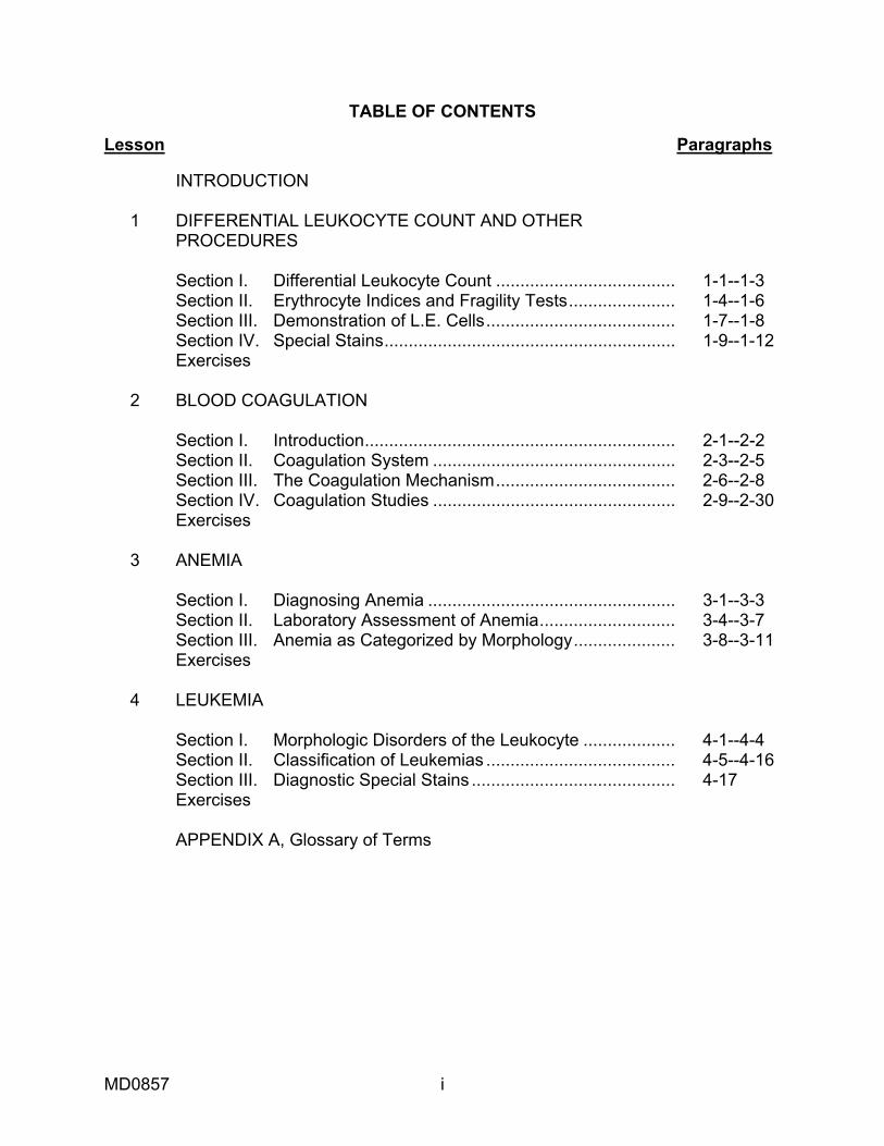
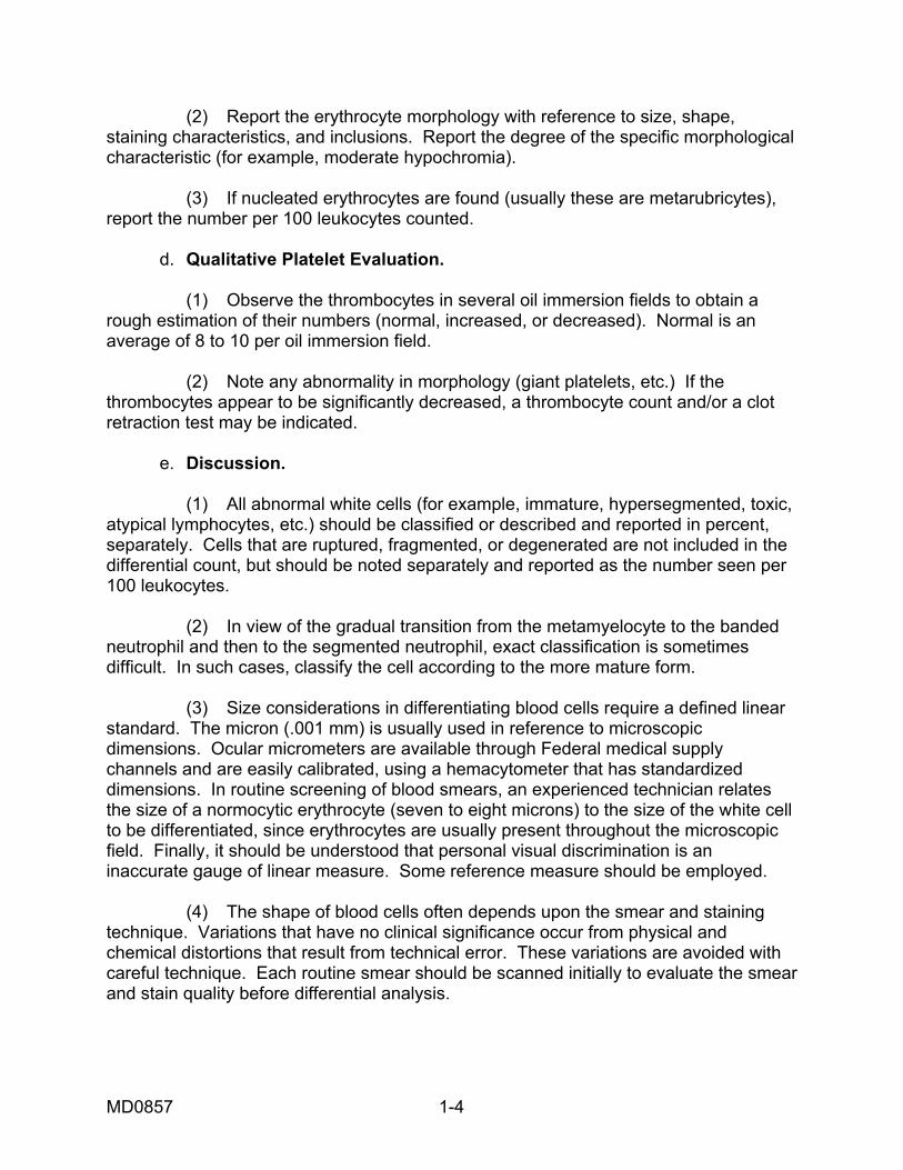
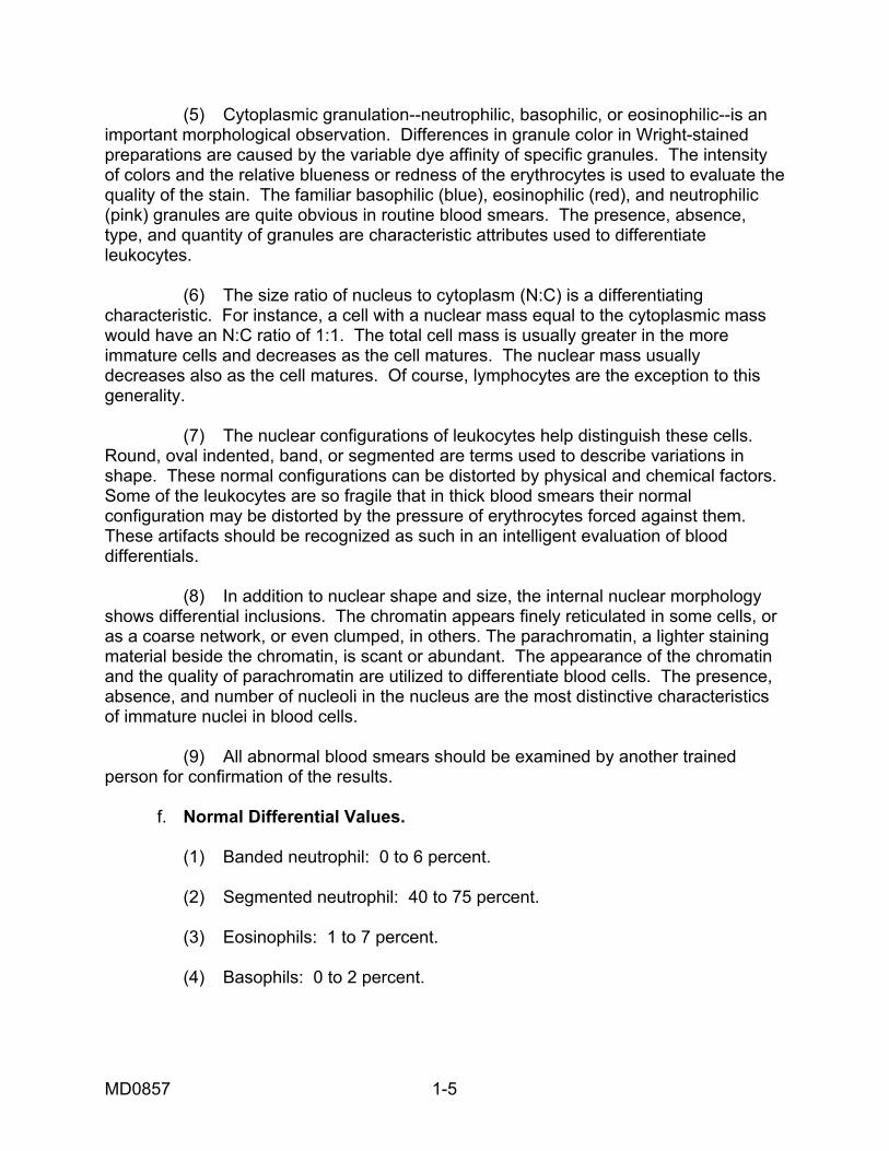
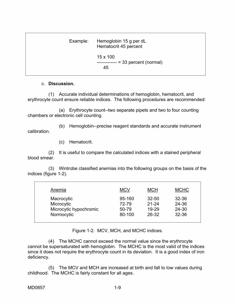
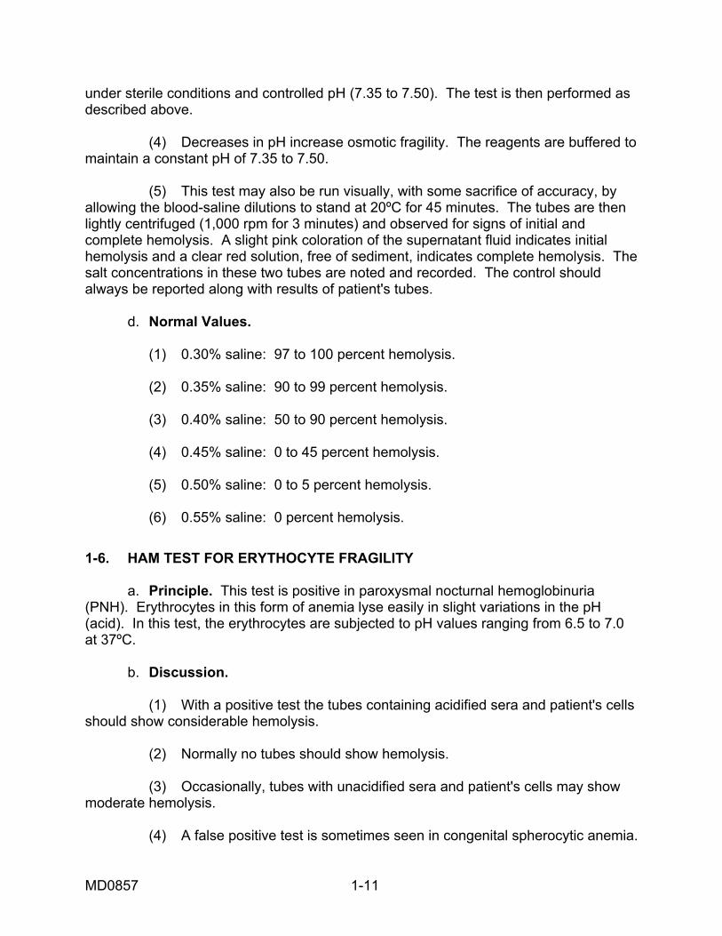
Example: Hemoglobin 15 g per dL Hematocrit 45 percent 15 x 100 ------------- = 33 percent (normal) 45 c. Discussion. (1) Accurate individual determinations of hemoglobin, hematocrit, and erythrocyte count ensure reliable indices. The following procedures are recommended: (a) Erythrocyte count--two separate pipets and two to four counting chambers or electronic cell counting. (b) Hemoglobin--precise reagent standards and accurate instrument calibration. (c) Hematocrit. (2) It is useful to compare the calculated indices with a stained peripheral blood smear. (3) Wintrobe classified anemias into the following groups on the basis of the indices (figure 1-2).
Anemia MCV MCH MCHC Macrocytic 95-160 32-50 32-36 Microcytic 72-79 21-24 24-36 Microcytic hypochromic 50-79 19-29 24-30 Normocytic 80-100 26-32 32-36
Figure 1-2. MCV, MCH, and MCHC indices.
(4) The MCHC cannot exceed the normal value since the erythrocyte cannot be supersaturated with hemoglobin. The MCHC is the most valid of the indices since it does not require the erythrocyte count in its deviation. It is a good index of iron deficiency. (5) The MCV and MCH are increased at birth and fall to low values during childhood. The MCHC is fairly constant for all ages.
MD0857 1-10
d. Normal Values. (1) Mean corpuscular volume: 80 to 96 fl. (2) Mean corpuscular hemoglobin: 27 to 32 micromicrograms. (3) Mean corpuscular hemoglobin concentration: 32 to 36 percent. 1-5. ERYTHROCYTE OSMOTIC FRAGILITY TEST (DACIE) a. Principle. A specific amount of blood is introduced into a series of tubes containing different concentrations of buffered salt solutions. The amount of hemolysis is then determined by examining the supernatant fluid either visually or with a spectrophotometer. b. Sources of Error. (1) The concentration of the NaCl in the solutions is critical. The salt must be chemically pure and dried before weighing. It is advisable to dry the salt in a 100ºC oven and store it in a desiccator. Store the NaCl solutions in a glass-stoppered, tightly sealed bottle. (2) Inaccurate preparation of the dilutions causes inaccurate results. (3) Maintain the pH of the solution at an interval of 7.35-7.50. A different pH range causes invalid results. (4) Rough handling of the blood specimen causes hemolys is which leads to invalid results. c. Discussion. (1) In hypotonic salt solutions, erythrocytes take up water, swell to a spheroid shape and burst. In congenital spherocytic anemia the red cells with defective structure more rapidly rupture at salt concentrations closer to isotonicity (0.85 percent). These cells thus show an increased osmotic fragility. In contrast, the flat or thin but otherwise normal red cells of hypochromic anemia show a decreased osmotic fragility and do not hemolyze until lower salt concentrations are reached. (2) When hemolysis begins beyond the range of the prepared solutions or when intermediate dilutions are desired, the additional dilutions are readily prepared using the 1 percent sodium chloride stock solution. (3) In cases where the results of the fragility test are borderline, the following procedure is recommended to enhance any latent abnormality in fragility. Incubate samples of defibrinated blood (control and patient's) at 37ºC for 24 hours
MD0857 1-11
under sterile conditions and controlled pH (7.35 to 7.50). The test is then performed as described above. (4) Decreases in pH increase osmotic fragility. The reagents are buffered to maintain a constant pH of 7.35 to 7.50. (5) This test may also be run visually, with some sacrifice of accuracy, by allowing the blood-saline dilutions to stand at 20ºC for 45 minutes. The tubes are then lightly centrifuged (1,000 rpm for 3 minutes) and observed for signs of initial and complete hemolysis. A slight pink coloration of the supernatant fluid indicates initial hemolysis and a clear red solution, free of sediment, indicates complete hemolysis. The salt concentrations in these two tubes are noted and recorded. The control should always be reported along with results of patient's tubes. d. Normal Values. (1) 0.30% saline: 97 to 100 percent hemolysis. (2) 0.35% saline: 90 to 99 percent hemolysis. (3) 0.40% saline: 50 to 90 percent hemolysis. (4) 0.45% saline: 0 to 45 percent hemolysis. (5) 0.50% saline: 0 to 5 percent hemolysis. (6) 0.55% saline: 0 percent hemolysis.
1-6. HAM TEST FOR ERYTHOCYTE FRAGILITY a. Principle. This test is positive in paroxysmal nocturnal hemoglobinuria (PNH). Erythrocytes in this form of anemia lyse easily in slight variations in the pH (acid). In this test, the erythrocytes are subjected to pH values ranging from 6.5 to 7.0 at 37ºC. b. Discussion. (1) With a positive test the tubes containing acidified sera and patient's cells should show considerable hemolysis. (2) Normally no tubes should show hemolysis. (3) Occasionally, tubes with unacidified sera and patient's cells may show moderate hemolysis. (4) A false positive test is sometimes seen in congenital spherocytic anemia.

MD0857 1-12
(5) If congenital spherocytic anemia is suspected, the test should be repeated, using acidified serum previously inactivated at 56ºC for 30 minutes. (6) Since erythrocytes of PNH require complement for hemolysis, the modified test (item 5 above) will be negative in PNH and will remain positive in spherocytosis. c. Interpretation. Hemolysis in the acidified tube is indicative of paroxysmal nocturnal hemoglobinuria.
Section III. DEMONSTRATION OF L.E. CELLS 1-7. GENERAL INFORMATION a. Persons having lupus erythematosus, one of the "collagen" diseases, have an abnormal plasma protein that causes swelling and breakdown of certain blood cell nuclei in vitro. This degenerated nuclear material attracts phagocytic cells, particularly segmented neutrophils, which engulf this nuclear mass. The resulting phagocyte and inclusion material is termed an "L.E." cell. b. Two methods of demonstrating the L.E. cell and antinuclear antibodies are the rotary bead method and fluorescent antibody method. The rotary bead method is positive in 75-80 erythematosus. The fluorescent antibody method is positive in 95-100 patients with lupus erythematosus. The rotary bead method is presented in the next paragraph. The fluorescent antibody method requires equipment that limits its use to larger laboratories. 1-8. ROTARY BEAD METHOD a. Principle. Leukocytes are broken down in vitro allowing the abnormal plasma protein to react on the altered nuclear material. Incubation enhances the nuclear deterioration and phagocytosis. Slides are prepared and examined for the peculiar "L.E." cell. b. Discussion. (1) Lupus erythematosus is a chronic, sometimes fatal, disease of unknown etiology. The peculiar skin eruption across the nose and cheeks (butterfly rash) and arthritis can be accompanied by various visceral manifestations. Often the rash is not present, and diagnosis depends on demonstration of the L.E. cell. Frequently the earliest symptoms appear after intense exposure to sunlight. Leukopenia, thrombocytopenia, and an elevated sedimentation rate are some of the clinical signs of the disease.
MD0857 1-13
(2) Free masses of lysed nuclear material, with or without polymorphonuclear leukocytes clustered about them (rosette formation), are suggestive of the L.E. phenomenon. Observing "rosettes" should encourage the technician to repeat examinations and further search for the true "L.E." cells. A positive report should not be made without the identification of this cell. The inclusion body with the leukocyte is homogeneous and has no chromatin pattern. This feature distinguishes the true "L.E." cell from the "tart" cell (nucleophagocytosis). This latter cell contains an engulfed, damaged nucleus, usually that of a lymphocyte, which still contains a recognizable chromatin pattern and a distinct nuclear membrane. c. Interpretation. (1) These cells are seen as large polymorphonuclear (segmented) leukocytes which contain large ingested nuclear fragments in their cytoplasm. (2) The inclusion body is a purplish-staining, smoky, homogeneous mass of material that is so large that it usually pushes the nucleus to one side of the cell.
Section IV. SPECIAL STAINS 1-9. PEROXIDASE STAIN (KAPLOW) a. Principle. The members of the granulocytic series contain an enzyme, peroxidase, which liberates the oxygen from hydrogen peroxide. This enzyme is more prominent in mature forms. A benzidine derivative is used as an indicator of peroxidase activity. The indicator is oxidized and precipitates in the form of brown to blue granules. This stain is used to help differentiate leukemias. NOTE: Follow manufacture's instructions for all special stains. b. Interpretation. Peroxidase positive cells are identified by yellow-green to blue and brown-green granules. Cells of the granulocyte series from the promyelocyte through the segmented neutrophil are peroxidase positive. The degree of peroxidase activity increases as the granulocytes mature. Monocytes may show a weak reaction. All other cells are negative. c. Discussion. (1) The oxidizing enzyme in the granules of the granulocytic leukocytes deteriorates rapidly in vitro. It is, therefore, necessary to use fresh blood in making the preparation. (2) Smears should be prepared within one hour of obtaining the specimen and stained within three hours after they are prepared.
MD0857 1-14
(3) The monocyte is thought to be slightly peroxidase positive through the phagocytization of peroxidase positive granules of ruptured cells. (4) Myeloblasts can show weak peroxidase activity using this method. (5) Addition of 4.9 mg of sodium cyanide to the stain inhibits peroxidase activity in all granulocytes except eosinophils. (6) If greater nuclear detail is required, counter stain with laqueous cresyl violet acetate for one minute or in freshly prepared Giemsa stain for 10 minutes. (7) Giemsa stain is prepared as follows: Mix 3.8 g Giemsa stain powder and 200 ml glycerin. Incubate at 60ºC for two hours. Add 312 ml absolute methanol; dilute the staining solution 1:10 with 5 sodium carbonate before use. 1-10. LEUKOCYTE ALKALINE PHOSPHATASE a. Principle. Blood smears are fixed and stained for alkaline phosphatase activity. b. Scoring. (1) Count two slides (100 cells per slide) on each patient, rating the segmented neutrophils according to how much black staining of the granules is observed. If no staining is noted, the rating is 0; if slight black staining is noted, the rating is 1+, if a medium amount of black staining is noted, the rating is 2+, if a heavy amount of dark black staining is observed, the rating is 3+, and if there is heavy black staining covering all the cytoplasm, the rating is 4+. (2) After 100 cells per slide are rated, figure the score-giving cells counted as 0--no score; cells rated as 1+ get a score of 1 each; cells rated as 2+ get a score of 2 each, etc. (3) Report the total number of cells, giving their ratings and score. Report the total score for each individual slide. Average the two total scores and report the average. Also, always report the normal score range. c. Discussion. (1) Patients with infections, polycythemia, and myeloproliferative disorders demonstrate increased alkaline phosphatase activity. (2) In patients with acute or chronic granulocytic leukemia, alkaline phosphatase activity is decreased.
MD0857 1-15
d. Normal Values. Scores of 13 to 130 have been obtained in healthy adults. However, the attending physician should interpret whether values are normal or abnormal. 1-11. HEINZ-BODY STAIN a. Principle. Blood is mixed with methyl violet solution and a smear is prepared. Heinz-bodies stained with methyl violet are purple, round or oval granules, one-two microns in diameter within the erythrocytes. b. Interpretation. Iron granules present in erythrocytes stain blue. c. Discussion. (1) Heinz-bodies are invisible in Wright-stained preparation. (2) They can be observed in reticulocyte preparations and by the use of phase microscopy. (3) Heinz-bodies are thought to be denatured hemoglobin. They are usually demonstrated in hemolytic anemias caused by toxic agents, including vegetable and animal poisons. 1-12. SIDEROCYTE STAIN a. Principle. Siderocytes are erythrocytes containing iron granules. The granules are blue when stained with Prussian blue. b. Interpretation. Iron granules present in erythrocytes stain blue. c. Discussion. (1) Siderocytes occur in several anemias, lead poisoning, and after splenectomy. (2) On Wright-stained preparation, the granules are bluish-purple and are called Pappenheimer bodies.
Continue with Exercises
MD0857 1-16
EXERCISES, LESSON 1 INSTRUCTIONS: Answer the following exercises by marking the lettered response that best answers the exercise, by completing the incomplete statement, or by writing the answer in the space provided at the end of the exercise. After you have completed all of the exercises, turn to "Solutions to Exercises" at the end of the lesson and check your answers. For each exercise answered incorrectly, reread the material referenced with the solution. 1. A critical examination of a stained blood smear includes the differential count that quantitates the three types of: a. Thrombocytes. b. Granulocytes. c. Lympocytes. d. Leukocytes. 2. Which area of the blood smear is used for the differential leukocyte count? a. Thin end. b. Thick end. c. Inner portion. d. Peripheral area. 3. The ________________ objective lens is used to perform the differential leukocyte count. a. 10X (low power). b. 40X (high power). c. 100X (oil immersion).
MD0857 1-17
4. When nucleated erythrocytes are located on a blood smear, they are reported by the ______________________________ counted. a. Number per 100 leukocytes. b. Number per 100 erythrocytes. c. Percentage of all leukocytes. d. Percentage of all erythrocytes. 5. What is the normal average number of thrombocytes counted per oil immersion field when performing a qualitative platelet evaluation of a blood smear? a. 0-2. b. 4-6. c. 6-8 d. 8-10. 6. Which test is indicated when the amount of thrombocytes appear to be decreasing significantly on an oil immersion field blood smear? a. Rosettes. b. Alkali denaturation. c. Clot retraction. d. pH acid. 7. Which white blood cells are counted as part of the 100 in a differential leukocyte count and reported in a separate category in percent? a. Immature leukocyte. b. Ruptured leukocyte. c. Fragmented leukocyte. d. Degenerated leukocyte.
MD0857 1-18
8. A cell with a nuclear mass twice as great as the cytoplasmic mass would have an N:C ratio of: a. 1:2. b. 2:3. c. 1:1. d. 2:1. 9. What has the second highest value in the normal differential count? a. Monocytes. b. Eosinophils. c. Lymphocytes. d. Segmented neutrophils. e. Neutrophilic band cells. 10. The myeloid-erythroid (M:E) ratio of the bone marrow is the ratio of the granulocytic white blood cells to the: a. Red blood cells. b. Mature red blood cells. c. Nucleated red blood cells. d. Bone marrow cells other than granulocytes. 11. The bone marrow study should be accompanied by a: a. Hematocrit. b. Red blood cell count. c. Peripheral blood evaluation. d. Total white blood cell count.
MD0857 1-19
12. What is the normal M:E (myeloid-erythroid) ratio of the bone marrow? a. 1:1. b. 2:1 to 3:1. c. 3:1 to 4:1. d. 4:1 to 7:1. 13. The hematocrit and the RBC count are needed to calculate the: a. MCV. b. MCH. c. MCHC. 14. When calculating the MCV in femtoliters, what is the divisor after multiplying the hemotocrit and 10? a. RBC count (millions). b. Hematocrit (percent). c. WBC count (thousands). d. Hemoglobin concentration (g/dl). 15. What is the MCV if the hematocrit is 44 percent, the RBC count is 5.2 million per cu mm, and the hemoglobin concentration is 14 g/dl? a. 1.2 fl. b. 8.5 fl. c. 12 fl. d. 85 fl.
MD0857 1-20
16. What is the MCV if the hematocrit is 36 percent. the RBC is 4.6 million per cu mm, and the hemoglobin concentration is 11 g/dl? a. 1.2 fl. b. 78 fl. c. 118 fl. d. 783 fl. 17. The RBC count and the hemoglobin concentration are needed to calculate the: a. MCV. b. MCH. c. MCHC. 18. To calculate the MCH in micromicrograms, the _________ is multiplied by to 10. a. RBC count (millions). b. WBC count (thousands). c. Hematocrit (percent). d. Hemoglobin concentration (g/dl). 19. If the hematocrit is 44 percent, the RBC is 5.2 million per cu mm, and the hemoglobin concentration is 14 g/dl, what is the MCH? a. 12 micromicrograms. b. 27 micromicrograms. c. 37 micromicrograms. d. 85 micromicrograms.
MD0857 1-21
20. If the hematocrit is 36 percent, the RBC is 4.6 million per cu mm, and the hemoglobin concentration is 11 g/dl, what is the MCH? a. 24 micromicrograms. b. 31 micromicrograms. c. 33 micromicrograms. d. 1/2 micromicrogram. 21. To calculate the MCHC, ___________________ is multiplied by 100, then divided by the Hemocrit. The result equals the percent of hemoglobin in the average RBC. a. RBC count (millions). b. WBC count (thousands). c. Hematocrit (percent). d. Hemoglobin concentration (g/dl). 22. If the hematocrit is 44 percent, the RBC is 5.2 million per cu mm, and the hemoglobin concentration is 14 g/dl, what is the MCHC? a. 12 percent. b. 27 percent. c. 32 percent. d. 37 percent.
MD0857 1-22
23. If the hematocrit is 36percelt, the RBC is 4.6 million per cu mm, and the hemoglobin concentration is 11 g/dl, what is the MCHC? a. 24 percent. b. 31 percent. c. 33 percent. d. 42 percent. 24. A mean corpuscular volume below 80 fl indicates that the erythrocytes are: a. Macrocytic. b. Normocytic. c. Microcytic. d. Megaloblastic. 25. The maximum value for the ______________ is included in its normal range. a. MCV. b. MCH. c. MCHC. 26. The normal range for the mean corpuscular volume of an erythrocyte is approximately: a. 62 to 82 fl. b. 70 to 80 fl. c. 80 to 97 fl. d. 90 to 100 fl.
MD0857 1-23
27. The osmotic fragility of erythrocytes is increased in: a. Thalassemia major. b. Sickle cell anemia. c. Iron deficiency (hypochromic) anemia. d. Congenital spherocytic (hemolytic) anemia. 28. When the osmotic fragility test is performed visually, the salt concentrations are recorded for the two tubes that show: a. 0 percent and 50 percent hemolysis. b. 0 percent and 100 percent hemolysis. c. Least and greatest hemolysis. d. Initial hemolysis and first complete hemolysis. 29. What is the normal percentage of hemolysis in 0.55 percent saline? a. 0 percent. b. 40 percent. c. 65 percent. d. 100 percent. 30. The Ham test is positive in: a. Polycythemia. b. Paroxysmal nocturnal hemoglobinuria. c. Chronic lymphocytic leukemia. d. All hemoglobinopathies.
MD0857 1-24
31. Erythrocytes in paroxysmal nocturnal hemoglobinuria lyse easily in serum which is slightly: a. Basic. b. Acidic. c. Hypotonic. d. Hypertonic. 32. A false-positive Ham test may occur in: a. Sickle cell anemia. b. Congenital spherocytic anemia. c. Severe iron deficiency anemia. d. Paroxysmal nocturnal hemoglobinuria. 33. When demonstrating "L.D." cells, which of the following has degenerative nuclear material that attracts phagocytic cells, particular segmented neutrophis? a. Jaundice. b. Leukemia. c. Lupus erythematossus. d. Pernicious amenia. 34. Which method is used to determine L.E. cell and antinuclear antibodies with a 75 to 80 percent accuracy rate? a. Rotary bead. b. Fluorescent antibody. c. a and b. d. None of the above.
MD0857 1-25
35. Lupus erythematosus is: a. A chronic, sometimes fatal, disease of unknown etiology. b. A regular skin eruption across the nose and mouth (butterfly rash), with arthritis that can be accompanied by various visceral manifestations. c. A rash, which is sometimes not present. Diagnosis depends on demonstration of the L.E. cell. d. Sometimes not diagnosed early because the early symptoms do not appear after intense exposure to sunlight. 36. Which statement is correct for the erythrocyte osmotic fragility test? a. In hypertonic salt solutions, erythrocytes take up water, swell to a spheroid shape and burst. b. In congenital spherocytic anemia, the WBCs with defective structure, will more rapidly rupture at salt concentrations closer to isotonicity (0.85 percent). c. The RBCs cells thus show an increased osmotic fragility. In contrast, the flat or thin but otherwise normal red cells of hypochromic anemia show a decreased osmotic fragility and do not hemolyze until lower salt concentrations are reached. d. When hemolysis begins within the normal range of the prepared solutions or when intermediate dilutions are desired, the additional dilutions are readily prepared using the 1 percent sodium chloride stock solution. 37. Which statement is correct for the erythrocyte osmotic fragility test? a. When the results of the fragility test are normal, one procedure is immediately followed to enhance any latent abnormality in fragility. b. Incubate samples of defibrinated blood (control and patient's) at 37ºC for 24 hours under sterile conditions and controlled pH (7.35 to 7.50). c. The test does not need to be performed. d. Increases in pH decrease osmotic fragility. The reagents are buffered to maintain a constant pH of 7.35 to 7.50.
MD0857 1-26
38. Which statement is correct for the erythrocyte osmotic fragility test? a. This test may also be run visually, with some sacrifice of accuracy, by allowing the blood-saline dilutions to stand at 20ºC for 45 minutes. b. The tubes are then vigorously centrifuged (1,000 rpm for 3 minutes) and observed for signs of initial and complete hemolysis. c. A rich dark pink coloration of the supernatant fluid indicates initial hemolysis and a cloudy red solution indicates complete hemolysis. d. Salt free concentrations in these two tubes are noted and recorded. e. The control does not have to be reported along with results of patient's tubes. 39. When there is no "butterfly rash," diagnosis of lupus erythematosus often depends upon demonstration of: a. Collagen. b. L.E. cells. c. Leukocytosis. d. Polycythemia. 40. In addition to L.E. cells, two characteristic phenomena in lupus erythematosus are: a. Free nuclear masses and rosettes. b. Rouleaux and Cabot rings. c. Basket cells and toxic granulation. d. Distorted lymphocytes and smudge cells.
MD0857 1-27
41. Which of the following is easily mistaken for an L.E. cell? a. Tart cell. b. Monocyte. c. Plasmocyte. d. Segmented neutrophil. 42. Which of the following is peroxidase negative? a. Lymphocytes. b. Promyelocytes. c. Neutrophilic myelocytes. d. Neutrophilic metamyelocytes. e. Neutrophilic band cells. f. Neutrophilic segmented cells. 43. Which of the following is peroxidase positive? a. Lymphocytes. b. Plasmocytes. c. Segmented neutrophils.
MD0857 1-28
44. With the alkaline phosphatase stain, a segmented neutrophil exhibiting no darkly stained granules is rated: a. 0. b. 1+. c. 2+. d. 3+. e. 4+. 45. Generally speaking, when leukocyte alkaline phosphatase is used, a patient with what score is considered to be a healthy adult? a. 10 to 50. b. 10 to 80. c. 13 to 100. d. 13 to 130. 46. Heinz bodies are often present in the erythrocytes of hemolytic anemia caused by: a. Toxic agents. b. Spherocytosis. c. Thalassemia major. d. Paroxysmal nocturnal hemoglobinuria. 47. What color are Heinz-bodies when stained with methyl violet? a. Colorless. b. Blue-green. c. Purple. d. Black.
MD0857 1-29
48. Heienz-bodies prepared with Wright-stain are what color? a. Green. b. Purple. c. Colorless. d. Orange. 49. Siderocytes are: a. Iron granules. b. Denatured hemoglobin. c. Nucleated erythrocytes. d. Erythrocytes containing iron granules. 50. Siderocytes may be observed: a. All of the below. b. In several anemias. c. After splenectomy. d. In lead poisoning.
Check Your Answers on Next Page
MD0857 1-30
SOLUTIONS TO EXERCISES, LESSON 1 1. d (para 1-1a) 2. a (para 1-2b(1), (2)) 3. d (para 1-2b(2)) 4. a (para 1-2c(3)) 5. d (para 1-2d(1)) 6. c (para 1-2d(2)) 7. a (para 1-2e(1) 8. d (para 1-2e(6)) 9. c (para 1-2f) 10. c (para 1-3b(3)) 11. c (para 1-3b(4)) 12. c (para 1-3d(4)) 13. a (para 1-4b(1)) 14. a (para 1-4b(1)) 15. d (para 1-4b(2)) 16. b (para 1-4b(2)) 17. b (para 1-4b(2)) 18. d (para 1-4b(2)) 19. b (para 1-4b(2)) 20. a (para 1-4b(2))
MD0857 1-31
21. d (para 1-4b(3)) 22. c (para 1-4b(3)) 23. b (para 1-4b(3)) 24. c (para 1-4c(3)) 25. c (para 1-4c(4)) 26. c (para 1-4d(1)) 27. d (para 1-5c(1) 28. d (para 1-5c(5)) 29. a (para 1-5d(6)) 30. b (para 1-6a) 31. b (para 1-6a) 32. b (para 1-6b(4)) 33. c (para 1-7a) 34. a (para 1-7b) 35. a (para 1-8b(1)) 36. c (para 1-5c(1)) 37. b (para 1-5c(3)) 38. a (para 1-5c(5)) 39. b (para 1-8b(1)) 40. a (para 1-8b(2))
MD0857 1-32
41. a (para 1-8b(2)) 42. a (para 1-9b) 43. c (para 1-9b) 44. a (para 1-10b(1)) 45. d (para 1-10d) 46. a (para 1-11c(3)) 47. c (para 1-11a) 48. c (para 1-11c(3)) 49. d (para 1-12a) 50. a (para 1-12c(1))
End of Lesson 1
MD0857 2-1
LESSON ASSIGNMENT LESSON 2 Blood Coagulation. TEXT ASSIGNMENT Paragraphs 2-1 through 2-30. LESSON OBJECTIVES After completing this lesson, you should be able to: 2-1. Select the statement that best describes the process of hemostasis and its relationship to the vascular system. 2-2. Select the statement that correctly describes the process and system of coagulation. 2-3. Select the statement that best describes the four components or stages of blood clotting. 2-4. Select the correct materials and procedures used in the different techniques and methods of blood collection for coagulation studies. 2-5. Select the statement that correctly describes the procedures, tests, and events during the stages of coagulation. 2-6. Select the statement that accurately describes normal value standards to follow and sources of errors that could occur during the coagulation process. 2-7. Select the statement that determines the adequacy of the thromboplastic complex. 2-8. Select the appropriate materials and procedures used to test for other coagulation deficiencies. SUGGESTION After completing the assignment, complete the exercises at the end of this lesson. These exercises will help you to achieve the lesson objectives.
MD0857 2-2
LESSON 2
BLOOD COAGULATION
Section I. INTRODUCTION 2-1. HEMOSTASIS
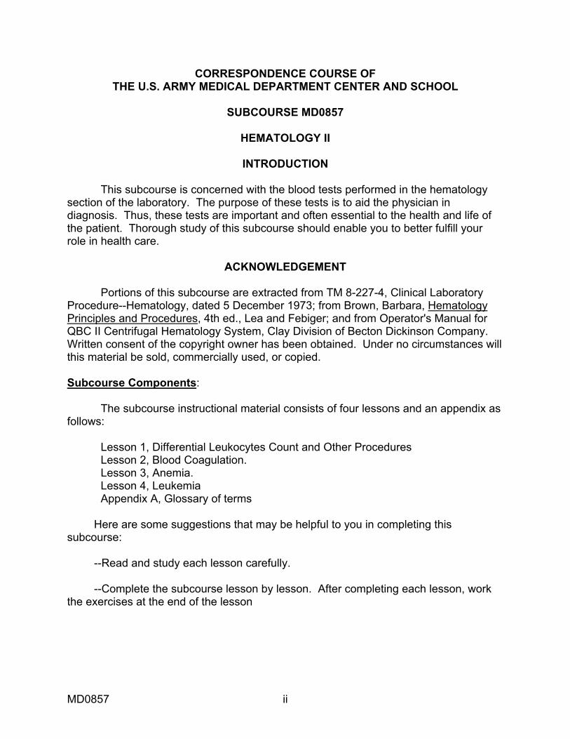
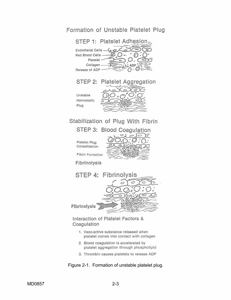
a. Coagulation of the blood is only one of the components in the larger function of stopping blood flow known as hemostasis. Hemostasis is a process in which there is a stoppage of blood flow from an injured blood vessel. It may be viewed as the combination of clotting and lysing mechanisms that maintain the integrity of the vascular system. There are two phases of hemostasis: Primary hemostasis, which is the formation of the platelet plug. Then there is secondary hemostasis, which is, the introduction of coagulation factors to form fibrin clot. b. Platelets play a major role in the hemostatic process. Within 1 to 2 seconds after injury to a blood vessel, platelets come in contact with and adhere to the injured tissues (platelet adhesiveness). As a result, the platelets become swollen and extend pseudopodia. Serotonin (5-hydroxytryptamine), ADP, catecholamines, and platelet factor 4 (a glycoprotein with antiheparin activity) are released by the platelets. The ADP released by the platelets and also by the injured tissues causes the platelets to stick to one another (known as platelet aggregation; when platelets attach to non-platelet surfaces, this is called platelet adhesion). Platelets continue to aggregate until the site of injury is healed. c. The vascular system also affects the hemostatic process through the function of vasoconstriction. The vascular mechanism involves the veins, arteries, and capillaries themselves. Their effectiveness depends on thickness of the vessel wall and its structure, contractibility, and retractibility. Bleeding into the tissues surrounding a wound increases perivascular pressure about small vessels, causing collapse and reduction of blood flow in larger vessels. Following the formation of a clot, clot retraction begins due to the action of actomyosin (thrombosthenin, the platelet contractile protein), which represents 15-20% of platelet protein. 2-2. COAGULATION SYSTEM a. Blood coagulation is the formation of a clot from liquid blood. When bleeding occurs, clotting is initiated by aggregation of platelets (see figure 2-1). The platelets congeal to plug the site of the injury. The congealing (viscous metamorphosis) process is stimulated by contact with collagen (the supporting tissue surrounding blood vessels) or by the formation of thrombin. Hemostasis is not achieved without the simultaneous formation of fibrin. Platelet and plasma factors are activated, and by a complex process, a fibrin clot is formed. The arrest of bleeding is attained when a firm fibrin network seals the blood vessel wound with enough strength to withstand the impact of intravascular pressure.
MD0857 2-3
Figure 2-1. Formation of unstable platelet plug.

MD0857 2-4
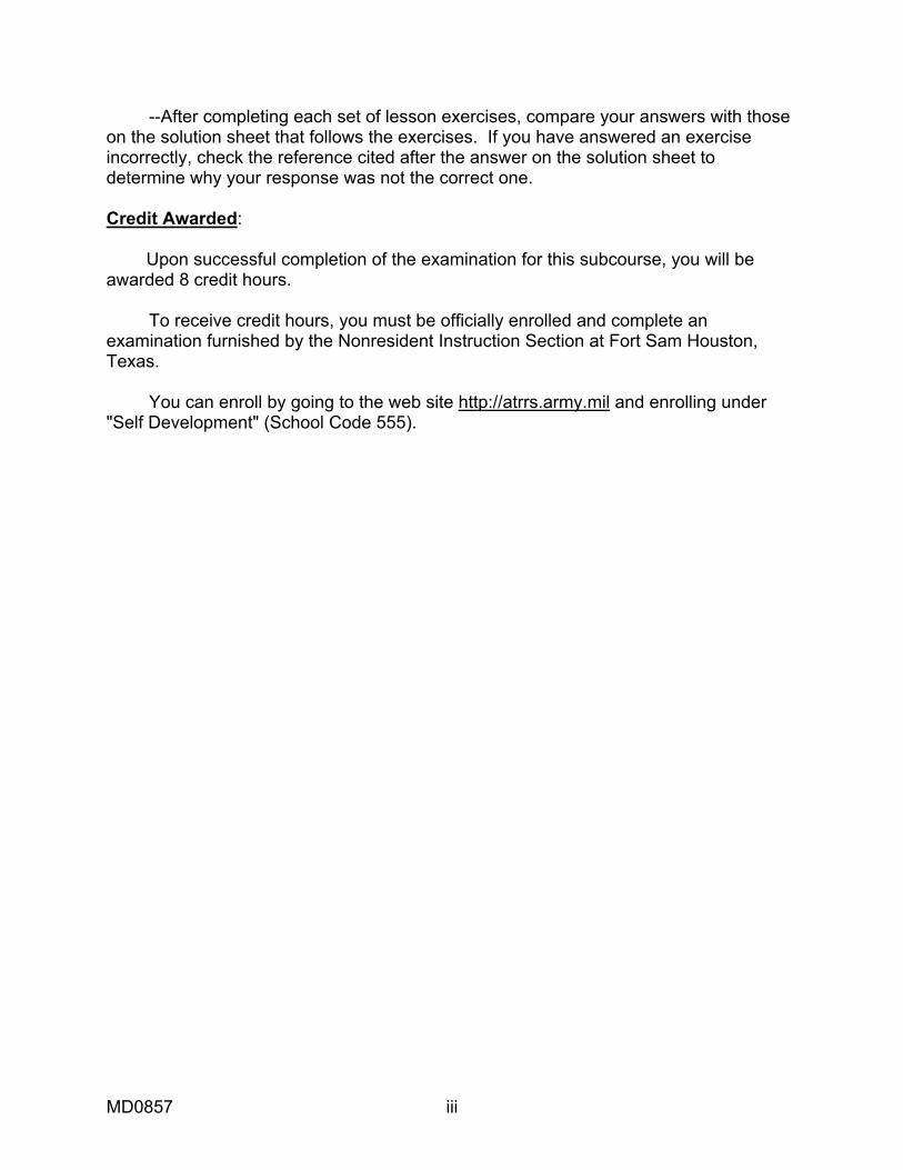
b. Bleeding disorders such as hemorrhaging and thrombosis, occur in the following instances: injury to the vascular system, inadequate number of platelets and/or dysfunctional platelets, inadequate fibrin clotting mechanisms, and inadequate fibroblastic repair. The laboratory performs a variety of tests that assist the physician in his investigation of blood coagulation. Several of these tests measure the overall coagulation process. The bleeding disorders are due to one or several of the many factors involved in this process. In most instances, prolonged bleeding is due to a deficiency of one factor or another. However, in some instances it is due to therapeutic anticoagulants that are intentionally injected to interfere with the coagulation mechanism. In a few rare instances, prolonged bleeding is due to a natural or antigenically stimulated increase in circulating anticoagulants produced in the body. c. The coagulation process is divided into two systems: the intrinsic pathway, which is the dominant pathway, all of the components are found in the circulating blood. The extrinsic pathway relies on thromboplastin, or tissue factor, (factor III), which is released from the damaged cells and tissues (see figure 2-2). The distinction between the intrinsic and extrinsic pathways becomes blurred upon deeper analysis. As more and more information is gathered, it shows how each interacts with the other and feedback mechanisms work in combination. Examples of such is how activated factor XII, will trigger factor VII to its active form. Additional crossovers show with the activation of factor XI by activated factor VII.
Figure 2-2. Coagulation systems.

MD0857 2-5
Section II. COAGULATION SYSTEM 2-3. COAGULATION FACTORS Table 2-1 contains a complete list of coagulation factors and their synonyms. Factor Numerical Description Name I Fibrinogen II .................. Prothrombin III ................. Tissue factor, tissue thromboplastin IV ................. Calcium V .................. Labile factor, proaccelerin, AC-globin VI…………….Accelerin, eliminated by the International Committee on Blood Clotting Factors VII ................ Proconvertin, stable factor VIII ............... Antihemophilic A factor (AHF) Antihemophilic globulin (AHG) IX ................. Plasma thromboplastin component (PTC), Christmas factor Antihemophilic B factor (AHB) X .................. Stuart-Prower factor, autoprothrombin III XI ................. Plasma thromboplastin antecedent (PTA) XII ................ Hageman factor, glass factor, contact factor XIII ............... Fibrin stabilizing factor (FSF), fibrinase Prekallikrein ...... Fletcher factor High molecular........... Fitzgerald factor, Williams factor, Flau Jeac factor, weight kininogen........ Contact activation cofactor (Designation) a .......... A factor that has been activated and is now functional
Table 2-1. Nomenclature of coagulation factors.
a. Factor I (Fibrinogen). Fibrinogen, a plasma glycoprotein, is converted into fibrin in the presence of thrombin. The major source of fibrinogen is the liver. A minimum of 50 to 100 mg/dL is required for normal coagulation. Bleeding due to a fibrinogen deficiency does not usually become manifest until the plasma concentration is below 75 mg per dl. Decreased levels of fibrinogen can be caused by several reasons -- decreased liver production is due to acute hepatitis or cirrosis; fibrinolysins, which attack both fibrin and fibrinogen molecules; and massive production of fibrin, as seen in disseminated intravascular coagulation (DIC). Replenishment can be achieved by administration of fresh frozen plasma or cryoprecipitates.
MD0857 2-6
b. Factor II (Prothrombin). This substance is a stable glycoprotein, synthesized in the liver if an adequate amount of vitamin K is available. Prothrombin (a proenzyme) is the inactive precursor of thrombin. When Vitamin K is absent, various types of prothrombin molecules still form, but the site on the molecule that would react in the coagulation scheme is left inactive and nonfunctional. Factors II, VII, IX, and X are part of the prothrombin complex; all are vitamin K dependent, meaning that these factors cannot be produced without the presence of vitamin K in the liver in adequate amounts. These factors are absorbed by both BaSO4 (barium sulfate) and Al(OH)3 (aluminum hydroxide). With the exception of Factor II, all are found in serum. c. Factor III (Tissue Thromboplastin). Thromboplastin, or tissue factor is a high-molecular-weight lipoprotein found in most of the body tissues with increased concentrations in the lungs and brain. It is probably the phospholipid content of tissue thromboplastin that makes platelets unnecessary in stage I of the coagulation process. Tissue thromboplastin requires calcium and factors V, VII, and X to convert prothrombin to thrombin. It is found in the brain, lung, vascular endothelium, liver, placenta, or kidneys. d. Factor IV ( Ionized Calcium). Calcium is an inorganic ion that is necessary for clotting to occur. The exact mechanism by which calcium acts in the coagulation process is not known. The fact that it is essential for coagulation makes possible the use of anticoagulants, which merely bind up the calcium and, therefore, completely inhibit coagulation. It is unlikely, however, that a bleeding tendency is ever caused by a deficiency of calcium, since clinical tetany occurs with higher levels of calcium than are necessary for coagulation. e. Factor V (Labile Factor Proaccelerin, Accelerator Globulin). Factor V is derived from plasma globulin, and it acts as an accelerator in the conversion of prothrombin to thrombin in the presence of tissue thromboplastin. Factor V is not present in serum because it is consumed during the clotting of blood. In addition, it rapidly is inactivated during storage. It is not absorbed by BaSO4 (barium sulfate). f. Factor VI (Accelerin). Factor VI has been eliminated as an entity by the International Committee on Blood Clotting Factors. g. Factor VII (Stable Factor, Proconvertin). Factor VII is stable to both heat and storage. It is thought to act as an accelerator in the conversion of prothrombin to thrombin. Factor VII is not consumed in the clotting process; therefore, it has a high concentration in serum and plasma. Factor VII activity may actually increase the coagulation process. A vitamin K dependent factor is manufactured in the liver.
MD0857 2-7
h. Factor VIII (Antihemophilic Factor, Antihemophilic Globulin). Factor VIII is essential to the formation of intrinsic blood thromboplastin in the first stage of clotting. Factor VIII is a combination of two subunits: VIII:C and VIII:vWF( von Willebrand’s factor). Deficiency of factor VIII results in the reduction of thromboplastin as well as decreased conversion of prothrombin. Factor VIII deficiency is a hereditary sex-linked disorder, which is transmitted by females and manifested almost exclusively in males (hemophilia A). The unit is measured by the activated partial thromboplastin time (APTT) test. i. Factor IX (Plasma Thromboplastin Component, Christmas Factor). Factor IX influences the amount of thromboplastin formed. This factor is not consumed in the clotting process; therefore, it is present in serum. Deficiency of factor IX is either hereditary or acquired and is known as hemophilia B or Christmas disease. j. Factor X (Stuart Prower Factor). It is found in both serum and plasma. Factor X aids in the prompt conversion of prothrombin to thrombin. Deficiency of factor X is either acquired or hereditary. k. Factor XI (Plasma Thromboplastin Antecedent). Factor XI aids in the formation of plasma thromboplastin. This factor is stable and is found in plasma or serum. It is synthesized in the liver and vitamin K is not required for production. Deficiency of factor XI is probably hereditary and results in a mild hemophilia. l. Factor XII (Hageman Factor, Glass Factor). This factor is not required for normal hemostasis, but it is important in the various in vitro assays of the clotting mechanisms. It is a plasma contact factor with glass and is absorbed onto glass. Factor XII is related to factor XI in the activation of thromboplastin, and behaves like an enzyme for which one substrate is factor XI. m. Factor XIII (Fibrin Stabilizing Factor, Fibrinase). Factor XIII converts a loosely linked, fibrin clot (in the presence of the calcium ions) into a tough gel. Its activity is greatly reduced in serum (as compared with plasma) because of its strong adsorption of fibrin. 2-4. PLATELET FACTORS Platelets are active in blood coagulation. They perform the following functions: aid in vasoconstriction and the formation of a hemostatic plug, thromboplastic activity, and clot retraction. When platelets contact a wettable surface, at first they adhere to one another and then rupture, releasing chemical factors. a. Platelet Factor 1. This factor accelerates prothrombin conversion and is actually blood factor V adsorbed on platelets. (No longer used conventionally.) b. Platelet Factor 2. Factor 2 accelerates fibrinogen clotting of thrombin. (No longer used conventionally.)
MD0857 2-8
c. Platelet Factor 3. A phospholipid substance, found in the platelet membrane, is involved in prothrombin activation. This is the most important factor and probably is an actual intrinsic component of platelets. d. Platelet Factor 4. This factor reacts to neutralize heparin. e. Platelet Factor 5. This factor is an adsorbed intrinsic fibrinogen. (No longer used conventionally.) f. Platelet Factor 6. This factor reduces fibrinolytic activity. (No longer used conventionally.) g. Platelet Factor 7. This factor is adsorbed blood factor VII. (No longer used conventionally.) 2-5. FIBRINOLYTIC FACTORS Fibrinolysis is the dissolution of a fibrin clot. The process is a necessary activity following clot formation. The mechanism of clot dissolution is complex and involves a variety of factors. In active circulating plasma profibrinolysin (plasminogen) is converted to its active form, fibrinolysin (plasmin), by tissue activators, streptokinase, urokinase, and other unknown activators. Fibrinolysin acts locally to dissolve the clot.
Section III. THE COAGULATION MECHANISM 2-6. INTRODUCTION a. The classical theory of Morowitz proposed that four components interact to form a clot as follows:
Thromboplastin
Prothrombin ----------------------------------> Thrombin Calcium
Thrombin
Fibrinogen ----------------------------------> Fibrin clot
b. From this concept, the modern theory was devised. The modern theory is based on four stages: (I) the formation of thromboplastin, (II) the conversion of prothrombin to thrombin, (III) formation of an insoluble fibrin clot through the interaction of fibrinogen and thrombin, and (IV) the lysis of the fibrin clot by fibrinolysin. These stages are illustrated in figure 2-3.

MD0857 2-9
Figure 2-3. Stages of coagulation.
MD0857 2-10
2-7. STAGES OF COAGULATION a. Stage I--The Generation of Plasma Thromboplastin. Stage I involves the information of intrinsic (plasma) thromboplastin. This stage is initiated by the platelets adhering and rupturing, releasing platelet factor 3. Platelet factor 3 reacts with factor XII, along with prekallikrein and high molecular weight kininogen (HMWK), PTA (factor XI), PTC (factor IX), AHF (factor VIII) and in the presence of calcium (factor IV), factor X, and factor V to form intrinsic thromboplastin. Tissue thromboplastin or extrinsic thromboplastin is released by the affected tissues. b. Stage II--The Formation of Thrombin from Prothrombin. In the intrinsic system, prothrombin is converted to thrombin in the presence of plasma thromboplastin, calcium, and factors V and X, and platelets. The extrinsic system requires the presence of an additional factor, factor VII, for the conversion of prothrombin to thrombin. c. Stage III--The Formation of Fibrin from Fibrinogen. After the thrombin is generated, it quickly reacts with fibrinogen to form a fine fibrin fiber. The fibrin fibers polymerize (form a mesh with other fibers with disulphide bonds) in conjunction with calcium ions and factor XIII, the fibrin-stabilizing factor, to form a stabilized clot. The stabilized clot is characterized by its insolubility in 5 M urea. d. Stage IV--Clot Lysis. This stage involves the fibrinolytic dissolution of the clot. Circulating plasminogen can be converted to its active form plasmin, by both intrinsic and extrinsic factors. The intrinsic factors, which are present in circulation plasma include: factor XII, prekallikrein, HMWK, and pro-urokinase. The extrinsic factors, which are present in body tissues are: tissue plasminogen activator (t-PA), urokinase (UK), and streptokinase (SK). Plasmin, which is a serine protease, hydrolyzes arginine and lysine bonds that are present in fibrin, fibrinogen, and factors V and VIII. Thus, the clot in the presence of plasmin is dissolved, forming a series of fragments called fibrin or fibrinogen degredation products (FDP) or fibrin or fibrinogen split products (FSP). These fragments FDP-FSP will inhibit platelet aggregation and conversion of fibrinogen by thrombin. 2-8. COAGULATION INHIBITORS In addition to the factors necessary for clot formation, inhibitors are present which control but do not prevent coagulation. Natural inhibitors have been described for virtually every clotting factor. Clotting activity is also inhibited by the administration of anticoagulants such as heparin and coumarin derivatives.
MD0857 2-11
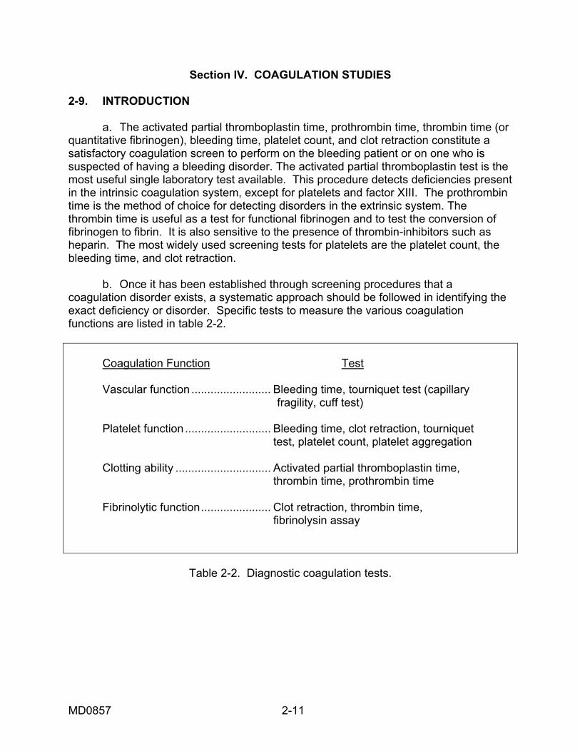
Section IV. COAGULATION STUDIES 2-9. INTRODUCTION a. The activated partial thromboplastin time, prothrombin time, thrombin time (or quantitative fibrinogen), bleeding time, platelet count, and clot retraction constitute a satisfactory coagulation screen to perform on the bleeding patient or on one who is suspected of having a bleeding disorder. The activated partial thromboplastin test is the most useful single laboratory test available. This procedure detects deficiencies present in the intrinsic coagulation system, except for platelets and factor XIII. The prothrombin time is the method of choice for detecting disorders in the extrinsic system. The thrombin time is useful as a test for functional fibrinogen and to test the conversion of fibrinogen to fibrin. It is also sensitive to the presence of thrombin-inhibitors such as heparin. The most widely used screening tests for platelets are the platelet count, the bleeding time, and clot retraction. b. Once it has been established through screening procedures that a coagulation disorder exists, a systematic approach should be followed in identifying the exact deficiency or disorder. Specific tests to measure the various coagulation functions are listed in table 2-2. Coagulation Function Test Vascular function ......................... Bleeding time, tourniquet test (capillary fragility, cuff test) Platelet function ........................... Bleeding time, clot retraction, tourniquet test, platelet count, platelet aggregation Clotting ability .............................. Activated partial thromboplastin time, thrombin time, prothrombin time Fibrinolytic function...................... Clot retraction, thrombin time, fibrinolysin assay
Table 2-2. Diagnostic coagulation tests.
MD0857 2-12
2-10. BLOOD COLLECTION a. Principle. There are general rules for using a "Vacutainer-type" blood collection system. Tests requiring sterility are always drawn first (blood culture). Next, obtain those tubes for specimens which do not require anticoagulants. This is followed by those specimens which do require anticoagulants. Coagulation specimens come first. b. Two-Tube Technique. To avoid contamination of blood by tissue juices, the two-tube (Vacutainer-type) venipuncture is usually employed for specialized coagulation tests using venous blood. The technique is as follows: (1) Place a tourniquet on the arm, no longer than 1 minute. (2) Observe the area for the most accessible vein. (3) Cleanse the site with 70 percent isopropyl alcohol and allow to dry. (4) Insert the needle and remove the tourniquet. (Quick removal of the tourniquet prevents stasis.) (5) Obtain specimens for all tests that require no anticoagulants first. If only coagulation work is specified, use a "red" top tube to withdraw about 5 ml of blood, and dispose of the "red" top tube. (6) Draw off the required specimen using a "blue" top coagulation tube. The ratio of blood to enclosed anticoagulant is 1:10. Allow the tube to fill to its capacity, do not remove until filling stops completely. (7) Blood samples needing anticoagulation should be mixed immediately following withdrawal from the needle. c. Anticoagulants. Anticoagulants for coagulation studies can be obtained in commercially-prepared vacuum tubes or prepared in the laboratory. The preparation and use of anticoagulants is as follows: (1) Sodium citrate, 3.2 percent. Dissolve 3.2 g of sodium citrate in 100 ml distilled water. Refrigerate. As an anticoagulant, combine one part 3.2 percent sodium citrate with 9 parts blood. (2) Sodium oxalate, 0.1 M. Dissolve 1.34 g sodium oxalate in 100 ml distilled water. Refrigerate. As an anticoagulant, combine one part 0.1 m. sodium oxalate with 9 parts blood.
MD0857 2-13
(3) EDTA. Dissolve 10 g of EDTA salt in 100 ml distilled water. Pipet 1 ml of this solution into a suitable test tube. Allow to dry in the oven at low temperature. For use as an anticoagulant, add 5 ml of venous blood and mix well. d. Glassware. All glassware for coagulation studies must be scrupulously clean. Used glassware should be free of chemicals and any traces of human blood components. All glassware should be cleaned in detergents free of organic solvents and rinsed several times with distilled water. The use of disposable syringes and needles eliminates the need for siliconizing glassware. CAUTION: Never use test tubes or pipets that are damaged. 2-11. TEMPLATE BLEEDING TIME (MIELKE MODIFICATION) a. Principle. (1) The bleeding time is an in vivo measurement of platelet participation in small vessel hemostasis. The introduction of the template bleeding time technique has improved the reproducibility and sensitivity of the test by controlling the length and depth of the incision. Control of the depth is important so that only the smaller vessels (capillaries) are incised. (2) The platelets initially adhere to the cut surface of the vessel wall; aggregation takes place. The platelets interdigitate and release the contents of their granules. This is followed by the formation of fibrin that stabilizes the hemostatic plug. (3) The purpose of the bleeding time test is to provide a measure of such platelet functions in small vessel hemostasis. A prolonged bleeding time does not in itself diagnose underlying platelet disorders, either qualitatively or quantitatively. It indicates the need for more quantifiable testing. b. Reagents and Materials. (1) 1 Surgicutt. (2) 1 70 percent alcohol pad. (3) Sphygmomanometer. (4) Stopwatch. (5) Filter paper (2 to 3 sheets). (6) Band-Aid ® or other bandage.
MD0857 2-14
c. Procedure (1) Position the patient's arm with the volar surface exposed. (2) Select a site avoiding surface veins, bruises, and edematous areas. (3) Place the sphygmomanometer on the upper arm. (4) Cleanse the test area with the 70 percent alcohol pad and let air-dry. (5) Inflate the cuff to 40 mm Hg., and hold at this exact pressure for the duration of the test. NOTE: The time between inflation and incision should be 30 to 60 seconds. (6) Open the sterile package and gently rest the Surgicutt surface on the patient's forearm. (7) Apply minimal pressure so that both ends of the instrument lightly touch the skin. (8) Gently push the trigger, starting the stopwatch simultaneously. (9) Remove the Surgicutt immediately after triggering. (10) After 30 seconds, wipe the flow of blood with the filter paper. (Bring the paper close to the incision, but do not touch the paper directly to the incision, so as not to disturb the formation of the platelet plug.) (11) Wipe the blood every 30 seconds thereafter, until no blood stains the paper. (12) Stop the timer when only clear fluid is absorbed onto the filter paper. The bleeding time is determined to the nearest 30 seconds. (13) Release the pressure of the sphygmomanometer. (14) Record the bleeding time. d. Sources of Error. (1) A horizontal incision, approximately 5 cm below the antecubital crease, 2.4MM depth, gives the best reproducibility. (2) If body hair will interfere, lightly shave the area.
MD0857 2-15
(3) Patient should be advised of a potential to produce a scar. This can usually be avoided by the use of a butterfly bandage applied for 24 hours. (4) Aspirin and aspirin-containing products may cause a prolonged bleeding time for up to two (2) weeks. (5) A standardized cut is necessary for valid results. Too little pressure on the device and the wound will be shallow or nonexistent. Too much pressure and the wound will be too deep. This is the one area where standardization has not been completely controlled. (6) Low skin temperature produces a constriction of the capillary vessels, resulting in decreased blood flow. e. Discussion. (1) The Template Bleeding Time is the method of choice because the blood pressure on the vessels is constant, the incision is uniform, and the arm offers an area for multiple determinations. (2) The bleeding time depends primarily on extravascular and vascular factors and, to a lesser degree, on the factors of coagulation. The chief factor controlling bleeding from a small cut is the constriction of the minute vessels following injury. Accuracy in this test is enhanced by blotting the drops of blood at shorter intervals of time as the drops of blood become progressively smaller. (3) Thrombocytes play an important part in the formation of the hemostatic plug that seals off a wound. In thrombocytopenic purpura there is a decrease in platelets resulting in a prolonged bleeding time due to a defective platelet plug. An additional factor prolonging the bleeding time in this condition is a defect in capillary contraction. It can be hereditary and acquired platelet dysfunction. (4) In hemophilia, the bleeding time is normal. This is explained by the fact that there are no vascular or extravascular abnormalities. However, the test should not be performed on a known hemophiliac, for delayed oozing of blood is a real hazard. f. Range of Values. (1) Normal values: 2 to 9 minutes. (2) Critical values: Less Than Greater Than Not applicable 15 minutes
MD0857 2-16
2-12. WHOLE BLOOD COAGULATION (CLOTTING) TIME (LEE-WHITE) a. Principle. The whole blood clotting time is a rough measure of all intrinsic clotting factors in the absence of tissue factors. Variations are wide and the test sensitivity is limited. Whole blood, when removed from the vascular system and exposed to a foreign surface, will form a solid clot. Within limits, the time required for the formation of the solid clot is a measure of the coagulation system. b. Reagents and Materials. (1) Stop watch equipment for collection of blood. (2) 2 plastic syringes. (3) 3 clean, dry glass test tubes (10 x 75 mm). (4) Water/dry bath (at 37oC). c. Procedure. (1) Label glass tubes #1, #2, and #3. (2) Collect at least 1 to 2 ml of blood in a plastic syringe. Discard this blood. (This prevents tissue thromboplastin from entering the blood sample.) Change syringes. (3) Collect at least 5 ml of blood in the second plastic syringe. (4) Approximately 1 ml of blood is placed in each of the three glass test tubes. (#3 first, then #2, then #1) (5) The stopwatch is started as soon as the blood enters the first tube #3. (6) All tubes are placed into the 37ºC water bath. (7) Gently tilt tube #3 (45 angle) every 30 seconds, until the blood in it clots. (8) Thirty seconds after tube #3 clots, proceed with tube #2, tilting every 30 seconds, until a clot is formed. (9) Thirty seconds after tube #2 is clotted, tube #1 is tilted until no flow of blood is observed on tilting. (10) Record the time. The coagulation time is the time required for the blood to clot in the last tube. (Tube #1)
MD0857 2-17
NOTE: This range should be between 5 to 10 minutes. d. Range of Values. (1) Normal values: 5 to 15 minutes. (2) Critical value: Less Than Greater Than Not applicable 15 minutes (3) Special notes. (a) The following variables tend to decrease the clotting time: rough handling of the blood specimen, presence of tissue fluids (traumatic venipuncture), frequent tilting of the tube, and unclean tubes. (b) The following variables tend to increase the clotting time: extreme increases in temperature, variation in pH, and performance of the test at room temperature. (c) This test is of value primarily as it was used to follow heparin therapy. Its use as a screening procedure is limited due to its poor sensitivity. (d) The whole blood clotting time is affected mainly by defects in the intrinsic pathway factors and by defects in fibrin and fibrinogen. It is not sensitive to platelet abnormalities. (e) A prolonged clotting time immediately indicates impaired coagulation, but a normal clotting time does not exclude many serious clotting defects. (f) One disadvantage of the whole blood clotting time is its relative lack of reproducibility. (g) This procedure has been replaced in most laboratories with the APTT, which is more reproducible and easily controlled. (h) The coagulation time is normal in thrombocytopenic purpura. This is explained by the fact that only a small number of thrombocytes need be present for normal coagulation to take place.
MD0857 2-18
2-13. CLOT RETRACTION TEST a. Principle. When blood coagulation is complete, the clot retracts and expresses serum as the clot becomes denser. Thrombosthenin, released by the platelets, is responsible for clot retraction. The number of platelets present also affects the clot retraction time. b. Reagents and Equipment. (1) Venous specimen, approximately 3 ml, using the two-tube technique. (2) 12 x 75 mm glass test tubes, 3. (3) 37ºC waterbath. (4) Timer. (5) 1 ml Pipettor or volumetric pipet. c. Procedure. (1) Withdraw 3 ml of venous blood using the two-tube technique. (2) Place 1 ml of blood into each of 3 glass test tubes and immediately incubate in a 37ºC water bath. (3) Set a timer for 1 hour. (4) At 1 hour, observe the clot and record results. (5) Inspect the tubes at 2, 4, and 24 hours, observe and record results. (6) Examine the tubes for retraction after incubation. Separation of the clot from the test tube is complete retraction (+4). d. Calculations. Approximate amount of shrinkage of clot. Either in percentage or using (+1 to +4) grading. e. Sources of Error. (1) Shaking or jarring of the tube of blood should be avoided. This may lead to a shortened clot retraction time. (2) Certain anemic patients with a low hematocrit value show increased clot retraction due to the formation of a small clot. Polycythemia vera may also affects results.
MD0857 2-19
f. Discussion. (1) Poor clot retraction occurs in thrombocytopenia, qualitative platelet deficiency, and in cases of increased red cell mass. (2) The clot retraction is normal in hemophilia since there are a normal number of platelets. However, the onset of contraction is often delayed in blood samples from hemophiliac patients. (3) Clot retraction varies inversely with the plasma fibrinogen concentration. That is, if the plasma fibrinogen level is elevated, clot retraction may be poor. (4) Generally, there is a small amount of what is termed red cell fallout during clot retraction. This is seen as a few red cells at the bottom of the tube that have fallen from the clot. The significance of an increased amount of red cell fallout is not known. Whenever red cell fallout is increased, a notation on the patient's report should be made. (5) Tubes from a completed Lee-White clotting time can be used to perform this test. g. Normal Values. Results are reported as the length of time it took for the clotted blood to retract. A normal clot retracts from the sides and bottom of the test tube within 1 to 2 hours. 2-14. TOURNIQUET TEST a. Principle. The fragility of capillaries is determined under increased pressure due to a sphygmomanometer. The pressure partially obstructs the venous return from the arm and increases intracapillary pressure. The number of petechial hemorrhages reflects the degree of capillary fragility. b. Equipment. Sphygmomanometer. c. Procedure. (1) Place a blood pressure cuff on the patient's arm. (2) Inflate it to a point midway between the systolic and diastolic pressure. (3) Maintain this pressure for 5 minutes. (4) Remove the cuff and wait 2 minutes. (5) Examine a representative area (a circle about 2.5 centimeters in diameter) on the hand or arm for the presence of petechiae.
MD0857 2-20
d. Calculations. Grade the number of petechiae as follows: 0 -10 = 1+ 10 -20 = 2+ 20 -50 = 3+ over 50 = 4+ e. Sources of Error. (1) Mistaking skin blemishes for petechia increases the number. NOTE: Check for skin blemishes before the test. (2) Capillary fragility varies at different sites. (3) Maintaining the pressure too long causes false positives. f. Normal Values. 0-10 petechiae per 2.5 cm area. g. Discussion. (1) Increased petechiae are observed with vascular purpura. (2) Platelet disorders also cause increased petechial formation. (3) The tourniquet test is a crude test to determine the ability of blood vessels to withstand trauma and should not be used as a screening test for surgery. (4) Increased vascular fragility is sometimes found in qualitative and quantitative platelet defects, vitamin C deficiency, dietary ascorbic acid deficiency, and in the various purpuras. The term "purpura" is not specific but applies to a number of affections characterized by bleeding into tissue. (5) The tourniquet test is most often performed by the physician. 2-15. PARTIAL THROMBOPLASTIN TIME (ACTIVATED) a. Principle. Normal citrated plasma contains all clotting factors except calcium ions and platelets. Calcium ions and a partial thromboplastin (platelet-like substance, a phospholipid) are added to the plasma and the clotting time recorded. An activator (such as ellagic acid, celite, or kaolin) is added to make activation of the plasma independent of the surface contact of the tube. As a result of optimal activation of the contact factors, the activated partial thromboplastin time is shorter and less variable than the partial thromboplastin time. The time required for the plasma to clot is the activated partial thromboplastin time.
MD0857 2-21
NOTE: The tube must have the exact required blood volume. A "short" draw will affect results. Ratio of blood to anticoagulant must be1:10. Most tubes draw 4.5 ml of whole blood to be mixed with 0.5 ml anticoagulant, sodium citrate. b. Reagents and Equipment. (1) Blue top, sodium citrated Vacutainer tube. (2) Kontact APTT (Curtin Matheson Supply (CMS)). (3) Liquid reagent calcium chloride, CMS. (4) Thromboscreen controls. (5) Levels I & II MLA 800 cuvettes 0.1 ml pipettor. (6) Pipettor tips. (7) MLA 800 automatic coagulation timer. c. Procedure. (1) Obtain the coagulation specimen by the two-tube method. (2) Centrifuge the specimen, at 2500 RPM, for 5 minutes, to obtain a platelet poor plasma (PPP) specimen. (3) Remove the specimen and if not tested, place in refrigerator. (4) Make sure that the correct reagents on set up on the instrument, since it performs both PT and APTT testing, using the same pumps. (5) Pump #2 should contain the activated partial thromboplastin reagent. (6) Pump #1 should contain the calcium chloride reagent. (7) Place a cuvette in the first well. (8) Place pipette into wells A and B, 0.1 ml of the same patient's PPP specimen. (9) Place the instrument in the APPT mode. (10) Press run on the instrument's touch panel.
MD0857 2-22
(11) Print out the results on both the screen and hard copy from wells A and B, in addition to the average, following the incubation period that the instrument is set up for, either three or five minutes. d. Calculations. All done by the instrument. e. Sources of Error. (1) Plasma must be prepared with care without disturbing the buffy coat. (2) If plasma is left at room temperature, the test must be performed within 45 minutes. Otherwise plasma can be stored at 4ºC for three hours. (3) Accurate pipetting is essential for valid results. (4) Do not use sodium oxalate as an anticoagulant. Sodium citrate is a better preservative and activates plasma faster. (5) Paired APTT values must be within plus or minus 10 percent of each other in the normal range. If not, instrument will beep and flag the results. f. Discussion. (1) This test is an overall screening test for all three stages of coagulation with the exception of factor VII, calcium, and platelet factor 3. (2) The partial thromboplastin time is prolonged in all deficiencies of prothrombin and factor V, as well as deficiencies of all the plasma factors in the intrinsic system. To exclude an abnormality in the extrinsic system, a prothrombin time test should be performed on all abnormal plasma. (3) In conjunction with the prothrombin time the following deficiencies can be determined: PTT PT Deficiency Abnormal Normal Stage I: factors VIII, IX, XI, XII Abnormal Abnormal Fibrinogen, prothrombin, factor V, and X Normal Abnormal Factor VII (4) A differential partial thromboplastin time is performed to detect the specific deficiency. (5) The partial thromboplastin test is much more sensitive to coagulation factor deficiencies than is the whole blood clotting time.
MD0857 2-23
g. Range of Values. Both Normal and Abnormal controls should be run on a daily or shift basis. Controls should come in within 2 S.D.'s of the established mean. (1) Normal range: 25 to 40 seconds. (2) Critical values: Less Than Greater Than APTT 20 secs. 100 secs. 2-16. ONE-STAGE PROTHROMBIN TIME a. Principle. When optimal amounts of thromboplastin, calcium, and citrated plasma are mixed under carefully controlled conditions, fibrin strands will normally form within a matter of seconds. The interval during which timed reaction occurs is the prothrombin time. This test measures the overall prothrombin activity of plasma. The prothrombin time measures the integrity of the extrinsic system. It is the most widely used measurement for monitoring patients on coumarin (oral anticoagulant) therapy. b. Reagents and Equipment. (1) Blue top, sodium citrated Vacutainer tube thromboscreen. (2) Thromboplastin (CMS),liquid thromboscreen controls, levels I & II MLA 800 cuvettes, and 0.1 ml pipettor pipettor tips. (3) MLA 800 automatic coagulation timer. (4) One tube of citrated blood (blue top tube), drawn by the two-tube method. NOTE: The tube must have the exact required blood volume. A "short" draw will affect results. Ratio of blood to anticoagulant must be 1:10. Most tubes draw 4.5 ml of whole blood to be mixed with 0.5 ml anticoagulant, usually sodium citrate. Tube should be centrifuged as soon as possible, or, preferably centrifuged within one hour. c. Procedure. (1) Obtain the coagulation specimen by the two-tube method. (2) Centrifuge the specimen, at 2500 RPM, for 5 minutes, to obtain a platelet poor plasma (PPP) specimen. (3) Remove the specimen and place in an ice bath or cold block.
MD0857 2-24
(4) Make sure that the correct reagents on set up on the instrument, since it performs both PT and APTT testing, using the same pumps. (5) Pump #1 should contain the thromboplastin reagent. (6) Pump #2 may contain the activated partial thromboplastin reagent, it does not matter. It will not be used during this procedure. (7) Place a cuvette in the first well. (8) Pipette into wells A and B, 0.1 ml of the same patient's PPP specimen. (9) Place the instrument in the PT mode. (10) Press run on the instrument's touch panel. (11) Following the two minute incubation period that the instrument is set up for, the results will be printed out on both the screen and on a hard copy. (12) The instrument will print out both results, from wells A and B. In addition, it will print out the average plus a ratio from a stored normal range value. d. Calculations. All done by the instrument. e. Source of Error. (1) Avoid traumatic venipuncture. (2) Tourniquet application must not be prolonged. (3) The test must be accomplished within three hours after collection of the blood specimen. If the test is not run immediately, refrigerate the plasma and thromboplastin. Refrigerated plasma is stable for a maximum of three hours. (4) The blood must be added to the anticoagulant immediately to avoid clot formation. (5) The use of sodium oxalate as anticoagulant decreases the activity of factor V. Use sodium citrate as stated in the procedure. (6) Hemolyzed plasma must not be used. f. Discussion. (1) Paired PT values must be within plus or minus 0.5 secs of each other in the normal range, or the instrument will beep and flag the results.
MD0857 2-25
(2) The prothrombin activity may also be reported as a ratio as well as in seconds by applying the patient's prothrombin time (in seconds) to a stored normal range prothrombin value. (3) The prothrombin activity of a patient's plasma has important diagnostic and prognostic significance in diseases of the liver, in vitamin K deficiency, specific coagulation deficiencies, and in the use of coumarin drugs as therapeutic anticoagulants. (4) Patients receiving coumarin drugs for thromboembolic disorders generally have prothrombin times of 20 to 30 seconds or 1.5 to 2.5 times their normal prothrombin time. (5) Prolongation of the one-stage prothrombin time does not measure prothrombin deficiency alone but rather indicates some failure of conversion of prothrombin to thrombin. Specifically, the test detects deficiencies of factors I, II, V, VII, or X. Varying reductions in any one, or combination, of these factors prolong the one-stage prothrombin time. (6) This test is well adapted for fibrometer use also. Prothrombin time Variation 12-20 seconds 0.5 second 20-30 seconds 1.0 second Over 30 seconds 2.0 second g. Range of Values. Both normal and abnormal controls should be run on a daily or shift basis. Controls should come in within 2 S.D.'s of the established mean. (1) Normal value: 12 to 14 seconds. (2) Critical value: Less Than Greater Than PT Not applicable 40 secs.
MD0857 2-26
2-17. PROTHROMBIN CONSUMPTION TIME a. Principle. Prothrombin is partially used up in the normal clotting process. The amount of prothrombin remaining in serum indicates the adequacy of the thromboplastin complex. The time required for a clot to form when optimal amounts of thromboplastin, fibrinogen, factor V, calcium, and serum are mixed is the prothrombin consumption time. The test is less sensitive than either TGT or the APTT. The test is not sensitive enough to exclude minor defects in the intrinsic pathway as for mildly deficient patients. Thrombocytopenia and certain other platelet defects will cause the PCT to be abnormal. b. Reagents and Specimen Required. (1) Thromboplastin, liquid thromboscreen controls, levels I & II MLA 800 cuvettes, and 0.1 ml pipettor pipettor tips. (2) MLA 800 automatic coagulation timer. (3) One tube of citrated blood (blue top tube), drawn by the two-tube method. (4) Normal adsorbed plasma reagent. (a) Mix 9 parts of freshly collected, normal blood with 1 part 3.2 percent sodium citrate. (b) Centrifuge at 2,000 rpm for 5 minutes and remove supernatant plasma. (c) For each ml of plasma, add 100 mg of chemically pure barium sulfate (see Federal Supply Catalog). (d) Mix well for 3 minutes and allow to stand at room temperature for an additional 2 minutes to complete adsorption. (e) Centrifuge the material and the upper three-quarters of the plasma. (f) Perform a prothrombin time on the adsorbed plasma. The prothrombin time should be greater than 60 seconds; if not, repeat the absorption. NOTE: Barium sulfate-adsorbed plasma is available commercially. Also available is a reagent containing fibrinogen, factor V, thromboplastin, and calcium.
MD0857 2-27
c. Procedure. (1) Draw 5 ml of blood by a nontraumatic venipuncture. (2) Place the blood in a test tube and allow it to clot at room temperature. (3) Place the clotted blood in a 37ºC water bath for 1 hour. (4) Centrifuge the specimen at 3,000 rpm for 3 minutes and transfer the serum to another test tube. (5) Place the serum, thromboplastin reagent, and barium sulfate- adsorbed plasma in a 37ºC water bath for 5 minutes to equilibrate to that temperature. (6) Pipet 0.1 ml of serum, 0.1 ml of barium sulfate-adsorbed plasma, and 0.2 ml of thromboplastin reagent into a Kahn tube in that order, Once items are added, start a stopwatch. (7) Determine the time for the clot to form as in the one-stage prothrombin time. (8) Repeat the procedure, commencing with step 6, for two other determinations. (9) The three determinations should agree as follows: Time Variation Over 30 seconds 2.0 seconds 20-30 seconds 1.0 second 12-20 seconds 0.5 second d. Sources of Error. See paragraph 2-16d. e. Disscussion. (1) Patients having decreases or defects in the thromboplastin complex (thrombocytopenia, hemophilia, deficiency in plasma thromboplastin antecedent or component) will not use up normal amounts of prothrombin in the clotting process. The excess of residual prothrombin in the serum will result in a serum prothrombin time of less than 20 seconds. If the content of any of the factors in the first phase is low, the rate will be decreased and, therefore, the prothrombin will not be consumed as rapidly as normal.
MD0857 2-28
(2) Administration of heparin will serve to shorten the prothrombin consumption time as well as prolong the "Lee-White" clotting time. Coumarin, on the other hand, will have no effect upon the serum prothrombin time, even while prolonging the plasma prothrombin time. This indicates that coumarin has no influence upon the thromboplastin complex. (3) Normal results are considered valid only if the one-stage prothrombin time is normal. (4) The test is abnormal in platelet deficiencies and deficiencies of factors VIII, IX, X, XI, and XII. (5) It is important that the barium sulfate-adsorbed plasma be deprothrombinated sufficiently to give a prothrombin time of over 1 minute. This reagent is a source of fibrinogen and factor V since they are used up in the formation of a clot. f. Range of Values. (1) Normal value: Longer than 30 seconds. (2) Doubtful: 20 to 30 seconds. (3) Abnormal: Less than 20 seconds. 2-18. THROMBOPLASTIN GENERATION TIME a. Principle. A potent thromboplastin is generated when platelets, prothrombin-free plasma, serum, and calcium are mixed. After generation of the thromboplastin, all factors necessary to produce a clot are present except for factor I and factor II. When these factors are added to a normal thromboplastin generation mixture, a clot is detected within 7-16 seconds. With an abnormal time, deficient factors are identified by substituting adsorbed patient plasma or aged patient serum. This procedure, which is quite complicated, tests the ability of the patient to generate adequate prothrombin activation through the intrinsic system pathway. b. Reagents and Specimen Required. (1) Normal plasma substrate. Available from the Federal Supply Catalog. This control is used as the source of factors I, II, V, VII, and X. (2) Partial thromboplastin (platelet-like substance). Available from commercial sources.
MD0857 2-29
(3) Normal adsorbed plasma reagent. See paragraph 6-17b(2). The adsorbed plasma is the source of factors V, VIII, XI, and XII. Dilute normal adsorbed plasma 1:5 in saline (0.85 percent NaCl) and allow to stand for 1 hour at 4ºC or place in an ice bath before use. (4) Normal aged serum. Add 2 ml of freshly-drawn blood to a clean test tube and allow to clot at 37ºC for 4 hours. After the incubation, centrifuge and remove the clot. Dilute the serum 1:10 with 0.85 percent NaCl (saline) and allow to stand for 1 hour before using. (5) Calcium chloride, 0.025 M. Add 0.277 g anhydrous calcium chloride to a 100 ml volumetric flask. Dilute to the mark with distilled water. (6) Patient's adsorbed plasma reagent. Prepare in the same manner as normal adsorbed plasma, substituting patient's plasma for normal plasma. (7) Patient's aged serum reagent. Prepare in the same manner as normal aged serum substituting patient's serum for normal serum. c. Procedure for Standardization of Control Reagents. (1) Pipet 0.1 ml of 0.025 M calcium chloride into three 12 x 75 mm test tubes. (2) Place test tubes of the following reagents in a 37ºC water bath: (a) Partial thromboplastin reagent. (b) Normal plasma substrate. (c) Normal adsorbed plasma. (d) Normal aged serum. (e) 2.0 ml of 0.025 M calcium chloride. (f) Four tubes containing 0.1 ml of 0.025 M calcium chloride. (3) Prepare a generation mixture by adding the following reagents to a clear 12 x 75 mm test tube: (a) 0.2 ml partial thromboplastin reagent. (b) 0.2 ml normal adsorbed plasma reagent.
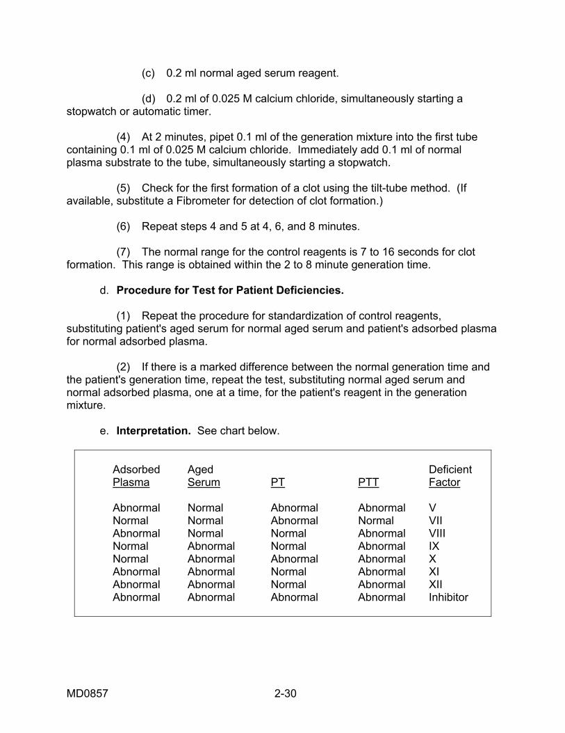
MD0857 2-30
(c) 0.2 ml normal aged serum reagent. (d) 0.2 ml of 0.025 M calcium chloride, simultaneously starting a stopwatch or automatic timer. (4) At 2 minutes, pipet 0.1 ml of the generation mixture into the first tube containing 0.1 ml of 0.025 M calcium chloride. Immediately add 0.1 ml of normal plasma substrate to the tube, simultaneously starting a stopwatch. (5) Check for the first formation of a clot using the tilt-tube method. (If available, substitute a Fibrometer for detection of clot formation.) (6) Repeat steps 4 and 5 at 4, 6, and 8 minutes. (7) The normal range for the control reagents is 7 to 16 seconds for clot formation. This range is obtained within the 2 to 8 minute generation time. d. Procedure for Test for Patient Deficiencies. (1) Repeat the procedure for standardization of control reagents, substituting patient's aged serum for normal aged serum and patient's adsorbed plasma for normal adsorbed plasma. (2) If there is a marked difference between the normal generation time and the patient's generation time, repeat the test, substituting normal aged serum and normal adsorbed plasma, one at a time, for the patient's reagent in the generation mixture. e. Interpretation. See chart below.
Adsorbed Aged Deficient Plasma Serum PT PTT Factor Abnormal Normal Abnormal Abnormal V Normal Normal Abnormal Normal VII Abnormal Normal Normal Abnormal VIII Normal Abnormal Normal Abnormal IX Normal Abnormal Abnormal Abnormal X Abnormal Abnormal Normal Abnormal XI Abnormal Abnormal Normal Abnormal XII Abnormal Abnormal Abnormal Abnormal Inhibitor
MD0857 2-31
f. Sources of Error. (1) All reagents must be freshly prepared each day. (2) Tubes of plasma substrate left at room temperature are unreliable. (3) All U.S.P. barium sulfate preparations are not standard in reaction. Factor IX at times may be incompletely removed. (4) Antithromboplastin activity in blood prolongs TGT, and accounts for some double deficiencies. (5) This is not a routine laboratory procedure; considerable technologist skill is required. (6) A clot can form when the generation mixture is prepared. This is removed with a wooden applicator stick. (7) Other sources of error can be found in paragraphs 2-15d and 2-16d. g. Discussion. (1) A platelet deficiency can be detected by the TGT. This is done by preparing platelet-rich plasma and substituting it for the partial thromboplastin reagent. An abnormal generation time is noted in a patient with platelet deficiency. (2) The TGT reveals abnormalities essential to the development of thromboplastin activity in the intrinsic blood system. (3) The TGT should be correlated with the PT and PTT to determine the specific abnormality. (4) Adsorbed plasma contains factors V, VIII, XI, and XII. If the TGT is prolonged when patient's adsorbed plasma is substituted for normal adsorbed plasma, a deficiency in one or more of these factors is indicated. (5) Aged serum contains factors IX, X, XI, and XII. If the TGT is prolonged when patient's aged serum is substituted for normal aged serum, a deficiency in one or more of these factors is indicated. (6) Factors XI and XII are present in adsorbed plasma and aged serum. A deficiency in either of these factors prolongs the TGT. Determination of the specific factor deficiency is done by the physician on the basis of clinical grounds. h. Normal Values. Clot formation time of 7 to 16 seconds within 8 minutes generation time.
MD0857 2-32
2-19. THROMBIN TIME a. Principle. A known amount of thrombin is added to plasma and the time required for clot formation is recorded. Results are affected by abnormal fibrinogen levels, heparin, and antithrombins. This is useful in the diagnosis of DIC. b. Reagents. (1) Thrombin, 5000 units per ml. Available from the Federal Supply Catalog. (2) Stock thrombin solution, 500 units per ml. Reconstitute the thrombin with 10 ml of saline (0.85 percent NaCl). (3) Working thrombin solution, 10 Units per ml. Dilute the stock solution 1:50 with saline (0.85 percent NaCl). (4) Control plasma. Available from the Federal Supply Catalog. c. Procedure. (1) Obtain 4.5 ml of venous blood and add to 0.5 ml sodium citrate. Mix well. (2) Centrifuge and separate the plasma. (3) Incubate patient plasma, control plasma, and thrombin solution in 37ºC water bath for 3 minutes. (4) Pipet 0.1 ml of control plasma and 0.1 ml of thrombin solution into a clean test-tube, simultaneously starting a stopwatch. Observe for clot formation. (5) Repeat step (4) for the patient's plasma. d. Sources of Error. See paragraph 6-16d. e. Discussion. (1) The thrombin time is prolonged in fibrinogen levels below 100 mg per dl, presence of fibrinolysins, and presence of circulating anticoagulants. (2) Low concentrations of heparin in the patient's plasma may not be observed by this test. To detect small amounts of heparin, thrombin is diluted, and normal patient's plasma thrombin times are determined. A prolongation of the patient's thrombin time over that of the normal at some dilution indicates an antithrombic substance.
MD0857 2-33
(3) To perform the antithrombin test, dilute the working thrombin 1:2, 1:4, 1:8, 1:16, and 1:32 with saline. Then follow the procedure for the thrombin time test using the dilutions. f. Normal Values. 11 to 15 seconds. 2-20. FIBRINOGEN ASSAY (SEMIQUANTITATIVE) a. Principle. Fibrinogen, a plasma globulin formed in the liver, is salted out by ammonium sulfate and measured with a spectrophotometer. b. Reagents. (1) Parfentjev reagent. Add 133.33 g ammonium sulfate, 10.0 g NaCl, and 0.025 g merthiolate to a 1-liter volumetric flask. Dilute to the mark with distilled water. (2) Saline (0.85 percent NaCl). Add 8.5 g sodium chloride to a 1-liter volumetric flask. Dilute to the mark with distilled water. (3) Fibrinogen, U.S.P. Available from the Federal Supply Catalog. (4) Fibrinogen stock standard, 500 mg per dl. Add 500 mg of fibrinogen to a 100-ml volumetric flask. Dilute to the mark with saline. c. Calibration Curve. (1) Prepare the following dilutions of the stock standard: Fibrogen Stock Standard Saline Concentration 10.0 ml 0 500 mg per dl 7.5 ml 2.5 ml 350 mg per dl 5.0 ml 5.0 ml 250 mg per dl 2.5 ml 7.5 ml 125 mg per dl (2) Set up the following cuvettes for each standard: Blank Cuvet Test Cuvette 0.5 ml standard 0.5 ml standard 4.5 ml saline 4.5 Parfentjev reagent
MD0857 2-34
(3) Three minutes after addition of the Parfentjev reagent, shake the cuvettes vigorously, and read the absorbances of the test cuvettes at 510 nm with the blank set at zero absorbance. (4) Plot absorbance versus concentration on linear graph paper. d. Procedure. (1) Draw 4.5 ml of fresh venous blood and add to a test tube containing 0.5 ml of 3.8 percent sodium citrate. (2) Centrifuge and separate the plasma. (3) Set up the following cuvettes for each unknown plasma: Blank Unknown 0.5 ml plasma 0.5 ml plasma 4.5 saline 4.5 Parfentjev reagent (4) Three minutes after addition of the Parfentjev reagent, shake the cuvets vigorously, and read the absorbances of the test cuvettes at 510 nm with the blank set at zero absorbance. (5) Obtain the fibrinogen concentration from the calibration curve. e. Sources of Error. (1) The Parfentjev reagent deteriorates after two weeks of storage. Prepare fresh every 2 weeks. (2) The fibrinogen has a potency for 60 months. Do not use outdated fibrinogen to prepare standards. (3) The procedure is limited as it is dependent on time, temperature, anticoagulant, and concentration of other proteins. f. Discussion. (1) Although the procedure is limited, a rapid determination can be obtained when adequate fibrinogen levels are present. (2) Screening procedures, in kit form, are available commercially.
MD0857 2-35
(3) Fibrinogen is essential to clot formation in stage 3 of the clotting mechanism. Bleeding is encountered when the plasma fibrinogen level falls below 75 mg per dl. (4) A colorimetric fibrinogen procedure is outlined in AFM 160-49. g. Normal Values. 200 to 400 mg per dl. 2-21. PLASMA RECALCIFICATION TIME a. Principle. Calcium chloride is added to plasma and the clotting time recorded. b. Reagents. (1) Calcium chloride, 0.025 M, information is at paragraph 2-18b(5). (2) Sodium citrate, 3.8 percent, is available from the Federal Supply Catalog. (3) Sodium chloride, 0.85 percent (w/v). c. Procedure. (1) Draw 4.5 ml of fresh venous blood and add to 0.5 ml of 3.8 percent sodium citrate. (2) Centrifuge at 2,500 rpm for 20 minutes and separate the plasma. (3) Incubate, at 37ºC for 2 to 3 minutes prior to each test, each of the following, in separate test tubes: (a) Patient's platelet-poor plasma. (b) Normal platelet-poor control plasma. (c) Calcium chloride, 0.025 M. (d) Sodium chloride, 0.85 percent. (4) Into a 13 x 100 mm test tube, in the 37ºC water bath, pipet 0.1 ml 0.85 percent sodium chloride and 0.1 ml of patient's plasma. Mix. (5) Blow in 0.1 ml 0.025 M calcium chloride and simultaneously start a stopwatch.
MD0857 2-36
(6) Allow the tube to remain in the 37ºC water bath for 90 seconds, tilting the tube gently every 30 seconds. (7) After 90 seconds, remove the tube from the water bath and gently tilt. Stop the watch as soon as a clot forms, and record the results. d. Sources of Error. (1) Platelet-poor plasma must be used in the test. Platelet-rich plasma shortens the recalcification time. (2) The test is dependent on the platelet count, concentration of plasma clotting factors, time of storage in glass, and the presence of circulating anticoagulants. (3) See paragraph 2-15d for other sources of error. e. Discussion. (1) This test is the basis for other coagulation procedures. (2) A decrease in any of the clotting factors present in the intrinsic system will cause a prolonged clotting time. f. Normal Values. 90 to 250 seconds. 2-22. DETECTION OF A CIRCULATING ANTICOAGULANT a. Principle. An abnormal recalcification time is not corrected by the addition of normal plasma if a circulating anticoagulant is present. b. Reagents. See paragraph 2-21b. c. Procedure. (1) Obtain citrated plasma from a normal donor and from the patient.
MD0857 2-37
(2) Set up the following mixture in 12 x 75 mm test tubes: Tube Patient's Normal % Patient's No. Plasma Plasma Plasma 1 0.20 ml 0.00 100% 2 0.15 ml 0.05 ml 75% 3 0.10 ml 0.10 ml 50% 4 0.05 ml 0.15 ml 25% 5 0.02 ml 0.18 ml 10% 6 0.00 0.20 ml Control (3) Add 0.2 ml of 0.025 M. CaCl2 to each tube and determine the plasma recalcification time (see para 2-21) on each tube. d. Interpretation. (1) The abnormal recalcification time is corrected by normal plasma if no circulating anticoagulant is present. (2) A circulating anticoagulant present in patient's plasma prolongs the recalcification time of normal plasma. 2-23. DETECTION OF FACTOR XIII DEFICIENCY a. Principle. Factor XIII converts a loosely-linked, fibrin clot (in the presence of calcium ions) into a tough gel. The well-formed clot is insoluble in 5 M urea. In the absence of factor XIII, the clot lyses within 3 hours. b. Reagents. (1) Sodium citrate, 3.8 percent. Available from the Federal Supply Catalog. (2) Calcium chloride, 0.05 M. Add 0.555 g anhydrous calcium chloride to a 100 ml volumetric flask. Dilute to the mark with distilled water. (3) Urea solution, 5 M. Add 30 g of urea to a 100-ml volumetric flask. Dilute to the mark with distilled water. c. Procedure. (1) Draw 4.5 ml of fresh venous blood and add to 0.5 ml of 3.8 percent sodium citrate. (2) Centrifuge at 2,500 rpm for 5 minutes and separate plasma.

MD0857 2-38
(3) Pipet 0.5 ml of patient's plasma into each of two test tubes. Repeat, pipeting 0.5 ml of normal control plasma into each of two additional tubes. (4) Add 0.5 ml of 0.05 M calcium chloride to the four tubes. (5) Incubate the resulting fibrin clots at 37ºC for 30 minutes. (6) Loosen the clots from the sides of the test tubes by gently tapping the sides of the tube. (7) Transfer one of the patient's clots and one of the normal control clots to respective tubes containing 5 ml of 5 M urea. Transfer both the remaining patient clot and the normal control clot to a third tube containing 5 ml of 5 M urea. (8) Allow the mixtures to stand at room temperature. (9) Check the clots at the end of 1 hour, 2 hours, 3 hours, and 24 hours, and note if the clots have dissolved. d. Interpretation. A clot that lyses within 3 hours is deficient in factor XIII. 2-24. PLATELET COUNT (UNOPETTE) a. Principle. Unopette Test is a stable in vitro diagnostic reagent system for the enumeration of leukocytes and platelets in whole blood. Whole blood is added to diluent, the tonicity of which lyses red cells but preserves platelets, leukocytes and reticuloytes. b. Reagent. Unopette reservoir- containing 1.98 ml of diluent mixture. 11.45 gm of ammonium oxalate. 1.0 gm sorensen’s phosphate buffer. 0.1 gm thimersat and purified water. Unopette capillary pipette. c. Procedure. (1) Puncture diaphram in unopette reservoir (2) Remove shield and draw blood into the unopette capillary tube. (3) Add the whole blood in capillary tube to diluent in reservoir, squeeze gently two to three times, let stand for 10 minutes, the diluted specimen is good for 3 hours at room temperature (4) Prepare a moist chamber by placing a moist piece of filter paper in the top of a Petri dish. Paper should be moist enough to adhere to the Petri dish.
MD0857 2-39
(5) Discharge a few drops from the pipet. Then charge both chambers of a hemacytometer. (6) Allow the platelets to settle for 10 minutes. To prevent drying, place the moist chamber over the hemacytometer. (7) Under 10x magnification, leukocytes will appear refractile, under 40x magnification platelets appear oval to round. (8) Count all (9) large squares of counting chamber. d. Calculations. Add 10 percent of count to total number of cells counted. Multiply by 100 to get total leukocyte count. The formula for calculation of indirect platelet count is as follows.
Plts/L = # cells counted X dilution factor X area factor X depth factor X 106
OR
Plts/L = # cells counted X "K" factor X 106
e. Sources of Error. (1) Platelet agglutinates invalidate the count. This is prevented by using scrupulously clean glassware. Therefore, the following precautions are recommended: (a) Boil counting chambers and coverslips for 2 hours in distilled water to which a small amount of sodium bicarbonate has been added. (b) Pipets must be very carefully cleaned. (c) The diluting fluid must be frequently filtered or centrifuged and stored at 2o to 4oC. (2) Other sources of error as listed under red and white blood cell counts also apply, including pipetting errors. These procedures should be consulted. (3) More accuracy may be obtained through use of the phase contrast microscope.
MD0857 2-40
f. Discussion. (1) No one method for the enumeration of thrombocytes is satisfactory in every respect. In experienced hands the direct procedure is more accurate than the indirect method, and venous blood samples are more representative and preferable to those obtained by capillary puncture. Direct procedures, however, require greater skill and experience for proper performance and interpretation. The error in a single count has been estimated to be about 10 percent and this margin of error can be reduced by multiple counts. (2) A major disadvantage of the direct thrombocyte count is that the platelets must be counted under high-power rather than oil-immersion magnification as is used in the indirect method. Because of the extremely small size of blood platelets, it is a common error to confuse them with yeasts, debris, and precipitated stain. (3) Due to the inherent errors in this procedure, it is recommended that a thrombocyte count be performed on a normal person as a control. g. Normal Values. 150,000 to 350,000 platelets per cu mm. NOTE: This range is generally acceptable, but it must be realized that there is a great variance in normal values which differ with each technique, laboratory, and technician. 2-25. PLATELET COUNT (PHASE MICROSCOPY) a. Principle. Blood is diluted with ammonium oxalate, and the diluted specimen is introduced into a counting chamber. The platelets appear round or oval, pink, purple, or even black under a phase condenser. b. Reagent. Ammonium oxalate, 1%. Add 1g of ammonium oxalate to a 100-ml volumetric flask. Dilute to the mark with distilled water. Store in the refrigerator and filter before use. c. Procedure. (1) Draw 5 ml of venous blood and immediately place in a test tube containing EDTA. (If capillary blood is used, immediately fill a red blood cell diluting pipet to the 1 mark.) (2) Fill a red blood cell diluting pipet to the 1 mark and then draw ammonium oxalate to the 101 mark. (3) Shake the pipet 10 to 15 minutes.

MD0857 2-41
(4) Discard from 1/3 to 1/2 the volume from an RBC pipet, then charge a special flat bottom-phase counting chamber. Place the counting chamber in a Petri dish containing moist gauze and let stand 15 to 20 minutes to ensure complete settling of platelets. (5) Count the platelets in all 25 squares of the area normally used for the red cell count. Multiply the results by 1,000. (6) Both sides of each chamber are filled with the same sample; the count from either side should not deviate more than 10% from the other count. If a greater than 10 percent deviation occurs, repeat the count using a fresh dilution and a second chamber. (7) Report the average of the two sides counted. d. Sources of Error. (1) The diluting fluid must be fresh and free of bacterial contamination. (2) Platelet clumping occurs if there is delay in adding blood to the anticoagulant or if the mixing is inadequate. Platelet clumps cause invalid results. (3) Occasionally extraneous material is mistaken for platelets. (4) Glassware that is not scrupulously clean causes platelets to attach to the debris on the glassware. (5) See paragraph 2-24e for other sources of error. e. Discussion. (1) Platelets sometimes show dendritic processes. Structures such as dirt, crystals, and WBCs are refractile. Platelets are not. The white cells are normally lysed; however, with patients that demonstrate excessive high white counts some cells will be observed in the platelet counting prep. (2) Ammonium oxalate ensures clearing of the background by hemolysis. (3) This method is more accurate than the Rees-Ecker. The main drawback to this method is that special equipment is required. (4) See paragraph 2-24f for further discussion. f. Normal Values. 150,000 to 350,000 per cu mm.

MD0857 2-42
2-26. MACROGLOBULIN DISTILLED WATER SCREENING TEST a. Principle. Macroglobulins are precipitated when brought into contact with distilled water. b. Procedure. (1) Collect 5 ml of blood from the patient by venipuncture. (2) Set the blood aside for 1 hour to clot, and then separate the serum by centrifuging at 2,000 rpm for 5 minutes. NOTE: Oxalated, citrated, or heparinized plasma is also satisfactory for the performance of this test. (3) Place 5 ml of distilled water in a Wassermann tube. (4) With a 1 ml pipet, draw up 0.5 ml of patient's serum or plasma. Holding the pipet tip close to the meniscus of the water, add 0.2 ml of the patient's serum or plasma to the distilled water. (5) Interpret results. c. Interpretation. (1) Observe the behavior of the drop of serum or plasma sinking in the water and the modifications of color of the solution. (2) If macroglobins are present: (a) The drop will sink leaving a smoky trace. (b) The entire distilled water medium will promptly assume a whitish color, due to the condensation of transparent, slimy masses forming a precipitate which quickly sinks to the bottom. (3) The separated precipitate will dissolve well when transferred to a test tube containing normal saline.

MD0857 2-43
2-27. CRYOGLOBULIN SCREENING TEST a. Principle. Cryoglobulins in serum or plasma are precipitated at 4ºC and dissolved by temperature elevation to 37oC. b. Procedure. (1) Collect 10 ml of blood by venipuncture and place a 5 ml portion in a tube containing anticoagulant (citrate, oxalate, or heparin) and place the other 5 ml portion into a clean, sterile test tube. (2) Incubate both tubes immediately for 1 hour at 37oC. (3) One hour later, separate the supernatant plasma from the tube containing the anticoagulant and the serum from the clot in the untreated tube. (4) Transfer aliquots of 1 ml of serum into 2 clean sterile Wassermann tubes. (5) Transfer aliquots of 1 ml of plasma into 2 clean sterile Wassermann tubes. (6) Incubate 1 serum specimen and 1 plasma specimen at 4ºC (refrigerator temperature) for 4 hours. (7) Incubate 1 serum specimen and 1 plasma specimen at 37ºC for 4 hours. (8) Read results. c. Interpretation. (1) The test is positive if the serum or plasma incubated at 37ºC shows no changes and the serum or plasma incubated at 4ºC shows the following changes: (a) The serum or plasma appears clotted in toto. (b) The plasma or serum appear divided into two layers; the upper one containing normal plasma or serum, faintly stained by bilirubin; the lower one represented by the cryoglobulin, whitish in color, which has precipitated to the bottom of the test tube. (2) The test is confirmed if the serum or plasma incubated at 4ºC is brought back to a temperature of 37ºC and its appearance returns to normal.
MD0857 2-44
2-28. FIBRIN/FIBRINOGEN DEGREDATION (SPLIT) PRODUCTS a. Principle. In the normal production of clot formation, fibrinogen is acted upon by thrombin, breaking off small molecules from the fibrinogen molecule. These small molecules are fibrinopeptides D and E (fibrinogen split products). Once these molecules are broken off, the main molecule that remains is the fibrin monomer (A & B). It is this monomer that polymerizes to form the fibrin threads of a clot. In addition to the split products from the action of thrombin on fibrinogen, additional split products are produced by the action of fibrinolysin on fibrinogen and plasma in both fibrinogen and fibrin clots. When large amounts of FSP's are produced or are present, prior to adsorption antifibrinogen antiserum with an affected individual's serum, they may interfere with polymerization of fibrin monomers, preventing clot formation (DIC). b. Specimen and Reagents. (1) Venous blood in a special blue top vacutainer-type tube does not contain the anticoagulant found in blue top tubes, but a clot activator, specific for FDP assay. The tube contains thrombin to promote clotting, and soybean trypsin inhibitor to prevent in vitro fibrinolysis. This is sufficient to collect 2 ml whole blood or urine. (2) Use the FDP Assay Kit from Wellcome Reagents Div., Burroughs Wellcome Co., Research Triangle Pk, NC 27709 c. Procedure. (1) Prepare dilutions of serum samples. Take 2- 12 x 75 mm test tubes and label them #1 and #2. (2) Mark 2 of the rings on the glass slide #1 and #2. (3) Using the graduated dropper provided with the bottle of buffer solution in the kit, place 0.75 ml of glycine buffer into each test tube. (4) Using 1 of the plastic droppers (provided with the kit), with the teat provided (blue), aspirate clear serum. (5) Place 5 drops of serum into test tube #1 and 1 drop of serum into test tube #2. (6) Mix the contents of each test tube: Dilution #1 = 1:5. #2 = 1:20. (7) Rinse the plastic dropper with glycine buffer.

MD0857 2-45
(8) Transfer 1 drop from test tube #2 to position #2 on the slide. (9) Now take 1 drop from test tube #1 and place it in position #1 on the slide. NOTE: Since you're going from a lesser to greater concentration, you must pipette in this order. (10) Mix the latex suspension by shaking the container vigorously for a few minutes. (11) Add 1 drop of latex suspension to each position on the slide. (12) Stir the serum/latex mixture with a wooded applicator, spreading the mixture to fill the entire circle. (13) Rock the slide gently back and forth for a maximum of 2 minutes, while observing for agglutination. Agglutination means that there is at least 2 μg/ml present. d. Calculations. (1) If agglutination is present, there is at least 2 μg/ml present in that position. (2) If agglutination occurs in #1, concentration is greater than 10 ug/ml. If agglutination occurs in #2, concentration is greater than 40 μg/ml. NOTE: Additional serial dilutions may be made and tested accordingly.
10 μg/ml 40 μg/ml Dilutions: 1:2 20 80 1:4 40 160
e. Controls. Controls should be run with each batch of tests. Positive control should contain 5 to 10 μg/ml FDP. Negative control should contain less than 2 μg/ml of FDP. f. Range of Values. Normal values are below 10 μg/ml. In cases of DIC, levels of FDP exceed 10 μg/ml, and in severe cases can exceed 40 μg/ml.
MD0857 2-46
2-29. FACTOR V ASSAY (SIMILAR FOR FACTORS VII & X) a. Principle. The patient's plasma is mixed with a commercially known deficient plasma. It is then tested, by doing a Prothrombin Time test on the mixture, and compared on a logarithmic chart. Factors V, VII, and X are synthesized in the liver and are decreased in severe liver disease. b. Material and Equipment Required. (1) Citrated plasma from a blue top, vacutainer-type tube. (2) Commercially prepared factor (specific) deficient plasma and Log-Log chart paper. (3) Any device for performing prothrombin time testing. c. Procedure. (1) Prepare the graph from normal citrated plasma (see chart below).
Control for making graph aliquots of normal citrated plasma.
100% -- Undiluted (100% = 0.2 ml plasma). 50% -- Diluted with imidazole buffered saline (50% = 0.2 ml plasma + 0.2 ml IBS). 25% -- Diluted with imidazole buffered saline (25% = 0.1 ml plasma + 0.3 ml IBS). 10% -- Diluted with imidazole buffered saline (10% = 0.1 ml plasma + 0.9 ml IBS).
(2) Set up a series of five 10 x 75 mm test tubes. a) 1st tube -- 0.01 ml undiluted plasma. (b) 2nd tube -- 0.01 ml 50% dilution. (c) 3rd tube -- 0.01 ml 25% dilution. (d) 4th tube -- 0.01 ml 10% dilution. (e) 5th tube -- no normal plasma (optional).
MD0857 2-47
(3) To the first 4 tubes, add 0.09 ml factor V (or VII or X) deficient plasma, and mix. (4) Proceed with performing a regular prothrombin time analysis. (5) The above mixtures are in a ratio of 1:9, where 1 is normal and 9 is deficient. (6) Record the results on a double-logarithmic (Log-Log) scale. (7) For each patient to be analyzed, use 100% and a 50% dilution. (8) Read the results off the graph. d. Controls. Any normal coagulation controls will do. Any abnormal coagulation controls will do. e. Normal Values. Normal patients should show values from 80 to 120 of the normal control. NOTE: Refrigeration preserves factor V, but after 24 hours, even at 4oC, factor V is greatly reduced. NOTE: Factor VII is a vitamin K dependent factor. 2-30. FACTOR VIII ASSAY (SIMILAR FOR FACTORS IX, XI, & XII) a. Principle. The patient's plasma is mixed with a commercially known deficient plasma. It is then tested, by doing a activated partial thromboplastin test on the mixture, and compared on a logarithmic chart. Factors VIII, IX, XI, and XII are analyzed using this procedure. b. Materials, Equipment, and Specimen Required. (1) Citrated plasma from a blue top, vacutainer-type tube. (2) Commercially prepared factor (specific) deficient plasma and Log-Log chart paper. (3) Any device for performing activated partial thromboplastin time testing.
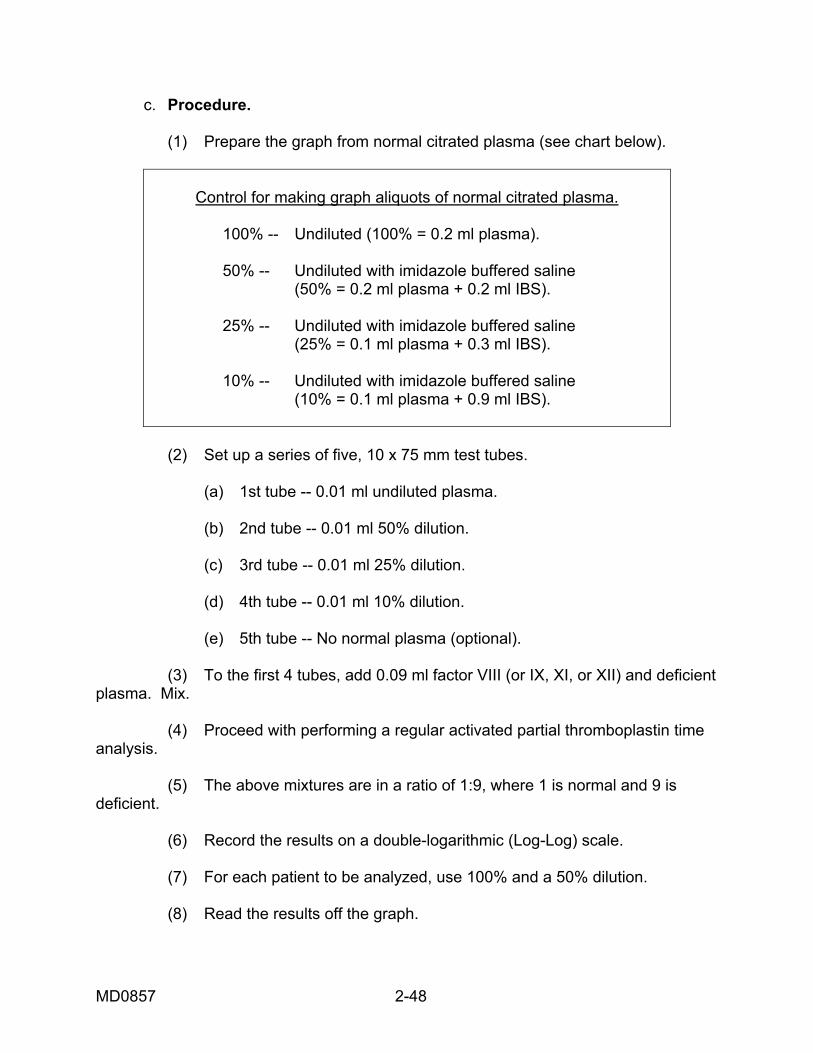
MD0857 2-48
c. Procedure. (1) Prepare the graph from normal citrated plasma (see chart below).
Control for making graph aliquots of normal citrated plasma.
100% -- Undiluted (100% = 0.2 ml plasma). 50% -- Undiluted with imidazole buffered saline (50% = 0.2 ml plasma + 0.2 ml IBS). 25% -- Undiluted with imidazole buffered saline (25% = 0.1 ml plasma + 0.3 ml IBS). 10% -- Undiluted with imidazole buffered saline (10% = 0.1 ml plasma + 0.9 ml IBS).
(2) Set up a series of five, 10 x 75 mm test tubes. (a) 1st tube -- 0.01 ml undiluted plasma. (b) 2nd tube -- 0.01 ml 50% dilution. (c) 3rd tube -- 0.01 ml 25% dilution. (d) 4th tube -- 0.01 ml 10% dilution. (e) 5th tube -- No normal plasma (optional). (3) To the first 4 tubes, add 0.09 ml factor VIII (or IX, XI, or XII) and deficient plasma. Mix. (4) Proceed with performing a regular activated partial thromboplastin time analysis. (5) The above mixtures are in a ratio of 1:9, where 1 is normal and 9 is deficient. (6) Record the results on a double-logarithmic (Log-Log) scale. (7) For each patient to be analyzed, use 100% and a 50% dilution. (8) Read the results off the graph.

MD0857 2-49
d. Control. Any normal coagulation controls will do. Any abnormal coagulation controls will do. e. Normal Values. Normal patients should show values from 80 to 120 percent of the normal control.
Continue with Exercises
MD0857 2-50
EXERCISES, LESSON 2 INSTRUCTIONS: Answer the following exercises by marking the lettered responses that best answers the exercise, by completing the incomplete statement, or by writing the answer in the space provided at the end of the exercise. After you have completed all of the exercises, turn to "Solutions to Exercises" at the end of the lesson and check your answers. For each exercise answered incorrectly, reread the material referenced with the solution. 1. Which of the following statements is correct? a. Hemostasis plays a small part in stopping the flow of blood. b. Hemostasis plays a large part in stopping the flow of blood. c. Hemostasis is a vascular system. d. Hemostasis represents 15 to 20 percent of platelet protein. 2. Coagulation is one component in: a. Fibrinolysis. b. Hemostasis. c. Disruption of the intrinsic system. d. Vascular dilatation. 3. What major role do blood platelets perform? a. In the hemostatic process, blood platelets swell, congeal, and adhere to blood vessel wall to plug the site of the injury. b. They contract. c. They stimulate collagen. d. Platelets extend pseudopodia.

MD0857 2-51
4. Which occurs last in the clotting process? a. Formation of thrombin. b. Formation of thromboplastin. c. Aggregation of platelets. d. Formation of fibrin network. 5. Which of the following lists coagulation factors I through IV in order? a. Calcium, fibrinogen, prothrombin, and Christmas. b. Fibrinogen, prothrombin, thromboplastin, and calcium. c. Prothrombin, calcium, fibrinogen, and thromboplastin. d. Thromboplastin, prothrombin, calcium, and fibrinogen. 6. Which is the Stuart Prower factor? a. Factor V. b. Factor VII. c. Factor X. d. Factor XIII. 7. Thromboplastin eliminates the need for what item in stage I? a. Globulin. b. The accelerator, proconvertin. c. High concentration of serum and plasma. d. Platelets.
MD0857 2-52
8. What are the two systems that make up the coagulation process? a. Vascular and hemolysis. b. Extrinsic and intravascular. c. Intravascular and vascular. d. Intrinsic and extrinsic. 9. Coagulation factor XIII produces a tough gel through: a. Increasing the serum content. b. Stabilizing serum. c. Converting a loosely linked fibrin clot with the help of calcium ions. d. Decreasing the conversion of prothrombin with the help of calcium ions. 10. The synthesis of prothrombin takes place in the liver and requires the presence of: a. Folic acid. b. Vitamin A. c. Vitamin B12. d. Vitamin K. 11. Prothrombin is the precursor of: a. Fibrin. b. Fibrinogen. c. Thrombin. d. Thromboplastin.
MD0857 2-53
12. A ___________ deficiency is unlikely to interfere with coagulation because clinical tetany would intervene. a. Calcium. b. Fibrinogen. c. Prothrombin. d. Factor VIII. 13. Which factor is thought to be an accelerator in the conversion of prothrombin to thrombin? a. Factor VII. b. Factor VIII. c. Factor IX. d. Factor XII. 14. Which of the following is consumed during the clotting process and is therefore not present in serum? a. Factor V. b. Factor VII. c. Factor IX. d. Factor XI. 15. Which platelet factor activates prothrombin? a. 2. b. 3. c. 4. d. 7.

MD0857 2-54
16. Hemophilias A and B are hereditary deficiencies of which coagulation factors, respectively? a. VII and IX. b. VIII and IX. c. XI and VIII. d. XI and IX. 17. Platelet factor 3 is found in the platelet _______________ and is a ______________ component of platelets. a. Membrane; extrinsic. b. Plasma; extrinsic. c. Membrane; intrinsic. d. Plasma; intrinsic. 18. A fibrin clot is ultimately formed by the interaction of fibrinogen and: a. Calcium. b. Thrombin. c. Thromboplastin. d. Platelets. 19. The formation of thrombin from prothrombin occurs in what stage? a. Stage I. b. Stage II. c. Stage III. d. Stage IV.
MD0857 2-55
20. During the clot lysis stage, what happens to the blood clot when plasmin interacts with it? a. Enlarges. c. Shrivels up like an old red blood cell. c. Enlongates like an ameoba. d. Dissolves into fragments. 21. One anticoagulant that inhibits the clotting activity of blood is: a. Coumarin derivitives. b. Fibrin. c. Throboplastin. d. Proconvertin. 22. In the Template method for determining the bleeding time, the blood pressure cuff is inflated to: a. 20 mm Hg. b. 30 mm Hg. c. 40 mm Hg. d. 50 mm Hg. 23. The normal bleeding time with the Template method is: a. Up to 9 minutes. b. 3 to 10 minutes. c. 10 to 15 minutes. d. 15 to 20 minutes.
MD0857 2-56
24. What should be the temperature of the water bath for the whole blood clotting time? (NOTE: The whole blood clotting time discussed in paragraph 2-12 of the text is also referred to as the coagulation time or clotting time.) a. 10oC. b. 20oC. c. 37oC. d. 56oC. 25. Which of the variables listed below does NOT decrease the clotting time of whole blood? a. Excessive agitation of the blood specimen. b. Air bubbles in the blood. c. Tissue fluid in the blood. d. Performing test at room temperature. 26. Using the Lee-White method for whole blood clotting, what should be done to tube #1 after tube #2's blood has clotted for 30 seconds? a. Place it straight up for no blood to flow. b. Tilt it until no flow blood is observed. c. Tilt it upside down for blood to flow. d. Gently tilt it upward. 27. The clot retraction is normal in hemophilia because of: a. A reduced number of platelets. b. A normal number of platelets. c. Increased prothrombin activity. d. The presence of calcium.
MD0857 2-57
28. To measure clot retraction, we can use tubes previously used to determine: a. Coagulation time. b. Prothrombin time. c. Prothrombin consumption time. d. Thromboplastin generation time. 29. The tourniquet test is positive in: a. Purpuras. b. Hemophilia. c. Polycythemia. d. Agranulocytosis. 30. The partial thromboplastin time cannot be used to reveal a deficiency of: a. Factors V or X. b. Factors XI or XII. c. Prothrombin or fibrinogen. d. Factor VII or platelet factor 3. 31. Which of the following values for the activated partial thromboplastin time is normal? a. 30 seconds. b. 50 seconds. c. 1 minute, 10 seconds. d. 1 minute, 30 seconds.
MD0857 2-58
32. The anticoagulant used in preparing the patient's plasma for the one-stage prothrombin time is: a. 0.2 ml of EDTA. b. 0.5 ml of 3.2 percent sodium citrate. c. 0.5 ml of 1.34 percent sodium citrate. d. 0.1 ml of 5 percent potassium oxalate. 33. The prothrombin time is sensitive to a deficiency of factor: a. VII. b. VIII. c. IX. d. XI. e. XII. 34. A prothrombin consumption time of less than 20 seconds indicates a deficiency in: a. Fibrinogen. b. Prothrombin. c. Factor V, VII, or X. d. Platelet and factors VIII, IX, XI, or XII. 35. What is the most important difference between the procedures for the one-stage prothrombin time and the prothrombin consumption time? a. Sensitivity. b. Sources of error. c. Blood fraction tested.
MD0857 2-59
36. Deficiencies of which two factors cannot be distinguished from each other by using the thromboplastin generation time, the prothrombin time, and the partial thromboplastin time? a. V and VII. b. VIII and IX. c. X and XI. d. XI and XII. 37. Which of the following is NOT used to determine the thrombin time? a. Patient's plasma. b. Thrombin solution. c. Thromboplastin reagent. d. A 37ºC water bath. 38. The thrombin time is sensitive to a deficiency of: a. Factor V. b. Factor VII. c. Fibrinogen. d. Prothrombin. 39. Which of the following plasma concentrations of fibrinogen falls in the normal range? a. 50 mg/dl. b. 100 mg/dl. c. 300 mg/dl. d. 500 mg/dl.
MD0857 2-60
40. The plasma recalcification time is used in which of the following? a. Coagulation time. b. Thrombin time. c. Detection of factor XIII deficiency. d. Detection of a circulating anticoagulant. 41. If a clot lyses within three hours after being placed in 5M urea, which factor is deficient? a. Fibrin. b. Platelets. c. Factor VIII. d. Factor XIII. 42. In the Rees-Ecker platelet count, what is the platelet count if a total of 100 platelets are counted in the two center 1-sq mm areas? a. 50,000 per cu mm. b. 100,000 per cu mm. c. 150,000 per cu mm. d. 200,000 per cu mm.
MD0857 2-61
43. During the microscopy phase of the platelet count, what is the appearance of the platelets in the counting chamber after the blood has been diluted with ammonium oxalate? a. Round or oval. b. Pink. c. Purple. d. Or even black under a phase condenser. e. All of the above. f. None of the above. 44. Once the diluted blood has been left to stand for 15 to 20 minutes, what method is used to count the platelets and what are the results multiplied by? a. Count the platelets in all 25 squares of the area normally used for the RBCs and then multiply those results by 1,000. b. Count the platelets in 6 squares of the area normally used for the WBCs and then multiply those results by 1,000. c. Count the platelets and then multiply those results by 2,000. d. Count the platelets in all 40 squares of the area normally used for the RBCs and then multiply those results by 500. 45. What other items could be used to precipitate macroglobulins besides using distilled water? a. Oxalated or heparinized plasma or calcium dioxide. b. Oxalated, citrated, or heparinized plasma. c. Cresyl blue, citrated, or heparinized plasma. d. Oxalated, citrated, or heparinized plasma or formaldehyde.
MD0857 2-62
46. When interpretating the results of the macroglobulin distilled water screening test, what do you observe? a. The behavior of the drop of serum or plasma sinking in the water and the modifications of color of the solution. b. The behavior of the drop of serum or plasma remaining stationary in the water and the modifications of color of the solution. c. The behavior of the drop of serum or plasma rising in the water and the modifications of color of the solution. d. The behavior of the drop of serum or plasma sinking in the water and the retention of the color of the solution. 47. What confirms that serum or plasma tests positive when incubated at 4ºC during the cryoglobulin screening test? a. If the serum or plasma turns a bright green color upon returning to a temperature of 37oC, then the test is confirmed. b. The test is confirmed if the serum or plasma is brought back to a temperature of 37ºC and its appearance returns to normal. c. If the serum or plasma turns a dull yellow color upon returning to a temperature of 37oC, then the test is confirmed. d. The test is confirmed if the serum or plasma is brought back to a temperature of 44ºC and its appearance returns to normal. 48. What is a range of values for the fibrin/frininogen degredation products? a. Normal is above 10 ug/ml. b. DIC levels of FDP are below 8 ug/ml. c. Severe cases exceed 40 ug/ml. d. Normal levels are at 10ug/ml.
MD0857 2-63
49. In performing a factor V assay, what percentage of dilution should the second tube be with 0.01 ml of imidazole buffered saline? a. 100. b. 60. c. 50. d. 25. 50. Which factors are analyzed using the factor VIII assay procedure? a. Factors II and III. b. Factors V, VI, and VII. c. Factors I and IX. d. Factors VIII, IX, XI, and XII.
Check Your Answers on Next Page
MD0857 2-64
SOLUTIONS TO EXERCISES, LESSON 2 1. b (para 2-1a) 2. b (para 2-2a) 3. a (para 2-1(b)) 4. d (para 2-2a) 5. b (paras 2-3a-d, table 2-1) 6. c (para 2-3j) 7. d (para 2-3c) 8. d (para 2-2c) 9. c (para 2-3m) 10. d (para 2-3b) 11. c (para 2-3b) 12. a (para 2-3d) 13. a (para 2-3g) 14. a (para 2-3e) 15. b (para 2-4c) 16. b (paras 2-3h, i) 17. c (para 2-4c) 18. b (para 2-7c, figure 2-1) 19. b (para 2-7b) 20. d (para 2-7d)
MD0857 2-65
21. a (para 2-8) 22. c (para 2-11c(5)) 23. a (para 2-11f) 24. c (para 2-12b) 25. d (para 2-12d NOTES (1), (2)) 26. b (para 2-12c(9)) 27. b (para 2-13f(2)) 28. c (para 2-13(a)) 29. a (para 2-14g(4)) 30. c (para 2-15f) 31. a (para 2-15g(1)) 32. b (para 2-16b) 33. a (para 2-16f(5)) 34. d (paras 2-17e(1), (4)) 35. c (paras 2-16f(5), 2-17) 36. d (paras 2-18e, g(6)) 37. c (paras 2-19b, c) 38. c (para 2-19e(1)) 39. c (para 2-20g) 40. d (paras 2-22a, c(3))
MD0857 2-66
41. d (para 2-23d) 42. b (para 2-24d) 43. e (para 2-25a) 44. a (para 2-25c) 45. b (para 2-26b(1) NOTE) 46. a (para 2-26c(1)) 47. b (para 2-27c(2)) 48. c (para 2-28f) 49. c (para 2-29c(2)(b)) 50. d (para 2-30)
End of Lesson 2
MD0857 3-1
LESSON ASSIGNMENT LESSON 3 Anemia TEXT ASSIGNMENT Paragraphs 3-1 through 3-11. LESSON OBJECTIVES After completing this lesson, you should be able to: 3-1. Identify major laboratory manifestations of anemia’s. 3-2. Identify erythrocyte indices. 3-3. Identify morphology of RBCs associated with elevated, normal, and decreased indices. 3-4. Calculate the RBC indices and interpret the results. 3-5. Identify laboratory procedures used for assessment of anemia. 3-6. Identify pathophysiological categories of anemia. 3.7. Identify classification of anemia’s by morphology. 3.8. Identify macrocytic anemia and list the lab findings associated with it. 3-9. Identify normocytic anemia and lab findings associated with normocytic anemia. 3.10. Identify microcytic anemia and lab findings associated with the microcytic anemia. SUGGESTION After completing the assignment, complete the exercises at the end of this lesson. These exercises will help you to achieve the lesson objectives.
MD0857 3-2
LESSON 3
ANEMIA
Section I. DIAGNOSING ANEMIA
3-1. INTRODUCTION a. Physicians and clinical laboratory scientists counted RBC’s in measured volumes to detect anemia or polycythemia. Anemia means loss of oxygen-carrying capacity and is often reflected in a reduced RBC count. Polycythemia means an increased RBC count related to increase body RBC mass. b. Major laboratory manifestations of anemia: (1) Decreased hemoglobin (Hgb) concentration. (3) Reduced packed cell volume hematocrit (Hct). (3) Decreased number of RBCs/L. c. Laboratory testing procedures for assessing anemia: (1) Hemoglobin and hematocrit - most widely used. (2) Red blood cell count. (3) Red blood cell indices: (a) Calculations used to define the size of and hemoglobin concentration within red blood cells. (b) May be calculated manually but are usually calculated by automated instruments. (c) RBC indices, combined with an examination of the RBCs on a stained smear, tell the examiner whether the RBCs are normocytic, microcytic, macrocytic (size related), and normochromic, or hypochromic (Hgb content related). 3-2. ERYTHROCYTE INDICES a. Mean Corpuscular Volume (MCV): (1) Indicates the average volume of the RBCs in femtoliters (fL). (2) Expressed in SI units as femtoliter (fL; 1 fL = 10–15 L).
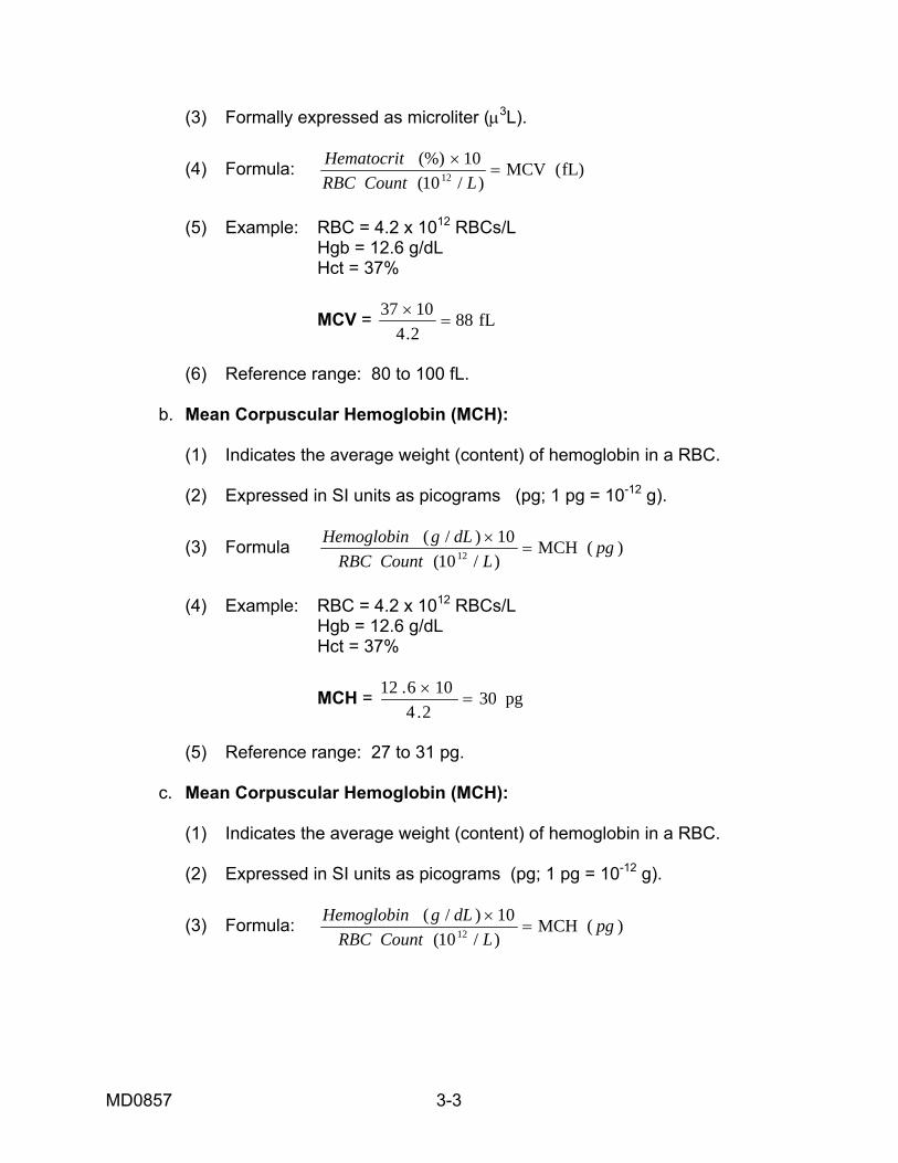
MD0857 3-3
(3) Formally expressed as microliter (μ3L).
(4) Formula: fL)(MCV)/10(
10(%)12 =×
LCountRBCHematocrit
(5) Example: RBC = 4.2 x 10¹² RBCs/L Hgb = 12.6 g/dL Hct = 37%
MCV = fL882.41037
=×
(6) Reference range: 80 to 100 fL. b. Mean Corpuscular Hemoglobin (MCH): (1) Indicates the average weight (content) of hemoglobin in a RBC. (2) Expressed in SI units as picograms (pg; 1 pg = 10-12 g).
(3) Formula )(MCH)/10(10)/(
12 pgLCountRBC
dLgHemoglobin=
×
(4) Example: RBC = 4.2 x 10¹² RBCs/L Hgb = 12.6 g/dL Hct = 37%
MCH = pg302.4
106.12=
×
(5) Reference range: 27 to 31 pg. c. Mean Corpuscular Hemoglobin (MCH): (1) Indicates the average weight (content) of hemoglobin in a RBC. (2) Expressed in SI units as picograms (pg; 1 pg = 10-12 g).
(3) Formula: )(MCH)/10(10)/(
12 pgLCountRBC
dLgHemoglobin=
×
MD0857 3-4
(4) Example: RBC = 4.2 x 10¹² RBCs/L Hgb = 12.6 g/dL Hct = 37%
MCH = pg302.4
106.12=
×
(5) Reference range: 27 to 31 pg. d. Mean Corpuscular Hemoglobin Concentration (MCHC): (1) Calculated from Hemoglobin and Hematocrit and is an expression of the average concentration of hemoglobin in the RBC. (2) Expressed in SI units as g/dL. (3) Formerly expressed as percent (%).
(4) Formula: )%/(MCHC(%)
100)/( ordLgHematocrit
dLgHemoglobin=
×
(5) Example: RBC = 4.2 x 10¹² RBCs/L Hgb = 12.6 g/dL Hct = 37%
MCHC = %org/dL34%371006.12
=×
(6) Reference range: 31 to 36 g/dL (or %). 3-3. MORPHOLOGY OF RBC’S ASSOCIATED WITH VARYING INDICES a. MCV and Expected RBC Morphology. (1) Elevated MCV (↑ 100 fL) – Macrocytic RBCs. (2) Normal MCV (80 – 100 fL) – Normocytic RBCs. (3) Decreased MCV (↓ 80 fL) – Microcytic RBCs.
MD0857 3-5
b. MCH and Expected RBC Morphology. (1) Elevated MCH (↑ 31 pg) - usually macrocytic/normochromic RBCs. (2) Normal MCH (27 – 31 pg) - usually normocytic/normochromic RBCs. (3) Decreased MCH (↓ 27 pg) - variable macrocytic to microcytic / normochromic to hypochromic RBCs. NOTE: MCH rarely used alone; results should be correlated with MCV and MCHC. c. MCHC and Expected RBC Morphology. (1) Elevated MCHC (↑ 36 g/dL) – Normochromic never use the misnomer “hyperchromic”: (a) Hereditary spherocytosis. (b) Usually MCHC does not rise above 37 g/dL - at 37 g/dL, hemoglobin becomes gel-like and at higher concentrations crystallization may occur. (c) If greater than 38 g/dL, check specimen for cold agglutinins and lipemia. Lipemia may cause falsely elevated hemoglobin values, thus elevating the MCHC calculations. (2) Normal MCHC (31 – 36 g/dL) -- normochromic RBCs. (3) Decreased MCHC (↓ 31 g/dL) -- hypochromic RBCs. NOTE: RBC color intensity is directly proportional to the cell's hemoglobin concentration. When reviewing a peripheral smear, the color of the RBCs should correspond to the MCHC value.
Section II: LABORATORY ASSESSMENT OF ANEMIA 3-4. INTRODUCTION a. To determine or detect the presence of anemia, the clinical laboratory professional performs a complete blood count on a hematology cell analyzer to determine the RBC count, hemoglobin, hematocrit, RBC indices, white blood cell count, and platelet count. In this section practice questions will be provided to reiterate how to calculate and interpret the usages of RBC indices in diagnosing anemia.
MD0857 3-6
b. Calculate the RBC indices. (1) A patient was treated in the Emergency Room for severe lacerations and possible abdominal injuries as a result of an automobile accident. Results of the initial CBC were: HGB: 10.5 g/dL HCT: 34% RBC: 3.8 x 10¹²/L WBC: 12.0 x 10⁹/L (2) A patient's CBC results were: HGB: 5.6 g/dL HCT: 24% RBC: 3.5 x 10¹²/L WBC: 10.5 x 10⁹/L 3-5. LABORATORY EVALUATIONS/PROCEDURES a. Red cell distribution width (RDW). (1) Indicates the degree of anisocytosis (RBC size variation). (2) Calculated value using the MCV and RBC count. (3) Useful in early detection of iron deficiency anemia but should not replace other diagnostic tests. b. RBC morphology. c. WBC count, platelet count, and morphology. (Manual Differential). d. Reticulocyte count. e. Examination of bone marrow smears. f. Malarial smears. g. Osmotic fragility test - measures the ability of the red cells to take up fluids without lysing. h. Sugar Water Screening Test. i. Ham’s Acidified Serum Test.
MD0857 3-7
j. Sickle cell testing and hemoglobin electrophoresis. k. Pyruvate kinase - enzyme in the Embden-Meyerhof pathway. l. Folic acid (folate) and vitamin B12 levels -- important in DNA synthesis. m. Serum Iron levels. 3-6. PATHOPHYSIOLOGICAL CATEGORIES OF ANEMIA a. Blood loss - acute or chronic. b. Decreased or impaired red blood cell production. (1) Damage to or suppression of the bone marrow. (2) Deficiency of erythropoietic growth factors such as erythropoietin. (3) Abnormal metabolism of iron or insufficient iron storage causing problems in heme synthesis. Includes Vitamin and mineral deficiencies. (4) Defect in globin synthesis. c. Accelerated destruction of the RBC. (1) Intrinsic defects within the RBC (hereditary). (a) Membrane defects or deficiencies. (b) Enzyme defects or deficiencies. (c) Hemoglobinopathies. (d) Defective globin synthesis. (e) Acquired paroxysmal nocturnal hemoglobinuria (PNH). (2) Extracorpuscular causes -- non-immune acquired hemolytic anemia. (a) Chemicals, toxins, venoms. (b) Physical trauma due to cardiac replacement valves, burns, disseminated intravascular coagulation (DIC). (c) Infectious agents such as malaria.
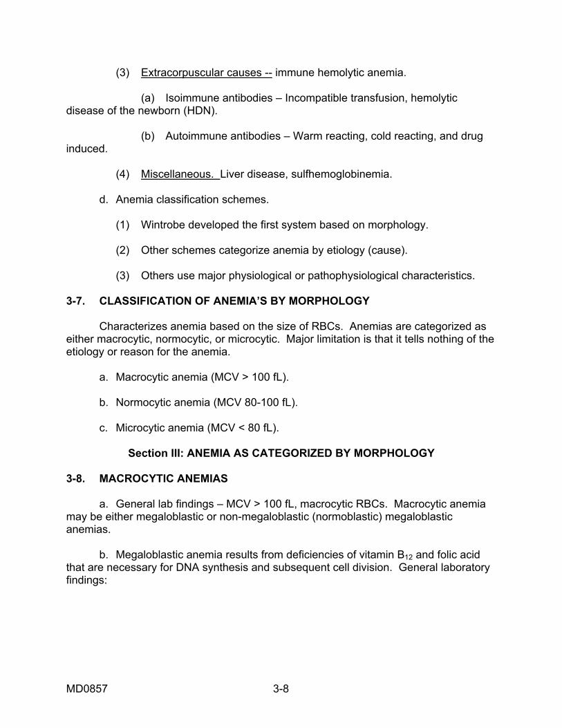
MD0857 3-8
(3) Extracorpuscular causes -- immune hemolytic anemia. (a) Isoimmune antibodies – Incompatible transfusion, hemolytic disease of the newborn (HDN). (b) Autoimmune antibodies – Warm reacting, cold reacting, and drug induced. (4) Miscellaneous. Liver disease, sulfhemoglobinemia. d. Anemia classification schemes. (1) Wintrobe developed the first system based on morphology. (2) Other schemes categorize anemia by etiology (cause). (3) Others use major physiological or pathophysiological characteristics. 3-7. CLASSIFICATION OF ANEMIA’S BY MORPHOLOGY Characterizes anemia based on the size of RBCs. Anemias are categorized as either macrocytic, normocytic, or microcytic. Major limitation is that it tells nothing of the etiology or reason for the anemia. a. Macrocytic anemia (MCV > 100 fL). b. Normocytic anemia (MCV 80-100 fL). c. Microcytic anemia (MCV < 80 fL).
Section III: ANEMIA AS CATEGORIZED BY MORPHOLOGY 3-8. MACROCYTIC ANEMIAS a. General lab findings – MCV > 100 fL, macrocytic RBCs. Macrocytic anemia may be either megaloblastic or non-megaloblastic (normoblastic) megaloblastic anemias. b. Megaloblastic anemia results from deficiencies of vitamin B12 and folic acid that are necessary for DNA synthesis and subsequent cell division. General laboratory findings:
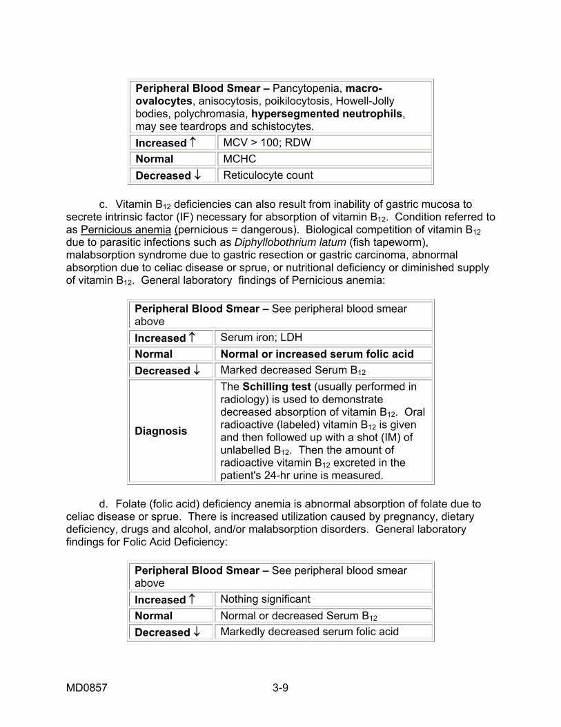
MD0857 3-9
Peripheral Blood Smear – Pancytopenia, macro-ovalocytes, anisocytosis, poikilocytosis, Howell-Jolly bodies, polychromasia, hypersegmented neutrophils, may see teardrops and schistocytes. Increased ↑ MCV > 100; RDW Normal MCHC Decreased ↓ Reticulocyte count
c. Vitamin B12 deficiencies can also result from inability of gastric mucosa to secrete intrinsic factor (IF) necessary for absorption of vitamin B12. Condition referred to as Pernicious anemia (pernicious = dangerous). Biological competition of vitamin B12 due to parasitic infections such as Diphyllobothrium latum (fish tapeworm), malabsorption syndrome due to gastric resection or gastric carcinoma, abnormal absorption due to celiac disease or sprue, or nutritional deficiency or diminished supply of vitamin B12. General laboratory findings of Pernicious anemia:
Peripheral Blood Smear – See peripheral blood smear above Increased ↑ Serum iron; LDH Normal Normal or increased serum folic acid Decreased ↓ Marked decreased Serum B12
Diagnosis
The Schilling test (usually performed in radiology) is used to demonstrate decreased absorption of vitamin B12. Oral radioactive (labeled) vitamin B12 is given and then followed up with a shot (IM) of unlabelled B12. Then the amount of radioactive vitamin B12 excreted in the patient's 24-hr urine is measured.
d. Folate (folic acid) deficiency anemia is abnormal absorption of folate due to celiac disease or sprue. There is increased utilization caused by pregnancy, dietary deficiency, drugs and alcohol, and/or malabsorption disorders. General laboratory findings for Folic Acid Deficiency:
Peripheral Blood Smear – See peripheral blood smear above Increased ↑ Nothing significant Normal Normal or decreased Serum B12 Decreased ↓ Markedly decreased serum folic acid
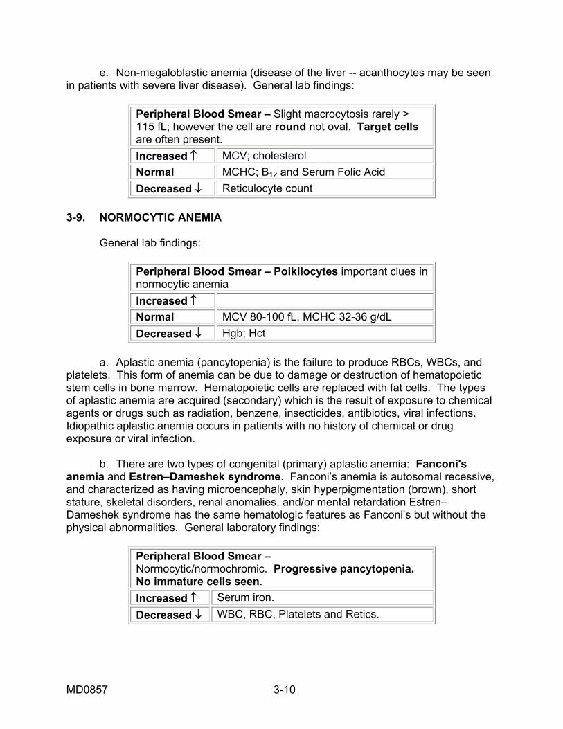
MD0857 3-10
e. Non-megaloblastic anemia (disease of the liver -- acanthocytes may be seen in patients with severe liver disease). General lab findings:
Peripheral Blood Smear – Slight macrocytosis rarely > 115 fL; however the cell are round not oval. Target cells are often present. Increased ↑ MCV; cholesterol Normal MCHC; B12 and Serum Folic Acid Decreased ↓ Reticulocyte count
3-9. NORMOCYTIC ANEMIA General lab findings:
Peripheral Blood Smear – Poikilocytes important clues in normocytic anemia Increased ↑ Normal MCV 80-100 fL, MCHC 32-36 g/dL Decreased ↓ Hgb; Hct
a. Aplastic anemia (pancytopenia) is the failure to produce RBCs, WBCs, and platelets. This form of anemia can be due to damage or destruction of hematopoietic stem cells in bone marrow. Hematopoietic cells are replaced with fat cells. The types of aplastic anemia are acquired (secondary) which is the result of exposure to chemical agents or drugs such as radiation, benzene, insecticides, antibiotics, viral infections. Idiopathic aplastic anemia occurs in patients with no history of chemical or drug exposure or viral infection. b. There are two types of congenital (primary) aplastic anemia: Fanconi's anemia and Estren–Dameshek syndrome. Fanconi’s anemia is autosomal recessive, and characterized as having microencephaly, skin hyperpigmentation (brown), short stature, skeletal disorders, renal anomalies, and/or mental retardation Estren–Dameshek syndrome has the same hematologic features as Fanconi’s but without the physical abnormalities. General laboratory findings:
Peripheral Blood Smear – Normocytic/normochromic. Progressive pancytopenia. No immature cells seen. Increased ↑ Serum iron. Decreased ↓ WBC, RBC, Platelets and Retics.

MD0857 3-11
c. Pure red cell aplasia is characterized has having suppressed RBC production with normal WBC and platelet production. The types are congenital erythroid hypoplasia (Diamond-Blackfan anemia) defective erythroid stem cell; slow, progressive anemia. Acute acquired pure RBC aplasia occurs as a result of hemolytic anemia, infections, malnutrition, and drug therapy, and chronic acquired pure RBC aplasia, an autoimmune disease in which IgG antibody attacks erythroid precursors. Thymoma (tumor of the thymus gland) is a frequent finding. General laboratory findings:
Peripheral Blood Smear – Severe Normocytic/normochromic to slightly macrocytic anemia. Increased ↑ MCV Normal WBC and Platelet count Decreased ↓ RBC and Reticulocyte count
d. Anemia of chronic disorders (ACD) is the term used for anemia associated with chronic infection, inflammation or malignancy. ACD is the second most common cause of anemia and may be the most common cause of anemia in hospitalized patients. The general causes are decreased erythropoietin level (cytokine produced by activated macrophages during the acute phase of inflammation inhibits erythropoietin), iron block (cytokine also stimulates neutrophils to release lactoferrin, which binds iron. This complex is phagocytized by marrow macrophages, which makes iron unavailable for hemoglobin synthesis.), shortened RBC survival (possibly due to increased activity of macrophages), and suppression of erythropoiesis. General laboratory findings:
Peripheral Blood Smear – Normocytic/normochromic anemia may develop slight hypochromia. Increased ↑ ESR; Serum ferritin Normal WBC and Platelet count Decreased ↓ Retics; Serum iron; TIBC

MD0857 3-12
e. Anemia of chronic renal disease is due to failure of kidneys to produce erythropoietin and decreased bone marrow response to erythropoietin. The severity of anemia directly related to level of blood urea nitrogen levels and uremic plasma is highly toxic to cells. General laboratory findings:
Peripheral Blood Smear – Normocytic/normochromic anemia. Fragmented RBCs and Burr Cells. Basophilic stippling.
Increased ↑ Blood urea nitrogen; Creatinine; ESR; slight neutrophilia;
Normal Platelets Normal to slightly increased, however platelets are often impaired; WBC and Retics.
Decreased ↓ Hct 15 – 30% f. Anemia of endocrine disease is due to a deficiency of hormones that regulate erythropoiesis. (1) Hypothyroidism - erythropoietin production by the kidney depends on the tissue oxygen tension, which is influenced by thyroid hormones. In a hypothyroid condition there will be a decreased tissue oxygen tension resulting in a decreased need for erythropoiesis. Hypothyroidism, when associated with a folic acid or vitamin B12 deficiency, may result in an anemia that is more macrocytic than normocytic such as adrenal abnormalities – Addison disease or Cushing’s disease. (2) Hypogonadism – androgens (testosterone) promote erythropoiesis: by increasing the production of erythropoietin by the kidney and by directly stimulating the marrow in conjunction with erythropoietin. (3) Hypopituitarism - pituitary gland is responsible for secretion of TSH and gonadotropins, which control the production androgens. General laboratory findings:
Peripheral Blood Smear – Normocytic/normochromic anemia. Increased ↑ Normal Retic Decreased ↓

MD0857 3-13
g. Myelophthisic anemia (leukoerythroblastosis) is characterized as having abnormal cells, (e.g., leukemia, replaced bone marrow). General laboratory findings:
Peripheral Blood Smear – Normocytic/normochromic anemia. Polychromasia, Basophilic stippling. Granulocytic shift to the left. Increased ↑ NRBCs, Reticulocytes. Normal Decreased ↓
h. Anemia caused by acute blood loss is usually associated with traumatic conditions such as accident or severe injury. It does not produce an immediate anemia which means there is no change in hematocrit and hemoglobin during the first hours after blood loss due to vasoconstriction. However, severe hemorrhage reduces an individual's total blood volume and produces a condition of shock. The body then adjusts itself by expanding the circulatory volume (fluid from the extravascular spaces enters the blood circulation and has a diluting effect on the remaining cells) which produces the subsequent anemia. General laboratory findings:
Peripheral Blood Smear – Normocytic/normochromic anemia. WBCs exhibit shift to the left. Following severe bleeding, large polychromatic RBCs and NRBCs will be present. Increased ↑ Retics, WBC and Platelet count. Normal Decreased ↓ Serum ferritin
i. Anemia caused by chronic blood loss is indicative in situations where blood loss of small amounts occurs over an extended period of time -- usually after several months. Instead of a dilution problem as with acute blood loss, chronic blood loss results in an iron deficiency anemia. This type is frequently associated with disorders of the gastrointestinal (GI) tract (bleeding ulcer), or extremely heavy menstrual flow. General laboratory findings:
Peripheral Blood Smear – Initially normocytic/normochromicanemia until iron stores are depleted, and then develop into microcytic/hypochromic anemia; Polychromasia. Increased ↑ RDW Normal Leukocytes
Decreased ↓ WBC; Retics; MCV, MCH, MCHC; Serum Iron;

MD0857 3-14
j. Hemolytic anemia- caused by an increase in RBC destruction and/or a decrease in the normal average life span of the RBC. k. RBC membrane defects are discussed below. (1) Hereditary spherocytosis (HS) characterized by a defect in RBC membrane protein (spectrin) composition. It results in weakening of the membrane skeleton and loss of membrane surface area. This is an autosomal dominant trait. The spherocytic cells are abnormally permeable to sodium ion (Na+) causing an influx of sodium at 10 times the normal rate. The cells have reduced cellular flexibility (less deformable than normal) and have shortened survival time due to destruction by the spleen. Some patients compensate and have only slight symptoms while others may be severely anemic. General laboratory findings:
Peripheral Blood Smear – Micro spherocytes; reticulocytes.
Increased ↑ MCHC > 36%; Osmotic fragility; serum bilirubin; retics.
Normal Negative DAT Decreased ↓ Plasma haptoglobin (may be absent) Diagnosis Osmotic Fragility Test
(2) Hereditary elliptocytosis (HE) exists where there is an abnormality in the red cell structural membrane protein spectrin. This is an autosomal dominant trait. The mechanism of hemolysis involves membrane loss, decreased RBC deformability and shortened RBC survival due to splenic destruction. General laboratory findings:
Peripheral Blood Smear –Normocytic/Normochromic; Oval and elongated (elliptocytes) RBCs > 25%; NRBCs and retics are normal in appearance Increased ↑ Serum bilirubin; retics Normal Osmotic fragility test
Decreased ↓ Serum haptoglobin levels when hemolysis is present
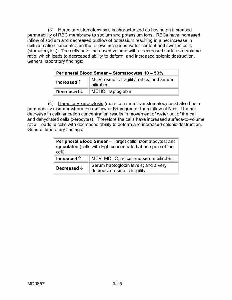
MD0857 3-15
(3) Hereditary stomatocytosis is characterized as having an increased permeability of RBC membrane to sodium and potassium ions. RBCs have increased inflow of sodium and decreased outflow of potassium resulting in a net increase in cellular cation concentration that allows increased water content and swollen cells (stomatocytes). The cells have increased volume with a decreased surface-to-volume ratio, which leads to decreased ability to deform, and increased splenic destruction. General laboratory findings:
Peripheral Blood Smear – Stomatocytes 10 – 50%.
Increased ↑ MCV; osmotic fragility; retics; and serum bilirubin.
Decreased ↓ MCHC; haptoglobin (4) Hereditary xerocytosis (more common than stomatocytosis) also has a permeability disorder where the outflow of K+ is greater than inflow of Na+. The net decrease in cellular cation concentration results in movement of water out of the cell and dehydrated cells (xerocytes). Therefore the cells have increased surface-to-volume ratio - leads to cells with decreased ability to deform and increased splenic destruction. General laboratory findings:
Peripheral Blood Smear – Target cells; stomatocytes; and spiculated (cells with Hgb concentrated at one pole of the cell). Increased ↑ MCV; MCHC; retics; and serum bilirubin.
Decreased ↓ Serum haptoglobin levels; and a very decreased osmotic fragility.
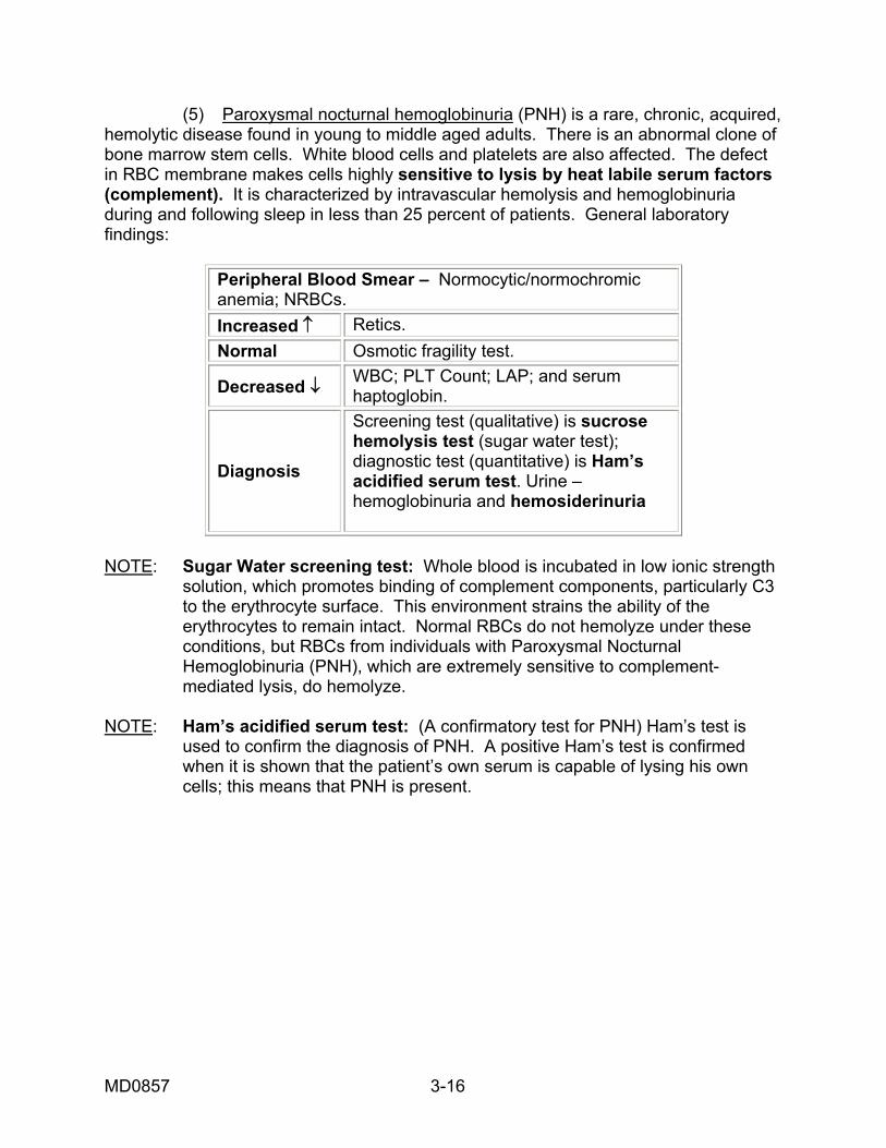
MD0857 3-16
(5) Paroxysmal nocturnal hemoglobinuria (PNH) is a rare, chronic, acquired, hemolytic disease found in young to middle aged adults. There is an abnormal clone of bone marrow stem cells. White blood cells and platelets are also affected. The defect in RBC membrane makes cells highly sensitive to lysis by heat labile serum factors (complement). It is characterized by intravascular hemolysis and hemoglobinuria during and following sleep in less than 25 percent of patients. General laboratory findings:
Peripheral Blood Smear – Normocytic/normochromic anemia; NRBCs. Increased ↑ Retics. Normal Osmotic fragility test.
Decreased ↓ WBC; PLT Count; LAP; and serum haptoglobin.
Diagnosis
Screening test (qualitative) is sucrose hemolysis test (sugar water test); diagnostic test (quantitative) is Ham’s acidified serum test. Urine – hemoglobinuria and hemosiderinuria
NOTE: Sugar Water screening test: Whole blood is incubated in low ionic strength solution, which promotes binding of complement components, particularly C3 to the erythrocyte surface. This environment strains the ability of the erythrocytes to remain intact. Normal RBCs do not hemolyze under these conditions, but RBCs from individuals with Paroxysmal Nocturnal Hemoglobinuria (PNH), which are extremely sensitive to complement- mediated lysis, do hemolyze. NOTE: Ham’s acidified serum test: (A confirmatory test for PNH) Ham’s test is used to confirm the diagnosis of PNH. A positive Ham’s test is confirmed when it is shown that the patient’s own serum is capable of lysing his own cells; this means that PNH is present.
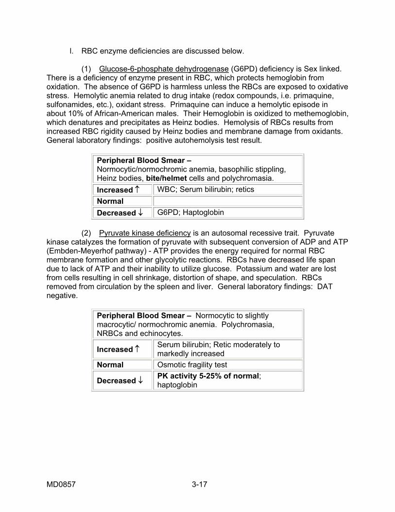
MD0857 3-17
l. RBC enzyme deficiencies are discussed below. (1) Glucose-6-phosphate dehydrogenase (G6PD) deficiency is Sex linked. There is a deficiency of enzyme present in RBC, which protects hemoglobin from oxidation. The absence of G6PD is harmless unless the RBCs are exposed to oxidative stress. Hemolytic anemia related to drug intake (redox compounds, i.e. primaquine, sulfonamides, etc.), oxidant stress. Primaquine can induce a hemolytic episode in about 10% of African-American males. Their Hemoglobin is oxidized to methemoglobin, which denatures and precipitates as Heinz bodies. Hemolysis of RBCs results from increased RBC rigidity caused by Heinz bodies and membrane damage from oxidants. General laboratory findings: positive autohemolysis test result.
Peripheral Blood Smear – Normocytic/normochromic anemia, basophilic stippling, Heinz bodies, bite/helmet cells and polychromasia. Increased ↑ WBC; Serum bilirubin; retics Normal Decreased ↓ G6PD; Haptoglobin
(2) Pyruvate kinase deficiency is an autosomal recessive trait. Pyruvate kinase catalyzes the formation of pyruvate with subsequent conversion of ADP and ATP (Embden-Meyerhof pathway) - ATP provides the energy required for normal RBC membrane formation and other glycolytic reactions. RBCs have decreased life span due to lack of ATP and their inability to utilize glucose. Potassium and water are lost from cells resulting in cell shrinkage, distortion of shape, and speculation. RBCs removed from circulation by the spleen and liver. General laboratory findings: DAT negative.
Peripheral Blood Smear – Normocytic to slightly macrocytic/ normochromic anemia. Polychromasia, NRBCs and echinocytes.
Increased ↑ Serum bilirubin; Retic moderately to markedly increased
Normal Osmotic fragility test
Decreased ↓ PK activity 5-25% of normal; haptoglobin
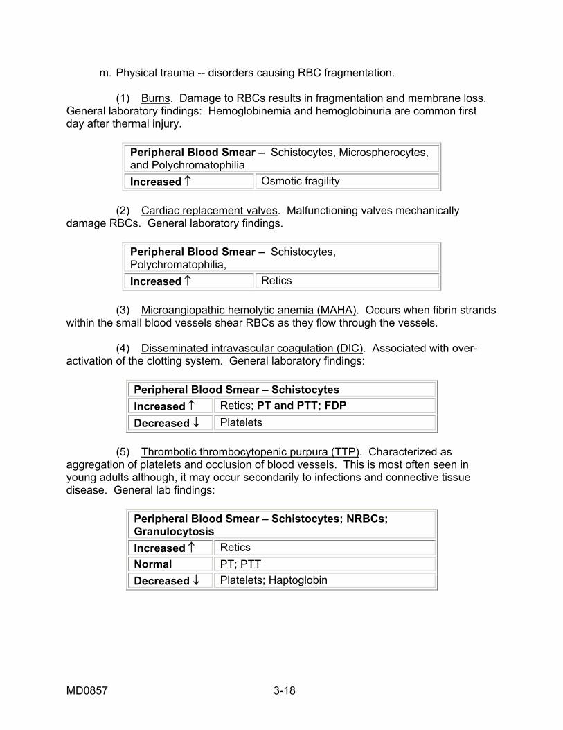
MD0857 3-18
m. Physical trauma -- disorders causing RBC fragmentation. (1) Burns. Damage to RBCs results in fragmentation and membrane loss. General laboratory findings: Hemoglobinemia and hemoglobinuria are common first day after thermal injury.
Peripheral Blood Smear – Schistocytes, Microspherocytes, and Polychromatophilia Increased ↑ Osmotic fragility
(2) Cardiac replacement valves. Malfunctioning valves mechanically damage RBCs. General laboratory findings.
Peripheral Blood Smear – Schistocytes, Polychromatophilia, Increased ↑ Retics
(3) Microangiopathic hemolytic anemia (MAHA). Occurs when fibrin strands within the small blood vessels shear RBCs as they flow through the vessels. (4) Disseminated intravascular coagulation (DIC). Associated with over-activation of the clotting system. General laboratory findings:
Peripheral Blood Smear – Schistocytes Increased ↑ Retics; PT and PTT; FDP Decreased ↓ Platelets
(5) Thrombotic thrombocytopenic purpura (TTP). Characterized as aggregation of platelets and occlusion of blood vessels. This is most often seen in young adults although, it may occur secondarily to infections and connective tissue disease. General lab findings:
Peripheral Blood Smear – Schistocytes; NRBCs; Granulocytosis Increased ↑ Retics Normal PT; PTT Decreased ↓ Platelets; Haptoglobin
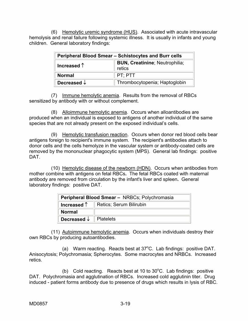
MD0857 3-19
(6) Hemolytic uremic syndrome (HUS). Associated with acute intravascular hemolysis and renal failure following systemic illness. It is usually in infants and young children. General laboratory findings:
Peripheral Blood Smear – Schistocytes and Burr cells
Increased ↑ BUN, Creatinine; Neutrophilia; retics
Normal PT; PTT Decreased ↓ Thrombocytopenia; Haptoglobin
(7) Immune hemolytic anemia. Results from the removal of RBCs sensitized by antibody with or without complement. (8) Alloimmune hemolytic anemia. Occurs when alloantibodies are produced when an individual is exposed to antigens of another individual of the same species that are not already present on the exposed individual’s cells. (9) Hemolytic transfusion reaction. Occurs when donor red blood cells bear antigens foreign to recipient's immune system. The recipient's antibodies attach to donor cells and the cells hemolyze in the vascular system or antibody-coated cells are removed by the mononuclear phagocytic system (MPS). General lab findings: positive DAT. (10) Hemolytic disease of the newborn (HDN). Occurs when antibodies from mother combine with antigens on fetal RBCs. The fetal RBCs coated with maternal antibody are removed from circulation by the infant's liver and spleen. General laboratory findings: positive DAT.
Peripheral Blood Smear – NRBCs; Polychromasia Increased ↑ Retics; Serum Bilirubin Normal Decreased ↓ Platelets
(11) Autoimmune hemolytic anemia. Occurs when individuals destroy their own RBCs by producing autoantibodies. (a) Warm reacting. Reacts best at 37oC. Lab findings: positive DAT. Anisocytosis; Polychromasia; Spherocytes. Some macrocytes and NRBCs. Increased retics. (b) Cold reacting. Reacts best at 10 to 30oC. Lab findings: positive DAT. Polychromasia and agglutination of RBCs. Increased cold agglutinin titer. Drug induced - patient forms antibody due to presence of drugs which results in lysis of RBC.
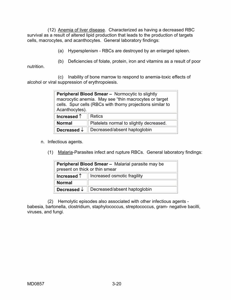
MD0857 3-20
(12) Anemia of liver disease. Characterized as having a decreased RBC survival as a result of altered lipid production that leads to the production of targets cells, macrocytes, and acanthocytes. General laboratory findings: (a) Hypersplenism - RBCs are destroyed by an enlarged spleen. (b) Deficiencies of folate, protein, iron and vitamins as a result of poor nutrition. (c) Inability of bone marrow to respond to anemia-toxic effects of alcohol or viral suppression of erythropoiesis.
Peripheral Blood Smear – Normocytic to slightly macrocytic anemia. May see “thin macrocytes or target cells. Spur cells (RBCs with thorny projections similar to Acanthocytes). Increased ↑ Retics Normal Platelets normal to slightly decreased. Decreased ↓ Decreased/absent haptoglobin
n. Infectious agents. (1) Malaria-Parasites infect and rupture RBCs. General laboratory findings:
Peripheral Blood Smear – Malarial parasite may be present on thick or thin smear Increased ↑ Increased osmotic fragility Normal Decreased ↓ Decreased/absent haptoglobin
(2) Hemolytic episodes also associated with other infectious agents - babesia, bartonella, clostridium, staphylococcus, streptococcus, gram- negative bacilli, viruses, and fungi.
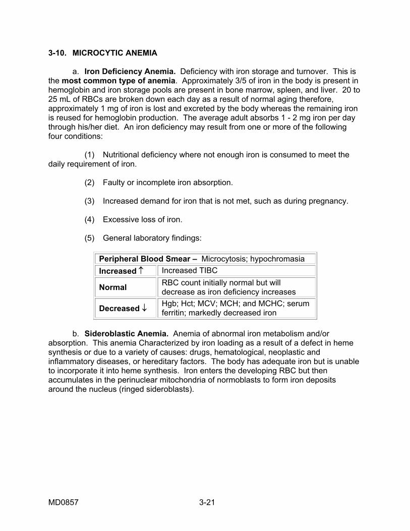
MD0857 3-21
3-10. MICROCYTIC ANEMIA a. Iron Deficiency Anemia. Deficiency with iron storage and turnover. This is the most common type of anemia. Approximately 3/5 of iron in the body is present in hemoglobin and iron storage pools are present in bone marrow, spleen, and liver. 20 to 25 mL of RBCs are broken down each day as a result of normal aging therefore, approximately 1 mg of iron is lost and excreted by the body whereas the remaining iron is reused for hemoglobin production. The average adult absorbs 1 - 2 mg iron per day through his/her diet. An iron deficiency may result from one or more of the following four conditions: (1) Nutritional deficiency where not enough iron is consumed to meet the daily requirement of iron. (2) Faulty or incomplete iron absorption. (3) Increased demand for iron that is not met, such as during pregnancy. (4) Excessive loss of iron. (5) General laboratory findings:
b. Sideroblastic Anemia. Anemia of abnormal iron metabolism and/or absorption. This anemia Characterized by iron loading as a result of a defect in heme synthesis or due to a variety of causes: drugs, hematological, neoplastic and inflammatory diseases, or hereditary factors. The body has adequate iron but is unable to incorporate it into heme synthesis. Iron enters the developing RBC but then accumulates in the perinuclear mitochondria of normoblasts to form iron deposits around the nucleus (ringed sideroblasts).
Peripheral Blood Smear – Microcytosis; hypochromasia Increased ↑ Increased TIBC
Normal RBC count initially normal but will decrease as iron deficiency increases
Decreased ↓ Hgb; Hct; MCV; MCH; and MCHC; serum ferritin; markedly decreased iron

MD0857 3-22
c. Hereditary Sideroblastic Anemia. Decreased d-aminolevulinic acid activity (the first enzyme in the heme synthesis pathway). General lab findings:
Peripheral Blood Smear – Marked hypochromia and microcytosis, target cells, basophilic stippling, and dimorphic RBC population (hypochromic-microcytic and normochromic- normocytic), WBC and PLT counts usually normal
Increased ↑ Increased serum iron;(ringed siderloblasts seen in bone marrow when stained with Prussian blue)
Normal Decreased ↓ Hgb; Hct
d. Primary Idiopathic Sideroblastic Anemia (Refractory Anemia) With Ringed Sideroblasts, Idiopathic Acquired Sideroblastic Anemia. Low d-aminolevulinic acid synthesis or heme synthase (ferrochetalase) activity. General lab findings:
Peripheral Blood Smear – Generally normocytic to slightly macrocytic RBCs. May find small group of hypochromic- microcytic RBCs. Ringed sideroblasts present
Increased ↑
Increased serum iron;(ringed siderloblasts seen in bone marrow when stained with Prussian blue). Serum iron usually increased
Normal WBC and PLT counts Decreased ↓
3-11. POLYCYTHEMIA a. Absolute Polycythemia. Signifies an above normal hemoglobin, hematocrit (greater than 50 percent) and RBC count. (1) Polycythemia vera is a stem cell disorder characterized by excessive proliferation of the erythroid, granulocytic, and megakaryocytic cells - distribution, morphology, and maturation of marrow cells is normal polycythemia. It is characterized by an increased platelet count along with increased blood viscosity which may cause clot formation. Individuals may develop bleeding problems due to abnormal platelet function. It is usually treated with therapeutic phlebotomy - patients develop a mild iron deficiency anemia, which later becomes marked and may terminate into acute leukemia. There are significantly increased hemoglobin, hematocrit, RBC count, WBC count, and platelet count. General laboratory findings:
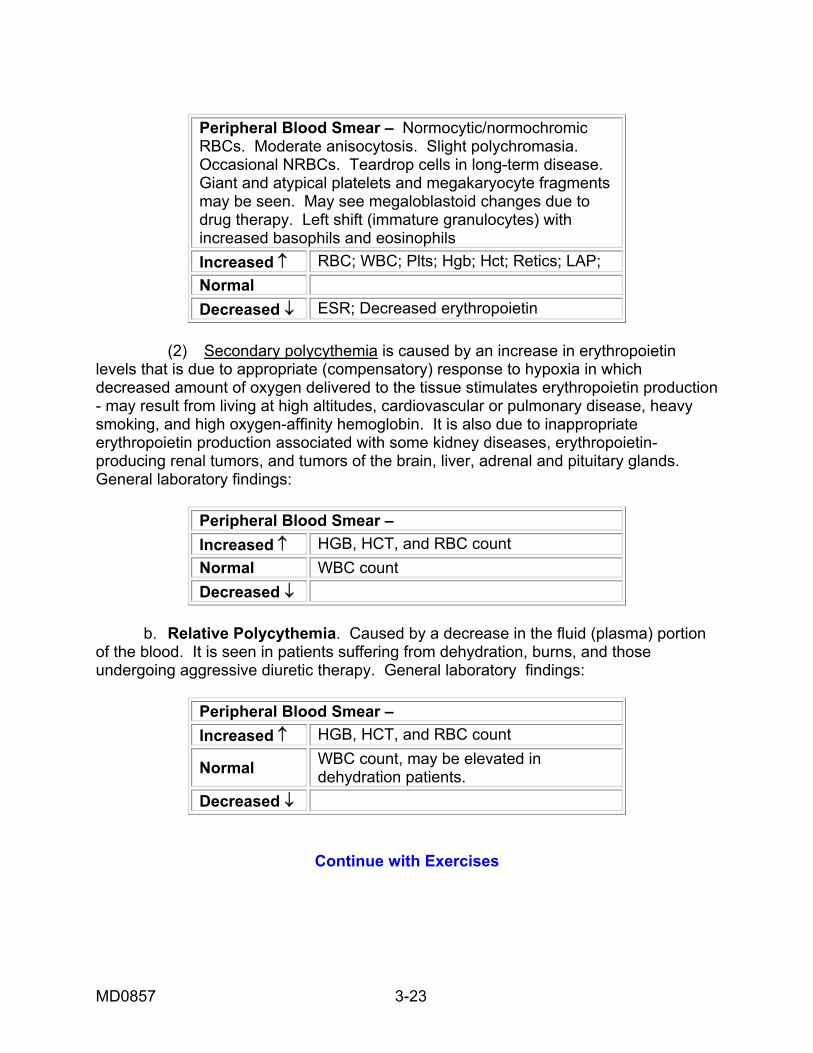
MD0857 3-23
Peripheral Blood Smear – Normocytic/normochromic RBCs. Moderate anisocytosis. Slight polychromasia. Occasional NRBCs. Teardrop cells in long-term disease. Giant and atypical platelets and megakaryocyte fragments may be seen. May see megaloblastoid changes due to drug therapy. Left shift (immature granulocytes) with increased basophils and eosinophils Increased ↑ RBC; WBC; Plts; Hgb; Hct; Retics; LAP; Normal Decreased ↓ ESR; Decreased erythropoietin
(2) Secondary polycythemia is caused by an increase in erythropoietin levels that is due to appropriate (compensatory) response to hypoxia in which decreased amount of oxygen delivered to the tissue stimulates erythropoietin production - may result from living at high altitudes, cardiovascular or pulmonary disease, heavy smoking, and high oxygen-affinity hemoglobin. It is also due to inappropriate erythropoietin production associated with some kidney diseases, erythropoietin-producing renal tumors, and tumors of the brain, liver, adrenal and pituitary glands. General laboratory findings:
Peripheral Blood Smear – Increased ↑ HGB, HCT, and RBC count Normal WBC count Decreased ↓
b. Relative Polycythemia. Caused by a decrease in the fluid (plasma) portion of the blood. It is seen in patients suffering from dehydration, burns, and those undergoing aggressive diuretic therapy. General laboratory findings:
Peripheral Blood Smear – Increased ↑ HGB, HCT, and RBC count
Normal WBC count, may be elevated in dehydration patients.
Decreased ↓
Continue with Exercises

MD0857 3-24
EXERCISES, LESSON 3 INSTRUCTIONS: Answer the following exercises by marking the lettered response that best answers the exercise or best completes the incomplete statement. After you have completed all the exercises, turn to "Solutions to Exercises" at the end of the lesson and check your answers. For each exercise answered incorrectly, reread the material referenced with the solution. 1. What are the three major laboratory manifestations of anemia? a. Increased hemoglobin concentration, decreased RBCs/L, increased hematocrit. b. Reduced hematocrit, decreased RBCs/L, decreased hemoglobin concentration. c. Decreased hemoglobin concentration, decreased hematocrit, increased RBCs/L. d. Increased hemoglobin concentration, increased hematocrit, increased RBCs/L. 2. Which of the following formulas should you use to calculate the mean corpuscular hemoglobin (MCH)? a. (Hct x 10)/RBC count. b. (Hgb x 10)/RBC count. c. (Hgb x 100)/Hct. d. (Hct x 100)/Hbg. 3. (Hgb x 100)/Hct is the formula used to determine? a. MCV (Mean Corpuscular Volume). b. MCH (Mean Corpuscular Hemoglobin). c. MCHC (Mean Corpuscular Hemoglobin Concentration). d. RDW (Red Cell Distribution Width).
MD0857 3-25
4. A patient had a CBC done , the test results were: Hgb 10.0 g/dL; Hct 35%; RBC 4.2 x 10/L; WBC 10.0 x 10/L What is the MCH? _________pg a. 24. b. 83. c. 22. d. 88. 5. What laboratory procedure is not used for assessment of anemia? a. Sugar water screening test. b. Malarial smears. c. Thyroid function test. d. Reticulocyte count. 6. Intrinsic defects within the RBC, extracorpuscular causes, and liver disease fall under which pathophysiological categories of anemia? a. Decreased or impaired red blood cell production. b. Accelerated destruction of the RBC. c. Blood Loss. d. Accelerated repair of the WBC. 7. What kind of RBC morphology would you expect to see on a blood smear from a patient with a MCV of 83 fL? a. Normocytic. b. Macrocytic. c. Microcytic. d. Normochromic.
MD0857 3-26
8. Macroovalocytes, pancytopenia, hypersegmented neutrophils, increased MCV, and decreased reticulocyte count are normally associated with: a. Sideroblastic. b. Megaloblastic. c. Aplastic. d. Myelophthistic. 9. Which of the following is a macrocytic anemia? a. Aplastic anemia. b. Folic acid deficiency. c. Iron deficiency anemia. d. Chronic blood loss. 10. What categories of anemia are aplastic anemia, anemia of endocrine disease, and myelophthisic anemia? a. Normocytic anemia. b. Macrocytic anemia. c. Microcytic anemia. d. Monochromic anemia. 11. The laboratory findings on a peripheral blood smear are burr cells, increased blood urea nitrogen and decreased Hct of 22%. What type of anemia would you suspect? a. Anemia of endocrine disease. b. Anemia caused by blood loss. c. Hemolytic anemia. d. Anemia of chronic renal disease.
MD0857 3-27
12. An increase in RBC destruction and a decrease in the normal average life span of the RBC is defined as: a. Myelophthisic anemia. b. Microcytic anemia. c. Hemolytic anemia. d. Macrocytic anemia. 13. Which of the following is/are hemolytic anemia caused by an RBC enzyme deficiency? a. Pernicious anemia and thalessemia. b. Iron deficiency anemia and folic acid deficiency. c. Pyruvate kinase deficiency and g6pd deficiency. d. Pure red cell aplasia. 14. A patient whose anemia was caused by physical trauma to the cells would usually have _____________ in his peripheral blood smear. a. Stacked cells. b. Acanthocytes. c. Spherocytes. d. Schistocytes. 15. Sideroblastic anemia is commonly: a. Macrocytic. b. Normocytic. c. Microcytic. d. Spherocytic.
MD0857 3-28
16. ________________ RBCs are seen in iron deficiency anemia. a. Normocytic, hyperchromic. b. Macrocytic, normochromic. c. Normocytic, normochromic. d. Microcytic, hypochromic. 17. Secondary polycythemia and polycythemia vera are examples of: a. Absolute polycythemia. b. Myelophthisic anemia . c. Relative polycythemia. 18. Relative polycythemia is caused by: a. Increased erythropoietin. b. A decrease in the plasma portion of the blood. c. Stem cell disorder. d. Decreased erythropoietin. 19. The ________________ is used to screen for cells which are normally sensitive to lysis by complement. a. Bone marrow examination. b. Reticulocyte count. c. Sugar water screening test. d. Activated partial thromboplastin.

MD0857 3-29
20. The reference range for the mean corpuscular hemoglobin concentration (MCHC) is _________ g/dL. a. 31 to 36. b. 27 to 31 . c. 80 to 100. d. 4 to 11. 21. HGB 7 g/dL, RBCs 3.9 x 10¹²/L, HCT 27%, WBCs 11.1 x 10 9/L Based on these indices result, what is the patients MCH? a. 18. b. 67. c. 22. d. 27. 22. Which of the following lab findings is usually associated with anemia caused by acute blood loss? a. Target cells and basophilic stippling. b. Extreme microcytosis. c. Low WBC count and decreased reticulocytes. d. Increased platelet and WBC counts; reticulocytosis. 23. ___________ anemia results from deficiencies of vitamin B12 and folic acid in the body. a. Sideroblastic. b. Megaloblastic. c. Aplastic. d. Myelopthisic.
MD0857 3-30
24. What laboratory findings are associated with paroxysmal nocturnal hemoglobinuria (PNH)? a. Macrocytic. b. Normocytic. c. Microcytic. d. Megaloblastic. 25. Laboratory findings normally associated with hereditary sideroblastic anemia include: a. Acanthocytes, normal to increased reticulocytes. b. Spherocytes, polychromasia, and reticulocytes. c. Dimorphic population of RBCs, target cells, and basophilic stippling. 26. The normal range for the mean corpuscular volume of an erythrocyte is: a. 62-82 fl. b. 70-80 fl. c. 80-97 fl. d. 90-100 fl. 27. A great suppression of RBC production with normal WBC and platelet production is a depiction of: a. Pyruvate kinase deficiency. b. Pure red cell aplasia. c. Paroxysmal nocturnal hemoglobinuria. d. Vitamin B12.
MD0857 3-31
28. Which anemia is usually associated with traumatic conditions such as accidents or severe injury? a. Sideroblastic anemia. b. Hereditary spherocytosis. c. Anemia due to acute blood loss. d. Iron deficiency anemia. 29. Hereditary xerocytosis is characterized as having which of these problems? a. Outflow of Na+ is greater than inflow of K+ and the net decrease in cellular cation concentration results in movement of water out of cell which dehydrates the cell. . b. Outflow of K+ is greater than inflow of Na+ and the net decrease in cellular cation concentration results in movement of water out of cell which dehydrates the cell. c. Outflow of K+ is greater than inflow of Ca+ and the net decrease in cellular cation concentration results in movement of water out of cell which dehydrates the cell. . d. Outflow of K+ is less than inflow of Na+ and the net increase in cellular cation concentration results in movement of water into the cell which hydrates the cell. 30. Acanthocytes are predominantly seen in patients with: a. Severe liver disease. b. Vitamin B12 deficiency. c. Chronic hemolytic anemia. d. Sideroblastic anemia.
MD0857 3-32
31. Celiac disease and sprue are two conditions that affect the body and create an abnormal absorption of what vitamin? a. Thiomine. b. Ferritin. c. Folate. d. Ascorbic acid. 32. Failure of the body to produce RBCs, WBCs, and platelets is known as: a. Anemia. b. Hemoglobin deficiency. c. Pancytopenia. d. Polycythemia vera. 33. Which condition is characterized by decreased tissue oxygen tension resulting in a decreased need for erythropoiesis? a. Hypopituitarism. b. Hypogonadism. c. Hypothyroidism. d. Hyperplasia. 34. Which disorder is characterized by aggregation of platelets and occlusion of blood vessels, most often seen in young adults, and may occur secondarily to infections and connective tissue disease? a. Disseminated intravascular coagulation (DIC). b. Thrombotic thrombocytopenic purpura (TTP). c. a and b. d. None of the above.
MD0857 3-33
35. Sideroblastic anemia is: a. An abnormal iron metabolism and/or absorption where iron loading is a result of a defect in heme synthesis. b. A regular skin eruption across the nose and mouth (butterfly rash), with arthritis that can be accompanied by various visceral manifestations. c. A chronic, sometimes fatal, disease of unknown etiology. d. Sometimes not diagnosed early because the early symptoms do not appear after intense exposure to sunlight. 36. Which is normally present in the clinical findings for macrocytic anemia? a. MCV < 80 pg. b. MCV > 100 fL. c. MCV > 100 pg. d. MCV < 80 fL. 37. What laboratory diagnostic test is calculated using the MCV and RBC count, indicates the degree of anisocytosis, and is useful in the early detection of iron deficiency anemia? a. Complete blood count. b. Ham’s acidified serum test. c. Mean corpuscular hemoglobin concentration. d. RDW.
MD0857 3-34
38. How many types of congenital aplastic anemia have been discovered? a. 5. b. 3. c. 4. d. 2. 39. Which method is the most widely used as a screening test for paroxysmal nocturnal hemoglobinuria (PNH)? a. Ham’s acidified serum test. b. Serum iron levels. c. Sugar water test. d. Schillings test. 40. If a MCHC result is > 38, what is the possible cause of this erroneous result? a. Clotted specimen. b. Cold agglutinins and lipemia. c. Contaminated specimen. d. Decreased platelet count. 41. When exposed to primaquine, which enzyme deficiency results in 10 percent of African Americans experiencing a hemolytic episode? a. Glucose-6-phosphate dehydrogenase (G6PD). b. Pyruvate kinase. c. Protease. d. Lipase.
MD0857 3-35
42. The normal and expected morphology for an elevated MCH is: a. Normochromic/hypochromic RBCs. b. Normochromic/normocytic RBCs. c. Macrocytic/normochromic RBCs. d. Microcytic/hypochromic RBCs. 43. Which parasites infect and rupture RBCs? a. Flea. b. Mosquito. c. Tick. d. Malarial parasites. 44. Increased erythropoietin levels due to compensatory response to hypoxia while living at high altitudes can lead to: a. Autoimmune hemolytic anemia. b. Stem cell disorder. c. Secondary polycythemia. d. Acute hemolytic anemia.
MD0857 3-36
45. Alloimmune hemolytic anemia is caused by: a. Alloantibodies produced by an individual exposed to antigens of another individual that are not already present on the exposed individual’s cells. b. Alloantibodies produced by an individual exposed to antibodies of another individual that are not already present on the exposed individual’s cells. c. Alloantibodies produced by an individual exposed to antigens of the fetus that are already present on the exposed individual’s cells. d. Alloantibodies produced by an individual exposed to antibodies of the fetus that are not already present on the exposed individual’s cells. 46. When antibodies transfer from the mother to the antigens of the fetus’s RBCs what disorder will occur? a. Autoimmune hemolytic anemia. b. Hemolytic disease of the newborn. c. Hemolytic transfusion reaction. d. Immune hemolytic anemia. 47. The first classification scheme was developed by: a. Wintrobe. b. Collins. c. T. Canemia. d. Embden-Meyerhof.
MD0857 3-37
48. Anemia can manifest from all of the following EXCEPT: a. Blood loss. b. Defect in hemoglobin synthesis. c. Healthy diet and exercise. d. RBC membrane defect. 49. Anemia due to Vitamin B12 deficiency may be the result of: a. Over-productive bone marrow processes. b. Inability of iron to be absorbed by the muscles. c. Decreased/suppressed liver function. d. Inability of gastric mucosa to secrete intrinsic factor. 50. Which statement is correct? a. The average adult absorbs 0.1-0.5 mg iron per day through diet. b. 75- 80 mL RBCs are broken down each day as a result of normal aging. c. Platelets possess carbon dioxide and metabolic systems, expend energy, and respond to stimuli. d. Approximately 3/5 of iron in the body is present in hemoglobin and iron storage pools in bone marrow.
Check Your Answers on Next Page

MD0857 3-38
SOLUTIONS TO EXERCISES, LESSON 3 1. b (para 3-1a) 2. b (para 3-2b(3)) 3. c (para 3-2c(3)) 4. a (para 3-2c) 5. c (para 3-5) 6. b (para 3-6c(1)) 7. a (para 3-7a(2)) 8. b (para 3-8b) 9. b (para 3-8d) 10. a (para 3-9) 11. d (para 3-9e) 12. c (para 3-9j) 13. c (para 3-9l(1)(2)) 14. d (para 3-9m) 15. c (para 3-10b) 16. d (par 3-10a) 17. a (para 3-12a) 18. b (para 3-12b) 19. c (para 3-9k(5)NOTE) 20. a (para 3-2d(5))
MD0857 3-39
21. a (para 3-2c(3)) 22. d (para 3-8h) 23. b (para 3-8(c)(d)) 24. b (para 3-9k(5)) 25. a (para 3-10c) 26. b (para 3-2a(6) para 3-3a(2)) 27. b (para 3-9c(1)) 28. c (para 3-9h) 29. b (para 3-9k(4)) 30. a (para 3-8e) 31. c (para 3-8d) 32. c (para 3-9a) 33. c (para 3-9f) 34. b (para 3-9m5) 35. a (para 3-10b) 36. b (para 3-7a(1) para 3-8a ) 37. d (para 3-5a (1-3)) 38. d (para 3-9b) 39. c (para 3-9k(5)) 40. b (para 3-3c(1c))
MD0857 3-40
41. a (para 3-9l(1)) 42. c (para 3-3b(1)) 43. d (para 3-9n(1)) 44. c (para 3-12a(2)) 45. a (para 3-9m(8)) 46. b (para 3-9m(10)) 47. a (para 3-5b(1)) 48. c (para 3-6(a-c)) 49. d (para 3-8c) 50. d (para 3-10a)
End of Lesson 3

MD0857 4-1
LESSON ASSIGNMENT LESSON 4 Leukemias TEXT ASSIGNMENT Paragraphs 4-1 through 4-17. LESSON OBJECTIVES After completing this lesson, you should be able to: 4-1. Identify abnormalities in cells associated with Alder-Reilly anomaly, Chediak-Higashi syndrome, May-Hegglin anomaly, Pelger-Huet anomaly, Gaucher’s disease, and Niemann-Pick disease. 4-2. Compare and contrast infectious mononucleosis, cytomegalovirus infection, and toxoplasmosis. 4-3. Identify the differences between reactive and malignant lymphocytes. 4-4. Select characteristics of acute leukemia with chronic leukemia.
4-5. Identify predominant and/or characteristic cells associated with each type of acute nonlymphoblastic leukemia (ANLL). 4-6. Identify disease states and general characteristic of myelodysplastic syndromes. 4-7 Select characteristics of chronic myeloproliferative disorders. 4-8 Identify laboratory findings seen in the peripheral blood associated with chronic myelocytic leukemia, idiopathic myelofibrosis, and essential thrombocythemia.
4-9. Identify predominate cell population for acute lymphoblastic leukemia and the laboratory findings according to the French American British classification.
MD0857 4-2
4-10. Identify predominant and characteristic cells associated with each of the chronic lymphoproliferative disorders. 4-11. Match the malignant lymphomas disorder with the affected lymphoid tissue or organ and their characteristic cell. 4-12. Identify causes of multiple myeloma, plasma cell leukemia, and Waldenström’s macroglobulinemia and the laboratory findings associated with each of these disorders. 4-13. Identify the function of each special cytochemical stain and its diagnostic use. SUGGESTION After completing the assignment, complete the exercises at the end of this lesson. These exercises will help you to achieve the lesson objectives.
MD0857 4-3
LESSON 4
LEUKEMIAS
Section I. MORPHOLOGIC DISORDERS OF THE LEUKOCYTE 4-1. INTRODUCTION Some inherited morphologic changes are clinically insignificant and of interest only to morphologists, whereas others indicate a life-threatening disorder. Most acquired morphological changes to the granulocytic cells may be used as indicators of disease states in conjunction with their clinical description. 4-2. DISORDERS OF THE GRANULOCYTES (MORPHOLOGIC ABNORMALITIES- CYTOPLASMIC CHANGES) a. Alder-Reilly Anomaly. This prevents normal breakdown of mucopolysaccharides. Mucopolysaccharides are deposited in the cytoplasm of neutrophils, monocytes, and lymphocytes. They appear as abnormally large azurophilic granules resembling severe toxic granulation and contain peroxidase-positive deposits. b. Chediak-Higashi Syndrome. This occurs when large, fused lysosomes are produced and the cells cannot release their contents after bacterial digestion. They appear as large acidophilic granules in granulocytes. This condition leads to abnormal phagocyte function whereas, they do not move well in response to chemotactic stimuli (e.g., poor directional motility), as well as, recurrent infections, bleeding tendencies, hypopigmentation, anemia, neutropenia, thrombocytopenia, and abnormal platelet aggregation. c. May-Hegglin Anomaly. This occurs when large, blue cytoplasmic inclusions resembling giant Dohle bodies in granulocytes and monocytes are present. These inclusions consist mainly of RNA. There is marked thrombocytopenia, instead large, bizarre-shaped platelets and variable neutropenia is observed. Many patients are asymptomatic, some may develop bleeding tendencies. d. Pelger-Huet Anomaly. This is consistent with neutrophilic changes. The neutrophil nucleus fails to segment properly. All neutrophils have bi-lobed or non-segmented nuclei.
MD0857 4-4
4-3. QUALITATIVE DISORDERS a. Defective motility (e.g., Job’s syndrome, lazy leukocyte syndrome). b. Defect in ability to kill microorganisms. (1) Chronic Granulomatous disease. (a) Neutrophils and monocytes ingest, but cannot kill, catalase-positive microorganisms due to a deficiency of membrane-bound oxidase or failure of the membrane surface to stimulate respiratory burst. (b) Bacteria multiply within the cell. (c) Recurrent, life-threatening infections and pneumonia common. (2) Myeloperoxidase deficiency. (a) Absence of myeloperoxidase enzyme from neutrophils and monocytes but not eosinophils - myeloperoxidase mediates oxidative destruction of microorganisms by H2O2. (b) Infections usually are not serious due to compensatory increase in respiratory burst activity - killing of bacteria is slowed but complete. (3) Monocyte-macrophage disorders. (a) Gaucher’s disease. Problem in cellular lipid metabolism - deficiency of enzyme (e.g., beta-glucocerebrosidase) results in inability of monocyte to degrade glucocerebrosides. Cerebroside accumulates in macrophages - Gaucher’s cells. Gaucher’s cell typically is large with one to three eccentric nuclei and wrinkled cytoplasm. There is a decrease in production of RBCs and WBCs as these abnormal cells infiltrate into the bone marrow. (b) Niemann-Pick disease. Problem in cellular lipid metabolism - deficiency of enzyme (e.g., sphingomyelinase) that cleaves sphingomyelin. Sphingomyelin accumulates in macrophages - Niemann-Pick cells (large macrophages with foamy-looking cytoplasm; cytoplasm swollen by numerous small, uniform lipid droplets). This is the most prevalent form of disease develops in infancy and is fatal within the first few years of life.

MD0857 4-5
4-4. NONMALIGNANT LYMPHOCYTIC DISORDERS a. Infectious Mononucleosis. Contagious, viral disease that affects young adults and teenagers - symptoms include fever, sore throat, lymphadenopathy. (1) Epstein-Barr virus (EBV) infects B lymphocytes; T lymphocytes become activated and restrain viral replication in B lymphocytes. (2) Normal to slightly elevated WBC count; lymphocytosis common. (3) Variant (reactive or atypical) lymphocytes: greater than 20 percent (represent T lymphocytes responding to the infection). (4) Positive heterophile antibody test (e.g., Monospot) or EBV-specific serologic test. b. Cytomegalovirus Infection. Closely resembles infectious mononucleosis but patients do not have sore throat or enlarged lymph nodes. (1) Reactive lymphocytosis is present; heterophile antibody is negative. (2) Increased cytomegalovirus titer. c. Toxoplasmosis (Toxoplasma gondii infection). Disease resembles infectious mononucleosis. There is a relative increase in lymphocytes, reactive lymphocytes present on peripheral blood smear; heterophile antibody is negative and there is an elevation of Toxoplasma antibodies. d. Discussion. Differentiation between reactive and malignant lymphocytes. (1) Malignant cells are usually clonal and all abnormal cells appear very similar to one another. (2) Reactive lymphocytes are morphologically more variable - within a single specimen you have both large and small cells, basophilic and pale cells, cells with immature chromatin and cells with densely clumped chromatin.
MD0857 4-6
Section II. CLASSIFICATION OF LEUKEMIAS 4-5. INTRODUCTION Most malignancies of the hematopoietic system are acquired genetic diseases, meaning that most patients are not born with the illness, but acquire sometime later in life. Most WBC malignancies or disorders are not localized but rather are systemic at the initiation of the malignant process. A single leukemia cell arising the bone marrow can obtain passage into the bloodstream and travel to any and all locations of the body, so most treatments given for curative intent of leukocyte neoplasms are not localized but must by nature be systemic-type treatment. The duration of the untreated disease is categorized as either acute or chronic. 4-6. ACUTE LEUKEMIA a. Sudden onset, symptoms of short duration. b. Total WBC count usually elevated, although some patients may have normal to decreased counts. c. Anemia and thrombocytopenia usually present. d. Prognosis of untreated disease - several weeks to several months. NOTE: Median survival time for untreated acute leukemia is three months. Seventy percent of adults and 90 percent of children have at least one remission. 4-7. CHRONIC LEUKEMIA a. Symptoms of long duration. b. Total WBC count ranges from extremely elevated to lower than normal. c. Anemia is not usually present until late in the disease; platelet counts usually normal. d. Prognosis of untreated chronic forms - months to years. e. Type of cells involved: (1) Acute leukemia. Predominance of immature cells (blast and “pro” stage). (2) Chronic leukemia. Predominance of mature cells.
MD0857 4-7
4-8. MYELOPROLIFERATIVE DISORDERS Acute nonlymphoblastic leukemias (ANLL). General characteristics: a. Mutant stem cells give rise to a monoclonal population of myeloid cells - impaired ability to differentiate beyond early forms of cells. b. Blasts and other early forms replace normal cells in bone marrow - increased M:E ratio. c. Symptoms: fever, malaise, fatigue, bleeding problems, and may include organ involvement. 4-9. UNIQUE CELLS OF ACUTE NONLYMPHOBLASTIC LEUKEMIA (ANLL) a. M0 - Acute Myeloblastic Leukemia with Minimal Differentiation. (1) Predominant cells - blasts with no granulocytic maturation. (2) Myeloid antigens present on blasts, lymphocyte associated antigens absent. (3) Classification of M0 cannot be made solely on WBC morphological characteristics – cytochemical staining is required. b. M1 - Acute Myeloblastic Leukemia without Maturation. (1) Predominant cells - poorly differentiated myeloblasts without granulation (90% or more of nonerythroid cells). (2) Fine nuclear chromatin and one or more distinct nucleoli. (3) Moderate amount of cytoplasm. (4) Auer rods are rare. (5) Leukocytosis in more than 1/3 of patients, with total WBC > 100 X 109/L. c. M2 - Acute Myeloblastic Leukemia with Maturation. (1) Predominant cells - myeloblasts (30 to 89 percent of nonerythroid cells). (2) Round or oval nuclei with one or more prominent nucleoli and fine reticular chromatin. (3) Basophilic cytoplasm with azurophilic granules.
MD0857 4-8
(4) Auer rods may be present. (5) Promyelocytes, myelocytes, metamyelocytes, and mature granulocytes present. (6) Leukocytosis common. d. M3 - Acute Promyelocytic Leukemia. (1) Predominant cells - promyelocytes (greater than 30 percent). (2) Immature nucleus, bi-lobed or kidney-bean shaped. (3) Large cytoplasmic granules that may obscure nucleus. (4) Auer rods may be present in multiples or in bundles. (5) Disseminated intravascular coagulation (DIC) may occur. (6) Leukopenia to leukocytosis, leukopenia frequently seen. e. M4 - Acute Myelomonocytic Leukemia. (1) Predominant cells - myeloblasts and monocytes (30 percent or more). (2) Leukopenia to leukocytosis with WBC count rarely exceeding 100 X 109/L. (3) Monocytic cells must account for 20 to 80 percent of the total WBCs in the bone marrow. f. M5 - Acute Monocytic Leukemia. (1) Predominant cells - 80 percent monoblasts, promonocytes, and monocytes. (2) Less than 20 percent granulocyte precursor cells. (3) WBC count: 15-100 X 109/L. (4) Forms: (a) M5a. Poorly differentiated - large monoblasts with lacy, delicate chromatin and 3 to 5 nucleoli (80 percent or more of monocytic cells are monoblasts). (b) M5b. Differentiated form - high proportion of mature monocytes present in peripheral blood (less than 80 percent of all monocytic cells are monoblasts).

MD0857 4-9
g. M6 - Erythroleukemia (Di Guglielmo's Syndrome). (1) Predominant cells - myeloblasts, immature granulocytic cells, immature RBCs including normoblasts. (2) WBC count - slightly decreased to moderately elevated. (3) Abnormal proliferation of erythroid and myeloid precursor cells. (4) RBC morphology - oval macrocytes, schistocytes, or mixed populations of hypochromic and normochromic red cells. (5) Bone marrow - decreased M:E ratio, 50 percent or more nucleated bone marrow cells are erythroblasts, 30 percent or more nucleated bone marrow cells are myeloblasts; see erythroblasts with bizarre morphology including megaloblastic changes, may see ringed sideroblasts with iron stain. (6) Erythroid cells exhibit various degrees of bizarre morphology (called dyserythropoietic changes) -- multi-lobed nuclei, multiple nuclei, nuclear fragments, gigantism, vacuolization, and megaloblastoid features. (7) Frequently progresses to M1, M2, or M4 leukemia. h. M7 - Acute Megakaryocytic Leukemia. (1) Predominant cells – megakaryocytes. (2) Pancytopenia. (3) Peripheral blood - undifferentiated blasts and megakaryocyte fragments. (4) Bone marrow - 30 percent or more of blasts are megakaryoblasts. 4-10. MYELODYSPLASTIC SYNDROMES (MDS) a. Myelodysplastic Syndromes. General characteristics: (1) Disorders that result from clonal abnormalities of hematopoietic pluripotent stem cells. (2) Hypercellular bone marrow. (3) Abnormalities in maturation of the following blood cells: Granulocytes, RBCs, megakaryocytes, monocytes. (4) Referred to as “preleukemia.”
MD0857 4-10
b. FAB classification. (1) RA - refractory anemia. Anemia with oval macrocytes, decreased hemoglobin levels caused by an impaired release of erythrocytes from the bone marrow. (a) Peripheral blood - Less than 1 percent blasts and decreased reticulocyte count. (b) Bone marrow - Less than 5 percent myeloblasts and less than 15 percent of erythroblasts are ringed sideroblasts. (2) RARS - refractory anemia with ringed sideroblasts. Similar to refractory anemia. (a) Peripheral blood – hypochromic microcytes and ovalomacrocytes. (b) Bone marrow - 15 percent or more of NRBC’s are ringed sideroblasts (demonstrates failure of cell to incorporate iron into hemoglobin molecule). (3) RAEB - refractory anemia with excess blasts. (a) Abnormalities in RBCs, granulocytes, and platelets - abnormalities more severe than in RA or RARS. (b) Peripheral blood - Less than 5 percent myeloblasts. (c) Bone marrow – 5 to 20 percent myeloblasts. NOTE: This condition is classified as an MDS rather than an ANLL because the percentage of myeloblasts in the bone marrow is between 5 and 20 percent. The cytopenias and dyspoiesis morphology separates RAEB from chronic granulocytic leukemia. (4) RAEB-T - refractory anemia with excess blasts in transformation. Abnormalities in RBCs, granulocytes, and platelets. (a) Peripheral blood - Less than 5 percent blasts. (b) Bone marrow – between 21 to 30 percent myeloblasts, may see Auer rods. (c) Highest rate of progression to ANLL.
MD0857 4-11
NOTE: RAEB-T is the designation used for cases in which many of the morphologic features suggest a diagnosis of ANNL but the percentage of bone marrow myeloblasts is less than 30 percent. (5) CMML - chronic myelomonocytic leukemia. Abnormalities in RBCs, granulocytes, and platelets. Similar to RAEB but with an increase in promonocytes. (a) Peripheral blood. (b) Less than 5 percent blasts. (c) Monocytosis. (d) WBC count greater than 1000 x 109/L. (e) Bone marrow. (f) 5 to 20 percent myeloblasts. (g) Increased serum and urine lysozyme (muramidase) levels due to release of enzyme from monocyte and neutrophil granules. 4-11. CHRONIC MYELOPROLIFERATIVE DISORDERS The general characteristics of chronic myeloproliferative disorders are: a. Acquired malignant disorder. b. Develop from the proliferation of an abnormal pluripotent stem cell. c. Excessive production of normal-appearing mature cells. d. Specific classification depends on the predominant cell type involved. e. Asymptomatic or fever, general weakness, bleeding, splenomegaly. f. Usually affect middle-aged to older people. g. Acute leukemia is usually the end stage.
MD0857 4-12
4-12. CHRONIC MYELOCYTIC LEUKEMIA (CML) a. Chronic Myelocytic Leukemia (CML). General causes: (1) Stem cell disorder affecting the granulocytic, monocytic, erythrocytic, and megakaryocytic cell lines. . (2) Philadelphia chromosome (Ph1) - translocation of long arm of chromosome 22 to long arm of chromosome 9, present in 70 to 90 percent of CML patients. (3) Laboratory findings on the peripheral smear: (a) RBCs - normocytic/normochromic anemia, NRBCs, anisocytosis, basophilic stippling. (b) Platelets - increased or decreased, may have giant platelets and megakaryocytic fragments. (c) WBCs -Moderately to markedly elevated, usually 50 to 300 X 109 WBCs/L at time of diagnosis. Myeloblasts to mature neutrophils present with a predominance of myelocytes and segmented neutrophils. Increased eosinophils and basophils (may see increased blood histamine levels). Frequently, leukocyte and platelet counts are inversely related: the higher the leukocyte count, the lower the platelet count. NOTE: This effect is probably due to a squeezing out of marrow megakaryocytes by the over abundance of granulocytic cells. Bone marrow with myeloid hyperplasia and increased M:E ratio (10:1). Majority of patients die of complications (infection and/or hemorrhage) associated with blast crisis - blast crisis is usually myeloid but may be lymphoid. NOTE: In blast phase, 60 percent of cell lines are myeloid. Lymphoblastic characteristics are seen in 25 to 30 percent of patients. Four percent have both lymphoid and myeloid markers, and the remainders are undifferentiated. b. Idiopathic Myelofibrosis. Arises from a stem cell defect resulting in an abnormal proliferation of granulocytes, erythrocytes and megakaryocytes. Characterized by: (1) Bone marrow fibrosis - proliferation of fibroblasts produce increased collagen in reaction to abnormal cells. (2) Extramedullary hematopoiesis or myeloid metaplasia of the spleen and liver.

MD0857 4-13
(3) Granulocytic and megakaryocytic proliferation in liver and spleen. (4) Leukoerythroblastosis - the presence of both NRBCs and immature neutrophils on the peripheral blood smear. (5) Teardrop cells in the peripheral blood due to passage of RBCs through enlarged spleen. (6) Laboratory findings on the peripheral smear. (a) WBCs: an increased WBC count in 50 percent of patients (most counts less than 30 X 109 WBCs/L but can be as high as 100 X 109 WBCs/L), immature granulocytes, increased basophils. (b) RBCs: teardrop-shaped RBCs, NRBCs, polychromatophilia, reticulocyte count – increased. (c) Platelets- platelet count - increased in about 50 percent of cases initially, but decreases as disease progresses, dwarf megakaryocytes or small megakaryoblasts often present, large, bizarre platelets, platelet dysfunction in up to 50% of patients. Premature death of defective platelets and megakaryocytes causes release of substances from platelet granules which stimulate growth of marrow fibroblasts - results in an increase in fibrous tissue. (7) May transform into acute leukemia. c. Essential Thrombocythemia. Caused by megakaryocytic hyperplasia in the bone marrow. (1) Megakaryocytes larger than normal and may be dysplastic in appearance. (2) Laboratory findings on the peripheral smear. (a) Platelets- marked thrombocytosis - platelet count 600 - 2500 X 109/L, giant platelets, microthrombocytes, platelet aggregates, abnormally granulated platelets, and megakaryocytic cytoplasmic fragments, and abnormal platelet function. (b) RBCs - normochromic, normocytic anemia; Howell-Jolly bodies, Pappenheimer bodies, target cells, acanthocytes. (c) Increased WBC count in 1/3 of patients.
MD0857 4-14
d. Lymphoproliferative Disorders--Acute Lymphoblastic Leukemia (ALL). Replacement of normal hematopoietic elements in the bone marrow by abnormal lymphoid cells. Results in decreased RBCs, phagocytes, and platelets. (1) General characteristics. Symptoms include lethargy, malaise, fever, and infection; many have bone pain in joints/extremities, bleeding problems. (2) Most common malignant disease in children. Most frequently occurs between the ages of 2 and 10. NOTE: Ninety percent of childhood leukemias in Western culture are the acute lymphoblastic variety. 4-13. FAB CLASSIFICATION OF ACUTE LYMPHOBLASTIC LEUKEMIA a. L1 - small, homogenous lymphoblasts. (1) Nucleus-round, regular in shape, not visible or inconspicuous nucleoli. (2) Cytoplasm- scanty amount, moderately basophilic. (3) Most common type of ALL found in children, responds best to therapy. b. L2 - large, heterogenous lymphoblasts. (1) Nucleus-irregular, often clefted or indented, large nucleoli present. (2) Cytoplasm- abundant, basophilic, primarily a disease of older children and adults. NOTE: This type accounts for 14 percent of childhood ALL cases and includes 64 percent of adult cases. c. L3 - “Burkitt's type” – large, homogenous lymphoblasts. (1) Nucleus- oval to round with fine chromatin structure, one or more prominent nucleoli. (2) Cytoplasm- moderately abundant, intensely basophilic, often with prominent vacuoles. (3) Laboratory findings: (a) WBCs-count elevated in 60 to 70 percent of patients (50-100 X 109/L), 50 percent of patients - blast forms predominate, and close to 100 percent lymphoblasts and lymphocytes.
MD0857 4-15
(b) Severe normocytic/normochromic anemia, decreased reticulocyte count. (c) Moderate to marked thrombocytopenia. (d) Lymphoblasts may be found within spinal fluid samples. (4) Causes of death - infection and bleeding . NOTE: This type accounts for about two percent of ALL cases and has a poor prognosis. d. Immunological marker classification. (1) Used to divide ALL into subtypes. (2) Based on specific cell markers on the cell membrane of the blasts. (a) Surface immunoglobulins. (b) Cytoplasmic immunoglobulins. (c) HLA surface antigens. (d) Surface markers detected with monoclonal antibodies. 4-14. CHRONIC LYMPHOPROLIFERATIVE DISORDERS a. Chronic Lymphoproliferative Disorders. Ninety-nine percent of these leukemias are clonal B-lymphocyte diseases. Proliferation and accumulation of clones of malignant B lymphocytes in the blood, bone marrow, lymph nodes, or other organs. Reduced rate of cell death (rather than an increased rate of cell production) appears to account for the accumulation of these cells. Symptoms include weakness, fatigue, weight loss, enlargement of spleen and/or lymph nodes. NOTE: A much smaller number of these leukemias are caused by T-lymphocyte proliferation. NOTE: It is important to distinguish B-cell malignancies from those caused by T cells, because patients with T-cell disease tend to have a more aggressive disease and poorer response to therapy.
MD0857 4-16
b. Chronic Lymphocytic Leukemia (CLL). Predominant cells - small mature lymphocytes (60 to 95 percent) which may show a small cleft or indentation in the nucleus. Often see smudge cells on peripheral smear - lymphocytes are more fragile than normal so they easily rupture during preparation. The WBC count - increased (20 to 200 X 109/L), and the RBC morphology displays a normocytic/normochromic anemia with normal or slightly decreased platelet count. Ninety percent of patients are over 50 years old. NOTE: Cytogenetic abnormalities have been noted in 50 percent of CLL cases. c. Prolymphocytic Leukemia (PLL). Predominant cells – prolymphocytes. Lymphocytosis exceeding 100 X 109 WBCs/L, anemia and thrombocytopenia. d. Hairy Cell Leukemia. Predominant cells - "hairy cells" (medium-sized lymphocytes 15 to 30 μm in diameter, the nucleus is round to oval with fine chromatin, with one to five distinct nucleoli, and a moderate amount of cytoplasm with hair-like or ruffled projections). e. Pancytopenia. Chronic, malignant lymphoproliferative disorder. 4-15. MALIGNANT LYMPHOMAS a. Malignant tumors of lymphoid tissue (lymph nodes and spleen). b. During advanced stages, malignant cells may spill into the blood circulation. c. Malignant lymphoma in which the cells reacting to the neoplasm (Reed Sternberg cells) predominant rather than the neoplastic cells. d. Moderately increased WBC count with monocytosis, neutrophilia, and variable eosinophilia; toxic granulation; large, bizarre platelets. (1) Hodgkin's disease (HD) -- painless enlargement of lymph node. This disorder distinguished from other lymphomas by the presence of the Reed-Sternberg cell. (2) Non-Hodgkin's lymphoma (NHL) -- painless lymph node enlargement. There is a proliferation of malignant lymphocytes arrested at certain stages of maturation, and abnormal lymphocytes (lymphoma cells) may be present on the peripheral smear. (3) Sézary syndrome -- condition affects the skin and may also involve the lymph nodes, liver, spleen, and lungs. Sézary cells are present in peripheral blood (larger than normal lymphocytes with scant cytoplasm and a cerebriform nucleus with or without nucleoli and with fine chromatin).
MD0857 4-17
NOTE: Reed-Sternberg cells are large and variable in size, 50 to 100 um. The cells often have two or more nuclei with large prominent, round or oval nucleolus and abundant purple-pink cytoplasm. NOTE: Together, Hodgkin’s disease and non-Hodgkin’s lymphoma are the seventh most common cause of death from cancer in the United States. 4-16. MULTIPLE MYELOMA & PLASMA CELL DISORDER a. Plasma Cell Disorders. Malignant disorders of differentiated end cells of B-lymphocytes. “monoclonal gammopathies” - malignant plasma cells all producing identical proteins. b. Multiple Myeloma (Plasma Cell Myeloma). Patients from 50 to 75 years old with bone pain, weakness, and fatigue. There is a malignant proliferation of atypical and immature forms of plasma cells, occurring in the bone marrow and increased plasma proteins. This disorder may impair function of platelets and interfere with coagulation factors to produce clotting abnormalities. There will be an increased ESR - may be greater than 100 mm/hr, Bence-Jones protein present in urine (increased production of light, monoclonal chains), increased albumin and gamma globulins on serum protein electrophoresis, and a moderate normocytic/normochromic anemia. The peripheral blood smear may show bluish tinge due to increased plasma proteins, marked rouleaux, myeloma cells (morphologically abnormal plasma cells) may be present (usually show bizarre abnormalities: eccentric round nucleus with one or two nucleoli, and multinucleated or lobulated nuclei). There will also be Russell bodies (red granules) in cytoplasm - represent accumulation of IgG in the sacs of the RER, and Mott cells, grape cells, or morula cells (plasma cells containing numerous Russell bodies), and flame cells with pinkish-red colored cytoplasm (represents an accumulation of IgA glycoprotein). The most common cause of death - bacterial infection due to lowered production of normal immunoglobulins. Patients treated with chemotherapy have a significant risk of developing acute myelogenous leukemia (AML). c. Plasma Cell Leukemia. Found only as a terminal stage in multiple myeloma. (1) Greater than 2 X 10⁹ abnormal plasma cells/L, appear small with little cytoplasm and pronounced nuclear: cytoplasmic asynchronism. (2) Moderate to marked anemia and thrombocytopenia in 70 percent of patients. (3) WBC count - slight to moderate elevation.
MD0857 4-18
d. Waldenström’s Macroglobulinemia. Symptoms include weakness, fatigue, bleeding, enlarged lymph nodes and spleen. This is caused by an infiltration of the bone marrow with a clone of B-lymphocytes in an intermediate stage of development between mature lymphocytes and early plasma cells. These patients develop macroglobulin (monoclonal IgM immunoglobulins of high molecular weight) - hyperviscosity syndrome. There is a normocytic, normochromic anemia present, thrombocytopenia, abnormal platelet function, abnormal coagulation studies, marked rouleaux on peripheral smear, increased ESR, and increased plasma viscosity.
Section III. DIAGNOSTIC SPECIAL STAINS 4-17. CYTOCHEMICAL STAINS a. Leukocyte Alkaline Phosphatase Stain (LAP). (1) Stains alkaline phosphatase present in the neutrophil. (2) Helpful in differentiating chronic myelogenous leukemia from a leukemoid reaction or polycythemia vera. b. Myeloperoxidase Stain (MPO). (1) Stains peroxidase present in primary granules of myeloid cells; demonstrates the presence of Auer rods. (2) Used to differentiate acute myelogenous or monocytic leukemias (+) from acute lymphocytic leukemia (-). c. Periodic Acid-Schiff Reaction (PAS). (1) Stains high molecular weight carbohydrates normally present in most blood cell types except pronormoblasts. (2) Used to help diagnose erythroleukemia where normoblasts may be strongly positive. d. Sudan Black B (SBB). (1) Stains phospholipids and lipoproteins present in granules of monocytes and granulocytes. (2) Used to differentiate acute myelogenous and myelomonocytic leukemias (+) from acute lymphocytic leukemia (-).
MD0857 4-19
e. Acid Phosphatase. (1) Stains acid phosphatase present in the lysosomes of all hematopoietic cells. (2) Used to identify T-cell acute lymphocytic leukemia. f. Acid Phosphatase (With Tartrate Resistance). (1) Acid phosphatase is present in all hematopoietic cells; -located in lysosomes. (2) Stain is helpful in diagnosing hairy cell leukemia. g. Nonspecific Esterase Stain. (1) Stains esterases in monocytes and macrophages. (2) Used to differentiate granulocytic leukemias (-) from monocytic leukemias (+). h. Specific Esterase (Naphthol AS-D Chloracetate) Stain (CAE). (1) Stains esterases present in mature and immature neutrophils and mast cells. (2) Used to differentiate neutrophilic cells (+) from monocytic cells (-). i. Toluidine Blue. (1) Stains the granules in basophils and mast cells. (2) Used to identify basophilic leukemia and mast cell disease.
Continue with Exercises
MD0857 4-20
EXERCISES, LESSON 4 INSTRUCTIONS: Answer the following exercises by marking the lettered response that best answers the exercise, by completing the incomplete statement, or by writing the answer in the space provided at the end of the exercise. After you have completed all the exercises, turn to "Solutions to Exercises" at the end of the lesson and check your answers. For each exercise answered incorrectly, reread the material referenced with the solution. 1. Patients with Chediak-Higashi syndrome have neutrophils containing large, fused lysosomes which appear as: a. Lipid droplets. b. Large acidophilic granules. c. Giant Dohle bodies. d. Chromosome fragments. 2. Patients with Alder-Reilly anomly have neutrophils and monocytes containing: a. Large azurophilic granules resembling severe toxic granulation. b. Large orange-red granules overlaying the nucleus. c. Phagocytized RBCs. d. Large blue inclusions resembling giant Dohle bodies. 3. What anomaly is characterized by large, blue cytoplasmic inclusions in granulocytes, abnormal giant platelets, and often thrombocytopenia? a. Pelger-Huet. b. Chediak-Higashi. c. Alder-Reilly. d. May-Hegglin.

MD0857 4-21
4. The accumulation of spingomyelin in the macrophage is indicative of: a. Niemann-Pick disease. b. May-Hegglin anamoly. c. Toxoplasmosis. d. Acute monocytic leukemia. 5. What non-malignant lymphocytic disorder has a positive heterophile antibody test? a. Cytomegalovirus. b. Toxoplasmosis. c. Infectious mononucleosis. d. Gaucher’s disease. 6. Which disease state is usually associated with an increase in lymphocytes with reactive lymphocytes present on a peripheral blood smear? a. Toxoplasmosis. b. Gaucher’s disease. c. Iron defiency anemia. d. Prolymphocytic leukemia. 7. What disease state is usually associated with reactive lymphocytosis and negative heterophile antibody test? a. Infectious mononucleosis. b. Cytomegalovirus infection. c. Acute nonlymphoblastic leukemia. d. Multiple myeloma.
MD0857 4-22
8. Which statement is true about reactive lymphocytes? a. Cells generally contain blue-black specific granules. b. Both large and small cells, basophilic and pale cells and cell with immature chromatin. c. Cells are only found in children. d. Cells are only found in the bone marrow and not in periphal smear. 9. In what type of leukemia are many immature cells found? a. Infectious. b. Occult. c. Acute. d. Chronic. 10. Which of the following is characterized by mostly mature cells? a. Acute leukemia. b. Chronic leukemia. c. Philadelphia leukemia. d. Burkitt’s lymphoma. 11. What type of leukemia is the prognosis of the patient with untreated disease usually from months to years? a. Acute. b. Infectious. c. Occult. d. Chronic.
MD0857 4-23
12. The predominant cell associated with acute myeloblastic leukemia with minimal differentiation (M0) is? a. Blast with no granulocytic maturation. b. Eosinophils. c. Monoblast. d. Immature granulocytes. 13. The FAB classification system of acute nonlymphoblastic leukemias classifies M1 as: a. Acute megakaryocytic leukemia. b. Acute myeloblastic leukemia without maturation. c. Hairy cell leukemia. d. Acute promelocytic leukemia. 14. Acute myeloblastic leukemia with maturation is which FAB classification? a. M0. b. M1. c. M2. d. M10. 15. The predominant cells associated with acute promyelocytic leukemia are: a. Promyelocytes. b. Monoblast. c. Immature RBCs. d. Megakaryocyte fragments.
MD0857 4-24
16. Erthroleukemia frequently progresses to what other FAB classifications? a. M0, M2, M4. b. M3, M6, M7. c. M1, M2, M4. d. M4, M5, M7. 17. Myelodysplastic syndromes (MDS) are disorders that result from clonal abnormalities of: a. Refractory anemia with ringed sideroblast. b. Refractory anemia with excess blast in transformation. c. Hematopoietic pluripotent stem cells. d. Refractory anemia with with excess blast. 18. Which of the myelodysplastic syndrome has the highest rate of progression to acute lymphyblastic leukemia (ANLL)? a. Chronic myelomonocytic leukemia (CMML). b. Refractory anemia with excess blast in transformation (RAEB-T). c. Refractory anemia. d. Refractory anemia with ringed sideroblast (RARS). 19. Chronic myeloproliferative disorders develop from: a. A problem in the cellular lipid metabolism. b. Proliferation of an abnormal pleuripotential stem cell. c. The infection of lymphocytes with Epstein Barr virus. d. Replacement of normal cells in the bone marrow by abnormal lymphoid cells.
MD0857 4-25
20. Which chronic myeloproliferative disorder is caused by myeloid hyperplasia in the bone marrow, and has giant platelets on the peripheral smear? a. Chronic myelomonocytic leukemia. b. Chronic megakarocytic leukemia. c. Idiopathic myelofibrosis. d. Malignant thrombocythemia. 21. _____________ is the result of megakaracytic hyperplasia in the bone marrow. a. Pernicious anemia. b. Cytomegalovirus infection. c. Prolymphocytic leukemia. d. Essential thrombocythemia. 22. A patient with idiopathic myelofibrosis characteristically has leukoerthroblastosis which is the presence of both ____________ and ____________ in the perphial blood. a. NRBCs, immature neutrophils. b. Reactive lymphocytes, crenated RBCs. c. Smudge cells, hairy cells. d. Mature monocytes, monoblast. 23. ___________ is a myeloproliferative disorder associated with dwarf megakarocytes; large bizarre platelets; and teardrop shaped RBCs. a. Essential thrombocythemia. b. Polymorphic leukemia. c. Pernicious anemia. d. Idiopathic myelofibriosis.
MD0857 4-26
24. Which of the following is normally associated with essential thrombocythemia? a. Increased platelet count. b. Normal platelet count. c. Decreased platelet count. d. Decreased WBC count. 25. What is the most common malignant disease seen in children from about age two to ten? a. Acute lymphoblastic leukemia (ALL). b. Acute myelocytic leukemia (AML). c. Chronic lymphoblastic leukemia (CML). 26. In the FAB system of classification of acute lymphoblastic leukemias, L1 is: a. Homogeneous lymphoblast. b. Heterogeneous lymphoblast. c. Large lymphoblast. d. Pleomorphic lymphoblast. 27. Acute lymphoblastic leukemias, “Burkitt’s type” lymphoma is designated as: a. L2. b. L3. c. M2. d. M3.
MD0857 4-27
28. Which acute lymphoblastic leukemia has abundant cytoplasm and an irregular nucleus? a. Small heterogenous lymphoblast. b. Small homogenous lymphoblast. c. Large heterogenous lymphoblast. d. Large homogenous lymphoblast. 29. The predominant cells found in chronic lymphocytic leukemia are: a. Myelocytes. b. Prolymphocytes. c. Lymphoblast. d. Small mature lymphocytes. 30. The presence of smudge cells on a perpherial blood smear is often associated with: a. Chronic lymohocytic leukemia. b. Hairy cell leukemia. c. Mast cell leukemia. d. Erythroleukemia. 31. What is the predominant cell associated with prolymphocytic leukemia? a. Hairy cell. b. Prolymphocytes. c. Lymphoblast. d. Myelocytes.
MD0857 4-28
32. Identify the leukemia that is characterized by lymphocytes that have a round oval nucleus with fine chromatin, one to five distinct nucleoli, and a moderate amount of cytoplasm with hair like projection. a. Prolymphocytic leukemia. b. Hairy cell leukemia. c. Chronic lymohocytic leukemia. d. Hodgkin’s disease. 33. What disorder is distinguished from other lymphomas by the presence of the Reed-Steinburg cell? a. Non-Hodgkins lymphoma. b. Hodgkins disease. c. Sezary syndrome. d. Multiple myeloma. 34. What type of lymphoma affects the skin and may also involve the lymph nodes, liver, spleen, and lungs? a. Non-Hodgkins lymphoma. b. Hodgkins lymphoma. c. Chronic lymohocytic leukemia. d. Sezary syndrome.
MD0857 4-29
35. _________ is caused by malignant proliferation of atypical and immature forms of plasma cells. a. Multiple myeloma. b. Acute monocytic leukemia. c. Acute myeloblastic leukemia. d. Idiopathic myelofibrosis. 36. On a patient with multiple myeloma, what would you see on the peripheral blood smear? a. Acanthocytes. b. Agglutination. c. Marked rouleaux. d. Sickle cells. 37. Lab findings associated with patients with plasma cell leukemia often include: a. Anemia, thrombocytopenia, and abnormal plasma cells. b. Hairy cells and panctopenia. c. Early myelogenous cell and monocytes. d. Undifferentiated blast and megakaryocyte fragments. 38. Waldenstrom’s macroglobulinemia is caused by: a. Growth of mature lymphocytes in the lymph nodes. b. Infiltration of the bone marrow with a clone of B lymphocytes. c. A stem cell defect resulting in abnormal growth of granulocytes, erythrocytes, and megakaryocytes. d. Mutant stem cells which produce a monoclonal population of myeloid cells.
MD0857 4-30
39. The leukocyte alkaline phosphate stain is useful in distinguishing chronic myelogenous leukemia from a __________ reaction. a. Blast crisis. b. Malignant. c. Leukomoid. d. Lymphoid. 40. The ________ stain is used to differentiate acute myelogenous or monocytic leukemias from acute lymphocytic leukemia. a. Toludine blue. b. Nonspecific esterase. c. Myeloperxidase. d. Acid phosphate. 41. The stain used to stain lipoproteins present in granuales of monocytes and granulocytes is: a. Toludine blue. b. Myeloperoxidase. c. Sudan black B. d. PAS. 42. What stain is helpful in diagnosing hairy cell leukemia? a. Leukocyte Alkaline Phosphate stain. b. Myeloperoxidase. c. Toludine blue. d. Acid phosphatase with tartrate resistance.
MD0857 4-31
43. Which stain differentiates granulocytic leukemias (-) from monocytic leukemias (+)? a. Sudan purple. b. Toludine blue. c. Acid phosphate. d. Nonspecific esterase. 44. What cytochemical stain will stain the granules in basophils and mast cells? a. Leukocyte alkaline phosphatase stain. b. Sudan black P. c. Toluidine blue. d. Methylene blue. 45. Lymphoma is abnormal growth of cell confined to the ___________ tissue. a. Brain. b. Bone. c. Lymphatic. d. Heart. 46. Which of the following cells are usually counted when determining the M:E ratio? a. Erthyocytes and megakarocytes. b. Lymphocytes and myelocytes. c. Megakarocytes and eosinophils. d. Granulocytes and red blood cells.
MD0857 4-32
47. The predominant cells associated with acute monocytic leukemia are: a. Mature and immature eosinophils, monocytes. b. Promyelocytes, metamyelocytes, and myelocytes. c. Myeloblasts, lymphoblast, prolymphocytes. d. Eighty percent monoblasts, promonocytes, and monocytes. 48. In __________, the predominant cells are small mature lymphocytes show a small cleft or indentation in the nucleus a. Chronic lymphocytic leukemia. b. Hair cell leukemia. c. Prolymphocytic leukemia. d. Hodgkin’s disease. 49. Which of the following is a lab finding for multiple myeloma? a. Decrease ESR. b. Decrease albumin and gamma globulins in the serum. c. The presence of the myeloma cells. d. Yellowish tinge to the peripherial blood smear. 50. Acute myelomonocytic leukemia is classified under the FAB system as: a. M3. b. M4. c. M5. d. M6.
Check Your Answers on Next Page
MD0857 4-33
SOLUTIONS TO EXERCISES, LESSON 3 1. b (para 4-2b) 2. a (para 4-2a) 3. d (para 4-2c) 4. a (para 4-3b(3)(b)) 5. c (para 4-4a(4)) 6. a (para 4-4c) 7. b (para 4-4b(1)) 8. b (para 4-4d(2)) 9. c (para 4-7e(1)) 10. b (para 4-7e(2)) 11. d (para 4-7d) 12. a (para 4-9a(1)) 13. b (para 4-9b) 14. d (para 4-9c) 15. a (para 4-9d(1)) 16. c (par 4-9g(7)) 17. c (para 4-10a(1)) 18. b (para 4-10b(4)) 19. b (para 4-11b) 20. a (para 4-12a)

MD0857 4-34
21. d (para 4-12c) 22. a (para 4-12b(6a-c)) 23. d (para 4-12b(6b-c)) 24. a (para 4-12c(2a)) 25. a (para 4-12d(2)) 26. a (para 4-13a) 27. b (para 4-13c) 28. c (para 4-13b(2)) 29. d (para 4-14b) 30. a (para 4-14b) 31. b (para 4-14c) 32. b (para 4-14d) 33. b (para 4-15d(3)) 34. d (para 4-15d(3)) 35. a (para 4-16b) 36. c (para 4-16b) 37. a (para 4-16c(1-3)) 38. b (para 4-16d) 39. c (para 4-17a(2)) 40. c (para 4-17b(2))
MD0857 4-35
41. c (para 4-17d(1)) 42. d (para 4-17f(2)) 43. d (para 4-17g(2)) 44. c (para 4-17i(1)) 45. c (4-15a) 46. d (4-9g(5)) 47. d (4-9f(1)) 48. a (4-14b) 49. c (4-16b) 50. b (4-9e)
End of Lesson 4
MD0857 A-1
GLOSSARY OF TERMS
A Agranulocyte: A leukocyte without definite cytoplasmic granules. Agranulocytosis: Complete or nearly complete absence of the granular leukocytes
from the blood and bone marrow. Aleukemic Leukemia: A fatal condition of the blood-forming tissues, characterized by
marked proliferation of immature cells in the bone marrow, without their presence, in any great numbers, in the blood steam.
Anemia: A condition in which the blood is deficient in quantity or quality of
erythrocytes. Anisocytosis: Variation in size of the erythrocytes. Anomaly: Abnormality. Anoxemia: Lack of normal proportion of oxygen in the blood. Antecubital Space: The area on the forearm frontal to the elbow. Anticoagulant: A substance that prevents the coagulation of blood. Commonly used
ones are potassium oxalate, sodium oxalate, sodium citrate, EDTA, and heparin. Aplasia: Incomplete or defective blood development; cessation of blood cell formation. Aplastic Anemia: Anemia characterized by incomplete or defective blood
development. Asynchronous: Uncoordinated development. Azurophilic Granule: Rounded, discrete, reddish-purple granule, smaller than the
granules of neutrophils; 0-10 are common in lymphocytes and they are very numerous and smaller in the cytoplasm of monocytes.
MD0857 A-2
B Band Form: In the Schilling classification, a neutrophil with the nucleus unsegmented
and ribbonlike; also stab, staff, nonfilamented. Basket Cell: A degenerated, primitive cell which has ruptured and in which the cell
nucleus appears as a pale staining smear without prescribed form or shape. Basopenia: An abnormal decrease in the number of basophils. Basophil: A granular leukocyte, the granules of which have an affinity for the basic dye
of Wright stain (methylene blue). The granules are large, irregular and blue-black in color.
Basophilia: An abnormal increase in the number of basophils. Basophilic: Staining readily with basic dyes, for example, blue with Romanovsky type
stains. Binary Fission: Simple cell division. Bleeding Time: The time required for a small standardized wound, made in the
capillary bed of the finger or ear lobe, to stop bleeding. Blood Dyscrasia: A disease of the blood or blood-forming organs. Buffy Coat: The layer of leukocytes that collects immediately above the erythrocytes in
sedimented or centrifuged whole blood.
C Cabot's Rings: Lines in the form of loops or figures-of-eight seen in erythrocytes in
severe anemias. Centriole: A minute cell organoid within the centrosome. Centrosome: An area of condensed cytoplasm active in mitosis. Chemotaxis: The phenomenon of movement of leukocytes caused by a chemical
influence. Chromatin: The more stainable portion of the cell nucleus contains genetic materials. Clot Retraction: The rate and degree of contraction of the blood clot.
MD0857 A-3
Coagulation Time: The time required for venous blood, in the absence of all tissue factors, to clot in glass tubes under controlled conditions.
Cocatalyst: A substance that works in tandem with another group of chemicals to
accelerate a reaction velocity without being used up in the reaction. Color Index: The ratio between the amount of hemoglobin and the number of red
blood cells. Complete Blood Count: A hematology study which consists of a red cell count, white
cell count, hematocrit, hemoglobin, and blood smear study including differential white cell count.
Congenital: Born with a person; existing at or before birth. Cooley's Anemia (Mediterranean Disease or Thalassemia): A chronic, progressive
anemia commencing early in life and characterized by many normoblasts in the blood, unusual facies, splenomegaly and familial, and racial incidence. Target type red blood cells are often present in the peripheral blood.
Crenation: The scalloped or notched appearance of the periphery of erythrocytes
found when the cells are suspended in a hypertonic solution. Also found in smears caused by dirty glassware, slow drying, and poor smearing technique.
Cytoplasm: Protoplasm of a cell excluding the nucleus.
D DNA: Deoxyribonucleic acid. Differential Count: An enumeration of the types of white blood cells seen on a stained
blood smear. Discrete: Separate. Dyscrasia: Abnormality.
MD0857 A-4
E Ecchymosis: Subcutaneous extravastion of blood covering a large area. Endothelial Leukocyte: Monocyte. Eosinopenia: An abnormal decrease in eosinophils. Eosinophil: A granular leukocyte, the granules of which have an affinity for the acid
dye of Wright's stain (eosin). The granules are large, round, uniform in size, red-orange in color, and are shiny and refractile.
Eosinophilia: A relative or absolute leukocytosis in which the main increase is in
eosinophils. Eosinophilic: Readily stained with eosin; red-orange stain. Epigastric: Pertaining to the upper middle portion of the abdomen. Erythremia: A disease marked by persistent polycythemia and increased blood
volume; also polychythemia vera. Erythrocyte: Red blood cell. Erythrocytosis: An increase in the total number of erythrocytes. Erythrogenic: Producing erythrocytes. Erythroleukemia: An abnormal condition characterized by proliferation of
erythroblastic and myeloblastic cells. Erythropenia: A decrease in the number of red cells in the blood. Erythropoiesis: The production of erythrocytes. Etiology: The theory of the causation of a disease. Extravascular: Occurring outside of the blood vessels. Extrinsic: Originating outside of the particular area.
MD0857 A-5
F Fibril: A microscopic filament often composed of fibrin. Fibrin: The end product of the clotting mechanism which forms a network of fibers that
enmesh the formed elements of blood. Fibrinogen: The precursor of fibrin which is present normally in the plasma and
produced by the liver. Fragility Test (Osmotic): A test devised to measure the resistance of the erythrocytes
to break down (hemolyze) when subjected to varying concentrations of hypotonic salt solutions.
Fulminating: Sudden and severe.
G Golgi Apparatus: A meshwork of lipid containing fibrils within the cytoplasmic portion
of a cell. Granulocyte: A white blood cell that contains specific cytoplasmic granules
(neutrophils, eosinophils, and basophils); these granules are peroxidase positive. Granulocytosis: The presence of increased numbers of granulocytes in the blood. Granulocytopenia (Granulopenia): A decrease in the number of granulocytes in the
blood. Granulopoiesis: The production of granulocytes.
H Hemacytometer: A calibrated chamber in which blood cells are counted. Hematin: A brown or blue-black amorphous iron substance which unites with globin
and forms hemoglobin. Hematocrit: The packed cell volume (PCV) of red blood cells obtained by
centrifugation of a blood specimen in a hematocrit tube. Hematology: The branch of medicine that deals with the study of blood cells, blood-
producing organs and the manner in which these cells and organs are affected in disease.
MD0857 A-6
Hematoma: Subcutaneous effusion of blood with resulting swelling, pain, and discoloration forming a tumorlike mass.
Hematopoietic (Hemopoietic): Blood forming. Hemoglobin: The coloring matter of the red blood cells. A complex iron-bearing
pigment which carries oxygen and carbon dioxide. Hemoglobinuria: The presence of free hemoglobin in the urine. Hemogram: The blood picture. Hemolysis: The dissolution or dissolving of the erythrocytes. Hemolytic Anemia: That type of anemia characterized by excessive intra-vascular
destruction of red cells. Hemophilia: A hereditary disease characterized by a prolonged coagulation time and
repeated hemorrhages, occurring only in males and transmitted only by females and affected males. The cause is a deficiency in a plasma factor (antihemophilic globulin or thromboplastinogen) resulting in a defect in thromboplastic activity.
Hemoptysis: The spitting of blood; coughing up blood. Hemostasis: The checking of the flow of blood, especially from a vessel. Hepatic: Originating from the liver. Heterozygous: Derived from germ cells unlike in respect to one or more factors. Homeostasis: Stability in normal body states. Homozygous: Derived from germ cells which are alike. Howell-Jolly Bodies: Small basophilic particles sometimes found in erythrocytes,
remnants of nuclear material. Hygroscopic: Readily taking up and retaining water. Hyperplasia: An increase in cell formation. Hypertonic: Greater than isotonic concentration. Hypertrophy: Enlargement of an organ or part due to increase in the size of the
constituent cells.
MD0857 A-7
Hypochromia: A decrease in color of the erythrocytes, hence a decrease in their hemoglobin content. Hypoplasia: A decrease in cell formation. Hypotonic: Less than isotonic concentration.
I Idiopathic: Disease of unknown cause. Inclusion: Usually lifeless, an accumulation of fats, proteins, crystals, pigments or
secretory granules within a cell cytoplasm. Inhibitor: A substance directed against a coagulation factor or factors which interferes
with the coagulation process. Intravascular: Occurring within the blood vessels. Intrinsic: Situated within the particular part. In Vitro: Within a test tube (glass, etc). In Vivo: Within the living organism, as in life. Isotonic: Solutions with the same osmotic pressure.
J Jaundice: Yellowness of the skin and eyes characterized by hyperbilrubinemia and
disposition of bile pigment in the skin; follows excessive destruction of the blood, obstruction of the bile passage, diffuse liver disease, certain infections, toxic chemical agents, and drugs.
Juvenile Cell: In the Schilling classification, the cell between the myelocyte and band
forms; also metamyelocyte.
K Karyolysis: Apparent destruction of the nucleus of a cell. Karyorrhexis: Fragmentation of the nucleus; a degenerative process usually followed
by karyolysis.
MD0857 A-8
L L.E. Cell: A large segmented neutrophil or eosinophil that contains ingested autolyzed nuclear fragments in its cytoplasm. Leukemia: An ultimately fatal disease of the blood-forming organs characterized by
increased numbers of leukocytes and associated anemia. Leukemoid Crisis or Reaction: A temporary appearance of immature leukocytes in
the blood stream, with a marked increase in the total white count. In the laboratory sometimes temporarily indistinguishable from leukemia.
Leukocyte: White blood cell. Leukocytosis: An increase in leukocytes in the blood. Leukopenia: A reduction in the number of leukocytes in the blood. Leukopoiesis: Leukocyte formation. Lymphoblast: The parent cell of the lymphocytic series. Lymphocyte: A white blood cell having a round or oval nucleus and sky blue
cytoplasm. The nuclear chromatin is densely clumped but separated by many clear areas giving a "hill and valley" effect. A few red-purple (azurophilic) granules may be present in the cytoplasm.
Lymphocytosis: A relative or absolute increase in the number of circulating
lymphocytes. Lymphopenia: An abnormal decrease in the number of lymphocytes. Lysis: Destruction by a specific agent.
M Macrocyte: An erythrocyte larger than normal. Macrocytosis: An increase in the number of macrocytes. Mast Cell: A basophil or a true tissue cell. Maturation Factor: A substance which will cause cells to ripen and come to maturity.
MD0857 A-9
Mean Corpuscular Hemoglobin (MCH): The average amount of hemoglobin in the red blood cell.
Mean Corpuscular Hemoglobin Concentration (MCHC): The average percent
hemoglobin saturation in the red blood cell. Mean Corpuscular Volume (MCV): The volume of the average red blood cell. Megakaryoblast: The parent cell of the megakaryocytic series. Megakaryocyte: An extremely large cell with an irregularly lobed, ring- or doughnut-
shaped nucleus which stains blue-purple. The cytoplasm is abundant, light blue, and is packed with fine azurophilic granules. This cell gives rise to thrombocytes.
Megaloblast: The type of red cell precursor found in pernicious anemia. This differs
from the normal erythrocyte precursor (normoblast) in that the megaloblast is larger and the nuclear chromatin has a fine meshwork or scroll design.
M:E Ratio: The ratio of myeloid to erythroid cells in the bone marrow. Mesentery: The fold of peritoneum which attaches the intestine to the posterior
abdominal wall. Metamyelocyte: Juvenile cell of Schilling. Metarubricyte: An erythrocyte with a pyknotic, contracted nucleus. Also called
orthochromatophilic normoblast. Methemoglobin: A spectroscopically detected compound of hemoglobin found in
nitrobenzol and other poisonings. The blood is a chocolate brown color to the eye. Microcyte: An erythrocyte smaller than normal. Microcytosis: An increase in the number of microcytes. Micron: One-thousandth of a millimeter, the common unit of microscopic measure. Mitochondria: Granular components of a cell cytoplasm active in oxidative processes. Mitosis: A series of changes through which the nucleus passes in indirect cell division.
A tissue showing many cells in mitosis indicates rapid growth of that tissue. Monoblast: The parent cell of the monocytic series.
MD0857 A-10
Monocyte: A large white blood cell with a pale blue-gray cytoplasm containing fine azurophilic granules. The nucleus is spongy and lobulated.
Monocytosis: A relative or absolute increase in the number of circulating monocytes. Mucosa: Mucous membrane. Myeloblast: The parent cell of the granulocytic or myelocytic series. Myelocyte: The stage in development of the granulocytic series which is characterized
by the first appearance of specific granules (eosinophilic, neutrophilic, or basophilic) and a round nucleus.
Myeloid Cells: The granular leukocytes and their stem cells. Myelopoiesis: Formation of bone marrow and the blood cells that originate in the bone
marrow. Myeloproliferative: Rapid production of bone marrow constituents. Necrosis: The death of a circumscribed portion of tissue. Simple necrosisgeneration
of the cytoplasm and nucleus without change in the gross appearance of the tissue.
N
Neutropenia: A decrease in the number of neutrophils in the blood. Neutrophil (Polymorphonuclear Neutrophil or Segmented Neutrophil): A
granulocyte having fine neutrophilic (pink-violet) granules in the cytoplasm. The nucleus is divided into two or more lobes; each lobe is usually connected by a filament.
Neutophilia: An increase in neutrophils. Normoblast: The nucleated precursor of the normal red blood cell. Also called a
rubriblast. Normocyte (Erythrocyte): A red blood cell of normal size. NRBC: Nucleated red blood cell, usually a metarubricyte when seen in the peripheral
blood smear. Nucleolus: An intranuclear pale blue body surrounded by a dense condensation of
chromatin.
MD0857 A-11
O Occult Blood: The presence of blood which cannot be detected except by special
chemical tests. Oligochromemia: A decrease in hemoglobin. Oligocythemia: A decrease in the number of erythrocytes. Organoid: Structures present in cells resembling organs. Ovalocyte: An elliptical erythrocyte. Oxyhemoglobin: The bright red hemoglobin that is loosely combined with oxygen and
found in arterial blood.
P Pancytopenia: A reduction in all three formed elements of the blood, namely, the
erythrocytes, leukocytes, and thrombocytes. Pathologic Increase (Or Decrease): Due to abnormal function or disease as
contrasted to physiological (due to normal body function). Pernicious Anemia: A chronic, macrocytic anemia caused by a defect in production of
"intrinsic factor" by the stomach. There is accompanying megaloblastic erythropoiesis, poikilocytosis, granulocytic hypersegmentation, achlorhydria, and neurological disturbances.
Petechiae: Small spots on the skin formed by subcutaneous effusion of blood (also
purpura and ecchymoses). Phagocytosis: The destruction of organisms and extraneous matter by a process of
envelopment and absorption. Plasma: The fluid portion of the blood composed of serum and fibrinogen obtained
when an anticoagulant is used. Plasma Cell: A lymphocyte-like cell with an eccentrically placed deep-staining nucleus.
The nuclear chromatin is distributed in a "wheel-spoke" fashion. The cytoplasm is deep blue with a lighter halo about the nucleus.
Platelet: Thrombocyte. Poikilocyte: A red blood cell having abnormal shape (pear-shape, sickle-shape, etc.).
MD0857 A-12
Poikilocytosis: Increased number of abnormally shaped erythrocytes. Polychromasia: Diffuse basophilia of the erythrocytes. Polychromatophilia: The presence in the stained blood smear of immature,
nonnucleated, bluish-staining red blood cells. Polycythemia: An increase in the total number of erythrocytes. (See erythremia.) Precursor: A substance from which another substance is formed. Promyelocyte: The precursor of the myelocyte having nonspecific azurophilic (red-purple) cytoplasmic granules. Prorubricyte: The second stage of development of the red cell. Prothrombin: The inactive precursor of thrombin which is formed in the liver and
present normally in the plasma. Its formation depends upon adequate vitamin K. Punctate Basophilia: Small basophilic aggregates in the erythrocytes that stain blue
with the basic dye of Wright's stain; also basophilic stippling. Purpura: Small spots on the skin formed by subcutaneous effusion of blood. Pyknosis: A condensation and reduction in size of the cell and its nucleus.
Q
R Reduced Hemoglobin: A combination of hemoglobin and carbon dioxide which is
found in venous blood. Reticulocyte: A red blood cell showing a reticulum or network when stained with vital
dyes (for example, brilliant cresyl blue). The stage between the nucleated red cell and the mature erythrocyte.
Reticulocytosis: An increase above normal values of reticulocytes in peripheral blood. RNA: Ribonucleic acid. Rouleaux Formation: The arrangement of red cells with their flat surfaces facing, in
which they appear as figures resembling stacks of coins. Rubricyte: Polychromatophilic normoblast.
MD0857 A-13
S Sedimentation Rate, Erythrocyte (ESR): The rate at which red cells will settle out in
their own plasma in a given time under controlled conditions. Serum: The fluid portion of the blood, after clot formation. Shift to the Left: A term used to designate that condition in which the immature forms
of the neutrophils are increased above their normal number. Shift to the Right: Increase in mature, pyknotic, and hypersegmented neutrophils. Sickle Cell: A sickle- or crescent-shaped erythrocyte. Sickle Cell Anemia: A hereditary and familial form of chronic, hemolytic anemia
essentially peculiar to Blacks. It is characterized clinically by symptoms of anemia, joint pains, leg ulcers, acute attacks of abdominal pain, and is distinguished hematologically by the presence of distinct hemoglobin, peculiar sickle-shaped and oat-shaped red corpuscles, and signs of excessive blood destruction and active blood formation.
Smudge Cell: A ruptured white cell; also basket cell, or degenerated cell. Spherocyte: A red blood cell which is more spherical, smaller, darker, and more fragile
than normal. Stasis: A stoppage of blood flow. Supravital Stain: A stain of low toxicity which will not cause death to living cells or
tissues. Synchronous: Occuring at the same time and in a regular pattern.
T Target Cell (Leptocyte): An abnormal, thin erythrocyte characteristic of Cooley's or
Mediterranean anemia. Triturate: To grind together. Thrombin: This is an enzyme formed from prothrombin which converts fibrinogen to
fibrin. This is not present in circulating blood. Thrombocyte: A blood platelet.
MD0857 A-14
Thrombocytopenia: A decrease in blood platelets; also thrombopenia. Thrombocytosis: An increase in blood platelets. Thromboplastin: The substance that initiates the process of blood clotting. It is
eleased from injured tissue and/or formed by the disintegration of platelets in combination with several plasma factors.
Thrombopoiesis: The production of thrombocytes. Thrombosis: Formation of a thrombus or blood clot.
U
V Vacuole: A space or cavity formed in the protoplasm of a cell. Venipuncture: The act of puncturing a vein in order to remove a sample of blood. Viscous Metamorphosis: Friction between molecules resulting in a structural change. Vitamin K: A vitamin constituent of the normal diet requiring bile salts for absorption.
This vitamin is utilized by the liver in the production of prothrombin.
W
X Xanthochromia: A yellowish discoloration, usually associated with spinal fluid.
Y
Z
End of Glossary