hematuria: from cytology to blue light cystoscopy...hematuria: from cytology to blue light...
TRANSCRIPT

Hematuria: From Cytology to Blue Light Cystoscopy
David F. Penson, MD, MPH Professor and Chair, Department of Urologic Surgery
Hamilton and Howd Chair of Urologic Oncology Director, Center for Surgical Quality and Outcomes Research
Vanderbilt University Medical Center Nashville, TN

Likelihood of most common diagnoses on evaluation of asymptomatic microhematuria
Point estimate* Confidence interval* Any Cancer 3.3% 2.2-5.0% Calculus 6.0% 3.8-9.2% Benign prostatic enlargement
12.9% 6.3-24.6%
Urethral stricture 1.4% 0.6-3.2%
*Meta-analysis of studies from January 1980 to November 2011. Includes studies of screening in healthy populations, and evaluation in referral populations.
35-65% of patients with hematuria are diagnosed with a urologic or renal condition, many of which require treatment

Malignancy in patients with microhematuria
• Referral studies: 4.0% – Higher in patients with risk factors (male, older, smoker, etc.)
• Re-evaluation in referral studies: 2.8% – of whom initial (often incomplete) workup was negative
• Screening studies: 2.6%
– of the roughly 6.5% (95% CI: 3.4-12.2%)* found to have MH
* Probably lower in reality, since there were outlier studies.

Grade I = Insufficient evidence to recommend

Evidence-based guidelines for evaluation of asymptomatic microhematuria


Definition of Microhematuria • 3 or greater RBC/hpf on UA with micro…
– Positive dipstick is sensitive, but insufficient to confirm diagnosis
– UA with micro is required – One positive test sufficient
• Mimics of hematuria
– Vaginal bleeding – Pigmenturia (myoglobinuria, beet-uria, dehydration)
• False negative UA if specific gravity < 308 mOsm

Ruling out Benign Causes
• Always gather evidence to support the presumed benign cause – UA with micro – Urine culture
• Always repeat the UA after resolution of the
presumed benign cause

Prior treatments in patients with bladder cancer initially presenting with LUTS
Henning et al. BJUI, 2013

+Microhematuria
Repeat UA after treatment of
confounding cause
Rule out confounding causes of MH, such as
infection
Consider concurrent Nephrologic work up if
proteinuria, red cell morphology or other signs indicate
nephrologic causes
H+P Cysto
Imaging (CTU)
Release from care
If unable to undergo CTU, options for imaging
parenchyma and collecting system:
MR Urogram or MRI plus retrograde pyelograms
Follow up with at least one UA/micro every 12
months for at least 2 years
Release from care
Follow persistent MH with annual UA. Consider
nephrologic evaluation. Repeat anatomic eval in 3-5 yrs or
sooner if clinically indicated
Treatment
Follow up as indicated by diagnosis.
Repeat UA after resolution of identified
condition.
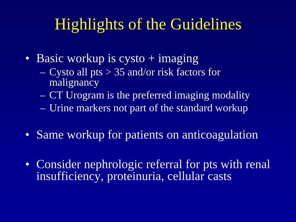
Highlights of the Guidelines
• Basic workup is cysto + imaging – Cysto all pts > 35 and/or risk factors for
malignancy – CT Urogram is the preferred imaging modality – Urine markers not part of the standard workup
• Same workup for patients on anticoagulation
• Consider nephrologic referral for pts with renal
insufficiency, proteinuria, cellular casts

Cystoscopy: “The Gold Standard” in Bladder Cancer Diagnosis
• Good but not perfect – Small lesions sometimes missed – False positives occur (inflammation/edema)
• Repeated testing is costly – Frequent surveillance cystoscopy and imaging is
required • Cystoscopy often causes patient discomfort
– Adherence to follow-up guidelines is poor

Can We Improve Upon (or Even Replace) Cystoscopy?
• Urinary Markers – Cytology – Protein based markers – Cellular markers – Genetic tests
• Endoscopic advances – Blue light cystoscopy – Narrow-band imaging

Cytology Advantages • High specificity in general • Reasonably high sensitivity
(33-95%) for high-grade disease
Disadvantages • Low sensitivity (4-31%) for
low-grade disease • Will still miss up to 60% of
high-grade tumors • Operator dependent • Results often equivocal
Lotan and Roehrborn, Urology, 2003 Karakiewicz, et al, BJUI, 2006

PROTEIN-BASED MARKERS BTA Stat® and NMP 22®

BTA stat®
Advantages • Point-of-care test • Improved sensitivity (50-
90%) compared to cytology • FDA approved • CLIA waived • Inexpensive
Disadvantages • Low specificity (50-73%)
– Some benign cells shed complement factor H
• Not tested in general hematuria population
Detects the presence of human complement factor H related protein which is present in the urine of patients with bladder cancer
Lotan and Roehrborn, Urology, 2003 Karakiewicz, et al, BJUI, 2006

NMP22 BladderChek® Detects nuclear mitotic apparatus protein 22 which is a matrix protein
more prevalent in cancer cells Advantages • Point-of-care test • Improved sensitivity (47-
90%) compared to cytology • FDA approved • CLIA waived • Inexpensive
Disadvantages • Moderate specificity (76-
83%) • Not tested in general
hematuria population
Lotan and Shariot, AUA Update, 2011

CELLULAR MARKERS ImmunoCyt®

ImmunoCyt®
Advantages • Improved sensitivity, even
for small tumors < 1 cm
Disadvantages • Performed in reference lab
(requires a fluorescence microscope and trained personnel)
• Specificity inferior to cytology alone – False positives associated
with immunofluorescence
• Expensive
Combines cytology with an immunofluorescence assay for CEA and two bladder cancer associated mucins
Messing, et al, J Urol 2005

GENETIC TESTS UroVysion®

UroVysion®
Advantages • Relatively high sensitivity (52-
100%) and specificity (62-83%)
• Sensitivity for upper tract tumors
• FDA-approved for use in bladder cancer and hematuria
Disadvantages • Expensive • Requires a reference lab • Still has a false positive rate
of 15-20% • 11% of tests are inconclusive
Fluorescence in-situ hybridization assay that detects increased numbers of chromosomes 3,7 and 17 as well as loss of the 9p21 locus
Mowatt, et al, Health Technol Assess, 2010 Schwarz, et al, J Clin Pathol, 2008
Bensalah, et al, Eur Urol, 2008


Sensitivity of Urinary Markers Stratified by Tumor Grade and Aggressiveness
Lotan and Shariot, AUA Update, 2011 Mowatt, et al, Health Technol Assess, 2010

Urinary Markers Alone CANNOT Replace or Avoid Cystoscopy
• Current AUA guidelines recommend AGAINST the use of urinary biomarkers in the routine work-up of patients with asymptomatic microscopic hematuria – Current AUA guidelines make no recommendations concerning the
use of urinary markers in the follow-up of NMIBC
• Current EAU guidelines suggest that “positive urine test results have a positive impact” on the quality of follow-up cystoscopy in NMIBC – While they don’t specify which test to use, they mention
that patients with negative cystoscopy and positive cytology should undergo guided bladder biopsy
Davis, et al, AUA guideline, 2012
Hall, et al, AUA guideline, 2010 Babjuk, et, EAU guideline, 2015

ADVANCES IN CYSTOSCOPY Blue-Light Cystoscopy and Narrow-Band Imaging

Enhancing Cystoscopy with Photodynamic Diagnosis
• Hexaminolevulinate (HAL, Cysview®)
– Lipophilic hexyl ester of 5-ALA which results in increased uptake of protoporphyrin IX in neoplastic tissue
– Illumination with blue-violet light (380-440 nm) results in red fluorescence from malignant tissue
• Used with the Karl Storz D-Light C Photodynamic Diagnostic (PDD).
• Not for repetitive use and is not a replacement for random bladder biopsies or other procedures used in the detection of bladder cancer.

Lerner and Goh, Cancer, 2014
Cysview- Papillary lesion

Cysview - CIS
Courtesy of Ashish Kamat, MD

Courtesy of Sam Chang, MD

US Pivotal Cysview (HAL) FDA registration study
Stenzl A et al. J Urol 2010

HAL Pivotal Study – Detection
• 286 patients: Ta or T1 bladder cancer – 16.4% of patients had one
or more Ta or T1 tumor with Cysview only (p=0.001)
• 41 patients: CIS – 32% of patients (13/41) had
CIS detected with Cysview only (p<0.0001)
• No difference in number of false positive results
Stenzl A et al. J Urol 2010; 184: 1907-1914. Burger M et al. EAU 2012.

HAL Pivotal Study – Recurrence
• Tumor recurrence rates over 9 months were 47% and 56% for the HAL and white light groups respectively (p=0.026)
• Relative reduction in recurrence rate was 16%
• Number of recurrences were lower at each timepoint (3, 6 and 9 months) in the BLC with HAL group compared with the white light group
Stenzl A et al. J Urol 2010 Burger M et al. EAU 2012.

HAL Meta-analyses • Prior meta-analysis were conflicting
– Kaush et al noted significant reduction in recurrence and improved tumor-free survival
– Shen et al noted the same reduction in recurrence rates but did not show an advantage in tumor-free survival
• Both meta-analyses had limitations – Both studies included studies with HAL and 5-ALA – Shen et al did not include key studies of HAL
• Recent meta-analysis from Burger et al overcomes these limitations
Kaush et al. Eur Urol, 2010
Shen, et al, BJUI, 2012 Burger, et al, Eur Urol, 2013

Meta-analysis included 2212 patients who underwent blue light cystoscopy
Burger, et al, Eur Urol, 2013

Meta-analysis: Ta/T1 tumor Detection Results
Burger, et al, Eur Urol, 2013
Group Patients N(%) in whom at least 1 Ta/T1 was detected with BL and not WL
Event rate
Total 168/790 (21.3%) 21.9%, p<0.001 (0.139-0.247)
Initial BC 68/346 (16.8%) 17.3%, p<0.001 (0.123-0.239)
Recurrence BC 110/444 (24.8%) 26.1%, p<0.001 (0.213-0.294)
High risk 83/377 (22%) 22.9%, p<0.001 (0.188-0.276)
Intermediate risk 79/237 (33.3%) 34.1%, p<0.001 (0.282-0.406)
Low risk 6/176 (3.4%) 4.6%, p<0.001; (0.022-0.097)
Prior intravesical treament 48/192 (25%) 25.3%, p<0.001; (0.196-0.319)
No prior intravesical treatment
88/434 (20.3%) 20.9% p<0.001; (0.173-0.291)
High risk: TaG3, T1 Medium risk: Multiple TaG1/G2 Low risk: Single TaG1/G2

Meta-analysis: CIS Detection Results
Burger, et al, Eur Urol, 2013
Group Patients (%) in whom CIS was detected with BL and not WL
Event rate, p-value, 95% CI
Total 61/256 (23.8) 24.6, p<0.001 (0.196-0.304)
Initial BC 30/110 (27.3) 23.8, p<0.001 (0.205-0.378)
Recurrent BC 31/146 (31.2) 21.8, p<0.001 (0.168-0.294)
Prior intravesical treatment
14/74 (18.9) 19.7, p<0.001 (0.120-0.307)
No prior intravesical treatment
29/128 (29.7) 27.4, p<0.001 (0.196-0.369)

Meta-analysis: Pooled Recurrence Rates
Burger, et al, Eur Urol, 2013

Enhancing Cystoscopy and TURBT with Narrow-Band Imaging
• Optical imaging technology (Olympus) enhances visibility of vessels on mucosal surfaces.
• Filters the white light into specific light wavelengths that penetrate only surface of human tissue are absorbed by hemoglobin.
• Bluish light enhances superficial capillary network and greenish light enhances deeper vessel visibility

White light cystoscopy vs. NBI
Herr and Donat, BJUI, 2008

White light cystoscopy vs. NBI
Herr and Donat, BJUI, 2008

Initial Results with NBI
Herr and Donat, BJUI, 2008

NBI may improve TURBT and reduce recurrence rates
• Cauberg et al, World J U, 2011 – Residual tumor after white light WL = 30% – Residual tumor after narrow band imaging NBI =
15% – p=0.03
• Naselli et al, EU, 2012 – 1 yr Recurrence Rate after NBI = 33% – 1 yr Recurrence Rate after WL = 51% – p= 0.01

RCT of White Light TUR vs. NBI TUR 254 pts with NMIBC total- 127 to each arm
Herr, Eur Urol, 2015

Narrow-band Imaging for Upper Urinary Tract Transitional-Cell CA
• Significant improvement in cancer detection and changes in management in 23% of patients. – Additional tumor detection
(14%) – Extended limits of ablation
(9%) – 11% - traditional white light
imaging MISSED the cancer
Traxer, J Endourol 2011
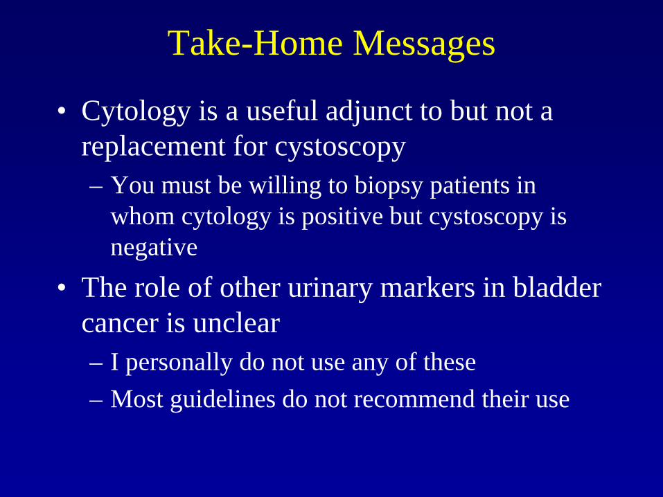
Take-Home Messages
• Cytology is a useful adjunct to but not a replacement for cystoscopy – You must be willing to biopsy patients in
whom cytology is positive but cystoscopy is negative
• The role of other urinary markers in bladder cancer is unclear – I personally do not use any of these – Most guidelines do not recommend their use

Take-Home Messages
• Enhanced cystoscopy with photodynamic diagnosis methods is very promising
• Both HAL (Blue light) and NBI have been shown to improve detection and reduce recurrence in studies
• HAL should not be used in all patients – Useful in patients with CIS or high-grade
disease, particularly after intravesical therapy – Useful in patients with negative cystoscopy and
positive cytology