hemodynamic response to familiar faces in children with adhd
TRANSCRIPT

RESEARCH Open Access
Hemodynamic response to familiar faces inchildren with ADHDKeiichi Shimamura1†, Takeshi Inoue1,2,3*† , Hiroko Ichikawa4, Emi Nakato5, Yuiko Sakuta6, So Kanazawa7,Masami K. Yamaguchi8, Ryusuke Kakigi9 and Ryoichi Sakuta2
Abstract
Background: School-age children with attention deficit hyperactivity disorder (ADHD) have difficulties in interpersonalrelationships, in addition to impaired facial expression perception and recognition. For successful social interactions, theability to discriminate between familiar and unfamiliar faces is critical. However, there are no published reports on therecognition of familiar and unfamiliar faces by children with ADHD.
Methods: We evaluated the neural correlates of familiar and unfamiliar facial recognition in children with ADHDcompared to typically developing (TD) children. We used functional near-infrared spectroscopy (fNIRS) to measurehemodynamic responses on the bilateral temporal regions while participants looked at photographs of familiar andunfamiliar faces. Nine boys with ADHD and 14 age-matched TD boys participated in the study. fNIRS data were Z-scored prior to analysis.
Results: During familiar face processing, TD children only showed significant activity in the late phase, while ADHDchildren showed significant activity in both the early and late phases. Additionally, the boys with ADHD did not showright hemispheric lateralization to familiar faces.
Conclusions: This study is the first to assess brain activity during familiar face processing in boys with ADHD usingfNIRS. These findings of atypical patterns of brain activity in boys with ADHD may be related to social cognitiveimpairments from ADHD.
Keywords: ADHD, Face recognition, Familiar face, fNIRS, The superior temporal sulci
BackgroundAttention deficit hyperactivity disorder (ADHD) is char-acterized by major symptoms of hyperactivity, impulsiv-ity, and inattention, which result in problems in theindividual’s quality of life [1, 2]. It is believed that it isdue to these symptoms that school-age children withADHD have difficulties in interpersonal relationships[3]; however, children with ADHD may have additionalsocial cognitive impairments [4]. These impairments re-lated to ADHD frequently lead to difficulties in daily lifeand may occasionally result in stressors. Understanding
the mechanisms of these impairments and stressors iscrucial in developing supports for children with ADHDthat prevent secondary disabilities such as school refusaland psychosomatic disorders.The early development of facial processing has been
considered fundamental to the development of socialabilities and theory of mind [5]. Several abilities com-pose social cognition, including the ability to perceivethe emotions of another person from affective prosody,faces, and body posture [4]. It has been shown repeat-edly that children with ADHD, particularly those thatare school-age, have impaired facial expression percep-tion and recognition [4, 6, 7].However, there is limited knowledge regarding basic fa-
cial processing in ADHD. Tye et al. (2013) were the firstto explore the face inversion effects and gaze processing inchildren with ADHD using event related potentials (ERPs)[8]. They found that children with ADHD displayed a
© The Author(s). 2019 Open Access This article is distributed under the terms of the Creative Commons Attribution 4.0International License (http://creativecommons.org/licenses/by/4.0/), which permits unrestricted use, distribution, andreproduction in any medium, provided you give appropriate credit to the original author(s) and the source, provide a link tothe Creative Commons license, and indicate if changes were made. The Creative Commons Public Domain Dedication waiver(http://creativecommons.org/publicdomain/zero/1.0/) applies to the data made available in this article, unless otherwise stated.
* Correspondence: [email protected]†Keiichi Shimamura and Takeshi Inoue contributed equally to this work.1Child Development and Psychosomatic Medicine Center, Dokkyo MedicalUniversity Saitama Medical Center, 2-1-50, Minami-Koshigaya, Koshigaya-shi,Saitama-Ken 343-8555, Japan2Department of Pediatrics, Dokkyo Medical University Saitama MedicalCenter, Saitama, JapanFull list of author information is available at the end of the article
Shimamura et al. BioPsychoSocial Medicine (2019) 13:30 https://doi.org/10.1186/s13030-019-0172-1

reduced face inversion effect on P1 latency compared withTD children. Sinzig et al. (2008) reported that the abilityof facial affect recognition is reduced in children sufferingfrom ADHD symptoms, both in autistic and ADHD chil-dren [9]. Several lines of evidence have suggested differen-tial processing between familiar and unfamiliar faces withbehavioral as well as neural findings in typical adult stud-ies [10, 11]. Therefore, we hypothesized that ADHD chil-dren, who present with social cognitive impairments, mayhave different familiar and unfamiliar facial recognitioncompared with TD children.fNIRS is a neuroimaging device which provides an
index of brain function by measuring changes in theconcentration of oxygenated hemoglobin (oxy-Hb), de-oxygenated hemoglobin (deoxy-Hb), and total-hemoglobin (total-Hb) using observation of the diffusetransmittance of near-infrared light at an appropriaterange of wavelengths [12, 13]. fNIRS has been utilizedfor more than a decade to examine brain activity duringvarious cognitive tasks in children with neurodevelop-mental disorders [7, 14–19]. Compared to other neuro-imaging devices such as functional magnetic resonanceimaging (fMRI), fNIRS has clear advantages, includingthat it is a completely silent process, has a high time.resolution, and provides a non-intrusive environment
requiring less restriction of the body and head [7, 20].fNIRS has been adopted in studies examining the brainactivity of children with ADHD while they perform cog-nitive tasks [16–19, 21]. These studies measured thehemodynamic response in the prefrontal areas, whichplay an important role in processing executive cognitivefunctions. With regard to facial recognition, fNIRS alsohas been utilized to measure neural activities in the tem-poral area, including the superior temporal sulci (STS),which is known to play an important role in processingfaces [22]; this is consistent with other hemodynamicevidence of facial processing in the temporal cortex ofthe brain [7, 20, 23–27].To our knowledge, there are no published reports on
the recognition of familiar and unfamiliar faces in chil-dren with ADHD. Thus, the purpose of this study is tocompare the neural correlates involved in familiar(mother) and unfamiliar facial recognition in childrenwith and without ADHD using a fNIRS system, whichwas placed over the bilateral temporal regions.
Materials and methodsParticipantsSeventeen ADHD boys and 16 TD boys were recruited.All diagnoses were guided by the DSM-5 [2] and weremade by two pediatric neurologists who worked withneurodevelopmental disorders for over 5 years. Exclu-sion criteria included learning, language, neurological ordevelopmental disabilities. Current study protocols were
approved by the Ethical Committee of the DokkyoMedical University Saitama Medical Center (hosp-k24,016). We obtained written informed consent fromthe participants and their parents. The experimentswere conducted according to the Declaration ofHelsinki.After excluding subjects whose fNIRS data failed the
motion criteria (frequent sharp changes in the time seriesof the raw fNIRS data), 23 subjects remained: 9 boys withADHD (mean age, 9 years 9months; SD, 1 year 6months;range, 8–13 years), and 14 TD boys (mean age, 9 years 4months; SD, 1 year 6months; range, 7–12 years) (Table 1).All participants were right-handed. Six out of nine boyswith ADHD received methylphenidate, and one ADHDboy received atomoxetine. All of them participated in theexamination for 4–6 h following medication. ADHD andTD subjects were not different according to age (seeTable 1). The ADHD-Rating Scale scores were 34.2(range, 11–52; SD, 13.9) in the ADHD boys and 9.8(range, 2–18; SD, 4.5) in the TD boys. The WechslerIntelligence Scale of Children Third Edition (WISC-III)full IQ scores of the TD boys (mean, 111.1; SD, 10.0;range, 90–129) were significantly higher (t = 3.68, p =0.048) than those of the ADHD participants (mean, 94.1;SD, 16.2; range, 78–125).
Table 1 Demographic and clinical characteristics of ADHD andTypical boys. The participants were 23 Japanese boys aged 7–13 years: 9 boys with ADHD and 14 TD boys. Fourteen TD boyswere matched with the ADHD subjects according to age. Theparticipants were limited to boys
ADHD Typical
ID age Full IQ ADHD-RS ID age Full IQ ADHD-RS
1 8y2m 106 41 10 7y3m 124 13
2 8y4m 91 38 11 7y9m 110 12
3 8y10m 82 52 12 7y9m 129 13
4 9y0m 110 11 13 8y0m 106 5
5 9y7m 125 15 14 8y5m 115 10
6 10y0m 78 29 15 8y7m 90 12
7 10y0m 84 48 16 8y8m 107 13
8 10y6m 79 34 17 8y9m 124 3
9 13y4m 92 40 18 10y1m 113 7
19 10y4m 105 10
20 10y5m 102 18
21 10y11m 110 12
22 11y0m 107 7
23 12y6m 113 2
range 8y-13y 78-125 11-52 range 7y-12y 90-129 2-18
mean 9y9m 94.1 34.2 mean 9y4m 111.1 9.8
SD 1y6m 16.2 13.9 SD 1y6m 10.0 4.5
Shimamura et al. BioPsychoSocial Medicine (2019) 13:30 Page 2 of 9
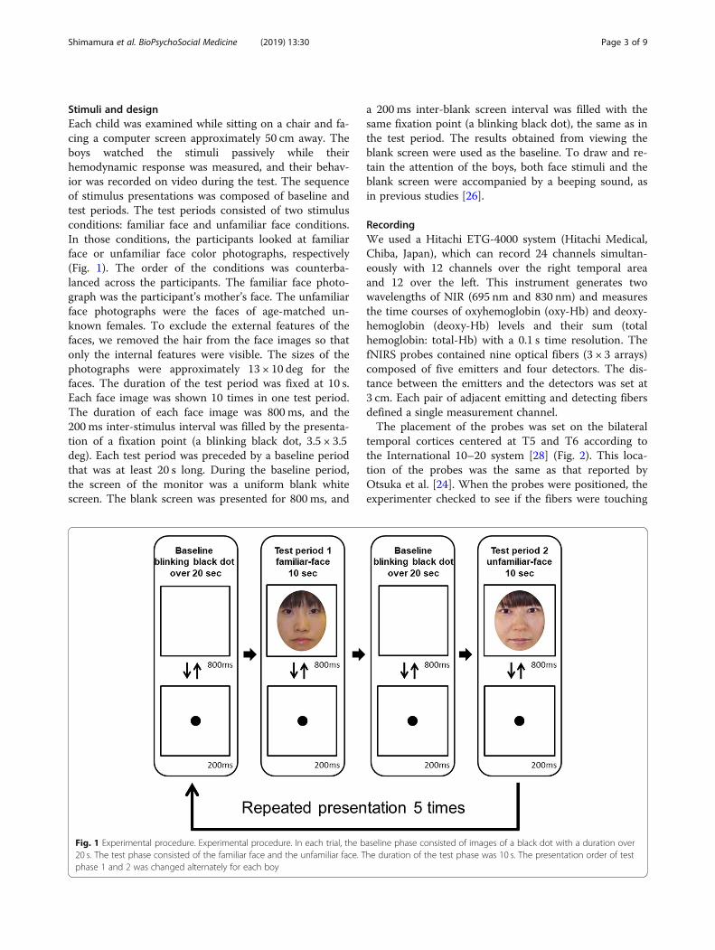
Stimuli and designEach child was examined while sitting on a chair and fa-cing a computer screen approximately 50 cm away. Theboys watched the stimuli passively while theirhemodynamic response was measured, and their behav-ior was recorded on video during the test. The sequenceof stimulus presentations was composed of baseline andtest periods. The test periods consisted of two stimulusconditions: familiar face and unfamiliar face conditions.In those conditions, the participants looked at familiarface or unfamiliar face color photographs, respectively(Fig. 1). The order of the conditions was counterba-lanced across the participants. The familiar face photo-graph was the participant’s mother’s face. The unfamiliarface photographs were the faces of age-matched un-known females. To exclude the external features of thefaces, we removed the hair from the face images so thatonly the internal features were visible. The sizes of thephotographs were approximately 13 × 10 deg for thefaces. The duration of the test period was fixed at 10 s.Each face image was shown 10 times in one test period.The duration of each face image was 800 ms, and the200 ms inter-stimulus interval was filled by the presenta-tion of a fixation point (a blinking black dot, 3.5 × 3.5deg). Each test period was preceded by a baseline periodthat was at least 20 s long. During the baseline period,the screen of the monitor was a uniform blank whitescreen. The blank screen was presented for 800 ms, and
a 200 ms inter-blank screen interval was filled with thesame fixation point (a blinking black dot), the same as inthe test period. The results obtained from viewing theblank screen were used as the baseline. To draw and re-tain the attention of the boys, both face stimuli and theblank screen were accompanied by a beeping sound, asin previous studies [26].
RecordingWe used a Hitachi ETG-4000 system (Hitachi Medical,Chiba, Japan), which can record 24 channels simultan-eously with 12 channels over the right temporal areaand 12 over the left. This instrument generates twowavelengths of NIR (695 nm and 830 nm) and measuresthe time courses of oxyhemoglobin (oxy-Hb) and deoxy-hemoglobin (deoxy-Hb) levels and their sum (totalhemoglobin: total-Hb) with a 0.1 s time resolution. ThefNIRS probes contained nine optical fibers (3 × 3 arrays)composed of five emitters and four detectors. The dis-tance between the emitters and the detectors was set at3 cm. Each pair of adjacent emitting and detecting fibersdefined a single measurement channel.The placement of the probes was set on the bilateral
temporal cortices centered at T5 and T6 according tothe International 10–20 system [28] (Fig. 2). This loca-tion of the probes was the same as that reported byOtsuka et al. [24]. When the probes were positioned, theexperimenter checked to see if the fibers were touching
Fig. 1 Experimental procedure. Experimental procedure. In each trial, the baseline phase consisted of images of a black dot with a duration over20 s. The test phase consisted of the familiar face and the unfamiliar face. The duration of the test phase was 10 s. The presentation order of testphase 1 and 2 was changed alternately for each boy
Shimamura et al. BioPsychoSocial Medicine (2019) 13:30 Page 3 of 9

the boys’ scalps correctly. The Hitachi ETG-4000 systemautomatically detects whether the contact adequatelymeasures emerging photons for each channel.
Data analysisThe analysis of fNIRS data in the present study wassimilar to that reported by Nakato et al. [26]. Before dataanalysis, we monitored the video recording of the boys’behavior to evaluate good trials for analysis. We re-moved a trial from the analysis if either the accumulativelooking time within the trial did not reach 5 s or ifmovement artifacts were detected by the analysis ofsharp changes in the time-series of the raw fNIRS data.The raw data on oxy-Hb, deoxy-Hb, and total-Hb con-
centrations from each channel were digitally band-pass-filtered at 0.02–1.0 Hz to remove noise caused by heart-beat pulsations or any longitudinal signal drifts. Themean concentration of each channel within a subjectwas then calculated by averaging data across the trials ina time-series with a 0.1 s resolution.Based on mean concentrations in the time-series, we
calculated the Z-scores for oxy-, deoxy-, and total-Hb inthe familiar face and the unfamiliar face conditions foreach channel within a subject [7, 20, 24–27]. The Z-score at each time-point indicates the deviation ofhemodynamic response from the baseline during thepresentation of faces. The mean concentration of the Z-score at 3 s before the trial was used as a baseline. TheZ-scores were calculated separately for oxy-Hb, deoxy-Hb, and total-Hb in the familiar face and unfamiliar faceconditions.The Z-scores obtained from the 12 channels within
the left and right temporal areas were averaged acrosstrials for every subject in order to increase the signal-to-noise ratio. Although raw NIRS data consist of relative
values that cannot be averaged directly across subjectsor channels, normalized data including Z-scores can beaveraged regardless of the unit. This calculation of theZ-score is a reliable analysis for changes in Hb concen-tration in the brain regions under the sensors since theanalysis is independent of the differential path lengthfactor (DPF). On the basis of these methodological ad-vantages for fNIRS data, we applied the same analysis ofthe Z-score in the present study as in previous fNIRSstudies with children [7, 20, 24–27].Consistent with our previous studies using fNIRS
[7, 20, 24–27], we found a response peaking a fewseconds after the stimulus onset. The hemodynamic re-sponse of oxy-Hb in the familiar face condition increasedgradually from approximately 3 s after stimulus onset andreached a plateau approximately 10 s after stimulus onsetin ADHD boys (see Fig. 3). In contrast, the hemodynamicresponse of oxy-Hb in the familiar face condition in-creased gradually from approximately 10 s after stimulusonset in TD boys (see Fig. 3). Therefore, we performedstatistical analyses against the mean Z-scores for the 3–10s (the early phase) and 10–17 s (the late phase) durationsof the test period. A two-tailed one-sample t-test against achance level of 0 (baseline) was conducted for the meanZ-score during the early phase and late phase in both tem-poral areas.
ResultsAvailable trials for statistical analysisData from the 23 boys who looked at faces over morethan three trials in both the familiar face and unfamiliarface conditions were obtained. Available trials were aver-aged across trials for each condition (familiar face, mean5.06, SD 0.89; unfamiliar face, mean 4.97, SD 0.95). Themean number of removed trials for the familiar face
Fig. 2 Placement of NIRS probes. The placement of NIRS probes. The probes were placed over the left and right temporal areas centering at T5and T6 of the International 10–20 system. T5/T6 is in the yellow circle. The distance between the probes was set at 3 cm
Shimamura et al. BioPsychoSocial Medicine (2019) 13:30 Page 4 of 9
condition was 0.33 (SD 0.74) while that for the unfamil-iar face condition was 0.30 (SD 0.64). There was no sig-nificant difference between the numbers of availabletrials under each condition.
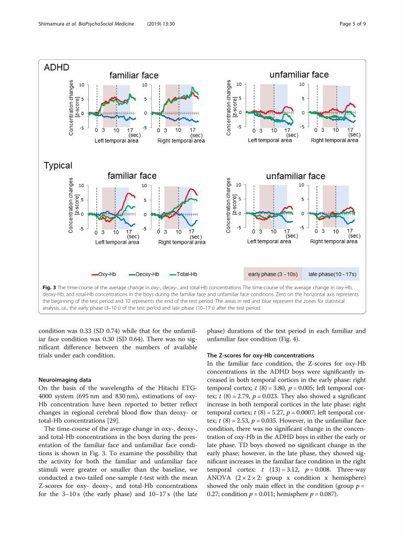
Neuroimaging dataOn the basis of the wavelengths of the Hitachi ETG-4000 system (695 nm and 830 nm), estimations of oxy-Hb concentration have been reported to better reflectchanges in regional cerebral blood flow than deoxy- ortotal-Hb concentrations [29].The time-course of the average change in oxy-, deoxy-,
and total-Hb concentrations in the boys during the pres-entation of the familiar face and unfamiliar face condi-tions is shown in Fig. 3. To examine the possibility thatthe activity for both the familiar and unfamiliar facestimuli were greater or smaller than the baseline, weconducted a two-tailed one-sample t-test with the meanZ-scores for oxy- deoxy-, and total-Hb concentrationsfor the 3–10 s (the early phase) and 10–17 s (the late
phase) durations of the test period in each familiar andunfamiliar face condition (Fig. 4).
The Z-scores for oxy-Hb concentrationsIn the familiar face condition, the Z-scores for oxy-Hbconcentrations in the ADHD boys were significantly in-creased in both temporal cortices in the early phase: righttemporal cortex; t (8) = 3.80, p = 0.005; left temporal cor-tex; t (8) = 2.79, p = 0.023. They also showed a significantincrease in both temporal cortices in the late phase: righttemporal cortex; t (8) = 5.27, p = 0.0007; left temporal cor-tex; t (8) = 2.53, p = 0.035. However, in the unfamiliar facecondition, there was no significant change in the concen-tration of oxy-Hb in the ADHD boys in either the early orlate phase. TD boys showed no significant change in theearly phase; however, in the late phase, they showed sig-nificant increases in the familiar face condition in the righttemporal cortex: t (13) = 3.12, p = 0.008. Three-wayANOVA (2 × 2 × 2: group x condition x hemisphere)showed the only main effect in the condition (group p =0.27; condition p = 0.011; hemisphere p = 0.087).
Fig. 3 The time-course of the average change in oxy-, deoxy-, and total-Hb concentrations The time-course of the average change in oxy-Hb,deoxy-Hb, and total-Hb concentrations in the boys during the familiar face and unfamiliar face conditions. Zero on the horizontal axis representsthe beginning of the test period and 10 represents the end of the test period. The areas in red and blue represent the zones for statisticalanalysis, i.e., the early phase (3–10 s) of the test period and late phase (10–17 s) after the test period
Shimamura et al. BioPsychoSocial Medicine (2019) 13:30 Page 5 of 9

The Z-scores for deoxy-Hb concentrationsThe Z-scores for deoxy-Hb concentrations of the ADHDboys showed no significant change in the familiar facecondition. However, in the unfamiliar face condition, theZ-scores of the ADHD boys were significantly decreasedin both the right and left temporal cortices in the earlyphase: right temporal cortex; t (8) = 2.47, p = 0.039; lefttemporal cortex; t (8) = 4.97, p = 0.001. In the late phase,they showed significant decreases in the left temporalcortex: t (8) = 2.42, p = 0.041. TD boys showed no signifi-cant change in the concentration of deoxy-Hb in eithercondition in either the early or late phase.
The Z-scores for total-Hb concentrationsThe Z-scores for the total-Hb concentrations in theADHD boys showed no significant change in either con-dition in the early phase. However, in the late phase,they showed significant increases in the right temporal
cortex (t [8] = 2.67, p = 0.021), but only in the familiarface condition. TD boys showed no significant change inthe concentration of the total-Hb in either condition inboth the early and late phases.
DiscussionThe current study compared the neural correlates ofviewing familiar and unfamiliar facial images in ADHDand TD boys using fNIRS. To measure the neural activ-ities of facial recognition, the fNIRS probes were set onthe bilateral temporal regions, including on the STS,which plays an essential role in processing faces. Thesemeasuring procedures were the same as in previousstudies [7, 20, 23–27]. We calculated the Z-scores forthe 12 channels within the temporal area from the rawdata in the familiar and unfamiliar face conditions.fNIRS monitors blood volume and oxygenation in the
brain. In this study, the hemodynamic response, as indicated
Fig. 4 The mean Z-score during the early phase and the late phase. The mean Z-score during the early phase and the late phase in the left andright temporal cortices. The vertical bar in the graphs represents one standard error (SE). The left graph shows the data during the early phaseand the right graphs shows the late phase. In the familiar face condition with ADHD, oxy-Hb concentrations (red bars) in the bilateral temporalcortex were significantly greater both in the early and late phase than the chance level of 0. In the TD boys, oxy-Hb concentrations of the righttemporal cortex during the late phase were increased in the familiar face condition (red bars) (* p < .05)
Shimamura et al. BioPsychoSocial Medicine (2019) 13:30 Page 6 of 9

by oxy-Hb, increased gradually during the test period inboth groups and was found to occur predominantly in theright hemisphere in TD boys. Neuroimaging studies have re-ported that the major aspects of facial processing arise pre-dominantly in the right hemisphere [30, 31]. Therefore,these findings suggest that the hemodynamic response inthis study indicates facial recognition in ADHD and TDboys. In the familiar face condition, Z-scores of oxy-Hb con-centrations were significantly increased in the early and inthe late phase for ADHD boys. In contrast, for TD boys,significant changes were only found in the late phase. In theunfamiliar face condition, we found no significantchanges in either group of boys. This indicates that thehemodynamic response in ADHD boys to familiar facesmay be earlier than TD boys. However, this researchfinding may not be related to the major ADHD symp-toms of hyperactivity, impulsivity, and inattention.Gobbini et al. documented that familiar faces are recog-nized by processes that operate outside the focus of at-tention and without visual awareness [32]. In addition,numerous studies have reported slower reaction timevariability among individuals with ADHD compare toTD individuals [33, 34]. Another possibility is that theADHD boys did not show adaptation for familiar faces.Generally, repeated facial presentation induces neuraladaptation [35].Hemodynamic responses to familiar faces were found to
occur predominantly in the right temporal area in TDboys. In contrast, ADHD boys showed significantly in-creased concentration of oxy-Hb to familiar faces in boththe right and left temporal areas compared with the base-line. The TD boys showed right hemispheric dominancein processing faces, which is consistent with many otherneuroimaging studies [30, 31]. Whereas the right hemi-spheric dominance in TD boys was indicated and success-fully monitored by fNIRS, ADHD boys did not show righthemispheric lateralization to familiar faces. This finding isconsistent with previous studies [6, 7]. Ichikawa et al.showed that the brain activity stimulated by seeing ahappy face is significantly increased in both the right andleft temporal areas compared with the baseline in ADHDboys.Aside from the major symptoms of ADHD, boys with
ADHD have been documented to have social cognitiveimpairments with impaired facial recognition and expres-sion perception [4, 6, 7]. Additionally, in terms of basic fa-cial processing, ADHD boys show a reduced faceinversion effect on P1 latency compared to TD childrenusing ERPs [8]. The inversion effect is an important indexof facial expertise. These findings suggest that individualswith ADHD may have impairments of facial perceptionand recognition, which is consistent with the results ofour study. There are a few limitations in the present study.First, we had difficulties with the measurement of non-
medicated ADHD boys because of motion artifacts bytheir hyperactivity. It is better to remove the effect ofmedication, however, the effect of medication in facialperception and recognition is unclear in children withADHD. Some previous studies showed that medicationdid not affect the recognition of facial expression inADHD children [36] or slightly improved it [6], and therewere some studies that did not examine the effect ofmedication [37, 38]. The second limitation was the smallnumber of participants. After removing participantswhose fNIRS data failed the motion criteria, only 69.7% ofthe subjects who were recruited remained in the study. Fi-nally, although this is the first study to evaluate the neuralcorrelates of familiar and unfamiliar facial recognition inchildren with ADHD, we recruited only boys in thecurrent study. Future studies should examine a largernumber of participants, recruit both genders, and considerthe effect of medication.In conclusion, we have demonstrated that differential
brain activity occurs between ADHD and TD boys in re-sponse to the sight of a familiar face. During familiarface processing, ADHD children showed significant ac-tivity in both the early and late phase, while TD childrenshowed significant activity only in the late phase. Add-itionally, the dominance of the right temporal areas forfacial processing was found only in TD children duringthe familiar face presentation. These findings of atypicalpatterns of brain activity in ADHD boys may be relatedto social cognitive impairments in ADHD. We believethat these insights make a number of important addi-tions to the literature and would be of considerableinterest to researchers of biopsychosocial medicine.
AcknowledgmentsWe would like to thank all participants and their families who took part inthe study. We also thank Dr. Margot J. Taylor for review of this manuscriptand Mr. M. Fujiwara (Hitachi Medical Cooperation) for his technical supportregarding the fNIRS measurement of children.
Authors’ contributionsSo Kanazawa, Masami K. Yamaguchi and Ryoichi Sakuta conceived andplanned the experiments. Keiichi Shimamura, Takeshi Inoue, Hiroko Ichikawa,Emi Nakato and Yuiko Sakuta carried out the experiments. Hiroko IchikawaEmi Nakato and Ryusuke Kakigi designed the model and the computationalframework and Keiichi Shimamura, Takeshi Inoue and Hiroko Ichikawaanalysed the data. Keiichi Shimamura, Takeshi Inoue and Hiroko Ichikawawrote the manuscript with support from Emi Nakato, Yuiko Sakuta, SoKanazawa, Masami K. Yamaguchi, Ryusuke Kakigi and Ryoichi Sakuta. Allauthors read and approved the final manuscript
FundingThis study was supported by Grant-in-Aid for Scientific Research on Innova-tive Areas, ‘Face perception and recognition’ from MEXT KAKENHI (20119002)and JSPS KAKENHI (25285204). The funding was used in a proofreading feeand article-processing charges.
Availability of data and materialsThe datasets analyzed in this study are available from the correspondingauthor upon reasonable request.
Shimamura et al. BioPsychoSocial Medicine (2019) 13:30 Page 7 of 9

Ethics approval and consent to participateThis study was approved by the Ethical Committee of the Dokkyo MedicalUniversity Saitama medical center (hosp-k 24016) and by the Ethical Committeeof Chuo University. Current study contains volunteer’s image in Fig. 2, and wehave received consent for publication.
Consent for publicationOur submission is not currently under review elsewhere and all authors haveread and approved the manuscript.
Competing interestsThe authors declare that they have no competing interests in this section.
Author details1Child Development and Psychosomatic Medicine Center, Dokkyo MedicalUniversity Saitama Medical Center, 2-1-50, Minami-Koshigaya, Koshigaya-shi,Saitama-Ken 343-8555, Japan. 2Department of Pediatrics, Dokkyo MedicalUniversity Saitama Medical Center, Saitama, Japan. 3Department ofDiagnostic Imaging, Program in Neurosciences & Mental Health, Hospital forSick Children, Toronto, Ontario, Canada. 4Faculty of Science and Technology,Tokyo University of Science, Chiba, Japan. 5Department of Clothing, OsakaShoin Women’s University, Osaka, Japan. 6Faculty of Human Life Sciences,Jissen Women’s University, Tokyo, Japan. 7Department of Psychology, JapanWomen’s University, Kanagawa, Japan. 8Department of Psychology, ChuoUniversity, Tokyo, Japan. 9Department of Integrative Physiology, NationalInstitute for Physiological Sciences, Aichi, Japan.
Received: 28 May 2019 Accepted: 14 November 2019
References1. Subcommittee on Attention-Deficit/Hyperactivity Disorder, Steering
Committee on Quality Improvement and Management, Wolraich M, BrownL, Brown RT, DuPaul G, et al. ADHD: Clinical Practice Guideline for theDiagnosis, Evaluation, and Treatment of Attention-Deficit/HyperactivityDisorder in Children and Adolescents. Pediatrics. 2011;128:1007–22.
2. American Psychiatric Association., American Psychiatric Association. DSM-5Task Force. Diagnostic and statistical manual of mental disorders : DSM-5.
3. Landau S, Moore LA. Social skills deficits in children with attention-deficithyperactivity disorder. School Psych Rev. 1991;20:235–51.
4. Uekermann J, Kraemer M, Abdel-Hamid M, Schimmelmann BG, HebebrandJ, Daum I, et al. Social cognition in attention-deficit hyperactivity disorder(ADHD). Neurosci Biobehav Rev. 2010;34:734–43.
5. Dawson G, Webb SJ, McPartland J. Understanding the nature of faceprocessing impairment in autism: insights from behavioral andelectrophysiological studies. Dev Neuropsychol. 2005;27:403–24.
6. Williams LM, Hermens DF, Palmer D, Kohn M, Clarke S, Keage H, et al.Misinterpreting emotional expressions in attention-deficit/hyperactivitydisorder: evidence for a neural marker and stimulant effects. Biol Psychiatry.2008;63:917–26.
7. Ichikawa H, Nakato E, Kanazawa S, Shimamura K, Sakuta Y, Sakuta R, et al.Hemodynamic response of children with attention-deficit and hyperactivedisorder (ADHD) to emotional facial expressions. Neuropsychologia. 2014;63:51–8.
8. Tye C, Mercure E, Ashwood KL, Azadi B, Asherson P, Johnson MH, et al.Neurophysiological responses to faces and gaze direction differentiatechildren with ASD, ADHD and ASD + ADHD. Dev Cogn Neurosci Elsevier.2013;5:71–85.
9. Sinzig J, Morsch D, Lehmkuhl G. Do hyperactivity, impulsivity andinattention have an impact on the ability of facial affect recognition inchildren with autism and ADHD? Eur Child Adolesc Psychiatry. 2008;17:63–72.
10. Johnston RA, Edmonds AJ. Familiar and unfamiliar face recognition: areview. Memory. 2009;17:577–96.
11. Itz ML, Schweinberger SR, Kaufmann JM. Effects of Caricaturing in Shape orColor on Familiarity Decisions for Familiar and Unfamiliar Faces. PLoS One.2016;11:e0149796 Pavlova MA, editor.
12. Watanabe E, Yamashita Y, Maki A, Ito Y, Koizumi H. Non-invasive functionalmapping with multi-channel near infra-red spectroscopic topography inhumans. Neurosci Lett Elsevier. 1996;205:41–4.
13. Schroeter ML, Bücheler MM, Müller K, Uludağ K, Obrig H, Lohmann G, et al.Towards a standard analysis for functional near-infrared imaging.Neuroimage. 2004;21:283–90.
14. Fukuda M. Near-infrared spectroscopy in psychiatry. Brain Nerve. 2012;64:175–83.15. Ichikawa H, Kitazono J, Nagata K, Manda A, Shimamura K, Sakuta R, et al.
Novel method to classify hemodynamic response obtained using MULTI-channel fNIRS measurements into two groups: exploring the combinationsof channels. Front Hum Neurosci. 2014;8:480.
16. Monden Y, Dan I, Nagashima M, Dan H, Uga M, Ikeda T, et al. Individualclassification of ADHD children by right prefrontal hemodynamic responsesduring a go/no-go task as assessed by fNIRS. NeuroImage Clin. 2015;9:1–12.
17. Araki A, Ikegami M, Okayama A, Matsumoto N, Takahashi S, Azuma H, et al.Improved prefrontal activity in AD/HD children treated with atomoxetine: aNIRS study. Brain Dev. 2015;37:76–87.
18. Nagashima M, Monden Y, Dan I, Dan H, Mizutani T, Tsuzuki D, et al.Neuropharmacological effect of atomoxetine on attention network inchildren with attention deficit hyperactivity disorder during oddballparadigms as assessed using functional near-infrared spectroscopy.Neurophotonics. 2014;1:025007.
19. Monden Y, Dan H, Nagashima M, Dan I, Tsuzuki D, Kyutoku Y, et al. Rightprefrontal activation as a neuro-functional biomarker for monitoring acuteeffects of methylphenidate in ADHD children: an fNIRS study. NeuroImageClin. 2012;1:131–40.
20. Inoue T, Sakuta Y, Shimamura K, Ichikawa H, Kobayashi M, Otani R, et al.Differences in the Pattern of Hemodynamic Response to Self-Face andStranger-Face Images in Adolescents with Anorexia Nervosa: A Near-InfraredSpectroscopic Study. PLoS One. 2015;10:e0132050 Nishijo H, editor.
21. Ehlis A-C, Bähne CG, Jacob CP, Herrmann MJ, Fallgatter AJ. Reduced lateralprefrontal activation in adult patients with attention-deficit/hyperactivitydisorder (ADHD) during a working memory task: a functional near-infraredspectroscopy (fNIRS) study. J Psychiatr Res. 2008;42:1060–7.
22. Allison P. McCarthy. Social perception from visual cues: role of the STSregion. Trends Cogn Sci. 2000;4:267–78.
23. Lloyd-Fox S, Blasi A, Everdell N, Elwell CE, Johnson MH. Selective corticalmapping of biological motion processing in young infants. J CognNeurosci. 2011;23:2521–32.
24. Otsuka Y, Nakato E, Kanazawa S, Yamaguchi MK, Watanabe S, Kakigi R.Neural activation to upright and inverted faces in infants measured by nearinfrared spectroscopy. Neuroimage. 2007;34:399–406.
25. Kobayashi M, Otsuka Y, Kanazawa S, Yamaguchi MK, Kakigi R. Size-invariantrepresentation of face in infant brain: an fNIRS-adaptation study.Neuroreport. 2012;23:984–8.
26. Nakato E, Otsuka Y, Kanazawa S, Yamaguchi MK, Watanabe S, Kakigi R.When do infants differentiate profile face from frontal face? A near-infraredspectroscopic study. Hum Brain Mapp. 2009;30:462–72.
27. Nakato E, Otsuka Y, Kanazawa S, Yamaguchi MK, Kakigi R. Distinctdifferences in the pattern of hemodynamic response to happy and angryfacial expressions in infants--a near-infrared spectroscopic study.Neuroimage. 2011;54:1600–6.
28. Klem GH, Lüders HO, Jasper HH, Elger C. The ten-twenty electrode systemof the international federation. The International Federation of ClinicalNeurophysiology. Electroencephalogr Clin Neurophysiol Suppl. 1999;52:3–6.
29. Yamashita Y, Maki A, Koizumi H. Wavelength dependence of the precisionof noninvasive optical measurement of oxy-, deoxy-, and total-hemoglobinconcentration. Med Phys. 2001;28:1108–14.
30. Sergent J, Ohta S, MacDonald B. Functional neuroanatomy of face and objectprocessing. A positron emission tomography study. Brain. 1992;115 Pt 1:15–36.
31. Tsuchiya N, Kawasaki H, Oya H, Howard MA, Adolphs R. Decoding faceinformation in time, frequency and space from direct intracranial recordingsof the human brain. PLoS One. 2008;3:e3892 Lauwereyns J, editor.
32. Gobbini MI, Gors JD, Halchenko YO, Rogers C, Guntupalli JS, Hughes H, et al.Prioritized Detection of Personally Familiar Faces. PLoS One. 2013;8:e66620Valdes-Sosa M, editor.
33. Kofler MJ, Rapport MD, Sarver DE, Raiker JS, Orban SA, Friedman LM, et al.Reaction time variability in ADHD: a meta-analytic review of 319 studies.Clin Psychol Rev. 2013;33:795–811.
34. Tamm L, Narad ME, Antonini TN, O’Brien KM, Hawk LW, Epstein JN. Reactiontime variability in ADHD: a review. Neurotherapeutics. 2012;9:500–8.
35. Kobayashi M, Otsuka Y, Kanazawa S, Yamaguchi MK, Kakigi R. Theprocessing of faces across non-rigid facial transformation develops at 7month of age: a fNIRS-adaptation study. BMC Neurosci. 2014;15:81.
Shimamura et al. BioPsychoSocial Medicine (2019) 13:30 Page 8 of 9

36. Schwenck C, Schneider T, Schreckenbach J, Zenglein Y, Gensthaler A,Taurines R, et al. Emotion recognition in children and adolescents withattention-deficit/hyperactivity disorder (ADHD). ADHD Atten DeficitHyperact Disord. 2013;5:295–302.
37. Miller M, Hanford RB, Fassbender C, Duke M, Schweitzer JB. Affectrecognition in adults with ADHD. J Atten Disord. 2011;15:452–60.
38. Da Fonseca D, Seguier V, Santos A, Poinso F, Deruelle C. Emotionunderstanding in children with ADHD. Child Psychiatry Hum Dev. 2009;40:111–21.
Publisher’s NoteSpringer Nature remains neutral with regard to jurisdictional claims inpublished maps and institutional affiliations.
Shimamura et al. BioPsychoSocial Medicine (2019) 13:30 Page 9 of 9