hemoglobin structure and function. conformational changes conformational changes in proteins are...
TRANSCRIPT
Hemoglobin
Structure and function
Conformational changes
•Conformational changes in proteins are critical for their functions and regulations. Changes in protein conformation facilitate movement, catalysis of molecules (oxygen), binding.
•Regions in proteins must be able to allow the structural changes (breaking and forming non
covalent interactions) .•Allosteric (from the Greek allos, other)
interactions or changes in conformations of a region in a protein (or another protein) which induces a change in shape in another part of the same molecule (or another molecule).
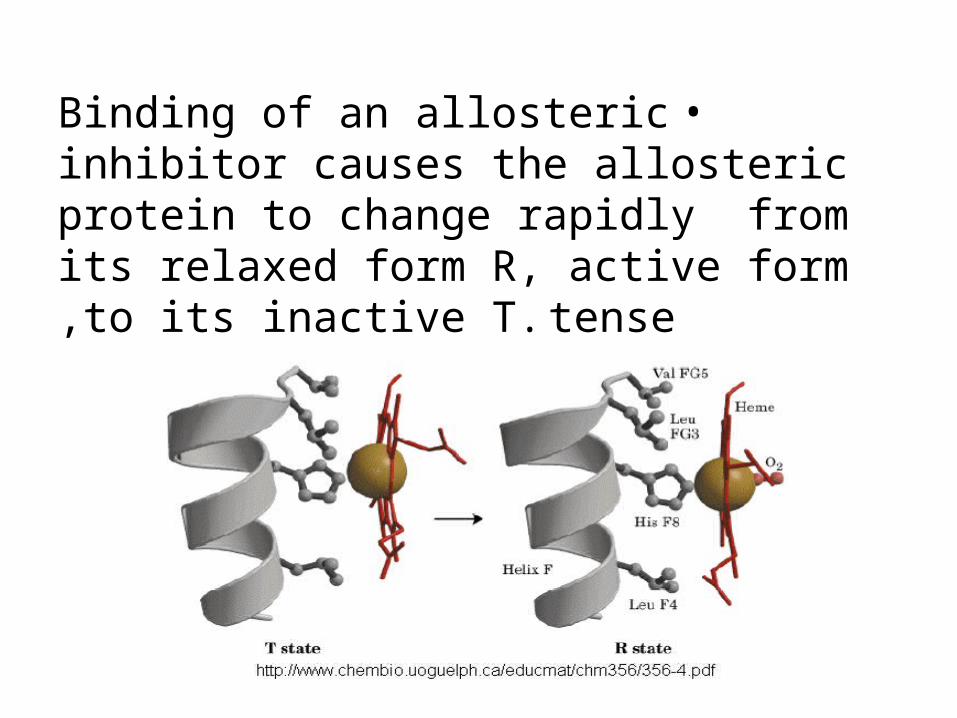
•Binding of an allosteric inhibitor causes the allosteric protein to change rapidly from its relaxed form R, active form to its inactive T. tense,
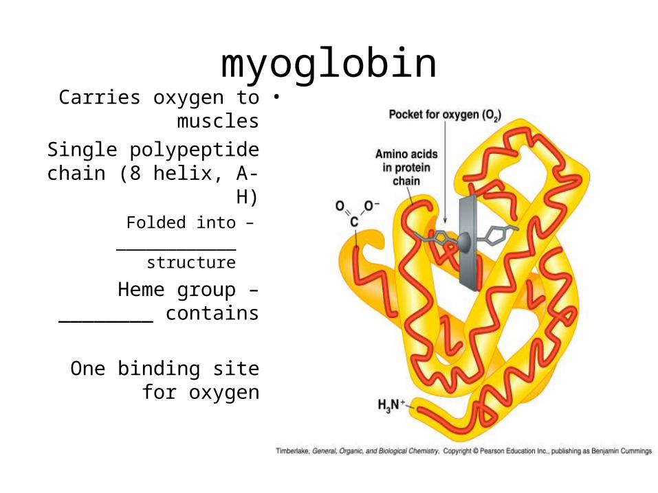
myoglobin•Carries oxygen to
muscles
•Single polypeptide chain (8 helix, A-H)
–Folded into ____________ structure
•Heme group – contains ________
•One binding site for oxygen
Continued
•O2 has relatively low solubility in water.•Myoglobin and hemoglobin are both oxygen-
carrying proteins which facilitate adequate delivery of O2 to cell .
•Myoglobin transport of oxygen in muscle and serves to store oxygen in muscle. It is single polypeptide chain of 153 residues.
•Fe atom of prosthetic group, heme, directly bond to N of His side chains .
Hemoglobin
•Hemoglobin, oxygen carrier within red blood cells and also transport H+ and carbon dioxide.
•It contains alpha (141 amino acids, 7 helix) and beta(146) subunits .
•Cooperativity: Change in conformation of one molecule which induces a change in another molecules.
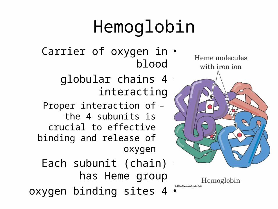
Hemoglobin •Carrier of oxygen in blood•4 globular chains interacting
–Proper interaction of the 4 subunits is crucial to effective
binding and release of oxygen
•Each subunit (chain) has Heme group
•4 oxygen binding sites
Types of Hb
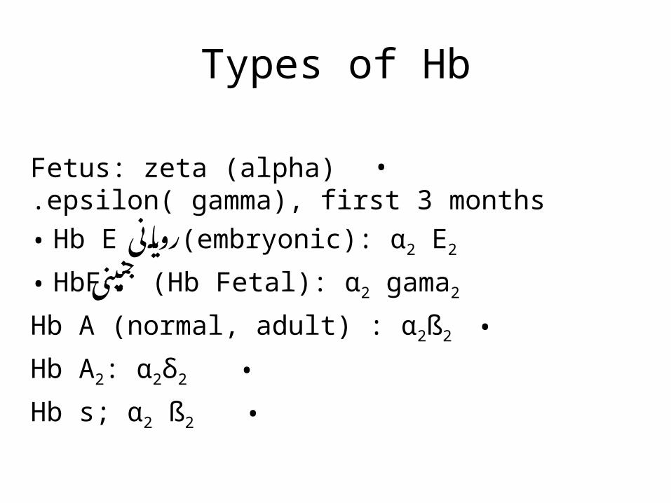
•Fetus: zeta (alpha) epsilon( gamma), first 3 months.
• Hb E رویانی(embryonic): α2 E2
• HbFجنینی (Hb Fetal): α2 gama2
•Hb A (normal, adult) : α2ß2
•Hb A2: α2δ2
•Hb s; α2 ß2
Continued
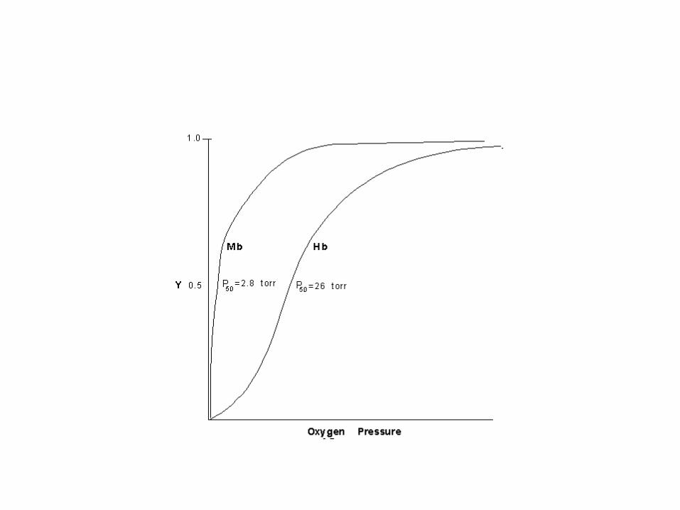
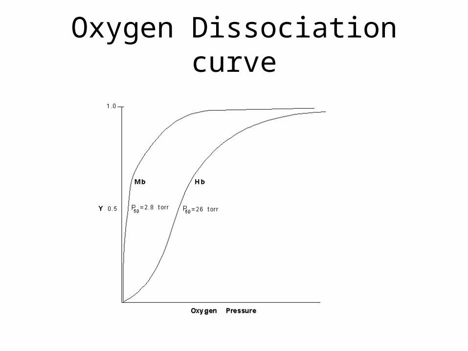
•The fraction of each protein saturated with oxygen (Y), is plotted against the partial pressure of oxygen,PO2.
•The oxygen-binding curve of myoglobin is hyperbolic, with half-saturation (Y=0.5) at
an oxygen pressure of 2.8 torr. •The oxygen-binding curve of hemoglobin
is sigmoidal (allostric), with half-saturation at an oxygen pressure of 26 torr.
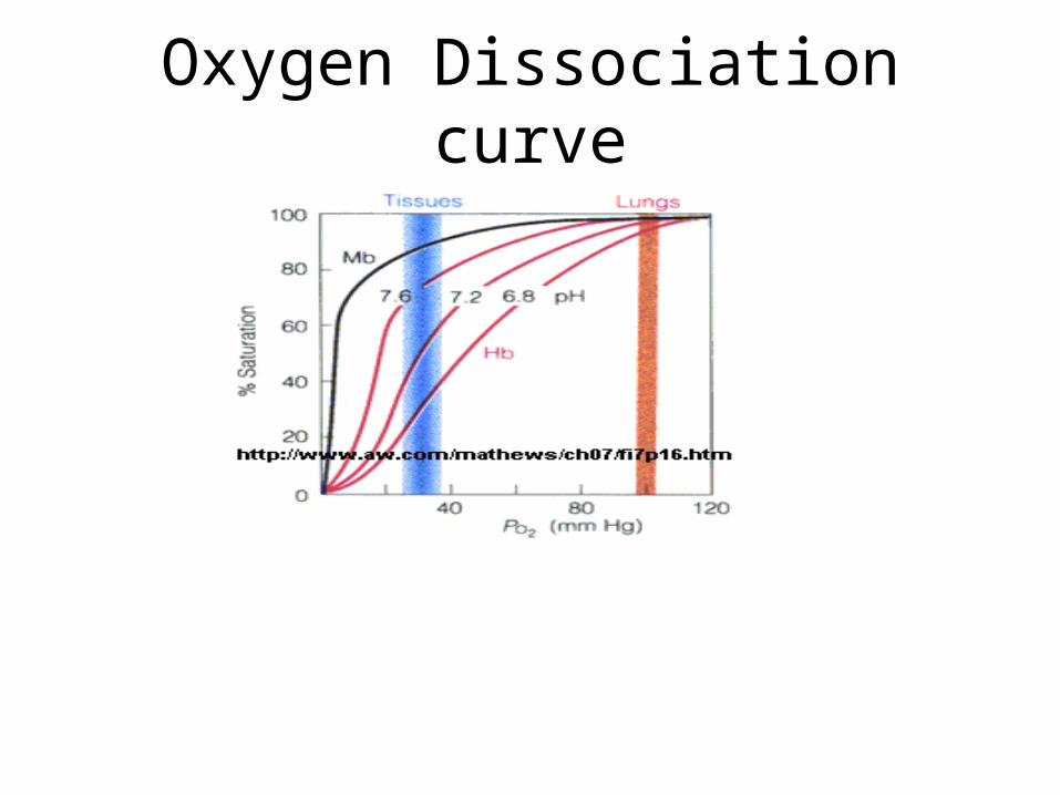
Oxygen Dissociation curve
Continued
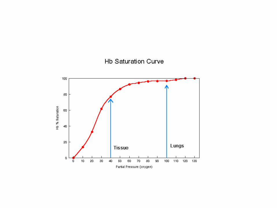
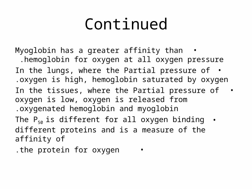
•Myoglobin has a greater affinity than hemoglobin for oxygen at all oxygen pressure .
•In the lungs, where the Partial pressure of oxygen is high, hemoglobin saturated by oxygen.
• In the tissues, where the Partial pressure of oxygen is low, oxygen is released from oxygenated hemoglobin and myoglobin.
•The P50 is different for all oxygen binding different proteins and is a measure of the affinity of
• the protein for oxygen.
•In hemoglobin the binding of O2 is cooperative, binding of O2 enhances binding of additional O2.Binding of O2 to one of the subunits or monomers alters affinity of other subunits for O2.
•The oxygen affinity of the hemoglobin increases as each oxygen molecules is bond.
Continued
Mechanism of oxygen binding in Hb
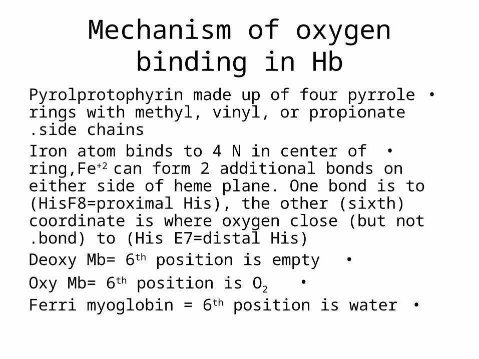
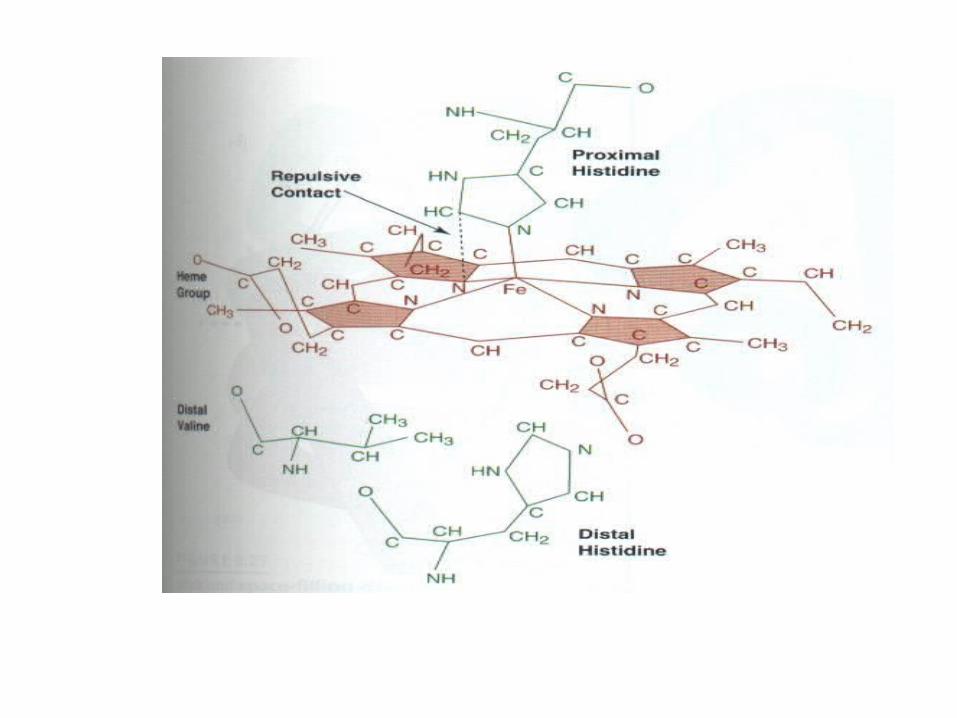
•Pyrolprotophyrin made up of four pyrrole rings with methyl, vinyl, or propionate side chains.
•Iron atom binds to 4 N in center of ring,Fe+2 can form 2 additional bonds on either side of heme plane. One bond is to (HisF8=proximal His), the other (sixth) coordinate is where oxygen close (but not bond) to (His E7=distal His).
•Deoxy Mb= 6th position is empty•Oxy Mb= 6th position is O2
•Ferri myoglobin = 6th position is water
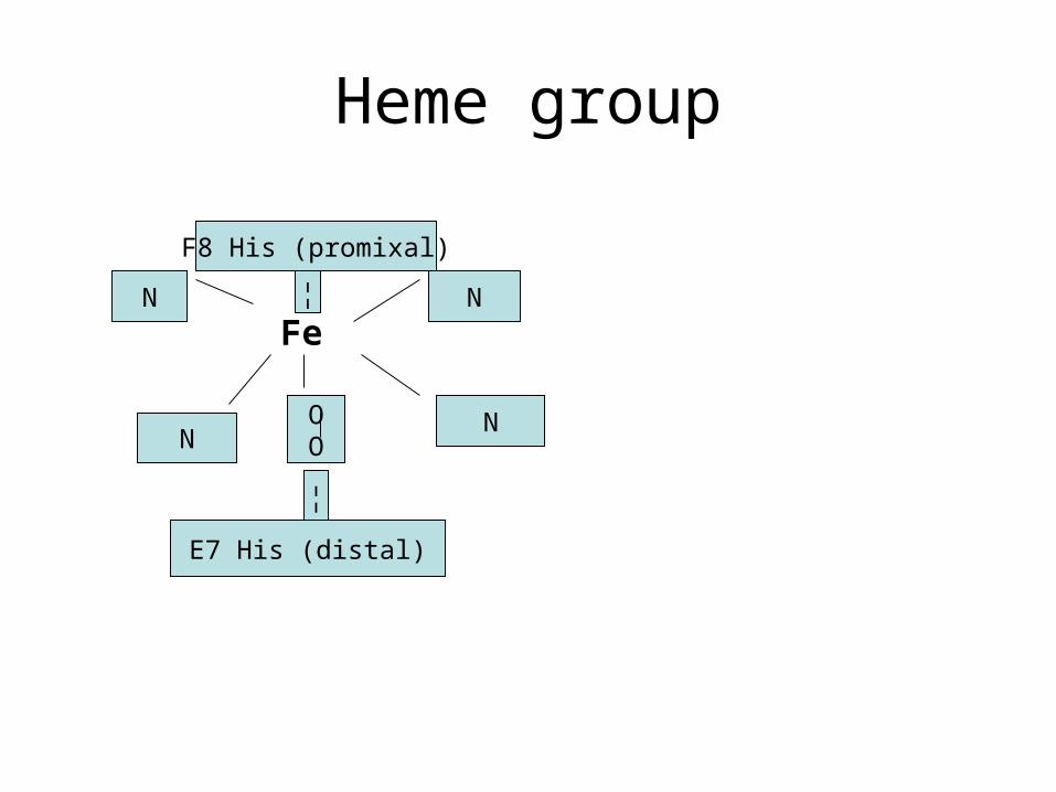
Heme group
Fe
F8 His (promixal)
E7 His (distal)
OO
N
NN
N
¦
¦
Continued
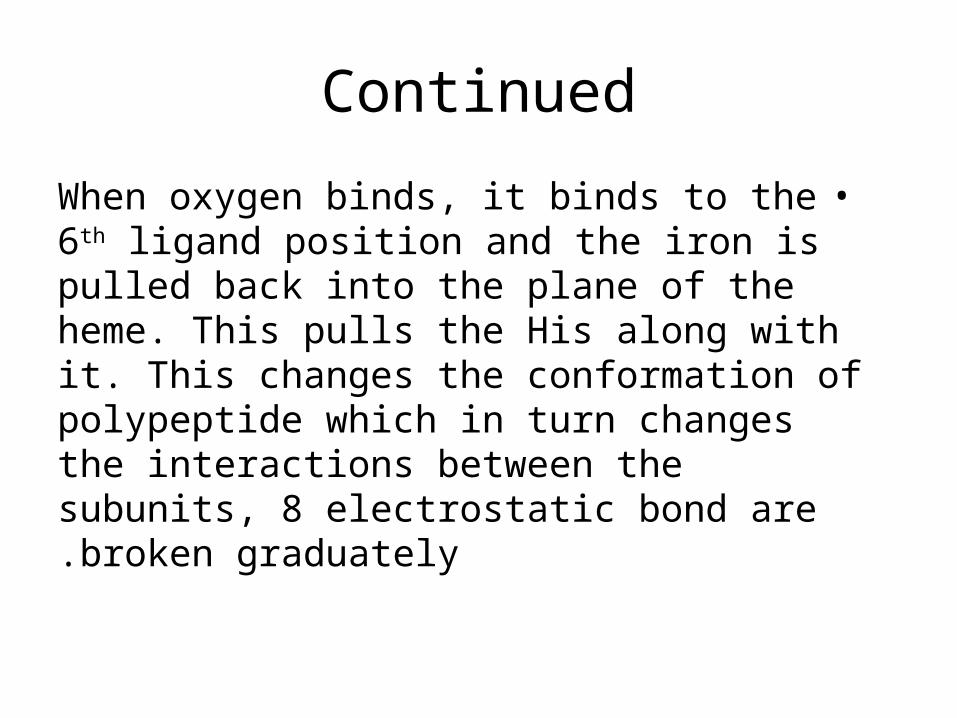
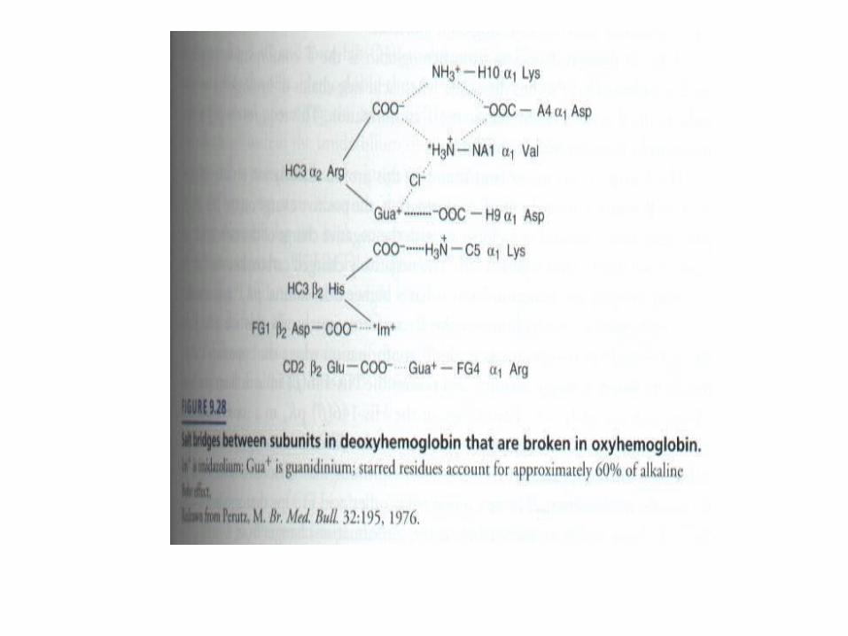
•When oxygen binds, it binds to the 6th ligand position and the iron is pulled back into the plane of the heme. This pulls the His along with it. This changes the conformation of polypeptide which in turn changes the interactions between the subunits, 8 electrostatic bond are broken graduately.
Effect of PH,CO2,BPG
•In lung oxy Hb is formed and H+,CO2 were released.
•In tissues Hb deoxy is formed ( oxygen is used for oxidative phosphorylation or stored via Mb).
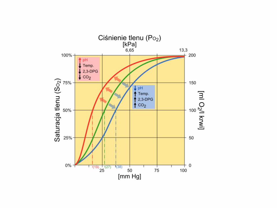
•At low PH (high H+), Bohr effect:• Hb O2-binding curve shifts to the right.
•H+ bind to His imidazole, favor release O2= Hb deoxy. Leads release of O2 in tissues.
•Bohr effect: hydrogen ions and carbon dioxide promote the release of oxygen (curve to right side)
Oxygen Dissociation curve
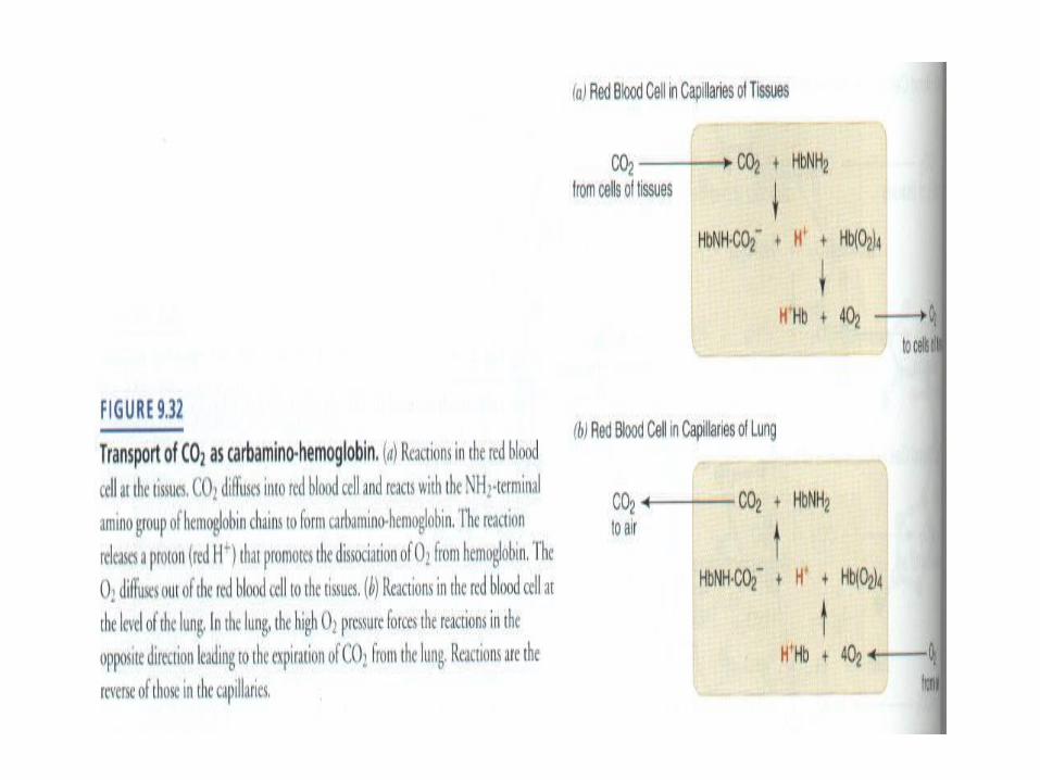
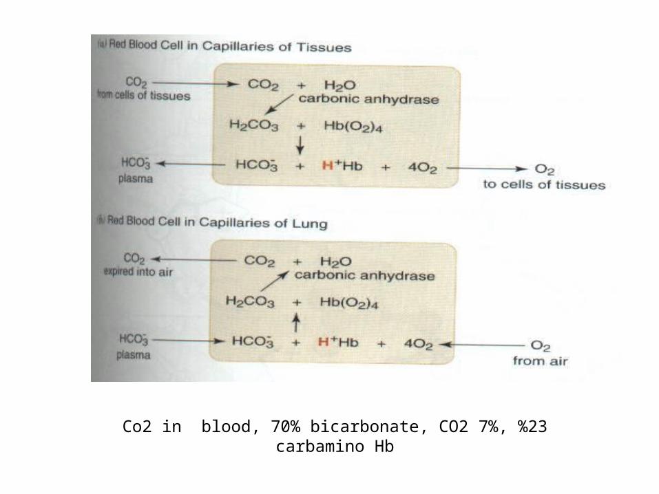
Co2 in blood, 70% bicarbonate, CO2 7%, %23 carbamino Hb
Continued
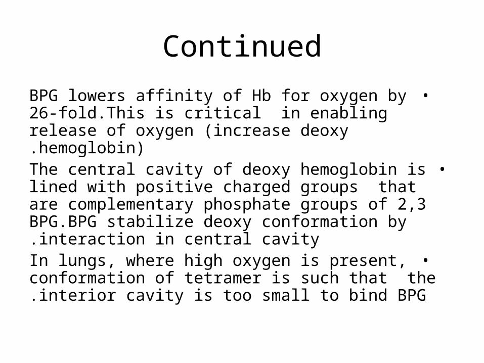
•BPG lowers affinity of Hb for oxygen by 26-fold.This is critical in enabling release of oxygen (increase deoxy hemoglobin).
•The central cavity of deoxy hemoglobin is lined with positive charged groups that are complementary phosphate groups of 2,3 BPG.BPG stabilize deoxy conformation by interaction in central cavity.
•In lungs, where high oxygen is present, conformation of tetramer is such that the interior cavity is too small to bind BPG.
Adaptation to high altitude
•In high altitude an increase in the number of erythrocyte and BPG was occurred .
•Elevated BPG, lowers the affinity of Hb for O2 (decreases P50),which enhances
release of O2 at the tissue .
Models for Allosteric interactions in Hb
•Simple sequential Koshland): binding O2 to 1 subunit increase affinity of the other subunits for O2 (model allows hybrid tetramer of T/R permutations).
•Concentrated model (Monod-Wyman-Changeux)=binding O2 to 1 subunit increase the probability that entire tetramer will convert to altered conformation.
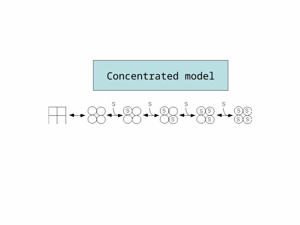
Concentrated model
Numerous Mutant Human Hemoglobins
.11 :When a mutation does compromise biological functions, the condition is termed hemoglobin pathy.
.2Met hemoglobin and Hb M
.3In met hemoglobinemia, the iron is ferric (sulfonamides). Met hemoglobin reductase reduce the Fe+3 to Fe+2.
.4F8 is replaced by Tyrosine In Hb M His
Sickle Cell anemia•Defective hemoglobin caused by mutation at one
amino acid on two of the four chains–Valine replaces glutamic acid
•What is the difference between the two amino acids?
–The Valine a.a. don’t like to interact with aqueous environment, so the molecules clump together
–Form long hemoglobin fibers cause sickle shape inside RBC
–Elongated cells clog capillaries–Loss of RBC anemia
–Can cause swelling, inflammation, pain, and cell death
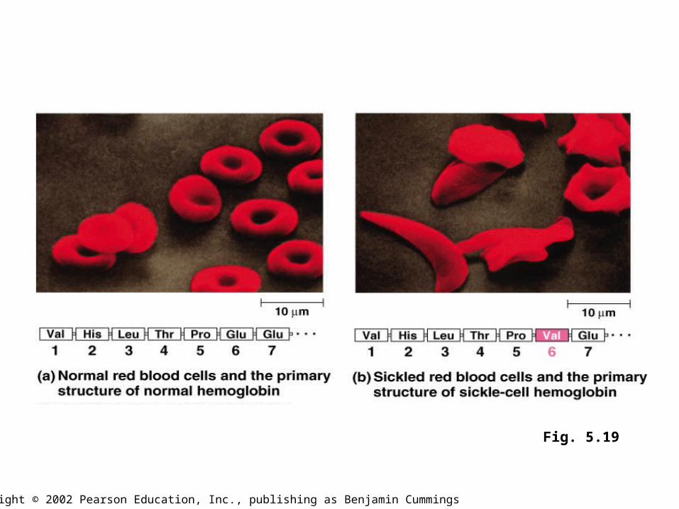
Fig. 5.19
Copyright © 2002 Pearson Education, Inc., publishing as Benjamin Cummings
Biomedical Implication•1(Myoglobinuria : Myoglobin released from damaged
muscle fiber colors the urine dark.•2 (Anemia, reduction in the number of red blood cells or Hb
in the blood ,can reflect impaired synthesis (iron deficiency) or impaired production of RBC. Diagnosis of anemia beings with measurement Hb.
•3(Thalassemias:Partial or total absence of 1 or more chains, alpha (alpha thalassemias) or beta (beta thalassemias) chains.
•4 (Glycosylated Hb (Hb A1c): when blood glucose enter the erythrocyte is glycosylated the amine of lysine and the amino terminals of Hb. The level of glycosylated Hb reflects the blood glucose over the preceding 6-8 weeks (diabetes).