hemolymph sugar homeostasis and starvation … the suppression of rpr-mediated cell death (e.g.,...
TRANSCRIPT

Copyright 2004 by the Genetics Society of America
Hemolymph Sugar Homeostasis and Starvation-Induced Hyperactivity Affectedby Genetic Manipulations of the Adipokinetic Hormone-Encoding
Gene in Drosophila melanogaster
Gyunghee Lee and Jae H. Park1
Department of Biochemistry and Cellular and Molecular Biology, University of Tennessee, Knoxville, Tennessee 37996
Manuscript received August 10, 2003Accepted for publication January 28, 2004
ABSTRACTAdipokinetic hormones (AKHs) are metabolic neuropeptides, mediating mobilization of energy sub-
strates from the fat body in many insects. In delving into the roles of the Drosophila Akh (dAkh) gene, itsdevelopmental expression patterns were examined and the physiological functions of the AKH-producingneurons were investigated using animals devoid of AKH neurons and ones with ectopically expressingdAkh. The dAkh gene is expressed exclusively in the corpora cardiaca from late embryos to adult stages.Projections emanating from the AKH neurons indicated that AKH has multiple target tissues as follows:the prothoracic gland and aorta in the larva and the crop and brain in the adult. Studies using transgenicmanipulations of the dAkh gene demonstrated that AKH induced both hypertrehalosemia and hyperli-pemia. Starved wild-type flies displayed prolonged hyperactivity prior to death; this novel behavioral patterncould be associated with food-searching activities in response to starvation. In contrast, flies devoid ofAKH neurons not only lacked this type of hyperactivity, but also displayed strong resistance to starvation-induced death. From these findings, we propose another role for AKH in the regulation of starvation-induced foraging behavior.
HOMEOSTATIC regulation of blood sugar levels Horst et al. 2001). Injection of the peptides into cock-is a fundamental physiological process in both roaches elevates levels of hemolymph trehalose, a non-
vertebrates and invertebrates. Failure to do so causes reducing disaccharide that is one of the major bloodserious health problems such as diabetes in humans. In sugar molecules in insects (Bedford 1977). Thus themammals, two important endocrine hormones, gluca- glucagon-like peptide in insects is referred to as hyper-gon and insulin, are key physiological effectors that trehalosemic hormone (HTH). However, injection ofregulate blood glucose levels. These peptide hormones this peptide into locusts elicits both carbohydrate andare synthesized by the endocrine glands in the pancreas lipid mobilization from the fat body, leading to theand released into the bloodstream in response to inter- alternative name adipok inetic hormone (AKH). Thesenal changes in sugar levels. In target tissues, such as peptide hormones form the largest neuropeptide familythe liver, these pancreatic hormones activate opposing in arthropods, including �30 isoforms identified in �80metabolic pathways (e.g., glycogen breakdown by gluca- species encompassing all major insect phyla and severalgon and glycogen synthesis by insulin), thereby main- crustacean species (Gade et al. 1997).taining steady-state glucose levels. Like other neuropeptides, AKHs are multifunctional.
Fundamental endocrine regulations of homeostatic Other known physiological effects observed for this sub-blood sugar levels are also conserved in insects. For stance include cardioacceleration in cockroaches (e.g.,instance, an insulin-related peptide, bombyxin, lowers Keeley et al. 1991) and migration of tegumentary andhemolymph sugar concentrations in a dose-dependent retinal distal pigments in crustaceans (Garfias et al.manner in the silkworm Bombyx mori (Satake et al. 1995; Porras et al. 2001). AKH also induces transcrip-1997). Recently, genes encoding Drosophila insulin-like tion of the cytochrome P450 gene in the fat body ofpeptides (dilp) have been identified (Brogiolo et al. cockroaches (Bradfield et al. 1991) and expression of2001), and transgenic ablation of dilp-producing neu- a gene encoding fatty acid binding protein in the flightrons results in the elevation of total blood sugar (Rulif- muscle of locusts (Chen and Haunerland 1994). Inson et al. 2002). addition, AKH peptides have excitatory effects on motor
Insects also produce peptide hormones that act as neurons in moths (Milde et al. 1995) and enhancefunctional homologs of vertebrate glucagons (Van der amplitudes of the electroretinogram in the crayfish
(Garfias et al. 1995).Despite the physiological studies just described, bio-
1Corresponding author: Department of Biochemistry and Cellular and logical functions of the AKH-encoding gene are un-Molecular Biology, M407, Walters Life Science Bldg., University ofTennessee, Knoxville, TN 37996. E-mail: [email protected] known, in part due to the lack of genetic variants involv-
Genetics 167: 311–323 (May 2004)

312 G. Lee and J. H. Park
For construction of r4-gal4 vector, a SpeI/XbaI fragment con-ing this substance. Drosophila AKH peptide and itstaining r4 was excised from a pUC/r4 vector (An and Wensinkencoding gene (dAkh) sequences have been previously1995) and subcloned at an XbaI site of pPTGAL. The cis-acting
reported (Schaffer et al. 1990; Noyes et al. 1995). To “r” element has been previously known to play a role in yolk-gain insight into in vivo roles of AKH in Drosophila, we protein gene expression in adult flies (An and Wensink 1995).
Since our data showed that r4-gal4 (an insertion in the thirdexamined anatomical details of AKH-expressing (AKH-chromosome) directed reporter gene expression specificallyergic) neurons in various developmental stages. Wein the fat body of both sexes, this driver was employed tothen carried out targeted ablation to obtain AKH-cell-effect fat body-targeted ectopic dAkh expression. The UAS-
deficient (AKH-CD) flies and ectopic dAkh expression, dAkh vector was constructed by subcloning dAkh cDNA (seefollowed by analyses of physiological and behavioral below) into NotI/XhoI sites of the pUAST vector (Phelps and
Brand 1998). A homozygous viable UAS-dAkh line (an inser-phenotypes resulting from these transgenic manipula-tion in the second chromosome) was used in this study.tions. The results show that AKH functions as a meta-
Whole-mount in situ hybridization: Since the transcriptionbolic stimulator causing both hypertrehalosemia andtermination site of the dAkh gene is unknown, we performed
hyperlipemia. Our data also suggest that AKH is in- 3�-rapid amplification of cDNA ends (RACE), using gene-volved in the regulation of starvation-induced locomo- specific primers in essentially the same manner as described
in Park and Hall (1998). As a result, we identified a consensustor activities, and such roles are likely to be associatedpolyadenylation signal (AATAAA) at an appropriate posi-with AKH’s metabolic roles to maximize the likelihoodtion (17-bp upstream of the 3�-end), validating our 3�-RACEof the fly’s survival when foods are scarce.experiment (not shown). On the basis of the sequence data,reverse transcription-PCR was carried out to obtain the full-length dAkh cDNA. The PCR product was subcloned into the
MATERIALS AND METHODS pGEM-T vector (Promega, Madison, WI), from which a SpeI/EcoRI fragment was reinserted into the pBluescript (Stra-Fly strains and genetic crosses: Flies were maintained attagene, La Jolla, CA). Subsequently, digoxigenin (dig)-labeled25� in light:dark (12-hr:12-hr) cycles on yeast-cornmeal-agarantisense dAkh riboprobes were generated by in vitro transcrip-media. Canton-S was used as the wild type and yellow whitetion, using the Genius-4 RNA labeling kit (Roche, Indianapo-(y w) as a genetic control for some experiments. For thelis), and applied for whole-mount in situ hybridizations asvisualization of GAL4-expressing cells, gal4 driver strains weredescribed in Lee et al. (2000).crossed to a reporter transgenic strain such as UAS-lacZ or
Histochemistry: To detect in situ lacZ expression, tissues wereUAS-NZ, which encodes cytoplasmic or nuclear �-galactosi-dissected, fixed, and stained with X-gal (e.g., Park et al. 2000).dase, respectively (Phelps and Brand 1998), or UAS-mCD8-After the desired staining was achieved, tissues were washedgfp, which encodes a membrane-bound derivative of greenin PBS, dehydrated, and mounted on a glass slide with afluorescent protein (GFP; Lee and Luo 1999). A UAS-reapermounting medium (1:6 mixture of methyl salycylate and per-(rpr) was used for cell-specific ablation and UAS-p35 was usedmount). For the visualization of GFP expression, dissectedfor the suppression of rpr-mediated cell death (e.g., Renn ettissues were fixed in 4% paraformaldehyde for 15 min on ice,al. 1999). To evaluate the absence of cells resulting from rpr-washed six times with PBS, and mounted with glycerol. Signalsinduced apoptosis, a double transgenic line containing bothwere viewed under an Olympus BX61 microscope equippedUAS-rpr and UAS-lacZ (hereafter, UAS-rpr:lacZ) was crossed towith a CC12 digital camera and images were acquired usinga dAkh-gal4 driver, and dAkh-expressing cells in the progenyanalySIS 3.0 software (Soft Imaging System). To detect AKHwere monitored by 5-bromo-4-chloro-3-indolyl �-d-galactosidepeptides in tissues, anti-AKH antibody was generated in rab-(X-gal) histochemistry. To rescue rpr-induced cell death, fliesbits, using keyhole limpet hemocyanin-conjugated syntheticdoubly homozygous for the dAkh-gal4 and UAS-p35 transgenesAKH peptide as an antigen (Genemed Synthesis, San Fran-were generated by recombination and crossed to the UAS-cisco). Tissues were incubated with diluted primary antiserumlacZ or UAS-rpr:lacZ. Ectopic dAkh expression in larval fat body(1:600) and subsequently with FITC-tagged secondary anti-was accomplished by crossing an r-tetramer (r4)-gal4 driverbodies (1:200; Jackson Immunoresearch Labs, West Grove,(see below) to a UAS-dAkh. Newly generated dAkh-gal4, r4-gal4,PA), and the resulting fluorescence signals were processed asand UAS-dAkh transgenic lines are described in the followingdescribed above. To visualize lipid droplets stored in the fatsection.cells, fat bodies were dissected from wandering third instarGeneration of transgenic fly strains: In constructing thelarvae, fixed with 4% paraformaldehyde for 10 min, rinsed indAkh promoter fused to the yeast transcription factor gal4 gene,PBS, and processed for Sudan Black-mediated staining (Zinkeforward (5�-GCTCTAGAACACGCGTCGACTGAGCTT-3�) andet al. 1999).reverse (5�-GCGGTACCTGAGTTCTATGCTGGTCCAC-3�)
Starvation-induced mortality assay: Eclosed flies were agedPCR primers were designed to encompass the 5� upstreamin a group for 7–14 days. For starvation tube preparation, glassregion extending from �1020 to �17 (�1 indicates the tran-tubes (6 � 50 mm; Fisher Scientific, Pittsburgh) were filledscription-start site, which was previously determined by Noyesup to one-third with 0.5% agarose. To synchronize starvationet al. 1995), on the basis of the nucleotide sequences flank-initiation for all genotypes tested, 40–60 flies of each genotypeing the dAkh gene (Berkeley Drosophila Genome Project;were transferred simultaneously into a vial containing 0.5%AC005814). The PCR product was subcloned into XbaI/KpnIagarose. Subsequently, each starvation tube was loaded withsites of the pPTGAL vector (Sharma et al. 2002). The resultinga single anesthetized fly, cotton plugged, and then placed inrecombinant vector was injected into y w embryos, using stan-a humidified chamber in a 25� incubator supplied with 12dard methods for P-element-mediated germ-line transforma-hr:12 hr light:dark cycles. Dead flies were counted every 6 ortion, leading to the establishment of the following three lines:12 hr and the survival rate for each genotype was plotted.dAkh-gal4, dAkh-gal4(2), and dAkh-gal4(3). A homozygous viable
Circadian locomotor activity behavior assays: For the circa-dAkh-gal4 line, which has an insertion in the third chromo-dian locomotor activity assay, individual flies were loaded intosome, was used primarily in this study. The other two homozy-a glass tube containing 4% sucrose in 2% Bactoagar medium,gous lethal strains (each of which carries an insertion in the
second chromosome) were used for some experiments. entrained for 3–7 days to 12-hr:12-hr light:dark (LD) cycles,
313Expression and Functions of dAkh
and then assessed for free-running locomotion for the next7 days in constant darkness (DD) at 25�. For the locomotoractivity assay under starvation, glass tubes containing 2% aga-rose only were used. The activities were monitored using aTrikinetics system interfaced with a PC computer, as describedby Konopka et al. (1994). Numbers of activity events wererecorded every half-hour (bin), and average numbers of activ-ity events per bin per fly were calculated using Microsoft Excelsoftware. Chi-square periodogram analysis (Sokolove andBushell 1978) was performed using a web-derived software(http://hawk.bcm.tmc.edu/) to detect periodicity.
Sugar and lipid measurements: A group of 6–12 third instarlarvae were washed twice with dH2O and blot dried. The hemo-lymph was allowed to bleed out on a dimple glass plate bytearing off the cuticle; 2 �l of hemolymph was rapidly with-drawn, mixed with 38 �l of PBS, and then centrifuged for 10min at 13,000 � g to precipitate blood cells and tissue debris.The supernatant was transferred to a fresh tube and an aliquot(2 �l) was applied for trehalose or glucose assays using acommercial kit (17-25; Sigma, St. Louis) as described in Rulif-son et al. (2002).
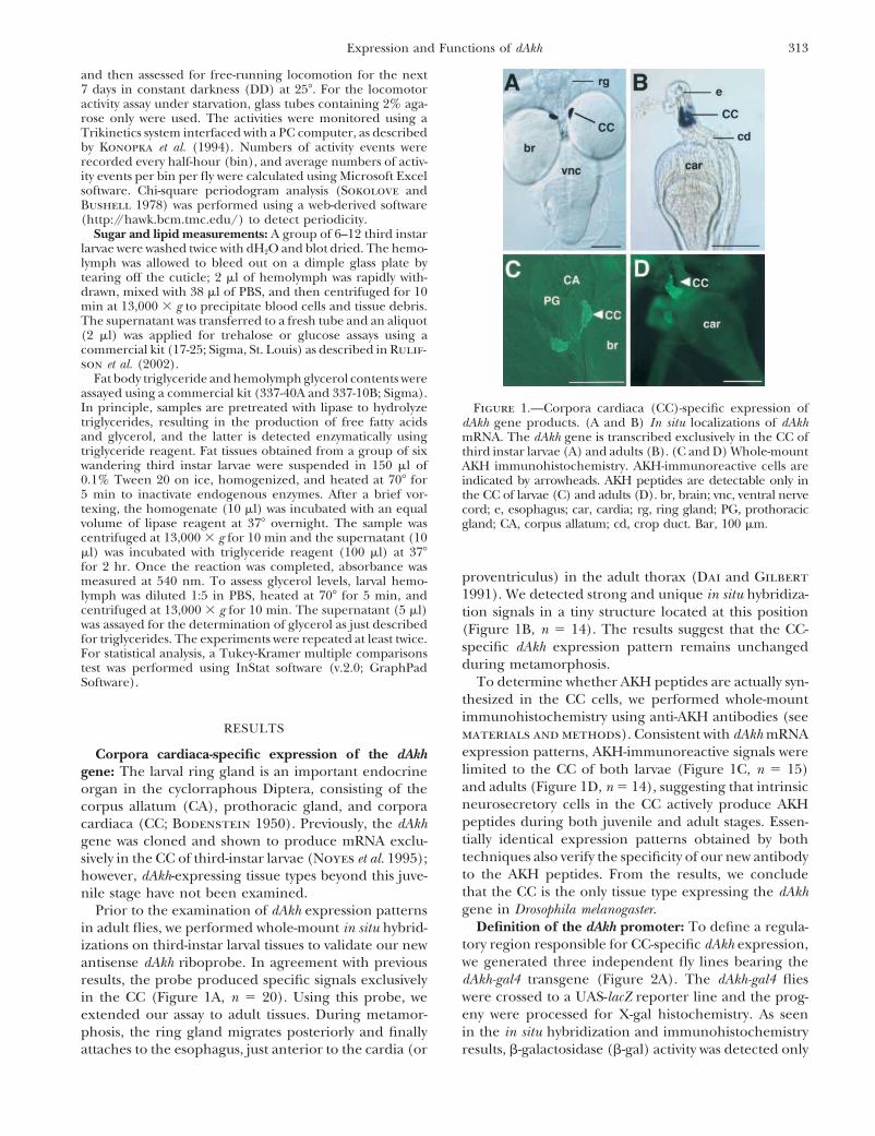
Fat body triglyceride and hemolymph glycerol contents wereassayed using a commercial kit (337-40A and 337-10B; Sigma).In principle, samples are pretreated with lipase to hydrolyze Figure 1.—Corpora cardiaca (CC)-specific expression oftriglycerides, resulting in the production of free fatty acids dAkh gene products. (A and B) In situ localizations of dAkhand glycerol, and the latter is detected enzymatically using mRNA. The dAkh gene is transcribed exclusively in the CC oftriglyceride reagent. Fat tissues obtained from a group of six third instar larvae (A) and adults (B). (C and D) Whole-mountwandering third instar larvae were suspended in 150 �l of AKH immunohistochemistry. AKH-immunoreactive cells are0.1% Tween 20 on ice, homogenized, and heated at 70� for indicated by arrowheads. AKH peptides are detectable only in5 min to inactivate endogenous enzymes. After a brief vor- the CC of larvae (C) and adults (D). br, brain; vnc, ventral nervetexing, the homogenate (10 �l) was incubated with an equal cord; e, esophagus; car, cardia; rg, ring gland; PG, prothoracicvolume of lipase reagent at 37� overnight. The sample was gland; CA, corpus allatum; cd, crop duct. Bar, 100 �m.centrifuged at 13,000 � g for 10 min and the supernatant (10�l) was incubated with triglyceride reagent (100 �l) at 37�for 2 hr. Once the reaction was completed, absorbance was
proventriculus) in the adult thorax (Dai and Gilbertmeasured at 540 nm. To assess glycerol levels, larval hemo-1991). We detected strong and unique in situ hybridiza-lymph was diluted 1:5 in PBS, heated at 70� for 5 min, and
centrifuged at 13,000 � g for 10 min. The supernatant (5 �l) tion signals in a tiny structure located at this positionwas assayed for the determination of glycerol as just described (Figure 1B, n 14). The results suggest that the CC-for triglycerides. The experiments were repeated at least twice. specific dAkh expression pattern remains unchangedFor statistical analysis, a Tukey-Kramer multiple comparisons
during metamorphosis.test was performed using InStat software (v.2.0; GraphPadTo determine whether AKH peptides are actually syn-Software).
thesized in the CC cells, we performed whole-mountimmunohistochemistry using anti-AKH antibodies (see
RESULTS materials and methods). Consistent with dAkh mRNAexpression patterns, AKH-immunoreactive signals wereCorpora cardiaca-specific expression of the dAkhlimited to the CC of both larvae (Figure 1C, n 15)gene: The larval ring gland is an important endocrineand adults (Figure 1D, n 14), suggesting that intrinsicorgan in the cyclorraphous Diptera, consisting of theneurosecretory cells in the CC actively produce AKHcorpus allatum (CA), prothoracic gland, and corporapeptides during both juvenile and adult stages. Essen-cardiaca (CC; Bodenstein 1950). Previously, the dAkhtially identical expression patterns obtained by bothgene was cloned and shown to produce mRNA exclu-techniques also verify the specificity of our new antibodysively in the CC of third-instar larvae (Noyes et al. 1995);to the AKH peptides. From the results, we concludehowever, dAkh-expressing tissue types beyond this juve-that the CC is the only tissue type expressing the dAkhnile stage have not been examined.gene in Drosophila melanogaster.Prior to the examination of dAkh expression patterns
Definition of the dAkh promoter: To define a regula-in adult flies, we performed whole-mount in situ hybrid-tory region responsible for CC-specific dAkh expression,izations on third-instar larval tissues to validate our newwe generated three independent fly lines bearing theantisense dAkh riboprobe. In agreement with previousdAkh-gal4 transgene (Figure 2A). The dAkh-gal4 fliesresults, the probe produced specific signals exclusivelywere crossed to a UAS-lacZ reporter line and the prog-in the CC (Figure 1A, n 20). Using this probe, weeny were processed for X-gal histochemistry. As seenextended our assay to adult tissues. During metamor-in the in situ hybridization and immunohistochemistryphosis, the ring gland migrates posteriorly and finally
attaches to the esophagus, just anterior to the cardia (or results, �-galactosidase (�-gal) activity was detected only

314 G. Lee and J. H. Park
confirmed by dAkh-gal4-driven gfp expression in “live”larvae (Figure 2F, n 7). The results suggest that cis-acting regulatory elements necessary for CC-specificdAkh expression are present within the 1-kb upstreamsequence.
Using the dAkh-gal4/UAS-lacZ system, we determinedthe earliest developmental stage of dAkh expression.The �-gal activity was at first faint in a paired structure inapproximately stage-14 embryos (Figure 2G) and thenbecame stronger in older embryos (Figure 2H). CC-specific expression was also observed in first-instar larvae(Figure 2I, n 7); however, projections from the CCneurons were undetectable (Figure 2B vs. 2I), sug-gesting that the dAkh neurons in first-instar larvae havenot yet been fully differentiated. Nevertheless, the over-all results suggest that normal dAkh gene functionsmight be necessary from late embryonic stages onward.
Anatomy and targets of dAkh-expressing neurons: Lit-tle is known about neuro-anatomical details of the in-trinsic neurosecretory cells in the CC of Drosophila.Since dAkh gene products could serve as a useful markerfor such cells, we further examined characteristics ofthese cells in great detail, using various transgenic ma-nipulations and histochemical assays. First, in determin-ing the number of dAkh-expressing cells, dAkh-gal4 flieswere crossed to a UAS-NZ reporter to express �-gal inthe nuclei of dAkh cells (Phelps and Brand 1998). Asa result, we identified �7 (0.1, SEM) cells per eachlobe of larval CC (Figure 3A, n 57). For adult CC,the total number of dAkh cells was counted from a wholeCC structure instead of per each lobe, since the bound-ary between lobes was not clearly recognizable, thushampering precise counting. This yielded an average
Figure 2.—Reporter gene expression mediated by dAkh of 13 (0.6, SEM) cells per CC (n 32), ranging frompromoter. (A) Schematics of dAkh gene structure and dAkh- 11 to 16 (Figure 3B). Since the counts in an adult CCgal4 construct. (B–I) X-gal histochemistry or gfp expression
are comparable to those observed in an entire larvalof the offspring of [dAkh-gal4 � UAS-lacZ or UAS-mCD8-gfp]CC, dAkh cells might be present persistently duringcross. (B) Third instar larval central nervous system. LacZ
expression is restricted to the CC. Processes innervating the metamorphosis.prothoracic gland (PG) are indicated by arrows. (C–E) LacZ To determine the population of dAkh cells in the CC,expression in adults. (C) Somata of adult AKHergic neurons we simultaneously marked somata of dAkh neurons byare present exclusively in the CC. Projections to the brain are
dAkh promoter-driven gfp expression and nuclei of en-denoted by white arrows and ones to the crop by black arrows.tire CC cells by 4�,6-diamidino-2-phenylindole (DAPI)(D) Projections arising from the posterior side of the CC
(arrows). A white arrowhead designates a projection leading staining. A majority of the DAPI-positive cells expressedto the crop. (E) Nerve terminals at the crop duct indicated gfp (Figure 3, C and D), suggesting that dAkh cells repre-by an arrowhead. (F) Expression of gfp in a “live” third instar sent most of the CC cells.larval progeny from a [dAkh-gal4 � UAS-mCD8-gfp] cross. Spe-
Stainings mediated by anti-AKH antibody and X-galcific GFP-mediated fluorescence signals are clearly visible inhistochemistry were examined at higher resolution toa paired structure (arrowhead) at a position where the CC
locate. (G and H) LacZ expression (arrowheads) during em- construct a fine neural mapping involving the AKHergicbryonic development. The expression (arrowhead) was first neurons. In larvae, we detected two potential targetsseen in stage-14 embryos (G) and became stronger in older innervated by AKHergic neurons, one of which is theembryos (stage 15; H). (I) CC-specific lacZ expression (arrow-
prothoracic gland located immediately adjacent to thehead) in first instar larva. Bars, 100 �m.CC and known to produce a molting hormone ecdyste-roid (e.g., Dai and Gilbert 1991). The AKHergic neu-rons sent two or three projections to this structure (Fig-in the CC of larvae (Figure 2B, n 25) and adults
(Figure 2C, n 16). Identical expression patterns were ures 2B and 3F). The other target is the aorta (or dorsalvessel) that is closely associated with the CC. Extensiveobtained from all three dAkh-gal4 lines. Lack of ectopic
expression sites directed by this promoter was further AKH-immunoreactive varicosities observed on the aorta

315Expression and Functions of dAkh
process whose target could not be identified (Figure2D). Nonetheless, this implies additional physiologicalfunctions attributed to AKH in adult flies.
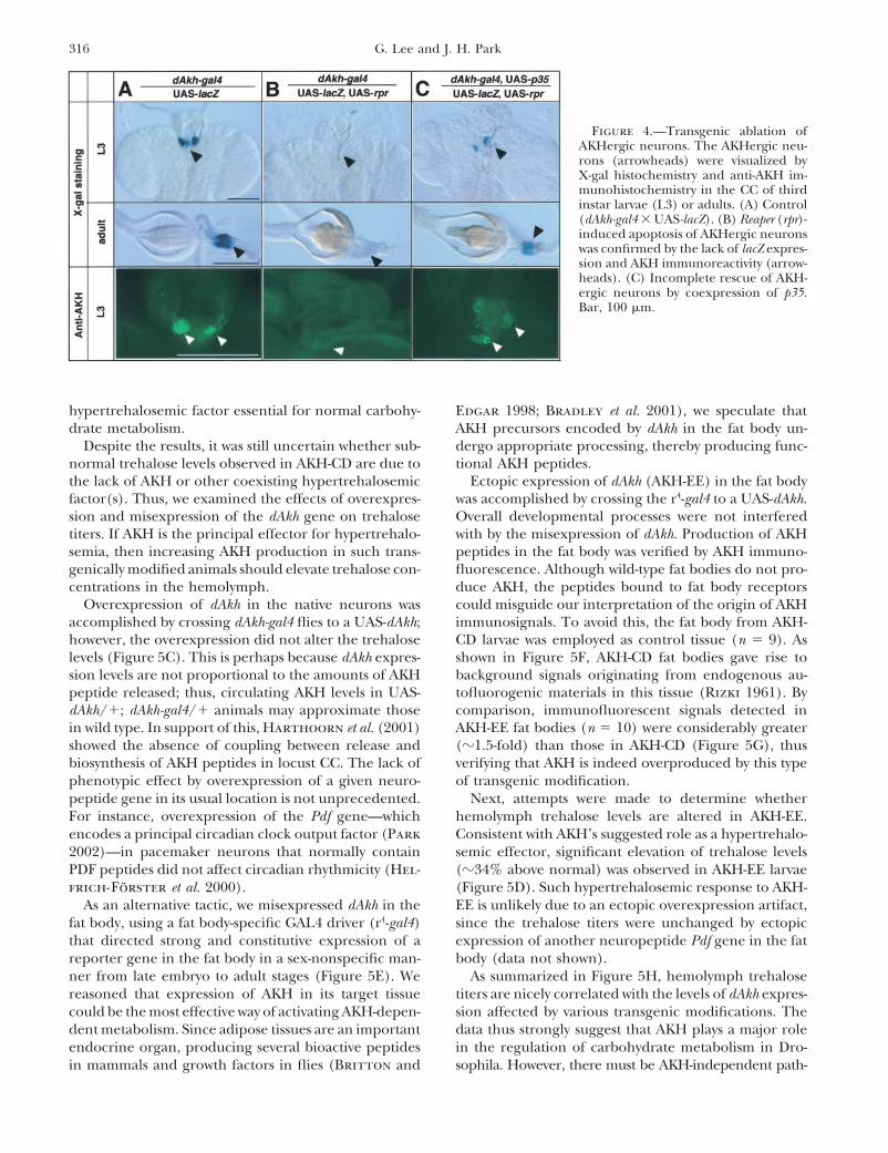
Targeted ablation of AKHergic neurons: To under-stand AKH functions in Drosophila, we obtained AKH-cell-deficient (AKH-CD) flies by expressing a pre-apoptotic gene, reaper (rpr ; White et al. 1996), in theAKHergic neurons. Ectopic expression of rpr in variouspeptidergic neurons has been successfully employed totrigger the apoptosis of these neurons (McNabb et al.1997; Renn et al. 1999; Park et al. 2003). The AKH-CDflies were generated by crossing dAkh-gal4 flies to a UAS-rpr or a double transgenic UAS-rpr:lacZ strain, and theirprogeny were examined histologically to confirm theabsence of the AKHergic neurons. The UAS-rpr:lacZ linewas useful particularly for checking ablation status, sincethe presence or absence of target neurons could beeasily judged by simple X-gal staining (Figure 4A vs.4B). Indeed, we were unable to detect any �-gal activityin the CC of the dAkh-gal4/UAS-rpr:lacZ larvae andadults, which therefore indicates a complete loss of dAkhFigure 3.—Cellular characteristics of AKHergic neurons.neurons. This was further confirmed by the lack of AKH(A and B) Dissected ring glands from the progeny of a [dAkh-
gal4 � UAS-NZ] cross were processed for X-gal histochemistry. immunoreactivities (Figure 4B). The rpr-mediated cellEach nuclear lacZ expression (arrowheads) represents individ- death was rescued partially by coexpression of the anti-ual neurons in third instar larval (A) and adult CC (B). Dotted apoptotic protein p35 (Hay et al. 1994), since fewerlines in A contour the brain. Bars, 100 �m. (C and D) The
numbers of X-gal-stained cells or less intensive AKHCC of larval offspring from a [dAkh-gal4 � UAS-mCD8-gfp]immunosignals were observed in the CC of p35-rescuedcross were stained by DAPI, and the fluorescence signals were
captured for either gfp (C) or DAPI (D). Note that most of animals compared with such stainings in the controlthe CC cells are AKHergic neurons. (E) A bright-field image animals (Figure 4C vs. 4A).of the same specimen shown in C and D. Bar, 25 �m. (F and
The AKHergic neurons apparently do not play a vitalG) AKH-immunoreactive projections in larval CC. Images wererole, since AKH-CD animals developed in an ostensiblycaptured from the same specimen at different focal plans to
show projections (arrows) innervating the prothoracic gland normal manner. No noticeable defects in growth, meta-(PG) and the aorta (a). Bar, 50 �m. morphosis, eclosion, and longevity were observed. Adult
AKH-CD flies also showed normal reproductive capabili-ties and courtship behavior (data not shown). The resultsindicate that AKHs are released into the circulatorysuggest that AKH functions are not essential for overallsystem (Figure 3G).development and reproduction at normal growth condi-Although it is not as clear as in larval CC, adult CCtions.also form a bilobed structure and the dAkh neurons are
Carbohydrate metabolism is affected by the ablationpresent in both lobes (Figure 1, B and D, Figure 2C).of AKHergic neurons and ectopic dAkh expression: Tre-Processes stemming from the anterior side of the lobeshalose is a disaccharide composed of two glucose mole-were traced proximate to the esophagus foramen wherecules and is the principal blood sugar in insects (Bed-they are likely to enter the protocerebrum. A pair offord 1977). Physiological studies in other insects havelong processes arising from the posterior side reachedshown that AKHs elevate hemolymph trehalose titers atthe crop duct at which the crop begins its expansionthe expense of glycogen storage in the fat body (e.g.,(Figure 2, C–E). In some insects, such as honeybees andPark and Keeley 1995). This prompted us to examineblow flies, the crop stores liquid foods (e.g., nectar orwhether AKH also plays a role in the regulation of carbo-soluble nutrients), and its volume is highly variable de-hydrate metabolism in Drosophila.pending on the amounts of liquid deposit (Dimitriadis
Hemolymph trehalose levels in AKH-CD larvae wereand Papamanoli 1992; Lorenz et al. 2001b). AKH-a mere 7–26% of normal, whereas the glucose levelshomologous peptides have been proposed to cause re-were unaffected (Figure 5, A and B). Moreover, thegurgitation of nectars from the crop in some wasp spe-trehalose titers in p35-rescued larvae were intermediatecies to increase hemolymph trehalose titers (Lorenz etbetween controls and AKH-CD (Figure 5B), thus reveal-al. 2001b). In this regard, our findings of AKH nerveing a positive correlation between the levels of dAkhterminals at the crop duct support the idea that AKHexpression and hemolymph trehalose concentrations.may control the crop volume in Drosophila. In addition
to the brain and the crop, we occasionally observed a The results suggest that the AKH neurons produce a
316 G. Lee and J. H. Park
Figure 4.—Transgenic ablation ofAKHergic neurons. The AKHergic neu-rons (arrowheads) were visualized byX-gal histochemistry and anti-AKH im-munohistochemistry in the CC of thirdinstar larvae (L3) or adults. (A) Control(dAkh-gal4 � UAS-lacZ). (B) Reaper (rpr)-induced apoptosis of AKHergic neuronswas confirmed by the lack of lacZ expres-sion and AKH immunoreactivity (arrow-heads). (C) Incomplete rescue of AKH-ergic neurons by coexpression of p35.Bar, 100 �m.
hypertrehalosemic factor essential for normal carbohy- Edgar 1998; Bradley et al. 2001), we speculate thatAKH precursors encoded by dAkh in the fat body un-drate metabolism.
Despite the results, it was still uncertain whether sub- dergo appropriate processing, thereby producing func-tional AKH peptides.normal trehalose levels observed in AKH-CD are due to
the lack of AKH or other coexisting hypertrehalosemic Ectopic expression of dAkh (AKH-EE) in the fat bodywas accomplished by crossing the r4-gal4 to a UAS-dAkh.factor(s). Thus, we examined the effects of overexpres-
sion and misexpression of the dAkh gene on trehalose Overall developmental processes were not interferedwith by the misexpression of dAkh. Production of AKHtiters. If AKH is the principal effector for hypertrehalo-
semia, then increasing AKH production in such trans- peptides in the fat body was verified by AKH immuno-fluorescence. Although wild-type fat bodies do not pro-genically modified animals should elevate trehalose con-
centrations in the hemolymph. duce AKH, the peptides bound to fat body receptorscould misguide our interpretation of the origin of AKHOverexpression of dAkh in the native neurons was
accomplished by crossing dAkh-gal4 flies to a UAS-dAkh; immunosignals. To avoid this, the fat body from AKH-CD larvae was employed as control tissue (n 9). Ashowever, the overexpression did not alter the trehalose
levels (Figure 5C). This is perhaps because dAkh expres- shown in Figure 5F, AKH-CD fat bodies gave rise tobackground signals originating from endogenous au-sion levels are not proportional to the amounts of AKH
peptide released; thus, circulating AKH levels in UAS- tofluorogenic materials in this tissue (Rizki 1961). Bycomparison, immunofluorescent signals detected indAkh/�; dAkh-gal4/� animals may approximate those
in wild type. In support of this, Harthoorn et al. (2001) AKH-EE fat bodies (n 10) were considerably greater(�1.5-fold) than those in AKH-CD (Figure 5G), thusshowed the absence of coupling between release and
biosynthesis of AKH peptides in locust CC. The lack of verifying that AKH is indeed overproduced by this typeof transgenic modification.phenotypic effect by overexpression of a given neuro-
peptide gene in its usual location is not unprecedented. Next, attempts were made to determine whetherhemolymph trehalose levels are altered in AKH-EE.For instance, overexpression of the Pdf gene—which
encodes a principal circadian clock output factor (Park Consistent with AKH’s suggested role as a hypertrehalo-semic effector, significant elevation of trehalose levels2002)—in pacemaker neurons that normally contain
PDF peptides did not affect circadian rhythmicity (Hel- (�34% above normal) was observed in AKH-EE larvae(Figure 5D). Such hypertrehalosemic response to AKH-frich-Forster et al. 2000).
As an alternative tactic, we misexpressed dAkh in the EE is unlikely due to an ectopic overexpression artifact,since the trehalose titers were unchanged by ectopicfat body, using a fat body-specific GAL4 driver (r4-gal4)
that directed strong and constitutive expression of a expression of another neuropeptide Pdf gene in the fatbody (data not shown).reporter gene in the fat body in a sex-nonspecific man-
ner from late embryo to adult stages (Figure 5E). We As summarized in Figure 5H, hemolymph trehalosetiters are nicely correlated with the levels of dAkh expres-reasoned that expression of AKH in its target tissue
could be the most effective way of activating AKH-depen- sion affected by various transgenic modifications. Thedata thus strongly suggest that AKH plays a major roledent metabolism. Since adipose tissues are an important
endocrine organ, producing several bioactive peptides in the regulation of carbohydrate metabolism in Dro-sophila. However, there must be AKH-independent path-in mammals and growth factors in flies (Britton and

317Expression and Functions of dAkh
Figure 5.—Effect of altered dAkh ex-pression on larval hemolymph sugar lev-els. Numbers in parentheses indicate thenumber of samples tested for each geno-type. (A) Ablation of AKHergic neurons(dAkh-gal4/UAS-rpr) caused significantreduction in trehalose levels. (B) Partialrescue of rpr-mediated ablation by p35(dAkh-gal4, UAS-p35/UAS-rpr) incom-pletely restored trehalose titers. (C)Hemolymph trehalose concentrationswere unchanged by overexpression of dAkhin AKHergic neurons (UAS-dAkh/�; dAkh-gal4/�), whereas (D) �34% increasewas triggered by ectopic dAkh expressionin the fat body (UAS-dAkh/�; r4-gal4/�). (E) Fat body-specific lacZ expressiondriven by r4-gal4 in larvae. (F and G)AKH immunohistochemistry of the fatbody. (F) Control, nonspecific autoflu-orescence in AKH-CD fat body. (G) AKHimmunofluorescence in AKH-EE fatbody. Note that the signals shown hereare much stronger than those in AKH-CD. (H) A summary for the trehalosephenotypes affected by various trans-genic manipulations of the dAkh gene.
ways for this type of physiological reaction, since detect- lipid droplets were substantially smaller and fewer inthe AKH-EE than in the controls (Figure 6, A–C, vs.able amounts of trehalose are still present in animals
devoid of AKHergic neurons (Figure 5, A and B). 6D). Consistent with the results, a quantitative assay alsorevealed a significant decrease of triglyceride content—Lipid metabolism induced by ectopic expression of
dAkh: Another well-documented physiological AKH the main storage form of lipids in the fat body—in AKH-EE (Figure 6E).function is to mobilize lipid storage from the fat body
via lipase activation; the resulting metabolites serve as Reduction of endogenous triglyceride levels in AKH-EE fat bodies could be a consequence of either subnor-energy substrates in locusts for long-term flight (Stone
et al. 1976; Van Der Horst et al. 1979; Van Heusden mal synthesis or supernormal degradation (hydrolysis)of the triglycerides. If the latter is the case, one canet al. 1984; Chino et al. 1989) and in tobacco hornworms
during periods of starvation (Ziegler et al. 1990; expect an increase of metabolites derived from the hy-drolysis of triglycerides (i.e., free fatty acids and glycerol)Ziegler 1991; Arrese and Wells 1997).
The fat body of Drosophila also stores large amounts in the serum of AKH-EE. In accordance with the predic-tion, hemolymph glycerol concentrations were signifi-of lipids, which are consumed rapidly upon starvation
(Zinke et al. 1999; Britton et al. 2002). However, it is cantly higher in AKH-EE than in controls (Figure 6F),thus supporting that reduction of triglyceride contentsunknown whether lipid metabolism is regulated by AKH
in Drosophila. To address this question, lipid droplets in the AKH-EE fat body is due to an enhanced lipolyticresponse to AKH.stored in the fat cells were visually examined in well-fed
AKH-EE and control larvae by using the Sudan Black If AKH is the sole effector for the hydrolysis of triglyc-erides, then complete suppression of lipolysis in AKH-staining method (for example, Zinke et al. 1999). The

318 G. Lee and J. H. Park
Figure 6.—Effect of AKH-EE on lipid contents within larvalfat cells. Lipid droplets were stained by Sudan Black. The sizesand amounts of the droplets (arrowheads) are significantlyreduced in AKH-EE (D) compared to those of genetic controls(A–C). Bar, 50 �m. (E and F) Quantitative measurements oftriglyceride (TG) content in the fat body (E) or glycerol inthe hemolymph (F). Numbers in parentheses designate thenumbers of samples tested. (E) Significant reduction of TG or(F) elevation of glycerol is observed in AKH-EE (UAS-dAkh/�;r4-gal4/�). Asterisks indicate statistical significance (P � 0.001).
CD would increase triglyceride storage in AKH-CD fatbody. Our data, however, showed that fat body triglycer-ide contents in AKH-CD were comparable to those ofcontrols (Figure 6E). The results indicate that lipid me-tabolism occurs normally in the absence of AKH, thus
Figure 7.—Survival curves in response to starvation. (A) Atforetelling the existence of alternative lipolytic pathways48 hr after starvation initiation, �50% of control flies died,that are independent of AKH.whereas most of the AKH-CD flies (dAkh-gal4/UAS-rpr) wereAKH-CD flies are resistant to starvation-induced death: still alive. (B) Both male (m) and female (f) AKH-CD flies
Since animals have to survive on nutrients stored in the display similar levels of tolerance to starvation. (C) Rescue ofbody when foods are not available, slower catabolism AKH-CD by coexpression of p35 (dAkh-gal4, UAS-p35/UAS-
rpr) shows intermediate levels of resistance to starvation. (D)of such limited resources would help them to surviveDegrees of resistance to starvation-induced death are indistin-longer. In this context, AKH-CD flies are expected toguishable between young (�30 hr after eclosion) and oldlive longer than wild type, as the foregoing results indi- AKH-CD flies. At least 40 flies were tested for each genotype
cate that catabolic activities are appreciably attenuated or sex.in AKH-CD. To test the hypothesis, mortalities of AKH-CD and control flies, when supplied only with water,were monitored. (Figure 7). Resistance to starvation-induced death was
consistently observed for all dAkh-gal4 transgenic linesRemarkably, AKH-CD flies survived for at least 24 hrlonger than wild type or any other genetic controls regardless of gender (Figure 7B). Of importance, the

319Expression and Functions of dAkh
survival rate of p35-rescued flies was intermediate be-tween those of controls and AKH-CD (Figure 7C). Thisnicely correlates with dAkh expression levels in the p35-rescued flies that are also intermediate between normaland fully ablated (Figure 4C). The data suggest thatdegrees of resistance to the starvation-induced deathare most likely AKH-dose dependent.
Extended longevity of AKH-CD flies under starvationmay be due to their abnormal feeding habits (for in-stance, more frequent feeding or a larger amount offood intake per meal) in response to the reductionof blood sugar levels, resulting in a larger amount ofnutrients taken in by AKH-CD flies than by wild types.If so, then young flies have less time to feed than theolder flies do, thereby storing relatively low energy re-serves. As a consequence, young AKH-CD flies could bemore sensitive to starvation than older AKH-CD flies.We tested this hypothesis by assessing the phenotype ofvery young flies (the majority of whom were youngerthan 30 hr after eclosion). Survival rates displayed byyoung AKH-CD flies (Figure 7D) were similar to thoseof older flies, suggesting that the feeding anomaly (pro-posed above) may not be an influential factor for the
Figure 8.—Circadian locomotor activity rhythms of fedphenotype exhibited by Drosophila ablated of theirflies. Flies were entrained to four cycles of 12 hr:12 hrAKH cells. light:dark (LD) and then proceeded to 7 days of constant
Clock-independent locomotor activity patterns upon darkness (DD). Bars represent average numbers of activitystarvation: Recent studies show that locomotor activities events per half-hour bin for the number of flies tested. Hori-
zontal open and solid boxes indicate the light-on and light-of honeybees and wasps are unable to be sustained inoff phases, respectively, and the shaded box denotes subjectivethe absence of available energy substrates (Lorenz et al.day under DD condition. Numbers on the x -axis are Zeitgeber2001a,b). The studies led us to propose that subnormal (ZT) times (light is on at ZT-0 and off at ZT-12). Chi-square
levels of energy substrates observed in AKH-CD may periodogram analysis revealed free-running rhythms with theaffect motility of these animals. To test this hypothesis, following periodicities: 24.0 0.5 (mean SEM) for wild-
type and 24.2 0.5 for AKH-CD flies. Most of flies tested inwe monitored the flies’ circadian locomotor activities,each group were rhythmic.using an infrared emitter-detector system as described
previously (Konopka et al. 1994).First we measured daily locomotor activities of wild-
(n 36), and dAkh-gal4/� (n 30)]. The hunger-driventype and AKH-CD flies fed on 4% sucrose-agar medium.prolonged hyperactivity may reflect avid (even desper-Under 12-hr:12-hr LD conditions, wild type showed typi-ate) search for food.cal bimodal activity peaks, one at dawn and the other
Intriguingly, the majority of AKH-CD flies (n 45)at dusk; in subsequent DD conditions, robust circadiandid not show pronounced starvation-induced hyperac-rhythmicity was sustained (Figure 8). Quite similar rhyth-tivity (Figure 9B), suggesting a role for AKH in themic activity patterns were observed in AKH-CD flies,regulation of this novel phenotype. Lack of hyperactivitysuggesting that normal functions of AKH are not in-in starved AKH-CD flies is unlikely due to their generalvolved in clock-controlled locomotor activity rhythms.weakness, since they are as robust as wild type whenWe extended our studies to detect any differences infood is ample (Figure 8). Instead, this could be a conse-locomotor activities between starved and fed wild-typequence of lower levels of energy substrates in the hemo-flies or between wild-type and AKH-CD in the absencelymph of AKH-CD. If this is true, then higher levels ofof food. In doing so, flies were provided with water onlyenergy substrates in the hemolymph of AKH-EE mayin a form of agarose block. Under this assay condition,cause them to be excessively hyperactive. However, star-the nonfeeding wild-type flies were persistently activevation-dependent activity patterns of AKH-EE were notat Zeitgeber times (in LD cycles) while feeding flies weremuch different from those of the control (data notnormally quiescent (Figure 9A). Most of the starvedshown), indicating that the fat body’s metabolic activityflies died after the onset of accentuated locomotion.may not be a causative factor for the accentuated loco-Although the durations and amplitudes of such hyperac-motive behavior. Perhaps neural inputs from the AKHtivities varied individually, this type of behavioral patternneurons (Figure 2C) play a role in the starvation-was observed in �90% of hungry wild-type flies (n 80)
and other genetic controls [y w (n 40), UAS-rpr/� induced behavioral change.

320 G. Lee and J. H. Park
in the intrinsic neurosecretory cells of the CC in manyinsect species including D. melanogaster (Noyes et al.1995; Diederen et al. 2002). Currently our studies haveexplored developmental regulation of Drosophila Akhgene expression, its essential roles in energy metabolism,and function associated with starvation-induced feedingbehavior.
AKHergic neurons: Unlike in other insect species,larval CC of Drosophila and other cyclorraphous dipter-ans are fused to other endocrine glands, forming a ring-like structure called the ring gland (Bodenstein 1950).Using dAkh as a marker gene for the CC, we summarizedetailed neuro-anatomical aspects of the CC in Dro-sophila. First, most (if not all) of the CC cells are AKH-positive; therefore, characteristics of AKHergic neuronsrepresent overall morphology of the CC at least in lar-vae. There are �7 AKHergic cells in each larval CC lobeand 13 such cells in the entire adult CC. The lattercount (of adult AKHergic cells) agrees with the electronmicroscopic observation, which estimated �12 intrinsiccells in the CC of Drosophila adults (Aggarwal andKing 1971). Second, the adult CC also form bilobedstructure (analogous to the larval version of this organ).The lobes are closely associated with each other, so thatthey often appear to be a single mass of tissue. Third,larval AKHergic neurons send projections into theaorta, where AKH is likely to be released into the circula-tory system to reach distantly located target tissues (e.g.,fat body). In addition, we found projections invadingthe prothoracic gland, which is the source of a moltinghormone ecdysteroid (Dai and Gilbert 1991). Thus,it is tempting to speculate that AKH has a role in meta-morphosing processes. However, since AKH-CD larvaeand pupae molted in a normal fashion, we currently donot know the neurological roles of the projections justdescribed. Fourth, adult AKHergic neurons project to
Figure 9.—(A) Three representative locomotor activity pat- the brain and the crop. These potential targets are likelyterns of starved wild-type (WT) flies. In contrast to rhythmic
to be associated with metabolism/feeding-related rolesactivities displayed by fed flies, clock-independent prolongedof AKH, as discussed below.hyperactivities (arrowheads) are evident in hungry flies prior
to starvation-mediated death (arrows). (B) Similar hyperactive Roles of Drosophila AKH in energy metabolism: Itbehaviors are observed in other genetic controls (y w or UAS- has been well documented that members of the AKHrpr/�), whereas this type of behavior is suppressed in the family play a pivotal role in the stimulation of intermedi-starved AKH-CD flies. Arrows indicate time points of immobili-
ary metabolism in the fat body of various insects (Vanzation as in A.der Horst et al. 2001). For instance, in locusts, AKH-mediated lipid and carbohydrate mobilizations from thefat body provide energy substrates for the flight muscles.DISCUSSIONIn the horse fly (Tabanus atratus), injection of AKH
Active food acquisition and storage and utilization of causes hyperlipemia but not hypertrehalosemia (Jaffeenergy substrates are critical for an animal’s survival. et al. 1989), and in the blow fly (Phormia terraenovae), itRecent studies in mammals suggest that neuro-peptider- causes hypertrehalosemia, but not the other (Gade et al.gic signals from the hypothalamic-pituitary system con- 1990). By comparison, our genetic data show that AKHtrolled by the brain play pivotal roles in the regulation induces both hyperlipemia and hypertrehalosemia in theof energy metabolism and behavioral aspects of feeding fruit fly (D. melanogaster). Perhaps the fruit flies may need(Rayner and Trayhurn 2001; Mattson 2002). The pars (as do locusts) a combination of carbohydrates and lipidsintercerebralis (PI)-CC complex in insects has been con- as energy sources for a variety of energy-requiring condi-sidered as the functional counterpart of the vertebrate tions such as starvation, flight, and other locomotor activi-hypothalamic-pituitary system (Veelaert et al. 1998). ties.
Insect AKH is apparently a functional homolog of verte-To date, AKH is the only known neuropeptide expressed
321Expression and Functions of dAkh
brate glucagon. Recently, Drosophila insulin-like pep- this would facilitate rapid consumption of energy re-sources. Conversely, suppression of such behavior maytide (dilp) has been shown to produce a physiological
activity opposite to AKH with respect to carbohydrate help animals to survive longer during periods of starva-tion. This is what we observed in AKH-CD flies, whichmetabolism (Rulifson et al. 2002). These studies com-
bined with our results suggest that hormonal regulatory not only lacked hyperactive locomotion, but also sur-vived �24 hr longer than wild type under starvationmechanisms for homeostatic carbohydrate metabolism
are conserved between Drosophila and vertebrates. Of condition. Assuming that average life spans for humansand flies, under normal living conditions, are 70 yearsinterest, nerve fibers from the dilp neurons project to
the AKHergic neurons, implicating intercellular inter- and 45 days, respectively (Ashburner 1989), 24 hr offly life is equivalent to �570 days of that in humans.actions between these cell types. If in fact this is true,
it will be interesting to determine whether these peptid- By comparison, timings of starvation-induced death ofAKH-EE flies did not deviate from those of wild type,ergic neurons regulate each other, so that only one
type of peptide is dominantly produced under a certain perhaps because AKH-EE flies displayed wild-type-likephysiological circumstance. Exploiting cellular and mo- hyperactivity patterns (data not shown). From theselecular mechanisms involved in sensing hemolymph data, we speculate that prolonged hyperactive locomo-sugar titers is another avenue of inquiry prompted by tion is causally associated with starvation-induced le-the results we now present. thality.
Although AKH-mediated carbohydrate metabolism in On the basis of our findings, we propose that AKHthe fat body is the principal cause of hyperglycemia in acts in two ways to regulate separate phenotypes in Dro-some insects, studies done in hymenopteran insects sophila; in one way, AKH stimulates intermediary metab-have proposed another mechanism of hyperglycemia olism in the fat body, leading to hypertrehalosemia andcaused by this peptide. Lorenz et al. (2001a) reported hyperlipemia. In the other way, AKH may carry out athat workers of bumblebees, honeybees, and Vespula central function involving hyperactive behavior in re-vulgaris store most carbohydrates in the crop and essen- sponse to starvation. Apparently the central brain con-tially lack fat body storage of carbohydrates. Despite trols the fly’s locomotor activities, because lack of pace-this, injection of AKH into well-fed animals (whose crops maker neurons or “behavioral output factor” (PDFwere presumably full) still elicited significant hypergly- peptide) normally possessed by such cells disrupts circa-cemia, whereas no such effect was found in the animals dian activity rhythms (Renn et al. 1999; Park 2002). Thewith empty crops (Lorenz et al. 2001b). The results fact that no motor neurons in the brain are responsiblesuggest that the crop is a principal carbohydrate storage for locomotion implies the presence of a complex neu-organ in certain insects and that AKH induces hypergly- ral network that controls the fly’s general locomotion.cemia perhaps by stimulating crop-emptying activity. In AKHergic neuronal projections entering the brain mayline with this, potential innervation of the crop by AKH- be a part of the network. Evidence from studies in otherergic neurons indicates that the crop could be another insects supports the central role of AKH for locomotion;source of AKH-dependent hyperglycemia in Drosoph- for instance, injection of AKH into the mesothoracicila. AKH may modulate crop muscle contractions, neuropile elicits marked motor response in a mothsqueezing out sugar-containing fluid into the midgut (Milde et al. 1995). Nevertheless, central functions offrom which sugar molecules are transported into the AKH seem to be complementary to its hormonal roles,hemocoel through the gut epithelium. since AKH-mediated prolonged hyperactivities (central
Implications of AKH function for coordinating adult role) are likely to be supported by AKH-dependent fatfeeding behavior: When foods are abundant, wild-type body metabolism (hormonal role). Therefore, suchflies show robust daily activity-rest rhythms that are gov- multidirectional AKH functions maximize the fly’s besterned by a circadian pacemaker system (Hall 2003). chances for survival particularly when the food sourceHowever, we are the first to demonstrate that the clock is limited.system fails to control normal rhythmicity when animals
We thank Pamela Monahan for experimental assistance and Cynthiaare stressed by adverse environmental conditions. Pro- Peterson, Dan Roberts, and Albrecht Von Arnim for the use of equip-longed hyperactivities displayed by starved wild-type flies ment. We also thank Jeffrey C. Hall, Bruce McKee, and Beth Mohrprior to death could be a desperate attempt to acquire for comments on the manuscript. This research was supported by
National Institutes of Health grant MH-63823 (to J.H.P.).food that would be the key to their survival. Food is notalways available in nature; thus, this kind of accentuatedlocomotion, regardless of the time of day, might bean important behavioral component for the survival of LITERATURE CITEDhungry animals. This theory is supported by evidence
Aggarwal, S. K., and R. C. King, 1971 An electron microscopicthat food availability is an important environmental fac- study of the corpus cardiacum of adult Drosophila melanogaster
and its afferent nerves. J. Morphol. 134: 437–445.tor that controls animals’ circadian behavior (reviewedAn, W., and P. C. Wensink, 1995 Integrating sex- and tissue-specificin Stephan 2002).
regulation within a single Drosophila enhancer. Genes Dev. 9:Intuitively, persistent hyperactive behavior may aug- 256–266.
Arrese, E. L., and M. A. Wells, 1997 Adipokinetic hormone-ment the likelihood of starvation-induced death, since

322 G. Lee and J. H. Park
induced lipolysis in the fat body of an insect, Manduca sexta: Konopka, R. J., M. J. Hamblen-Coyle, C. Jamison and J. C. Hall,1994 An ultrashort clock mutation at the period locus of Dro-synthesis of sn-1,2-diacylglycerols. J. Lipid Res. 38: 68–76.
Ashburner, M., 1989 Drosophila: A Laboratory Handbook. Cold Spring sophila melanogaster that reveals some new features of the fly’scircadian system. J. Biol. Rhythms 9: 189–216.Harbor Laboratory Press, Cold Spring Harbor, NY.
Bedford, J. J., 1977 The carbohydrate levels of insect hemolymph. Lee, G., M. Foss, S. F. Goodwin, T. Carlo, B. J. Taylor et al., 2000Spatial, temporal, and sexually dimorphic expression patternsComp. Biochem. Physiol. A 54: 83–86.
Bodenstein, D., 1950 The postembryonic development of Drosoph- of the fruitless gene in the Drosophila central nervous system. J.Neurobiol. 43: 404–426.ila, pp. 275–367 in Biology of Drosophila, edited by M. Demerec.
Wiley, New York. Lee, T., and L. Luo, 1999 Mosaic analysis with a repressible cellmarker for studies of gene function in neuronal morphogenesis.Bradfield, J. Y., Y. H. Lee and L. L. Keeley, 1991 Cytochrome P450
family 4 in a cockroach: molecular cloning and regulation by Neuron 22: 451–461.Lorenz, M. W., R. Kellner, W. Volkl, K. H. Hoffmann and J.hypertrehalosemic hormone. Proc. Natl. Acad. Sci. USA 88: 4558–
4562. Woodring, 2001a A comparative study on hypertrehalossaemichormone in the Hymenoptera: sequence determination, physio-Bradley, R. L., K. A. Cleveland and B. Cheatham, 2001 The
adipocyte as a secretory organ: mechanisms of vesicle transport logical actions and biological significance. J. Insect Physiol. 47:563–571.and secretory pathways. Recent Prog. Horm. Res. 56: 329–358.
Britton, J. S., and B. A. Edgar, 1998 Environmental control of the Lorenz, M. W., R. Kellner, W. Volkl, K. H. Hoffmann and J.Woodring, 2001b Hypertrehalosaemic peptides in the honey-cell cycle in Drosophila: nutrition activates mitotic and endorepli-
cative cells by distinct mechanisms. Development 125: 2149–2158. bee (Apis mellifera): purification, identification, and function. J.Insect Physiol. 45: 647–653.Britton, J. S., W. K. Lockwood, L. Li, S. M. Cohen and B. A. Edgar,
2002 Drosophila’s insulin/PI3-kinase pathway coordinates cellu- Mattson, M. P., 2002 Brain evolution and life span regulation:conservation of signal transduction pathways that regulate energylar metabolism with nutritional conditions. Dev. Cell 2: 239–249.
Brogiolo, W., H. Stocker, T. Ikeya, F. Rintelen, R. Fernandez et metabolism. Mech. Aging Dev. 123: 947–953.McNabb, S. L., J. D. Baker, J. Agapite, H. Steller, L. M. Riddifordal., 2001 An evolutionarily conserved function of the Drosophila
insulin receptor and insulin-like peptides in growth control. Curr. et al., 1997 Disruption of a behavioral sequence by targeteddeath of peptidergic neurons in Drosophila. Neuron 19: 813–823.Biol. 11: 213–221.
Chen, X, and N. H. Haunerland, 1994 Fatty acid binding protein Milde, J. J., R. Ziegler and M. Wallstein, 1995 Adipokinetic hor-mone stimulates neurons in the insect central nervous system. J.expression in locust flight muscle. Induction by flight, adipoki-
netic hormone, and low density lipophorin. Insect Biochem. Mol. Exp. Biol. 198: 1307–1311.Noyes, B. E., F. N. Katz and M. H. Schaffer, 1995 IdentificationBiol. 24: 573–579.
Chino, H., Y. Kiyomoto and K. Takahashi, 1989 In vitro study of and expression of the Drosophila adipokinetic hormone gene.Mol. Cell. Endocrinol. 109: 133–141.the action of adipokinetic hormone in locusts. J. Lipid Res. 30:
571–578. Park, J. H., 2002 Downloading central clock information in Dro-sophila. Mol. Neurobiol. 26: 217–233.Dai, J. L., and L. I. Gilbert, 1991 Metamorphosis of the corpus
allatum and degeneration of the prothoracic glands during the Park, J. H., and J. C. Hall, 1998 Isolation and chronobiologicalanalysis of a neuropeptide pigment-dispersing factor gene inlarval-pupal-adult transformation of Drosophila melanogaster : a cy-
tophysiological analysis of the ring gland. Dev. Biol. 144: 309–326. Drosophila melanogaster. J. Biol. Rhythms 13: 219–228.Park, J. H., and L. L. Keeley, 1995 In vitro hormonal regulationDiederen, J. H. B., R. C. H. M. Oudejans, L. F. Harthoorn and
D. J. Van der Horst, 2002 Cell biology of the adipokinetic of glycogen phosphorylase activity in fat body of the tropicalcockroach, Blaberus discoidalis. Gen. Comp. Endocrinol. 98: 234–hormone-producing neurosecretory cells in the locust corpus
cardiacum. Microsc. Res. Tech. 56: 227–236. 243.Park, J. H., C. Helfrich-Forster, G. Lee, L. Li, M. Rosbash et al.,Dimitriadis, V. K., and E. Papamanoli, 1992 Functional morphol-
ogy of the crop of Drosophila auraria. Cytobios 69: 143–152. 2000 Differential regulation of circadian pacemaker output byseparate clock genes in Drosophila. Proc. Natl. Acad. Sci. USA 97:Gade, G., H. Wilps and R. Kellner, 1990 Isolation and structure
of a novel charged member of the red-pigment-concentrating 3608–3613.Park, J. H., A. J. Schroeder, C. Helfrich-Forster, F. R. Jacksonhormone-adipokinetic hormone family of peptides isolated from
the corpora cardiaca of the blowfly Phormia terraenovae (Diptera). and J. Ewer, 2003 Targeted ablation of CCAP neuropeptide-containing neurons of Drosophila causes specific defects in execu-Biochem. J. 269: 309–313.
Gade, G., K. H. Hoffmann and J. H. Spring, 1997 Hormonal regula- tion and circadian timing of ecdysis behavior. Development 130:2645–2656.tion in insects: facts, gaps and future directions. Physiol. Rev. 77:
963–1032. Phelps, C. B., and A. H. Brand, 1998 Ectopic gene expression inDrosophila using GAL4 system. Methods 14: 367–379.Garfias, A., L. Rodriguez-Sosa and H. Arechiga, 1995 Modula-
tion of crayfish retinal function by red pigment concentrating Porras, M. G., A. M. Lopez-Colome and H. Arechiga, 2001 Redpigment-concentrating hormone induces a calcium-mediated re-hormone. J. Exp. Biol. 198: 1447–1454.
Hall, J. C., 2003 Genetics and molecular biology of rhythms in traction of distal retinal pigments in the crayfish. J. Comp. Physiol.A 187: 349–357.Drosophila and other insects. Adv. Genet. 48: 1–280.
Harthoorn, L. F., R. C. Oudejans, J. H. Diederen, D. J. Van de Rayner, D. V., and P. Trayhurn, 2001 Regulation of leptin produc-tion: sympathetic nervous system interactions. J. Mol. Med. 79:Wijngaart and D. J. Van der Horst, 2001 Absence of coupling
between release and biosynthesis of peptide hormones in insect 8–20.Renn, S. C. P., J. H. Park, M. Rosbash, J. C. Hall and P. H. Taghert,neuroendocrine cells. Eur. J. Cell Biol. 80: 451–457.
Hay, B. A., T. Wolff and G. M. Rubin, 1994 Expression of baculo- 1999 A pdf neuropeptide gene mutation and ablation of PDFneurons each cause severe abnormalities of behavioral circadianvirus P35 prevents cell death in Drosophila. Development 120:
2121–2129. rhythms in Drosophila. Cell 99: 791–802.Rizki, T. M., 1961 Intracellular localization of kynurenine in the fatHelfrich-Forster, C., M. Tauber, J. H. Park, M. Muhlig-Versen, S.
Schneuwly et al., 2000 Ectopic expression of the neuropeptide body of Drosophila. J. Biophys. Biochem. Cytol. 9: 567–572.Rulifson, E. J., S. K. Kim and R. Nusse, 2002 Ablation of insulin-pigment-dispersing factor alters behavioral rhythms in Drosophila
melanogaster. J. Neurosci. 20: 3339–3353. producing neurons in flies: growth and diabetic phenotypes.Nature 296: 1118–1120.Jaffe, H., A. K. Raina, C. T. Riley, B. A. Fraser, R. J. Nachman et
al., 1989 Primary structure of two neuropeptide hormones with Satake, S., M. Masumura, H. Ishizaki, K. Nagata, H. Kataoka etal., 1997 Bombyxin, an insulin-related peptide of insects, re-adipokinetic and hypotrehalosemic activity isolated from the cor-
pora cardiaca of horse flies (Diptera). Proc. Natl. Acad. Sci. USA duces the major storage carbohydrates in the silkworm Bombyxmori. Comp. Biochem. Physiol. B 118: 349–357.86: 8161–8164.
Keeley, L. L., T. K. Hayes, J. Y. Bradfield and S. M. Sowa, 1991 Schaffer, M. H., B. E. Noyes, C. A. Slaughter, G. C. Thorne andS. J. Gaskell, 1990 The fruitfly Drosophila melanogaster containsPhysiological actions by hypertrehalosemic hormone and adipoki-
netic peptides in adult Blaberus discoidalis cockroaches. Insect a novel charged adipokinetic-hormone-family peptide. Biochem.J. 269: 315–320.Biochem. 21: 121–129.

323Expression and Functions of dAkh
Sharma, Y., U. Cheung, E. W. Larsen and D. F. Eberl, 2002 Beenakkers, 1984 In vitro studies on hormone stimulated lipidmobilization from fat body and interconversion of hemolymphpPTGAL, a convenient Gal4 P-element vector for testing expres-lipoproteins of Locusta migratoria. J. Insect Physiol. 30: 685–693.sion of enhancer fragment in Drosophila. Genesis 34: 115–118.
Veelaert, D., L. Schoofs and A. De Loof, 1998 Peptidergic controlSokolove, P. G., and W. N. Bushell, 1978 The chi square periodo-of the corpus cardiacum-corpora allata complex of locusts. Int.gram: its utility for analysis of circadian rhythms. J. Theor. Biol.Rev. Cytol. 182: 249–302.72: 131–160.
White, K., E. Tahaoglu and H. Stella, 1996 Cell killing by theStephan, F. K., 2002 The “other” circadian system: food as a zeit-Drosophila gene reaper. Science 271: 805–807.geber. J. Biol. Rhythms 17: 284–292.
Ziegler, R., 1991 Changes in lipid and carbohydrate metabolismStone, J. V., W. Mordue, K. E. Batley and H. R. Morris, 1976 Struc-during starvation in adult Menduca sexta. J. Comp. Physiol. B 161:ture of locust adipokinetic hormone, a neurohormone that regu-125–131.lates lipid utilization during flight. Nature 263: 207–211.
Ziegler, R., K. Eckart and J. H. Law, 1990 Adipokinetic hormoneVan Der Horst, D. J., J. M. Van Doorn and A. M. T. Beenakkers,controls lipid metabolism in adults and carbohydrates metabo-1979 Effects of the adipokinetic hormone on the release and lism in larvae of Manduca sexta. Peptides 11: 1037–1040.turnover of hemolymph diglycerides and on the formation of the Zinke, I., C. Kirchner, L. C. Chao, M. T. Tetzlaff and M. J. Pank-
diglyceride-transporting lipoprotein system during locust flight. ratz, 1999 Suppression of food intake and growth by aminoInsect Biochem. 9: 627–635. acids in Drosophila : the role of pumpless, a fat body expressed gene
Van der Horst, D. J., W. J. Van Marrewijk and J. H. Diederen, with homology to vertebrate glycine cleavage system. Develop-2001 Adipokinetic hormones of insect: release, signal transduc- ment 126: 5275–5284.tion, and responses. Int. Rev. Cytol. 211: 179–240.
Van Heusden, M. C., D. J. Van Der Horst, J. Voshol and A. M. Communicating editor: J. J. Loros
