hemolytic-uremic syndrome in childhood - scielo · revie article 208 hemolytic-uremic syndrome in...
TRANSCRIPT

Review ARticle
208
Hemolytic-Uremic Syndrome in childhood
AuthorsMaria Helena Vaisbich1
1 School of Medicine, University of São Paulo.
Submitted on: 09/16/2013.Approved on: 09/25/2013.
Correspondence to:Maria Helena Vaisbich.Nephrology Unit - Children’s Institute - HCFMUSP.Rua Carlos Steinen, nº 280, apto 31.São Paulo, SP, Brazil.CEP: 04004-011.E-mail: [email protected]
There is a group of diseases that may manifest with thrombotic mi-croangiopathy and present clinical overlap. Among these we emphasize the thrombotic thrombocytopenic purpura and Hemolytic Uremic Syn-drome, and the latter can occur by the action of toxins, systemic diseases, overactivation of the alternative complement system pathway, which can occur due to changes in regula-tory proteins (atypical HUS) and fi-nally, idiopathic. You must carry out a series of tests to differentiate them. aHUS is a diagnosis of exclusion of other causes of MAT. The treatment of aHUS with plasma therapy, re-sults in most cases with good short-term response, especially hemato-logical; however, it is a progressive and devastating disease and can lead to death and terminal chronic renal disease. Treatment with plasma dis-plays great recurrence of long-term disease and renal insufficiency. Ecu-lizumab, a monoclonal antibody anti-C5, has been associated with hematological remission, benefits on renal function and no need of plas-ma therapy.
AbstrAct
Keywords: acute kidney injury; ane-mia; child; chronic disease.
IntroductIon
Hemolytic uremic syndrome (HUS) is a severe condition which accounts for 0.2 to 4.28 per 100,000 cases of pediatric acute renal failure glo-bally.1 HUS has been included in the differential diagnosis of thrombotic microangiopathy (TMA). TMA is
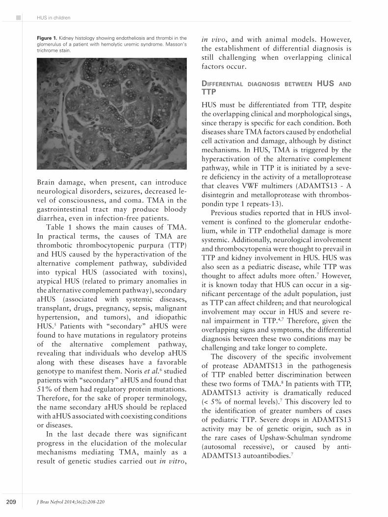
a term used to describe the forma-tion of thrombi that occlude the vessels in the microvasculature.2 Pathological factors include thicke-ning of the vessel walls, endothe-lial cell edema (endotheliosis) and detachment of the basement mem-brane, accumulation of debris in the subendothelial space, increased expression of the Von Willebrand Factor (VWF) - which attracts pla-telets and leads to the formation of microthrombi which partially or completely occlude the lumen of the vessels in the microvasculature. As a result, red blood cells are frag-mented by shearing. Figure 1 shows the renal histology of a patient wi-th HUS indicating the formation of thrombi.
Clinically, TMA has been asso-ciated with consumption throm-bocytopenia and nonautoimmune microangiopathic hemolytic ane-mia. It may also induce ischemia in different organs, with the kidneys and the brain topping the list, in addition to involving the gastroin-testinal tract and the heart, among others.3,4 Therefore, symptoms va-ry depending on which organs are affected. Patients often present with severe anemia caused by hemoly-sis, which introduces intense pallor, thrombocytopenia, and kidney da-mage, detected by the presence of edema, oliguria in addition to al-tered proteinuria, hematuria, im-paired renal, glomerular or tubular function, and acute renal failure.
DOI: 10.5935/0101-2800.20140032
J Bras Nefrol 2014;36(2):208-220
HUS in children
209
in vivo, and with animal models. However, the establishment of differential diagnosis is still challenging when overlapping clinical factors occur.
dIfferentIAl dIAgnosIs between Hus And ttP
HUS must be differentiated from TTP, despite the overlapping clinical and morphological sings, since therapy is specific for each condition. Both diseases share TMA factors caused by endothelial cell activation and damage, although by distinct mechanisms. In HUS, TMA is triggered by the hyperactivation of the alternative complement pathway, while in TTP it is initiated by a seve-re deficiency in the activity of a metalloprotease that cleaves VWF multimers (ADAMTS13 - A disintegrin and metalloprotease with thrombos-pondin type 1 repeats-13).
Previous studies reported that in HUS invol-vement is confined to the glomerular endothe-lium, while in TTP endothelial damage is more systemic. Additionally, neurological involvement and thrombocytopenia were thought to prevail in TTP and kidney involvement in HUS. HUS was also seen as a pediatric disease, while TTP was thought to affect adults more often.7 However, it is known today that HUS can occur in a sig-nificant percentage of the adult population, just as TTP can affect children; and that neurological involvement may occur in HUS and severe re-nal impairment in TTP.4,7 Therefore, given the overlapping signs and symptoms, the differential diagnosis between these two conditions may be challenging and take longer to complete.
The discovery of the specific involvement of protease ADAMTS13 in the pathogenesis of TTP enabled better discrimination between these two forms of TMA.8 In patients with TTP, ADAMTS13 activity is dramatically reduced (< 5% of normal levels).7 This discovery led to the identification of greater numbers of cases of pediatric TTP. Severe drops in ADAMTS13 activity may be of genetic origin, such as in the rare cases of Upshaw-Schulman syndrome (autosomal recessive), or caused by anti-ADAMTS13 autoantibodies.7
Figure 1. Kidney histology showing endotheliosis and thrombi in the glomerulus of a patient with hemolytic uremic syndrome. Masson’s trichrome stain.
Brain damage, when present, can introduce neurological disorders, seizures, decreased le-vel of consciousness, and coma. TMA in the gastrointestinal tract may produce bloody diarrhea, even in infection-free patients.
Table 1 shows the main causes of TMA. In practical terms, the causes of TMA are thrombotic thrombocytopenic purpura (TTP) and HUS caused by the hyperactivation of the alternative complement pathway, subdivided into typical HUS (associated with toxins), atypical HUS (related to primary anomalies in the alternative complement pathway), secondary aHUS (associated with systemic diseases, transplant, drugs, pregnancy, sepsis, malignant hypertension, and tumors), and idiopathic HUS.5 Patients with “secondary” aHUS were found to have mutations in regulatory proteins of the alternative complement pathway, revealing that individuals who develop aHUS along with these diseases have a favorable genotype to manifest them. Noris et al.6 studied patients with “secondary” aHUS and found that 51% of them had regulatory protein mutations. Therefore, for the sake of proper terminology, the name secondary aHUS should be replaced with aHUS associated with coexisting conditions or diseases.
In the last decade there was significant progress in the elucidation of the molecular mechanisms mediating TMA, mainly as a result of genetic studies carried out in vitro,

J Bras Nefrol 2014;36(2):208-220
HUS in children
210
tAble 1 cAuses of thRombotic micRoAngiopAthy (tmA)
Causes of TMA
Infection
STEC; Shigella dysenteriae Type I; Neuraminidase-producing Streptococcus pneumoniae; human immunodeficiency virus (HIV), invasive disease by other pathogens
Complement anomaliesGenetic anomalies in regulatory proteins; acquired defects such as anti-CFH antibodies
ADAMTS13 deficiency Genetic anomalies; anti-ADAMTS13 antibodies
Other
Systemic diseases such as SLE, altered cobalamin metabolism, APS Drugs such as cisplatin, tacrolimus, cyclosporin, rifampicin, clopidogrel Infection by parvovirus or cytomegalovirus and congenital infections Transplantation (rejection, drug toxicity) Pregnancy Bone marrow transplantation, radiation, graft-versus-host disease Glomerulopathies such as como a MPGN type II Malignant hypertension
STEC: Shiga toxin-producing Escherichia coli; SLE: Systemic lupus erythematosus; MPGN: Membranoproliferative glomerulonephritis; APS: Antiphospholipid syndrome; ADAMTS13: Metalloprotease responsible for cleaving the Von Willebrand Factor.
VWF is a multimeric glycoprotein produced in megakaryocytes and endothelial cells. It is secreted into plasma and plays a key role in platelet adhesion and aggregation. Cellular compartments contain larger amounts of large VWF than plasma, as ADAMTS13 cleavage occurs simultaneously with the secretion process. Larger multimers contain more platelet binding sites. In the microvasculature, blood flow leads to the unfolding of VWF and increases the exposure of platelet binding sites. Decreased ADAMTS13 activity leads to increased presence of large multimers in the circulation and hence a greater number of exposed sites for platelet adhesion, which induces the formation of platelet thrombi in the microvasculature and triggers the onset of TMA.7
In patients with the Upshaw-Schulman syndrome (USS) and anomalies in the ADAMTS13 gene, manifestations occur more often in the neonatal period, while subsequent episodes may be triggered by factors such as infection and immunization. Treatment is based on plasma transfusion to supply patients with ADAMTS13 protein. In cases of acquired disease, i.e., in the presence of anti-ADAMTS13 antibodies, manifestations are more delayed and treatment includes plasmapheresis, steroids and, in selected cases, rituximab (chimeric monoclonal
antibody directed against cell surface protein CD20).7 It is important to realize that these are severe cases which require immediate attention. Thus, plasmapheresis is indicated when it is not known whether the patient has circulating anti-ADAMTS13 antibodies. However, such procedure is not widely available and, in small children, it can be challenging to perform. In these cases, plasma transfusion must be offered promptly.
comPlement system
In HUS, TMA stems from the hyperactivation of the alternative pathway of the complement system. Therefore, the authors of this review de-cided to add a brief explanation of the mecha-nisms of activation of the complement system, its importance, and correlations with the coagu-lation system.
The complement system comprises plasma-soluble proteins and proteins expressed on the cell membrane which are part of the innate immune system. Their goal is to remove “damaged” cells and aid the adaptive immune defenses against pathogens through processes such as opsonization, chemotaxis, and cell lysis.6 There are three main activation pathways of the complement system: the classical pathway,

J Bras Nefrol 2014;36(2):208-220
HUS in children
211
bind to mannose, they are activated to cleave C4 and C2. The rest of the acti-vation pathway is similar to the classical pathway.4
3. The alternative pathway: It can be acti-vated in conjunction with the classical and lectin pathways, but may be consti-tutively activated at any time. The hydro-lysis of C3 triggers its activation to form C3a and C3b. C3b binds to the cell sur-face and interacts with Factor B, which is cleaved by Factor D to generate fragment Bb. Fragment Bb is capable of binding to other C3b molecules on the cell surface to form C3bBb (C3 convertase). C3 conver-tase triggers an amplification loop to in-crease the hydrolysis of C3. The ensuing surplus of C3b binds to C3 convertase and promotes conversion to C5 convertase (C3b2Bb). C5 convertase then cleaves C5 to form C5a and C5b, and the latter fac-tor (C5b) leads to the formation of MAC. C3a and C5a are anaphylatoxins.
A system of regulatory proteins, one of them being factor I, acts to prevent uncontrolled activation of the alternative pathway by inactivating C3b into C3bi (inactivated C3b). Factor H, the membrane cofactor protein (MCP, CD46), and thrombomodulin act as factor I cofactors to inactivate C3b (Figure 3).9 In the event these regulatory proteins fail to perform their duties, the alternative complement pathway is over-activated causing uncontrolled cell damage.
There is a correlation between the complement system and the coagulation cascade. Both systems are made of proteases, some of which have similar structure and target sites.10 There is evidence that components of the coagulation cascade can activate the complement system and vice versa. As an example, thrombin may directly cleave the complement C5 component,10,11 and anaphylatoxin C5a can activate the tissue factor, which, in turn, activates thrombin.10 Thrombomodulin (TM) is a protein that interacts with the complement system and the coagulation cascade to inactivate C3b of the factor I-mediated complement, in addition to binding to thrombin, thus activating anticoagulant protein C.12 These findings suggest a close
Figure 2. Complement system activation pathways. IC: Immune complexes (antigen-antibody); MBP: Mannose-binding protein; C3bi: C3b inactivated; MCP: Membrane cofactor protein; TM: Thrombomodulin; MAC: Membrane attack complex; ** Amplification loop.
the lectin pathway, and the alternative pathway. Figure 2 shows the mechanisms of activation of the complement system.
ActIvAtIon mecHAnIsms of tHe comPlement system
1. The classical pathway: It is activated when immune complexes (antigen-antibody) bind to the C1q component of the com-plement system. Subsequently there is the activation of C1r and C1s, both C1q-complexed proteases. Activated C1s clea-ves C4 to form C4a and C4b, which bind to the cell surface; C2 is cleaved into C2a which binds to C4b to form the C4b2a complex (C3 convertase). C3 convertase cleaves C3 to form C3b, which binds to C4b2a (C3 convertase) to form C4b2a3b complex (C5 convertase). C5 convertase in turn cleaves C5 into C5a and C5b, and the latter triggers the formation of the membrane attack complex (MAC, C5b-9), the final step in the complement cas-cade that leads to cell lysis. C4b and C3b promote opsonization, while C4a and C5a are anaphylatoxins with chemotactic properties and inflammatory response.
2. The lectin pathway: Activation of this pathway is similar to the activation of the classical pathway. It is triggered when mannose-binding proteins (MBP) found in complexes of lectin-binding proteases (MASP) recognize the mannose on the surface of a pathogen. As these proteins

J Bras Nefrol 2014;36(2):208-220
HUS in children
212
hemolytic anemia, thrombocytopenia, and acute kidney injury (AKI).13
The diagnosis of STEC-HUS can be do-ne through stool cultures, tests for the presen-ce of Shiga toxins (Stx) by immunoassays and PCR,16,17 and tests for the presence of serum an-tibodies against STEC.18
The pathogenesis of this condition is not fully understood and, therefore, there is no targeted therapy. Death occurs in 1% to 5% of the cases.19 Seventy-five percent of the pediatric patients recover with only support care measures, which include hydroelectrolytic and metabolic control, renal replacement therapy when indicated, hypertension management, and correction of anemia and thrombocytopenia when needed.9 However, approximately 30% of the patients have sequelae in the form of persistent proteinuria, hypertension, and end-stage renal disease, requiring permanent dialysis or kidney transplantation.20 Complications may occur years after the acute stages of the condition. Therefore, long-term follow-up is recommended.19 Clinical factors that help predict the risk of chronic renal involvement include the number of days of oliguria or time on dialysis, high leukocytosis, and need for plasma replacement.21 Brain involvement has also been associated with worse prognosis.22
Antibiotic therapy is usually not indicated for the treatment of infections by STEC,23 since it does not offer benefits to patients.24 Instead, it may increase the risk of HUS, particularly when administered in the early stages of the disease, by increasing the production and release of Stx, the main virulence factor of STEC involved in the pathogenesis of HUS.23,24
Other joint treatment options such as plasma infusion or plasmapheresis showed no benefits for individuals with STEC-HUS.25
HUS has also been observed in infections by Shighella dysinteriae type 1 and Campylobacter.26
Invasive infection by Streptococcus pneumoniae can also cause HUS. Previous studies described it as a rare cause of HUS associated with poor prognosis. More recently, HUS associated with invasive disease caused by pneumococcus was reported to account for 5% of the pediatric cases of HUS and for 40%
Figure 3. Regulatory mechanism for the activation of the alternative complement pathway. Factor I plays a key role in the inactivation of the alternative pathway; in order for it to function, cofactors must be found to the membrane: MCP (membrane cofactor protein, CD46) and TM (thrombomodulin) and circulating Factor H.
relationship between these two systems, which is certainly relevant for the onset of TMA.
tyPIcAl HemolytIc-uremIc syndrome
Ninety percent of the pediatric cases of HUS are triggered by Shiga toxin-producing Escherichia coli (STEC), particularly in children aged between two and six years.9 Incidence in children under the age of five has been recorded at 6.1/100,000 cases per year.13 Although E. coli O157:H7 is the most recognized serotype, other serotypes of this pathogen such as O26, O45, O111, O121, O103, and O145 account for approximately 71% of the outbreaks not caused by serotype O157:H7.14 Strain O104:H4 was present in one of the largest outbreaks in history, affecting 3,816 people and resulting in 845 cases of HUS and 54 deaths in Germany in 2011.15 Food borne contamination may result in a wide variety of clinical manifestations with different levels of severity, ranging from innocuous cases of diarrhea to hemorrhagic colitis and HUS. Three to eight days after contamination patients present with abdominal pain and profuse, occasionally bloody, watery diarrhea. Approximately 24 hours later, 10% to 15% of the patients develop
J Bras Nefrol 2014;36(2):208-220
HUS in children
213
of the cases not associated with STEC.27 The incidence of HUS after invasive pneumococcal infection ranges between 0.4% and 0.6%, but it is possible that these figures are underestimated. Unlike in other conditions, the direct Coombs test yields a positive result in specific cases of HUS triggered by pneumococcus. This is due to the important role the Thomsen-Friedenreich (T) antigen plays in the pathophysiology of HUS by pneumococcus.28
This antigen is normally “hidden” by neuraminic acid, but is exposed by pneumococcal neuraminidase. The T antigen binds to the surface of glomerular endothelial cells, platelets, and red blood cells. Preformed host antibodies can bind to the surface of cells expressing this antigen, triggering a series of events that lead to HUS. The direct Coombs test often detects these antibodies bound to T antigens and yields positive results in 90% of the cases of HUS by pneumococcus.29 The differential diagnosis between HUS triggered by invasive pneumococcal disease and sepsis with disseminated intravascular coagulation has not been definitively established.
Past studies reported high mortality rates, but more recent trials have revealed improvements in this area due to enhancements in intensive care. Copelovitch et al.30 looked into a series of 14 confirmed cases of HUS by pneumococcus and observed that 64% of the patients recovered without sequelae in the long-term follow-up, similarly to what had been seen in individuals with STEC-HUS. Higher risk for end-stage kidney disease was correlated with being on dialysis for more than 20 days, while death was correlated with meningitis, not pneumonia. The authors also drew attention to bacterial serotype 19A in patients with HUS.
Other relevant infections correlated with pediatric HUS include infection by the acquired immunodeficiency virus, the Epstein-Barr virus, and neonatal congenital viral infections. A case of HUS in a patient infected by Mycoplasma pneumoniae was recently reported.31
The mechanism by which these pathogens and consequently their toxins can trigger TMA and HUS is related to the activation of the alternative complement pathway. Evidence of such activation was first reported in 1980, when fragments of
the breakage of C3 and factor B, involved in the activation of the alternative pathway of the complement system, were detected in patients with STEC-HUS.32,33 Subsequently, lower serum C3 and decreased deposition of C3 were described in the kidneys of these patients.34 More recently, Thurman et al.35 found fragments of factor B and MAC (C5b -9) in the serum of 17 patients with STEC-HUS on admission and normal test results after 28 days, showing that activation of the alternative pathway occurs early in the course of the disease.
All evidence indicates that Stx binds and inhibits complement factor H, making cells vulnerable to the formation of MAC and leading to cell lysis. Therefore, factor H can be active in the subject’s bloodstream, but not at the level of the cell, as similarly observed in the case of mutations with loss of factor H function.9 Other studies confirmed the activation of the alternative complement pathway in infection by STEC-HUS, and this model can be transported to cases with other infectious agents.9 Therefore, toxins may potentially activate or hamper the control of the alternative complement pathway and trigger the onset of TMA.
Interestingly, only 10% to 15% of the patients with STEC infection develop HUS.13 Which factor triggers the disease? With that question in mind, Fang et al.36 reported the case of a child with confirmed STEC-HUS and MCP mutation (CD46). The authors speculated that aHUS, for which this child was genetically predisposed, may have been elicited by diarrheal disease caused by STEC.
AtyPIcAl HemolytIc-uremIc syndrome
Atypical HUS (aHUS), i.e., HUS without co-existing disease and not associated with STEC or pneumococcal neuraminidase, is seen in 5% to 10% of the cases. It affects people of all ages and may be sporadic or familial.37,38 Atypical HUS stems from chronic uncontrolled activation of the alternative complement pathway, which causes endothelial damage. The prognosis for patients with aHUS is poor. In its first clinical manifestation, 33% to 40% of the patients die or progress to ESRD.37,38 Within the first year of diagnosis, 65% of the patients die, require dialy-sis, or present permanent renal injury, despite plasmapheresis and/or plasma infusion.37
J Bras Nefrol 2014;36(2):208-220
HUS in children
214
tAble 2 some RegulAtoRy pRoteins of the AlteRnAtive complement pAthwAy, genes, pRoduction sites, Action sites, And contRibution to the occuRRence of AtypicAl hemolytic uRemic syndRome. modified fRom36
Protein Gene Source Location % in aHUS cases
Factor H CFH Liver Plasma ~ 15-30%
Factor I CFI Liver Plasma ~ 5-10%
Membrane cofactor protein (CD46) MCP Multiple sites Bound to membrane ~ 10-15%
Factor B CFB Liver Plasma < 5%
C3 C3 Liver Plasma ~ 5-10%
Anti-Factor H antibody CFHR1/CFHR3 Lymphocyte Plasma ~ 10%
Not identified 40% of the cases~ Approximately; CFH: Complement factor H; CFI: Complement factor I; CFB: Complement factor B.
In the last decade, a growing number of mutations in genes encoding proteins involved in the formation or regulation of the alternative pathway have been associated with aHUS.37-39 Therefore, aHUS is emerging as a paradigmatic disease caused by inefficient protection of the endothelium against complement attack. Dysregulation of C3 convertase induces excessive cleavage of C3 and subsequent exacerbated cleavage of C5, leading to endothelial cell damage, platelet recruitment, and formation of thrombi in the renal microvasculature, in a condition histologically characterized as TMA.40 Genetic anomalies in aHUS patients have been found to involve componentes of the alternative complement pathway, including factors H and I, membrane cofactor protein (MCP), factor B, complement component C3, and thrombomodulin.37-40 The mechanism of aHUS involves primarily the activation of MAC, C5b-9.
Mutations in regulatory proteins have been detected in about 61% of the patients with atypical aHUS.41 Table 2 shows the contribution of certain regulatory proteins, their sites of synthesis, responsible gene, location, and percent contribution to cases of aHUS. Atypical HUS may occur in the absence of identified mutations.
InvestIgAtIon of tHe comPlement system In AHus PAtIents
Recent guidelines recommend that HUS pa-tients be tested for complement anomalies, whenever possible.42,43 Tests can be used to measure total complement activity in se-rum (CH50), activity of the alternative pa-thway (AH50), and activity of complement system components C3, C4, factor H, and
factor I. One should bear in mind that most tests assess the presence of the protein, not its activity. Additionally, complement regula-tion anomalies may occur only at the level of the endothelial cell surface, and not systemi-cally. Therefore, serum levels of components of the complement system may be normal in patients with altered activity regulation, thus not allowing complement system genetic ano-malies to be ruled out. Ariceta et al.44 reported normal C3 and C4 levels in 80% of individuals with aHUS, while Noris et al.38 found normal levels of factor H in 40/46 (87%) patients with identified mutations on gene CFH.
Usually, small amounts of C3b are deposited on the cell surface, but these molecules are rapidly eliminated by Factor I with the aid of cofactors factor H, MCP (membrane cofactor protein, CD46), and thrombomodulin. In cases of mutation leading to loss of factor function, the C3b deposits are not completely wiped out, thus triggering uncontrolled activation of the complement system and subsequent cell damage, as seen with endothelial cells.
Mutations with function gains in molecules involved in the activation of the alternative pathway, such as factor B and C3, induce endothelial damage, even in the presence of functional regulators.38
In some patients, anti-factor H antibodies may contribute to the disease and have been associated with deletions in proteins 1 and 3 related to factor H (CFHR1/3).45
IncomPlete PenetrAnce In AHus
It is important to mention that some individuals with factor H, factor I, and MCP mutations do not develop the disease.6 This finding indicates

J Bras Nefrol 2014;36(2):208-220
HUS in children
215
that genetic alterations are not the only entity responsible for the occurrence of the disease, and points out to the need for an environmental fac-tor to act as the trigger of the complement casca-de and for the disease to occur. Caprioli et al.37 showed that in 77% of the patients with factor H, factor I, or MCP mutations the manifestation of clinical symptoms was preceded by symptoms of influenza, gastroenteritis, and other infections.
dIAgnosIng AHus
Atypical HUS is diagnosed by ruling out other causes of TMA, without the aid of a definitive diagnostic test; therefore, all relevant tests must be performed to ensure the assessment of the va-riables that may impact the diagnosis, including the search for coexisting conditions and comorbi-dities (Table 1). Atypical HUS has been diagnosed in patients with tumors, malignant hypertension, systemic disease, glomerulopathies, in associa-tion with pregnancy or use of calcineurin inhibi-tors,38 presenting anomalies in the proteins of the alternative complement pathway and identified mutations.
The differential diagnosis between aHUS and STEC-HUS is of great importance. STEC-HUS must be ruled out even for the 10% of the patient population that does not present diarrhea,46 and despite the fact that gastrointestinal tract TMA may cause diarrhea in the absence of infectious pathogens.38 The possibility of mutations in complement regulatory proteins must be considered for patients with suspected STEC-HUS, incompatible disease evolution patterns, and persistent TMA.
Specifically in the neonatal period, inborn errors of cobalamin metabolism due to alterations in the MMACHC gene responsible for the production of factor cobalamin-b (Clc-b), a key element in the metabolism of cobalamin, causing methylmalonic acidemia, must be ruled out.47 Without Clc-b, methylmalonic acid and homocysteine accumulate and lead to an increase in the levels of free radicals, thus introducing cell damage, increased platelet aggregation, increased binding of tissue plasminogen activator in the endothelium, and increased expression of local procoagulant factors.47
lAb workuP dIAgnostIc crIterIA for AHus
• Nonautoimmune hemolytic anemia: CBC showing low hemoglobin levels, nega-tive direct Coombs test, positive test re-sult for schistocytes in peripheral blood, low haptoglobin, increased serum lactate dehydrogenase (LDH) levels;
• Thrombocytopenia: CBC showing throm-bocytopenia (count < 150,000/mm3 or drop greater than 25% from previous measure);
• Renal involvement: may be present with hematuria, proteinuria, edema, oliguria, hypertension, increased serum levels of urea and creatinine;
• Tests for involvement of other organs and systems should be carried out depending on clinical findings; TMA can affect any organ, the neurological system, the gas-trointestinal tract, the heart, pancreas, li-ver etc.;48,49
• Negative test results for STEC-HUS; rule out other causes of typical HUS;
• ADAMTS13 activity > 5%; if value is under 5, look for anti-ADAMTS13 antibodies;
• AH50, CH50, C3, and C4 activity: aHUS may be accompanied by decreased serum C3 and reduced alternative pathway ac-tivity measured by AH50; these findings, however, are not definitive, as even nor-mal levels do not rule aHUS out.
Assessing components of the complement system
• Test for mutations in complement re-gulatory proteins. When available, it is valid to test for mutations of factors in-volved in complement system control as factor H, factor I, MCP, TM, C3, and factor B, regardless of serum levels. The presence of anti-factor H antibodies can be tested using enzyme-linked immuno-sorbent assays (ELISA). However, it is important to note that mutations have been identified in about 60% of the ca-ses of aHUS and, therefore, tests negati-ve for mutations cannot be used to rule aHUS out.41

J Bras Nefrol 2014;36(2):208-220
HUS in children
216
PrevIously AdvocAted tHerAPy for AtyPIcAl HemolytIc uremIc syndrome
plAsmApheResis And AdministRAtion of plAsmA
Before the advent of new treatment options for aHUS, such as complement blockade targeted therapy, plasma therapy was recommended des-pite the lack of controlled randomized trials.50
Cohort studies showed that plasmapheresis reduces mortality by 25%.6 Treatment with plasma therapy proved beneficial in the short term for the disease’s hematologic activity; however, the prognosis for renal involvement was poor.37
Plasma administration was deemed sufficient in cases of absent or anomalous complement system regulatory proteins. However, in the initial stages of disease without an established diagnosis, plasmapheresis was considered ideal as long as it was able to remove antibodies, when they were present. Plasma transfusion was recommended when plasmapheresis could not be offered, in order to replace the altered regulatory proteins of the alternative complement pathway.
After the initial period of treatment with plasmapheresis, reduced activation of complement system could be achieved with plasma transfusion, unless antibodies were present, which meant patients had to undergo plasmapheresis.
Previous guidelines from the European HUS44 study group recommended starting plasmapheresis within 24 hours of the diagnosis.44,51 Technically, the recommendation was to replace 1.5 volume (60 to 75 ml/kg) with fresh plasma. The guideline group claimed that plasmapheresis had to be done daily for five days, then in five sessions per week for two weeks, and then in three sessions per week for two weeks.44 In plasma transfusion in the absence of plasmapheresis, the recommendation was to start with 30 to 40 ml/kg and then move to 10 to 20 ml/kg per day.52
The best parameters to monitor response we-re platelet count, LDH and hemoglobin levels levels, which account for hematologic remission. Haptoglobin often stays at lower levels after he-matologic remission and, therefore, is not used as a parameter in the short term.
There was no parameter to assess time of treatment, but it was recommended to keep patients on treatment for at least two days after
complete remission.52 However, some patients with aHUS could become dependent on plasmapheresis; additionally, infections and immunization could trigger new TMA episodes, which required patients to return to plasmapheresis. Other factors that may triggered endothelial injury had to be controlled, such as hypercholesterolemia and hypertension.
Atypical HUS is a chronic disease and long-term treatment with plasma (plasma transfusion or plasmapheresis) leads to high rates of new TMA episodes, progression to ESRD, and death.38 Additionally, comorbidities such as infection and thrombosis, particularly in plasmapheresis with a central line, are more frequent, and antibodies appear with repeated plasma transfusions. It is worthwhile mentioning that plasmapheresis can be a challenging procedure to perform in young pediatric patients. Plasma transfusions in hypervolemic patients to replace altered proteins can also be difficult.
tRAnsplAntAtion
The prognosis for renal transplant patients wi-th aHUS is quite poor. Approximately 50% of the patients experience recurrence and lose their grafts. There is no single factor to predict recur-rence, though the use of calcineurin inhibitors has been associated with increased recurrence rates.53 Patients with aHUS are also more like-ly to develop acute rejection, which adversely affects graft survival.50 Knowledge of genetic de-fects may improve the prognosis. Patients with factor H mutations experience recurring disease in 75% to 90% of the cases; patients with fac-tor I mutations relapse 45% to 80% of the time; in C3 mutations the chance of recurrence ranges between 40% and 70%; patients with MCP mu-tations have a low probability of experiencing recurrence.6 In order to minimize the risk of re-currence, it is recommended to avoid prolonged ischemia and stay off calcineurin inhibitors.
One of the options to treat recurrence is plas-mapheresis and, when possible, prophylactic plasmapheresis sessions before and after trans-plantation.6 Some authors also advocate the use of simultaneous liver and kidney (SLK) trans-plants in cases with higher chances of recurrence, such as patients with known factor H or factor I mutations.54 However, SLK transplantation sig-nificantly increases morbimortality.
J Bras Nefrol 2014;36(2):208-220
HUS in children
217
Live donor kidney transplantation can only be offered if the donor is free of mutations; and only 60% of the mutations are currently known.41 Atypical HUS can be triggered in previously undiagnosed patients submitted to live donor transplants merely by the manipulation of the kidney during surgery. The most conservative approach is transplantation from a deceased donor.
Endothelial involvement causing aHUS recurrence may be triggered after kidney transplantation by the use of immunosuppressive drugs, viral infections, or rejection, even in subjects with mild genetic susceptibility.6
Although environmental factors account for most of the cases of recurrent aHUS, approximately 40% of the patients have genetic anomalies.55 To minimize the risk caused by environmental factors, it is recommended to adequately manage hypertension and hypercholesterolemia, and to offer calcineurin inhibitors judiciously.
A new erA In tHe treAtment of AHus - eculIzumAb
Increased knowledge on the pathogenesis of aHUS was accompanied by the emergence of eculizumab, a drug that acts as an inhibitor of the terminal pathway of the complement casca-de,56 considered the standard for the treatment of paroxysmal nocturnal hemoglobinuria.57 Eculizumab is a humanized monoclonal antibo-dy registered as the drug of choice for the treat-ment of aHUS by the FDA and the EMEA. The drug acts specifically by binding to complement factor C5, blocking the cleavage of C5 into C5b, and preventing the formation of anaphylatoxin C5a and MAC, C5b-9. Its use has been associa-ted with one significant adverse effect: increased risk of infection by Neisseria.58 As the clearan-ce of Neisseria meningitidis is highly dependent on MAC, patients treated with eculizumab are at a higher risk of infection by this pathogen. Therefore, patients must be given the polyvalent vaccine at least two weeks before starting treat-ment, and if the medication is used before this period, patients must be offered prophylactic an-tibiotic therapy. As the vaccine available in our area does not protect against all Neisseria sero-types, non-stop prophylaxis is recommended;58 patient family members and physicians must be aware of this diagnostic possibility.58
Eculizumab has been shown to alter the cour-se of aHUS. The drug has retrieved native kidney function and prevented post-transplant recurrence. Studies on series of aHUS patients on eculizumab reported increased platelet counts and improved renal function after the first dose. While on eculizu-mab, patients did not need dialysis or plasmaphere-sis, and tolerated the medication well.59,60 Legendre et al.61 looked into 37 patients with aHUS and ages above 12 years on eculizumab. Twenty of the pa-tients had longstanding disease (mean of 48.3 mon-ths, ranging from 0.7 to 285.8 months), mostly wi-th renal involvement, and were either on dialysis or in hematologic remission due to plasma therapy.
The remaining 17 patients had shorter course disease (mean of 9.7 months, ranging from 0.3 to 235.9 months) and signs of hematologic activity; some were on plasma therapy and had impaired renal function, while others were on dialysis. The most important finding described in this study was the possibility of retrieving renal function, even for patients on dialysis for months on end, achieving hematologic remission without signs of new episo-des of TMA, and improving quality of life during a follow-up of 26 to 62 weeks. No cases of meningo-coccal infection were reported, as all patients were immunized and offered prophylactic antibiotics.61 The reported adverse effects included hypertension (three cases), peritonitis (one case), venous sclero-sis at the site of infusion (one case), asymptomatic bacteriuria (one case), and infection by the influen-za virus (one case). Cases of meningococcal infec-tion were not observed in this study.61
Recently, Delmas et al.62 updated the study by Legendre et al., following patients with longstan-ding aHUS and chronic kidney disease (stages 3, 4, and 5) for three years. The authors reported hematologic and renal function improvements with the use of the drug. Another finding was greater numbers of patients free of new TMA episodes: 26 weeks into follow-up 16/20 (80%) were free of TMA events; 17/20 (85%) one ye-ar into follow-up; 19/20 (95%) after two years of follow-up; and 19/20 (95%) after three years of follow-up. The authors also reported a higher number of subjects with normal hematologic findings: 18/20 (90%) 26 weeks into follow-up; the ratio was kept unchanged three years into follow-up.
J Bras Nefrol 2014;36(2):208-220
HUS in children
218
Considering renal function, serum creatinine levels dropped by ≥ 25% in 15% of the patients after 26 weeks of treatment, in 35% of the patients after one year, in 55% of the cases after two years of treatment, and was kept constant from then until the third year of follow-up; glomerular filtration rates increased by ≥ 15 ml/min/1.73 m2 in 5% of the patients after 26 weeks of follow-up, and the ratio improved to 15% after one year, 40% after two years, and was kept stable after three years of drug therapy, with 60% of the patients improving from CKD by one or more stages by the third year of treatment. No cases of meningococcal infection or adverse side effects were reported.
A recent prospective study63 with 22 pediatric patients showed that the use of eculizumab provides rapid and sustained improvement (26 weeks of treatment) in hematological parameters and ongoing improvements in renal function. More specifically, complete remission from TMA was observed in 64% of the cases; normal hematologic parameters in 82%; increase in the GFR ≥ 15 ml/min/1.73 m2 in 86% of the patients (mean of 64 ml/min/1.73 m2); and nine of eleven patients could stop dialysis. No cases of meningococcal infection were reported and adverse effects were similar to those reported in previous studies, namely hypertension in two patients, upper airway infection in two individuals, viral gastroenteritis in two subjects, and fever in two patients. Based on the outcomes of this study, eculizumab has been recommended as the first line treatment for pediatric aHUS, supporting the recommendations of previous studies.
Transplantation protocols are advocating the use of eculizumab in patients with aHUS. Reports of live donor transplants support the combination of plasmapheresis the week prior to transplantation in the induction stage and after surgery during maintenance, with good outcomes.55 The literature on deceased donor transplants contains reports in which plasmapheresis was performed the day before and the day after transplant surgery in association with eculizumab;55 however, more recent case reports tell of good outcomes using eculizumab alone.64 In this situation, it is recommended to administer the first dose six hours before transplantation and repeat it the following day, then once a week for the next four weeks, and after that every 15 days; dosage must be adjusted based on the subject’s bodyweight.63
Notably, aHUS may occur in newborns and treatment in these cases is quite complicated, since plasmapheresis is difficult to perform in individuals in this age range. Additionally, dependence and antibody formation with successive plasma transfusions may occur, along with severe adverse events and sustained renal involvement. Reports have described the benefits in hematological recovery and renal function improvement in neonates treated with eculizumab.60,65
Eculizumab is not indicated for patients with typical HUS. However, in a recent outbreak in Germany, individuals with severe STEC-HUS and particularly patients with neurological impairment were offered the drug, and many adult patients benefitted from it.9 The drug did not impact the prognosis of the pediatric group followed during the outbreak, but the medication was administered later on. Therefore, it is not clear yet whether eculizumab is beneficial for patients with typical HUS.66
conclusIon
Differential diagnosis can be challenging in the group of diseases prone to manifesting TMA, as they may present overlapping clinical signs and symptoms. This group includes TTP and HUS, the latter of which may occur by the action of toxins, systemic diseases, uncontrolled activation of the alternative pathway due to alterations in pathway regulatory proteins (aHUS), or for unknown causes. A series of tests is required to differentiate these conditions. The diagnosis of aHUS is achieved by ruling out other causes of TMA. In most cases, patients respond well to the treatment of aHUS with plasma transfusion or plasmapheresis, particularly when it comes to short term hematologic parameters. However, aHUS is a severe chronic condition that may cause patients to die or develop end-stage renal disease.
Treatment with plasma is characterized by significant rates of recurrent disease in the long term and unfavorable renal outcome. Eculizumab, an anti-C5 monoclonal antibody, has emerged as a new hope for the improvement of patient short and long term prognosis, and has been recommended as the drug of choice in the treatment of aHUS.
references
1. Serna A 4th, Boedeker EC. Pathogenesis and treatment of Shi-ga toxin-producing Escherichia coli infections. Curr Opin Gas-troenterol 2008;24:38-47. DOI: http://dx.doi.org/10.1097/MOG.0b013e3282f2dfb8

J Bras Nefrol 2014;36(2):208-220
HUS in children
219
2. Zheng XL, Sadler JE. Pathogenesis of thrombotic microangio-pathies. Annu Rev Pathol 2008;3:249-77. DOI: http://dx.doi.org/10.1146/annurev.pathmechdis.3.121806.154311
3. Tsai HM. The molecular biology of thrombotic microangiopa-thy. Kidney Int 2006;70:16-23. PMID: 16760911 DOI: http://dx.doi.org/10.1038/sj.ki.5001535
4. Barbour T, Johnson S, Cohney S, Hughes P. Thrombotic microan-giopathy and associated renal disorders. Nephrol Dial Transplant 2012;27:2673-85. DOI: http://dx.doi.org/10.1093/ndt/gfs279
5. Nester CM, Thomas CP. Atypical hemolytic uremic syndrome: what is it, how is it diagnosed, and how is it treated? Hemato-logy Am Soc Hematol Educ Program 2012;2012:617-25. PMID: 23233643
6. Loirat C, Girma JP, Desconclois C, Coppo P, Veyradier A. Thrombotic thrombocytopenic purpura related to severe ADA-MTS13 deficiency in children. Pediatr Nephrol 2009;24:19-29. DOI: http://dx.doi.org/10.1007/s00467-008-0863-5
7. Furlan M, Robles R, Lämmle B. Partial purification and charac-terization of a protease from human plasma cleaving von Wille-brand factor to fragments produced by in vivo proteolysis. Blood 1996;87:4223-34. PMID: 8639781
8. Noris M, Remuzzi G. Atypical hemolytic-uremic syndrome. N Engl J Med 2009;361:1676-87. PMID: 19846853 DOI: http://dx.doi.org/10.1056/NEJMra0902814
9. Keir LS, Saleem MA. Current evidence for the role of comple-ment in the pathogenesis of Shiga toxin haemolytic uraemic syn-drome. Pediatr Nephrol 2013 Jul 11. [Epub ahead of print]. DOI: http://dx.doi.org/10.1007/s00467-013-2561-1
10. Amara U, Flierl MA, Rittirsch D, Klos A, Chen H, Acker B, et al. Molecular intercommunication between the complement and coagulation systems. J Immunol 2010;185:5628-36. PMID: 20870944 DOI: http://dx.doi.org/10.4049/jimmunol.0903678
11. Krisinger MJ, Goebeler V, Lu Z, Meixner SC, Myles T, Pryzdial EL, et al. Thrombin generates previously unidentified C5 pro-ducts that support the terminal complement activation pathway. Blood 2012;120:1717-25. PMID: 22802338 DOI: http://dx.doi.org/10.1182/blood-2012-02-412080
12. Delvaeye M, Noris M, De Vriese A, Esmon CT, Esmon NL, Ferrell G, et al. Thrombomodulin mutations in atypical hemolytic-uremic syndrome. N Engl J Med 2009;361:345-57. PMID: 19625716 DOI: http://dx.doi.org/10.1056/NEJMoa0810739
13. Scheiring J, Andreoli SP, Zimmerhackl LB. Treatment and ou-tcome of Shiga-toxin-associated hemolytic uremic syndrome (HUS). Pediatr Nephrol 2008;23:1749-60. DOI: http://dx.doi.org/10.1007/s00467-008-0935-6
14. Kalchayanand N, Arthur TM, Bosilevac JM, Schmidt JW, Wang R, Shackelford SD, et al. Evaluation of commonly used antimicrobial interventions for fresh beef inoculated with Shiga toxin-producing Es-cherichia coli serotypes O26, O45, O103, O111, O121, O145, and O157:H7. J Food Prot 2012;75:1207-12. PMID: 22980002 DOI: http://dx.doi.org/10.4315/0362-028X.JFP-11-531
15. Bielaszewska M, Mellmann A, Zhang W, Köck R, Fruth A, Bauwens A, et al. Characterisation of the Escherichia coli strain associated with an outbreak of haemolytic uraemic syndrome in Germany, 2011: a microbiological study. Lancet Infect Dis 2011;11:671-6.
16. He X, Patfield S, Hnasko R, Rasooly R, Mandrell RE. A Polyclo-nal Antibody Based Immunoassay Detects Seven Subtypes of Shiga Toxin 2 Produced by Escherichia coli in Human and Environmental Samples. PLoS One 2013;16;8:e76368. PMID: 24146860
17. Skinner C, Patfield S, Stanker L, He X. Development of Mono-clonal antibodies and immunoassays for sensitive and specific detection of Shiga toxin Stx2f. PLoS One 2013;8:e76563. DOI: http://dx.doi.org/10.1371/journal.pone.0076563
18. Palmeira P, Carbonare SB, Guth BE, Carbonare CB, Pontes GN, Tino-De-Franco M, et al. Acquisition of serum antibodies reactive with enterohemorrhagic Escherichia coli virulence-asso-ciated factors by healthy Brazilian children and adults. Pediatr Infect Dis J 2009;28:1089-94. DOI: http://dx.doi.org/10.1097/INF.0b013e3181aa6b2d
19. Spinale JM, Ruebner RL, Copelovitch L, Kaplan BS. Long-term outcomes of Shiga toxin hemolytic uremic syndrome. Pediatr Nephrol 2013;28:2097-105. DOI: http://dx.doi.org/10.1007/s00467-012-2383-6
20. Rosales A, Hofer J, Zimmerhackl LB, Jungraithmayr TC, Riedl M, Giner T, et al.; German-Austrian HUS Study Group. Need for long-term follow-up in enterohemorrhagic Escherichia coli-associated hemolytic uremic syndrome due to late-emerging sequelae. Clin Infect Dis 2012;54:1413-21. DOI: http://dx.doi.org/10.1093/cid/cis196
21. Gianviti A, Tozzi AE, De Petris L, Caprioli A, Ravà L, Edefonti A, et al. Risk factors for poor renal prognosis in children with hemolytic uremic syndrome. Pediatr Nephrol 2003;18:1229-35. DOI: http://dx.doi.org/10.1007/s00467-003-1262-6
22. Trachtman H, Austin C, Lewinski M, Stahl RA. Renal and neu-rological involvement in typical Shiga toxin-associated HUS. Nat Rev Nephrol 2012;8:658-69. DOI: http://dx.doi.org/10.1038/nrneph.2012.196
23. Wong CS, Jelacic S, Habeeb RL, Watkins SL, Tarr PI. The risk of the hemolytic-uremic syndrome after antibiotic treat-ment of Escherichia coli O157:H7 infections. N Engl J Med 2000;342:1930-6. PMID: 10874060 DOI: http://dx.doi.org/10.1056/NEJM200006293422601
24. Tarr PI, Gordon CA, Chandler WL. Shiga-toxin-producing Escherichia coli and haemolytic uraemic syndrome. Lancet 2005;365:1073-86. PMID: 15781103
25. Keir L, Coward RJ. Advances in our understanding of the pathoge-nesis of glomerular thrombotic microangiopathy. Pediatr Nephrol 2011;26:523-33. DOI: http://dx.doi.org/10.1007/s00467-010-1637-4
26. Michael M, Elliott EJ, Craig JC, Ridley G, Hodson EM. Interven-tions for hemolytic uremic syndrome and thrombotic thrombo-cytopenic purpura: a systematic review of randomized controlled trials. Am J Kidney Dis 2009;53:259-72. DOI: http://dx.doi.org/10.1053/j.ajkd.2008.07.038
27. Constantinescu AR, Bitzan M, Weiss LS, Christen E, Kaplan BS, Cnaan A, et al. Non-enteropathic hemolytic uremic syndrome: causes and short-term course. Am J Kidney Dis 2004;43:976-82. DOI: http://dx.doi.org/10.1053/j.ajkd.2004.02.010
28. von Vigier RO, Seibel K, Bianchetti MG. Positive Coombs test in pneumococcus-associated hemolytic uremic syndrome. A review of the literature. Nephron 1999;82:183-4. PMID: 10364712
29. von Vigier RO, Fossali E, Crosazzo L, Bianchetti MG. Positi-ve Coombs test in postpneumococcal hemolytic-uremic syndro-me. Pediatr Infect Dis J 2005;24:1028-9. DOI: http://dx.doi.org/10.1097/01.inf.0000187032.38556.b3
30. Copelovitch L, Kaplan BS. Streptococcus pneumoniae-associated hemolytic uremic syndrome: classification and the emergence of serotype 19A. Pediatrics 2010;125:e174-82. PMID: 20026500 DOI: http://dx.doi.org/10.1542/peds.2007-2017
31. Godron A, Pereyre S, Monet C, Llanas B, Harambat J. Hemolytic uremic syndrome complicating Mycoplasma pneumoniae infec-tion. Pediatr Nephrol 2013;28:2057-60. DOI: http://dx.doi.org/10.1007/s00467-013-2541-5
32. Monnens L, Molenaar J, Lambert PH, Proesmans W, van Muns-ter P. The complement system in hemolytic-uremic syndrome in childhood. Clin Nephrol 1980;13:168-71. PMID: 7379368
33. Kim Y, Miller K, Michael AF. Breakdown products of C3 and factor B in hemolytic-uremic syndrome. J Lab Clin Med 1977;89:845-50.
34. Robson WL, Leung AK, Fick GH, McKenna AI. Hypocomple-mentemia and leukocytosis in diarrhea-associated hemolytic ure-mic syndrome. Nephron 1992;62:296-9. PMID: 1436342 DOI: http://dx.doi.org/10.1159/000187063
35. Thurman JM, Marians R, Emlen W, Wood S, Smith C, Akana H, et al. Alternative pathway of complement in children with diarrhea--associated hemolytic uremic syndrome. Clin J Am Soc Nephrol 2009;4:1920-4. DOI: http://dx.doi.org/10.2215/CJN.02730409
36. Fang CJ, Fremeaux-Bacchi V, Liszewski MK, Pianetti G, No-ris M, Goodship TH, et al. Membrane cofactor protein mu-tations in atypical hemolytic uremic syndrome (aHUS), fatal Stx-HUS, C3 glomerulonephritis, and the HELLP syndrome. Blood 2008;111:624-32. PMID: 17914026 DOI: http://dx.doi.org/10.1182/blood-2007-04-084533
37. Caprioli J, Noris M, Brioschi S, Pianetti G, Castelletti F, Bettina-glio P, et al. Genetics of HUS: the impact of MCP, CFH, and IF mutations on clinical presentation, response to treatment, and outcome. Blood 2006;108:1267-79. PMID: 16621965 DOI: http://dx.doi.org/10.1182/blood-2005-10-007252

J Bras Nefrol 2014;36(2):208-220
HUS in children
220
38. Noris M, Caprioli J, Bresin E, Mossali C, Pianetti G, Gamba S, et al. Relative role of genetic complement abnormalities in spo-radic and familial aHUS and their impact on clinical phenotype. Clin J Am Soc Nephrol 2010;5:1844-59. DOI: http://dx.doi.org/10.2215/CJN.02210310
39. Roumenina LT, Loirat C, Dragon-Durey MA, Halbwa-chs-Mecarelli L, Sautes-Fridman C, Fremeaux-Bacchi V. Alternative complement pathway assessment in patients with atypical HUS. J Immunol Methods 2011;365:8-26. DOI: http://dx.doi.org/10.1016/j.jim.2010.12.020
40. Fakhouri F, Frémeaux-Bacchi V, Noël LH, Cook HT, Pickering MC. C3 glomerulopathy: a new classification. Nat Rev Nephrol 2010;8:494-9. DOI: http://dx.doi.org/10.1038/nrneph.2010.85
41. Fremeaux-Bacchi V, Fakhouri F, Garnier A, Bienaimé F, Dragon--Durey MA, Ngo S, et al. Genetics and outcome of atypical he-molytic uremic syndrome: a nationwide French series comparing children and adults. Clin J Am Soc Nephrol 2013;8:554-62. DOI: http://dx.doi.org/10.2215/CJN.04760512
42. de Jorge EG, Macor P, Paixão-Cavalcante D, Rose KL, Tedesco F, Cook HT, et al. PMID: 21148255.The development of atypical hemolytic uremic syndrome depends on complement C5. J Am Soc Nephrol 2011;22:137-45.
43. Pickering MC, de Jorge EG, Martinez-Barricarte R, Recalde S, Garcia-Layana A, Rose KL, et al. Spontaneous hemolytic ure-mic syndrome triggered by complement factor H lacking surfa-ce recognition domains. J Exp Med 2007;204:1249-56. PMID: 17517971 DOI: http://dx.doi.org/10.1084/jem.20070301
44. Ariceta G, Besbas N, Johnson S, Karpman D, Landau D, Licht C, et al.; European Paediatric Study Group for HUS. Guideli-ne for the investigation and initial therapy of diarrhea-negative hemolytic uremic syndrome. Pediatr Nephrol 2009;24:687-96. DOI: http://dx.doi.org/10.1007/s00467-008-0964-1
45. Moore I, Strain L, Pappworth I, Kavanagh D, Barlow PN, Herbert AP, et al. Association of factor H autoantibodies with deletions of CFHR1, CFHR3, CFHR4, and with mutations in CFH, CFI, CD46, and C3 in patients with atypical hemolytic uremic syndrome. Blood 2010;115:379-87. PMID: 19861685 DOI: http://dx.doi.org/10.1182/blood-2009-05-221549
46. Westra D, Wetzels JF, Volokhina EB, van den Heuvel LP, van de Kar NC. A new era in the diagnosis and treatment of atypi-cal haemolytic uraemic syndrome. Neth J Med 2012;70:121-9. PMID: 22516576
47. Menni F, Testa S, Guez S, Chiarelli G, Alberti L, Esposito S. Neo-natal atypical hemolytic uremic syndrome due to methylmalonic aciduria and homocystinuria. Pediatr Nephrol 2012;27:1401-5. DOI: http://dx.doi.org/10.1007/s00467-012-2152-6
48. Zuber J, Le Quintrec M, Sberro-Soussan R, Loirat C, Fré-meaux-Bacchi V, Legendre C. New insights into postrenal transplant hemolytic uremic syndrome. Nat Rev Nephrol 2011;7:23-35. DOI: http://dx.doi.org/10.1038/nrneph.2010.155
49. Dragon-Durey MA, Sethi SK, Bagga A, Blanc C, Blouin J, Ranchin B, et al. Clinical features of anti-factor H autoantibody-associated hemolytic uremic syndrome. J Am Soc Nephrol 2010;21:2180-7. DOI: http://dx.doi.org/10.1681/ASN.2010030315
50. Zuber J, Le Quintrec M, Morris H, Frémeaux-Bacchi V, Loirat C, Legendre C. Targeted strategies in the prevention and mana-gement of atypical HUS recurrence after kidney transplantation. Transplant Rev (Orlando) 2013;27:117-25. DOI: http://dx.doi.org/10.1016/j.trre.2013.07.003
51. Taylor CM, Machin S, Wigmore SJ, Goodship TH.; working party from the Renal Association, the British Committee for Standards in Haematology and the British Transplantation So-ciety. Clinical practice guidelines for the management of atypical haemolytic uraemic syndrome in the United Kingdom. Br J Hae-matol 2010;148:37-47. PMID: 19821824 DOI: http://dx.doi.org/10.1111/j.1365-2141.2009.07916.x
52. Zuber J, Fakhouri F, Roumenina LT, Loirat C, Frémeaux-Bac-chi V.; French Study Group for aHUS/C3G. Use of eculizumab for atypical haemolytic uraemic syndrome and C3 glomerulo-pathies. Nat Rev Nephrol 2012;8:643-57. DOI: http://dx.doi.org/10.1038/nrneph.2012.214
53. Jokiranta TS, Zipfel PF, Fremeaux-Bacchi V, Taylor CM, Goodship TJ, Noris M. Where next with atypical hemolytic uremic syndrome? Mol Immunol 2007;44:3889-900. PMID: 17768107 DOI: http://dx.doi.org/10.1016/j.mo-limm.2007.06.003
54. Loirat C, Fremeaux-Bacchi V. Hemolytic uremic syndro-me recurrence after renal transplantation. Pediatr Trans-plant 2008;12:619-29. DOI: http://dx.doi.org/10.1111/j.1399-3046.2008.00910.x
55. Nester C, Stewart Z, Myers D, Jetton J, Nair R, Reed A, et al. Pre-emptive eculizumab and plasmapheresis for renal transplant in atypical hemolytic uremic syndrome. Clin J Am Soc Nephrol 2011;6:1488-94. DOI: http://dx.doi.org/10.2215/CJN.10181110
56. Hillmen P, Hall C, Marsh JC, Elebute M, Bombara MP, Petro BE, et al. Effect of eculizumab on hemolysis and transfusion requi-rements in patients with paroxysmal nocturnal hemoglobinuria. N Engl J Med 2004;350:552-9. PMID: 14762182 DOI: http://dx.doi.org/10.1056/NEJMoa031688
57. Hillmen P, Muus P, Röth A, Elebute MO, Risitano AM, Schre-zenmeier H, et al. Long-term safety and efficacy of sustained ecu-lizumab treatment in patients with paroxysmal nocturnal haemo-globinuria. Br J Haematol 2013;162:62-73. DOI: http://dx.doi.org/10.1111/bjh.12347
58. Bouts A, Monnens L, Davin JC, Struijk G, Spanjaard L. Insu-fficient protection by Neisseria meningitidis vaccination alone during eculizumab therapy. Pediatr Nephrol 2011;26:1919-20. DOI: http://dx.doi.org/10.1007/s00467-011-1929-3
59. Gruppo RA, Rother RP. Eculizumab for congenital atypical he-molytic-uremic syndrome. N Engl J Med 2009;360:544-6. PMID: 19179329 DOI: http://dx.doi.org/10.1056/NEJMc0809959
60. Ariceta G, Arrizabalaga B, Aguirre M, Morteruel E, Lopez-Trasca-sa M. Eculizumab in the treatment of atypical hemolytic uremic syndrome in infants. Am J Kidney Dis 2012;59:707-10. DOI: http://dx.doi.org/10.1053/j.ajkd.2011.11.027
61. Legendre CM, Licht C, Muus P, Greenbaum LA, Babu S, Bedro-sian C, et al. Terminal complement inhibitor eculizumab in aty-pical hemolytic-uremic syndrome. N Engl J Med 2013;368:2169-81. DOI: http://dx.doi.org/10.1056/NEJMoa1208981
62. Delmas Y, Loirat C, Muus P, Legendre C, Douglas K, Hourmant M, et al. Sustained Efficacy and Safety of Eculizumab in Patients with Atypical Hemolytic Uremic Syndrome with Long Disease Duration and Chronic Kidney Disease: 3-Year Update. Pôster no Encontro Anual da American Society of Nephrology Kidney Week 2013, 5-10 de Novembro, Atlanta, Georgia.
63. Greenbaum LA, Fila M, Tsimaratos M, Ardissino G, Al-Akash S, Evans J, et al. Eculizumab Inhibits Thrombotic Microangiopathy and Improves Renal Function in Pediatric Patients with Atypi-cal Hemolytic Uremic Syndrome. Pôster no Encontro Anual da American Society of Nephrology Kidney Week 2013, 5-10 de Novembro, Atlanta, Georgia.
64. Román-Ortiz E, Mendizabal Oteiza S, Pinto S, López-Trascasa M, Sánchez-Corral P, Rodríguez de Cordoba S. Eculizumab long--term therapy for pediatric renal transplant in aHUS with CFH/CFHR1 hybrid gene. Pediatr Nephrol 2014;29:149-53. DOI: http://dx.doi.org/10.1007/s00467-013-2591-8
65. Besbas N, Gulhan B, Karpman D, Topaloglu R, Duzova A, Korkmaz E, et al. Neonatal onset atypical hemolytic uremic syndrome success-fully treated with eculizumab. Pediatr Nephrol 2013;28:155-8. DOI: http://dx.doi.org/10.1007/s00467-012-2296-4
66. Morigi M, Galbusera M, Gastoldi S, Locatelli M, Buelli S, Pezzot-ta A, et al. Alternative pathway activation of complement by Shiga toxin promotes exuberant C3a formation that triggers microvas-cular thrombosis. J Immunol 2011;187:172-80. PMID: 21642543 DOI: http://dx.doi.org/10.4049/jimmunol.1100491