heparin and related polysaccharides: synthesis using ... and related polysaccharides: synthesis...
TRANSCRIPT

MINI-REVIEW
Heparin and related polysaccharides: synthesis usingrecombinant enzymes and metabolic engineering
Matthew Suflita1 & Li Fu2 & Wenqin He3 & Mattheos Koffas1,3 & Robert J. Linhardt1,2,3,4
Received: 29 May 2015 /Revised: 1 July 2015 /Accepted: 3 July 2015 /Published online: 29 July 2015# Springer-Verlag Berlin Heidelberg 2015
Abstract Glycosaminoglycans are linear anionic polysaccha-rides that exhibit a number of important biological and phar-macological activities. The two most prominent members ofthis class of polysaccharides are heparin/heparan sulfate andthe chondroitin sulfates (including dermatan sulfate). Thesepolysaccharides, having complex structures and polydispersi-ty, are biosynthesized in the Golgi of most animal cells. Thechemical synthesis of these glycosaminoglycans is precludedby their structural complexity. Today, we depend on food an-imal tissues for their isolation and commercial production.Ton quantities of these glycosaminoglycans are used annuallyas pharmaceuticals and nutraceuticals. The variability ofanimal-sourced glycosaminoglycans, their inherent impuri-ties, the limited availability of source tissues, the poor controlof these source materials, and their manufacturing processessuggest a need for new approaches for their production. Overthe past decade, there have been major efforts in the biotech-nological production of these glycosaminoglycans. This mini-review focuses on the use of recombinant enzymes and
metabolic engineering for the production of heparin and chon-droitin sulfates.
Keywords Glycosaminoglycans . Chemoenzymatic .
Metabolic engineering . Recombinant enzymes . Heparin .
Chondroitin sulfate . Sulfotransferases .
Glycosyltransferases . PAPS
Introduction
Glycosaminoglycans (GAGs) are structurally complex mole-cules, with varying lengths, backbone sugars, and modifications(Fig. 1). GAGs are found primarily in the form of proteoglycans,composed of a core protein with varying numbers of GAGchains. The initial building blocks of a GAG are repeating di-saccharide units; in the case of heparan sulfate (HS), the disac-charide unit is composed of D-glucuronic acid (GlcA) linked toN-acetyl-D-glucosamine (GlcNAc) (Fig. 1a). GlcA can later beepimerized into L-iduronic acid (IdoA), and sulfo groups can besubstituted at the 2-hydroxyl groups of both IdoA and GlcA, aswell as the 3-hydroxyl and 6-hydroxyl groups and the 2-aminogroup of the glucosamine residue. HS also exhibits a domainstructure, with alternating NA and NS domains, composed ofcontiguous unsulfated N-acetyl regions and N-sulfated regions,respectively, and mixed NA/NS domains (Lindahl et al. 1998).Heparin (HP) is a highly sulfated form of HS and is often rep-resented as a single extended NS domain. Heparin has antico-agulant properties due to the presence of 3-O-sulfation, whichforms part of the antithrombin III binding site (Loganathan et al.1990; Esko and Lindahl 2001; Linhardt 2003). Chondroitin sul-fate (CS) is a similar sulfated glycosaminoglycan with the re-peating disaccharide unit of GlcA linked to N-acetyl-D-galactos-amine (GalNAc). The GalNAc residue can have sulfo groupssubstituted at the 4- and 6-hydroxyls of the GalNAc (Fig. 1b).
* Robert J. [email protected]
1 Department of Biology, Center for Biotechnology andInterdisciplinary Studies, Rensselaer Polytechnic Institute,Troy, NY 121806, USA
2 Department of Chemistry and Chemical, Center for Biotechnologyand Interdisciplinary Studies, Rensselaer Polytechnic Institute,Troy, NY 121806, USA
3 Department of Chemical and Biological Engineering, Center forBiotechnology and Interdisciplinary Studies, Rensselaer PolytechnicInstitute, Troy, NY 121806, USA
4 Department of Biomedical Engineering, Center for Biotechnologyand Interdisciplinary Studies, Rensselaer Polytechnic Institute,Troy, NY 121806, USA
Appl Microbiol Biotechnol (2015) 99:7465–7479DOI 10.1007/s00253-015-6821-9

The GlcA residue in CS can also be epimerized to IdoA, presentin dermatan sulfate (DS, also known as chondroitin sulfate B),and both the GlcA and IdoA residues can contain sulfo groups attheir 2-positions. CS-GAGs also have domain structures, similarto HS-GAGs (Mikami and Kitagawa 2013).
This diversity of structure poses a challenge for GAG analy-sis. While GAG sequencing and domain mapping methods ex-ist, they are still relatively difficult to perform. GAG composi-tion, however, can be readily obtained by several disaccharideanalysis methods. These techniques usually involve using en-zymes (heparin lyases I, II, and III, and chondroitinase ABC) tobreak down a GAG into its disaccharide building blocks, whichare then separated by liquid chromatography and detected byultraviolet spectroscopy or mass spectrometry. Comparison todisaccharide standards reveals the position and number of sulfogroups in each disaccharide. Options for chromatography stepsinclude hydrophilic liquid interaction, strong anion exchange,and ion-pairing reverse-phase chromatography. Sensitivity canbe increased to picomole detection limits by labeling with 2-aminoacridone, allowing analysis of GAG mixtures in biologi-cal samples (Yang et al. 2012; Sun et al. 2015). Liquidchromatography-mass spectrometry techniques can also be usedfor direct characterization of low molecular weight heparin oli-gosaccharides (Li et al. 2012). High-resolution one- and two-dimensional nuclear magnetic resonance has become an equallyimportant tool for GAG analysis, allowing determination of de-fined GAG structures (Zhang et al. 2011; Fu et al. 2013). Nu-clear magnetic resonance spectroscopy can also provide infor-mation about structural elements such as sulfation,
epimerization, and acetylation, and can be used to identify con-taminants in pharmaceutical heparin (Xu et al. 2011; Xiong et al.2013; Fu et al. 2014a; Guerrini et al. 2008). Additionally, mo-lecular weight distribution and polydispersity can be measuredby size exclusion chromatography.
The structural complexity of GAGs ismirrored by the diversefunctions they carry out, playing roles in signaling and develop-ment, blood coagulation, cancer and inflammation, woundhealing, as well as providing unique structural properties(Bernfield et al. 1999; Vlodavsky and Friedmann 2001;Linhardt. 2003; Lauder 2009; Cress et al. 2014). CS and HSproteoglycans are present in the extracellular matrix of animals,where theywork in concert with fibrous proteins tomaintain cellstructure. CS proteoglycans in cartilage and joints act as shockabsorbers, due to their tendency as polyanions to retain water.GAGs can also bind extracellular signaling molecules such asgrowth factors, morphogens, and other chemokines, acting as aBmolecular sponge.^ This allows GAGs to play twin roles insignaling, both stabilizing signal molecule gradients, and func-tioning as a reservoir of signal molecules, which can be releasedby GAG degrading enzymes (Taipale and Keski-Oja 1997). CSandHS proteoglycans are additionally found anchored at the cellsurface, where they make up a tissue-specific GAG coat; varia-tions in the composition and structure of this coat can be thoughtof as a kind of fingerprint (Li et al. 2015). Regulation of finestructure allows for different affinities to extracellular signalingmolecules, providing a level of fine tuning of GAG function(Kato et al. 1994; Shi and Zaia 2009). Endoglycosidases suchas heparanase are known to be important regulators of HS
Fig. 1 Structures of glycosaminoglycans and their oligosaccharides. aStructure and common domains of heparin (b ∼0.4, and a+d>c) orheparan sulfate (b<0.4, and c>a+d). b Common chondroitin sulfates. c
Low molecular weight heparin (Enoxaparin) and ultra-low molecularweight heparins (ULMWH and Arixtra)
7466 Appl Microbiol Biotechnol (2015) 99:7465–7479

function in cancer and inflammation. Breakdown of extracellularmatrix HS-GAGs by heparanase removes a physical barrier forleukocytes, which can transit into organs to exert an inflamma-tory response to injury. This mechanism is often co-opted bymetastasizing tumor cells, evidenced by the fact that HPSE ex-pression is a prognostic marker for high metastatic potential(Sato et al. 2004). Heparanase activity also releases boundgrowth factors that can drive tumor cell proliferation and angio-genesis (Parish et al. 2001; Li and Vlodavsky 2009; Ramaniet al. 2013). One of the most well documented and studiedfunctions of HS-GAGs is their anticoagulant activity. HS-GAGs containing 3-O-sulfation, primarily heparin, can bindstrongly to antithrombin, a serine protease inhibitor. This bind-ing causes a conformational change that activates antithrombin,inhibiting thrombin and related serine proteases involved in thecoagulation cascade (Linhardt. 2003).
The biosynthesis of CS and HS-GAGs starts out along asimilar pathway, beginning with the stepwise addition of fourmonosaccharides to a serine residue of a core protein (serine-xylose-galactose-galactose-glucuronic acid), through the in-volvement of high-energy UDP-activated sugars (Fig. 2a).
The subsequent addition of either a GlcNAc or GalNAc resi-due forms a pentasaccharide, determining whether an HS orCS chain is built, respectively (Mikami and Kitagawa 2013)(Fig. 2b, c). The CS and HS chains are then extended by theirrespective glycosyltransferases, and then modified by a hostof sulfotransferases and epimerases. Epimerization of GlcAresidues to IdoA occurs in both HP and HS, whileepimerization of CS GlcA residues forms DS. Thesulfotransferases use a universal sulfo group donor, 3′-phosphoadenosine-5′-phosphosulfate (PAPS), to transfer asulfate group to a specific hydroxyl or amino position of asugar residue. These modifying enzymes often have tissue-specific isoforms and expression patterns, giving rise to in-credible heterogeneity of structure and function (Sasisekharanand Venkataraman 2000; Mikami and Kitagawa 2013).
Because of an aging world population, the demand for gly-cosaminoglycan drugs such as heparin and chondroitin sulfatewill continue to climb. Heparin is an invaluable drug for treat-ment of coagulation and thrombotic disorders, and is listed asone of theWorldHealthOrganizations essential medicines. Hep-arin can also be used to create anticoagulant surfaces for test
Fig. 2 Biosynthesis of heparin/heparan sulfate and chondroitin sulfates. a Synthesis of the tetrasaccharide linker region. b Polymerization andmodification pathway of heparin/heparan sulfates. c Polymerization and modification pathway of chondroitin sulfates
Appl Microbiol Biotechnol (2015) 99:7465–7479 7467

tubes and in dialysis machines (Murugesan et al. 2008). Lowmolecular weight heparins (LMWHs), derived from heparin bydepolymerization, are used subcutaneously in treating deep veinthrombosis and are often used in cancer treatment due to theassociation between cancer and thromboembolic disease. Theuse of heparins results in additional survival benefits in cancertreatment, not attributable to their anticoagulant activities, main-ly due to inhibition of heparin binding growth factors that drivetumor growth, and inhibition of heparanase and selectin-mediated metastasis mechanisms (Castelli et al. 2004). CS hasbeen shown to relieve pain and stiffness associated with osteo-arthritis, one of the most common musculoskeletal conditions inthe world (Schiraldi et al. 2010). In addition to thechondroprotective properties that help to prevent and relieveosteoarthritis, CS also impact many pathologies by its anti-inflammatory response, in part by inhibition of the pro-inflammatory adipokine TNF-α (Tully et al. 2006; Ostermanand Lichtenstein 2007; Papoutsaki et al. 2013). In a clinical trial,CS-A and CS-E was shown to have dramatic impact on improv-ing psoriasis and colitis (Lauder 2009). Newmethods to preparelarge quantities of GAG-based drugs must be implemented tomeet growing demand. Synthetic methods must also be im-proved to allow preparation of GAGs with defined sequences,improving knowledge of structure-function relationships. Thisreview will focus on the use of recombinant enzymes and met-abolic engineering for the production of GAGs.
Current state of GAG preparation
Current heparins and CSs are derived from a variety of animaltissues. Animal source materials present serious concerns for thepossibility of transmission of viral and prion diseases, and thesusceptibility of animal populations to infectious disease oroverharvesting has potential to drastically reduce supply. More-over, seasonal, geographical, and subspecies variationsmay alterthe product obtained from a given animal species. The processof preparing pharmaceutical-grade heparin has been alteredsomewhat over time as the primary tissue source has changedfrom dog liver to beef lung and finally to porcine intestine(Linhardt. 2003). The preparation of heparin from ruminant tis-sues obtained at slaughterhouses present a special concern par-ticularly following the appearance of bovine spongiform en-cephalopathy (BSE, Bmad cow disease^) in both humans andcattle (Guerrini et al. 2008) and scrapies prion in sheep(Schonberger 1998). Thus, the use of bovine and ovine tissueproducts as injectable pharmaceuticals has declined and thesetissues are now rarely used in heparin production.
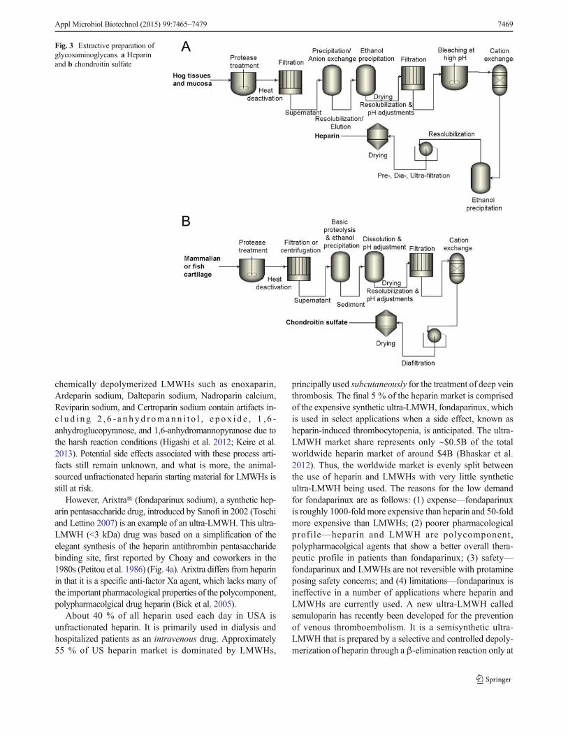
The methods used today for the commercial preparation ofheparin have changed from that used early in the twentieth cen-tury and involve five basic steps (Fig. 3a): (1) preparation oftissue; (2) extraction of heparin from tissue; (3) recovery ofraw heparin; (4) purification of heparin; and (5) recovery of
purified heparin (Evans and Mozen 1962; Williams 1967;Okuyama et al. 1975; Vidic 1981; Linhardt et al. 1992;Van Gorp et al. 1997; Bhaskar et al. 2012). However, tominimize the environmental impact of high-ash, high-biochemical oxygen demand hydrolyzed protein, raw heparinextraction typically takes place at the hog slaughtering facil-ity itself (not under current good manufacturing practices(cGMP) conditions). Additional high potency heparin maybe recovered by saving the waste brine solution of thehog casings operation (Vidic 1981).
There are growing concerns about porcine tissue nowa-days, especially after the heparin crisis that took place in2008. This crisis involved the introduction of an oversulfatedchondroitin sulfate into heparin produced from hogs in Chinaleading to the death of nearly 100 Americans (Liu et al. 2009).The lack of oversight and cGMP in slaughterhouses leaves theheparin supply chain open to this kind of adulteration, whichcan be difficult to detect. Bovine lung heparin can be distin-guished from porcine intestinal heparin because it contains adifferent distribution of structural variants of the antithrombinpentasaccharide binding site as well as other differences indisaccharide composition (Loganathan et al. 1990; Fu et al.2013). It is somewhat more difficult to distinguish bovineintestinal heparin or ovine intestinal heparin (Fu et al. 2013).Moreover, blends of pharmaceutical-grade heparins preparedfrom different species might make the content of non-porcineheparin even more difficult to assess.
Currently available commercial CS is mainly extractedfrom trachea, nasal septa, chicken keel, shark cartilage, andfish (Fig. 3b). As Bdietary supplements^ available in the USmarket, the overall quality of CS is poorly regulated. Someproducts contain much less CS than advertised, as low as 10%in some cases (Adebowale et al. 2000). Tracheal CS maysometimes be substituted for higher priced shark-derived CS(Sakai et al. 2007; Higashi et al. 2015). In addition to prob-lems of regulation and transmission of disease, overfishing ofshark populations (Higashi et al. 2015) and porcine epidemicssuch as blue ear pig disease and porcine epidemic diarrheavirus (Zhou et al. 2008) highlight the precarious nature ofthe supply chain for these GAGs.
Chemical synthesis and enzymatic depolymerizationto prepare heparin oligosaccharides
Low molecular weight heparins or fractionated heparins with amolecular weight of ∼3–8 kDa are a group of heparin-derivedanticoagulant/antithrombotic agents (Fig. 1c), and their develop-ment began approximately 30 years ago (Mousa and Fareed2001). Currently, the commercial preparation of LMWHs fromunfractionated heparin includes the controlled chemical depoly-merization of heparin by peroxidative cleavage, nitrous acidcleavage, and chemical β-elimination (Fig. 4b). These
7468 Appl Microbiol Biotechnol (2015) 99:7465–7479
chemically depolymerized LMWHs such as enoxaparin,Ardeparin sodium, Dalteparin sodium, Nadroparin calcium,Reviparin sodium, and Certroparin sodium contain artifacts in-c l u d i n g 2 , 6 - a n h y d r oman n i t o l , e p o x i d e , 1 , 6 -anhydroglucopyranose, and 1,6-anhydromannopyranose due tothe harsh reaction conditions (Higashi et al. 2012; Keire et al.2013). Potential side effects associated with these process arti-facts still remain unknown, and what is more, the animal-sourced unfractionated heparin starting material for LMWHs isstill at risk.
However, Arixtra® (fondaparinux sodium), a synthetic hep-arin pentasaccharide drug, introduced by Sanofi in 2002 (Toschiand Lettino 2007) is an example of an ultra-LMWH. This ultra-LMWH (<3 kDa) drug was based on a simplification of theelegant synthesis of the heparin antithrombin pentasaccharidebinding site, first reported by Choay and coworkers in the1980s (Petitou et al. 1986) (Fig. 4a). Arixtra differs from heparinin that it is a specific anti-factor Xa agent, which lacks many ofthe important pharmacological properties of the polycomponent,polypharmacolgical drug heparin (Bick et al. 2005).
About 40 % of all heparin used each day in USA isunfractionated heparin. It is primarily used in dialysis andhospitalized patients as an intravenous drug. Approximately55 % of US heparin market is dominated by LMWHs,
principally used subcutaneously for the treatment of deep veinthrombosis. The final 5 % of the heparin market is comprisedof the expensive synthetic ultra-LMWH, fondaparinux, whichis used in select applications when a side effect, known asheparin-induced thrombocytopenia, is anticipated. The ultra-LMWH market share represents only ∼$0.5B of the totalworldwide heparin market of around $4B (Bhaskar et al.2012). Thus, the worldwide market is evenly split betweenthe use of heparin and LMWHs with very little syntheticultra-LMWH being used. The reasons for the low demandfor fondaparinux are as follows: (1) expense—fondaparinuxis roughly 1000-fold more expensive than heparin and 50-foldmore expensive than LMWHs; (2) poorer pharmacologicalprofile—heparin and LMWH are polycomponent,polypharmacolgical agents that show a better overall thera-peutic profile in patients than fondaparinux; (3) safety—fondaparinux and LMWHs are not reversible with protamineposing safety concerns; and (4) limitations—fondaparinux isineffective in a number of applications where heparin andLMWHs are currently used. A new ultra-LMWH calledsemuloparin has recently been developed for the preventionof venous thromboembolism. It is a semisynthetic ultra-LMWH that is prepared by a selective and controlled depoly-merization of heparin through a β-elimination reaction only at
Fig. 3 Extractive preparation ofglycosaminoglycans. a Heparinand b chondroitin sulfate
Appl Microbiol Biotechnol (2015) 99:7465–7479 7469

the less hindered regions using a phosphazene base (Viskovet al. 2009). Due to its bulky structure, the base cleaves theheparin chain, leaving the crowded antithrombin-binding siteintact. Studies in patients showed the anti-factor Xa/anti-factorIIa ratio of semuloparin to be above 30, indicating nearly pureanti-factor Xa activity (Lassen et al. 2009). Although the prep-aration cost is significantly lower than that of fondaparinux, itis neither homogeneous nor structurally defined and, since it isstill derived from porcine intestinal heparin semuloparin,could be subject to contamination or adulteration.
Bioengineering approaches
Glycosyltransferases
The chemical syntheses of heparin or heparin-like drugs typical-ly involve numerous steps and result in low overall yields andhigh costs, which limits their clinical applications. Chemists arestarting to turn towards enzymatic or chemoenzymatic synthesisto circumvent these problems (Gijsen et al. 1996; Karst andLinhardt 2003; Deangelis et al. 2013). Unlike most chemical
Fig. 4 Chemical synthesis, depolymerization, and enzymaticdepolymerization of ultra-low molecular weight heparin and lowmolecular weight heparin. a Summary of a convergent multi-step
chemical synthesis of Arixtra from cellobiose derivative (reagents notshown). b Enzymatic (I) and chemical (III) depolymerization to preparelow molecular heparins from unfractionated heparin (II)
7470 Appl Microbiol Biotechnol (2015) 99:7465–7479
reactions, these enzymatic reactions are highly chemospecific,regiospecific, and stereospecific. Using recombinant technology,glycosyltransferases and heparin biosynthetic enzymes havebeen cloned and expressed, and are under study for the synthesisof heparin (Orellana et al. 1994; DeAngelis and White 2002).Initial efforts towards a chemoenzymatic preparation of heparinused C5-epimerase to convert the GlcA of the heparosan poly-saccharide to IdoA, but relied primarily on chemical modifica-tions for the introduction of N- and O-sulfo groups, creatingunwanted sulfation sites (Naggi et al. 2001). An enzymatic syn-thesis of an oligosaccharide based on the structure of HS hasbeen accomplished using the heparin/HS modification enzymes(Kuberan et al. 2003) and glycosyltransferases (Liu et al. 2010).
Enzymatic synthesis of polysaccharides and oligosaccharidesof defined lengths has recently become possible due to the avail-ability of many recombinantly expressed glycosyltransferases(Table 1). These enzymes use UDP-activated sugars producedby uridyltransferases such as GlmU, which can be used to pro-duce UDP-GlcNAc and UDP-GalNac in vitro, building blocksfor HS and CS backbones, respectively. GlmU is flexible in itssubstrate specificity and allows the synthesis of some unnaturalUDP-sugars possessing a tag, which can be polymerized intonovel glycosaminoglycans (Masuko et al. 2012). This techniquehas been used to incorporate labile N-trifluoroacetyl groups intoHS oligosaccharides, which can later be enzymatically sulfated
(Liu et al. 2010; Xu et al. 2011, 2014). Polymerizing enzymescan be processive, by addition of alternating UDP-sugars, ormay catalyze the addition of a single sugar, as in KfiA, aUDP-GlcNAc transferase that has been used to build heparinoligosaccharides in a controlled, stepwise manner (Liu et al.2010; Xu et al. 2011, 2014). Processive glycosyltransferasessuch as heparosan synthases 1 and 2 (PmHS1 and PmHS2) fromPasteurella multocida and chondroitin polymerase fromEscherichia coli K4, have been used to synthesize HS and CSbackbones of varying molecular weight (Sugiura et al. 2002;Sismey-Ragatz et al. 2007). Additionally, site-directed mutagen-esis studies have been able to isolate two single-actionP. multocida PmHS2 mutants, which can be used to build oli-gosaccharides in a stepwise manner (Chavaroche et al. 2012).
Sulfotransferases and epimerases
Many sulfotransferases involved in GAG biosynthesis havebeen expressed and characterized in vitro (Table 1). Unique toHS biosynthesis is the introduction of N-sulfo groups that iscarried out by N-sulfotransferase/N-deacetylases (NDSTs), bi-functional enzymes with two active sites (Berninsone andHirschberg 1998). While the bacterial recombinant expressionof activeN-deacetylase domain has been difficult, the bacteriallyexpressed N-sulfotransferase domain (NST) has been used in
Table 1 Enzymes utilized in heparin and chondroitin synthesis
Name Abbreviation Organism References
Chondroitin Polymerase/chondroitin synthase
K4CP Escherichia coli K4 Sugiura et al. 2002, 2012
N-acetyl-D-glucosaminyltransferase
KfiA Escherichia coli K5 Chen et al. 2006; Xu et al. 2011
Heparosan synthase 1 and 2 PmHS1,PmHS2
Pasteurellamultocida
Liu et al. 2010; Sismey-Ragatz et al. 2007; Xu et al. 2011
N-acetyl-glucosamine-1-phosphateuridyltransferase
GlmU Escherichia coli K5 Masuko et al. 2012
Arylsulfotransferase IV AST-IV Rattus norvegicus Bhaskar et al. 2015; Burkart et al. 2000
C5 epimerase C5 Epi Cricetulus griseus(CHO cell)
Bhaskar et al. 2015; Liu et al. 2010; Xu et al. 2011;Zhang et al. 2015b
2-O-sulfotransferase 1 2OST-1 Cricetulus griseus(CHO cell)
Bhaskar et al. 2015; Zhang et al. 2015b; Xu et al. 2011
6-O-sulfotransferase 1 6OST-1 Mus musculus Bhaskar et al. 2015; Liu et al. 2010; Restaino et al. 2013a; Xu et al. 2011
6-O-sulfotransferase 3 6OST-3 Mus musculus Bhaskar et al. 2015; Liu et al. 2010; Xu et al. 2011; Zhang et al. 2015a
3-O-sulfotransferase 1 3OST-1 Mus musculus Bhaskar et al. 2015; Liu et al. 2010; Moon et al. 2012; Xu et al. 2011
3-O-sulfotransferase 5 3OST-5 Mus musculus Liu et al. 2010
3-O-sulfotransferase 3 3OST-3 Mus musculus Moon et al. 2012
N-deacetylase/N-sulfotransferase NDST-1 Rattus norvegicus Liu et al. 2010; Saribaş et al. 2004
Chondroitin 4-sulfotransferase 1 C4ST-1 Homo sapiens Sugiura et al. 2012
Chondroitin 6-sulfotransferase 1 C6ST-1 Homo sapiens Sugiura et al. 2012
N-acetyl galactosamine 4-sulfate6-sulfotransferase
GalNAc4S-6ST
Homo sapiens Sugiura et al. 2012
Uronosyl 2-sulfotransferase UST Homo sapiens Sugiura et al. 2012
Appl Microbiol Biotechnol (2015) 99:7465–7479 7471

conjunction with N-trifluoroacetyl sugars to achieve the preciseintroduction of N-sulfo groups sites in heparin oligosaccharides(Liu et al. 2010; Xu et al. 2011, 2014). The presence of N-sulfogroups are a prerequisite for the further introduction of O-sulfogroups and for C5 epimerization; thus, NDST specificity con-trols the formation (or absence, as in heparin) of domain struc-tures in HS (Sheng et al. 2011). C5 epimerase, which producescritical IdoA residues in HS-GAGs, is thought to act irreversiblyin vivo, likely due to concurrent introduction of 2-O-sulfogroups by GAG-modifying enzyme complex of C5 epimeraseand 2-O-sufotransferase (2OST). The introduction of a 2-O-sulfo group blocks the reversible activity of C5 epimerasein vitro possibly due to steric hindrance, suggested from therecent crystallization of C5 epimerase in complex with a heparinoligosaccharide (Qin et al. 2015). There is only one 2OST iso-form identified in humans and it can act on both IdoA and GlcAresidues adjacent to an N-sulfo glucosamine (GlcNS) residuewithout a 6-O-sulfo group, with a preference for IdoA. A crys-tallization study elucidated themolecular basis of this specificity,showing favorable interactions with the N-sulfo group, and sug-gesting steric hindrance with the 6-sulfo groups of the adjacentresidue (Liu et al. 2014). Three 6-O-sulfotransferase isoforms(6OST-1, 6OST-2, 6OST-3) have been identified in humans,and found to have slightly different specificities; 6OST-1 and6OST-2 prefer to transfer a 6-O-sulfo groups to a GlcNS thatis next to an GlcA residue and IdoA2S residue, respectively(Bhaskar et al. 2012). There are at least six different isoformsof 3-O-sulfotransferases (3OSTs) with distinct substrate speci-ficities, two of which (3OST-1 and 3OST-3) have solved crystalstructures (Moon et al. 2004, 2012). Comparison of the twostructures reveals distinct binding modes for the two isoforms,suggesting a mechanism for recognition of fine saccharide struc-ture. It is thought that the presence of 3-O-sulfo groups canregulate many important HS functions. This is due to the mod-ification being critical for protein binding of at least two specificsaccharide sequences, the antithrombin-binding site and thebinding of the gD envelope protein of herpes simplex virus 1(Liu et al. 2002; Kusche-Gullberg and Kjellén 2003).
In addition to the HS sulfotransferases, there are severalrecombinant CS sulfotransferases with demonstrated in vitroactivity, including chondroitin-4-sulfotransferase 1 (C4ST-1),chondro i t i n -6 - su l fo t r ans f e r a s e 1 (C6ST-1 ) , N -acetylgalactosamine-4-sulfate 6-sulfotransferase (GalNAc4S-6ST), and uronosyl 2-sulfotransferase (UA2ST) (Sugiura et al.2012) (Table 1). Less is known about the CS sulfotransferasesand the two CS C5 epimerases (Silbert and Sugumaran 2002;Pacheco et al. 2009a, b; Thelin et al. 2013), but it is likely thatthe activities, specificity, and biosynthetic control parallel thatof the HS biosynthetic enzymes. Moreover, specific CS struc-tures seem to play prominent roles in nervous tissues and inbrain development and function (Higashi et al. 2015).
Finally, while not directly involved in GAG biosynthesis,arylsulfotransferase IV (AST-IV), a mammalian liver
detoxification enzyme involved in transferring sulfo groupsto the hydroxyl groups of phenols, has been indispensablefor chemoenzymatic synthesis of sulfated GAGs. While nor-mally catalyzing the transfer of a sulfo group from PAPS to aphenol, at high concentrations of p-nitrophenyl sulfate, AST-IV can be used to catalyze the reverse reaction transferring asulfate group from p-nitrophenyl sulfate to PAP, thus formingPAPS, the universal sulfate donor for sulfotransferases. Thisreverse reaction can be used as a cofactor regeneration systemwhen coupled to HS or CS sulfotransferase reactions andovercomes strong product inhibition of these sulfotransferasesby PAP (Burkart et al. 2000). This cofactor regeneration alsoproduces p-nitrophenol, a yellow-colored product which canbe easily monitored at a 400-nm wavelength, forming thebasis of a commonly used sulfotransferase assay (Burkartand Wong 1999; Sterner et al. 2014). Collectively, this cofac-tor regeneration system and colorimetric assay represent avaluable enzymatic toolbox for GAG synthesis.
Further structural elucidation of the GAG biosynthetic en-zymes and enzymes for cofactor recycling may lead to new,engineered forms with novel specificities, further expandingthe range of tools available. While protein engineering offersopportunities to improve the stability and activity of theserecombinant enzyme catalysts, the lack of crystal structuresfor many of these enzymes posses a barrier to progress. Fur-ther efforts to scale-up the production of these enzymes in fed-batch fermenters are underway and have been demonstratedfor four out of five of the HS sulfotransferases including2OST-1, C5 epimerase, 6OST-1, and 6OST-3 (Restaino et al.2013a; Zhang et al. 2015a, b). This opens the way for theindustrial-scale production of GAGs.
Chemoenzymatic synthesis/depolymerization of heparinoligosaccharides
Efforts to produce high-value oligosaccharide targets using thisenzymatic toolbox are underway. Two fondaparinux-like ultra-LMWHs (Figs. 1 and 5) that showed excellent in vivo andin vitro anticoagulant activity have been chemoenzymaticallysynthesized using heparin biosynthetic enzymes (Xu et al.2011). By using a chemoenzymatic approach, it is notable thatthese homogeneous heptasaccharides were synthesized throughan approach biomimetic to heparin biosynthesis and within 12steps at multi-milligram scale and in approximately 40% overallyield. Both heparin constructs were synthesized initially on aheparosan-derived disaccharide acceptor containing a ring-contracted anhydromannitol residue. Using the N-acetylglucosaminyltransferase (KfiA) and the heparosan synthase(pmHS2) (Sismey-Ragatz et al. 2007), the acceptor was elongat-ed stepwise from a disaccharide to a heptasaccharide, using theunnatural GlcN-trifluoroacetyl donor, which was laterdeprotected and N-sulfonated. KfiA transferred GlcN-trifluoroacetyl smoothly, demonstrating that uridine diphosphate
7472 Appl Microbiol Biotechnol (2015) 99:7465–7479

sugar is a compatible unnatural substrate for KfiA. Comparedwith chemical glycosylation, enzymatic glycosylationproceeded with over 80 % yield and in a stereospecific manner,giving the correct stereochemistry at each anomeric center.
The selective epimerization and sulfation of heparin oligosac-charide backbones are done using C5-epimerase and O-sulfotransferases, which converts GlcA into its C5-epimer IdoAand transfers sulfo groups to desired positions, respectively. Theselectivities of these modification enzymes provide excellentcontrol over products but require careful reaction scheme designand careful selection of the appropriate isoforms to obtain thedesired target structures. The most effective schemes are thosethat follow the reaction order found in natural heparin synthesis.Investigation of ideal reaction order and enzymatic activity,based on the heparin biosynthesis pathway, has shown thatC5-epimerase only converts GlcA residues between two GlcNSresidues (Liu et al. 2010) to IdoA and works best collaboratingwith 2OST, which locks the normally reversible epimerizationinto the IdoA conformation upon introduction of the 2-O-sulfogroup. This specificity requires the NST pre-treatment beforeC5-epimerase and 2OST (Sheng et al. 2012).
Recently, one-pot enzymatic synthesis has been exploredfor the preparation of certain heparin oligosaccharide targets(Chen et al. 2013). Chemoenzymatic strategies appear to bethe next step in the development of efficient syntheses ofheparin oligosaccharides having up to 20 saccharide units.
Sugiura and coworkers have chemoenzymatically synthe-sized various CS species with defined lengths and definedsulfate compositions, using bacterial chondroitin polymeraseand recombinant CS sulfotransferases, including C4ST-1,C6ST-1, GalNAc4S-6ST, and UA2ST. Chemoenzymatic syn-thesis enables the generation of CS chains of the desiredlengths, compositions, and distinct structures, and theresulting library will be a useful tool for studies of CS func-tions (Sugiura et al. 2012).
Another approach to oligosaccharide synthesis, used inpreparing LMWHs, involves the controlled enzymatic depo-lymerization of heparin using recombinant heparinases. Incontrast to chemical depolymerization, enzymatic depolymer-ization using recombinant heparin lyases was proven to be arelatively artifact-free method (Fu et al. 2014b). Enzymaticdepolymerization of heparin is scalable and potentially pro-vides more access to LMWHs with specific in vivo biologicaland pharmacological activities. LMWHs, such as tinzaparin,prepared through controlled heparinase treatment have al-ready been successfully commercialized.
Bioengineered heparin
A concerted effort is currently underway to chemoenzymaticallysynthesize a full-length bioengineered heparin, based on theoverexpression of the E. coli K5 capsular polysaccharide(CPS) heparosan, and subsequent modification with
recombinant HP biosynthetic enzymes (Zhang et al. 2008). Sucha bioengineered heparin might one day be approved as a genericheparin and also used in the preparation of LMWHs, increasingthe supply and eliminating the risks that come with drugs de-rived from animal tissues (Liu et al. 2009; Wang et al. 2011;Bhaskar et al. 2012). Small amounts of bioengineered heparinhave been prepared from this E. coli heparosan in several labo-ratories (Kuberan et al. 2003; Lindahl et al. 2005; Zhang et al.2008). Over the past 5 years, research has focused on developinga scalable process capable of producing sufficient quantities of abioengineered heparin for pre-clinical and clinical evaluation.Even greater challenges are anticipated to meet global demand(over 100 tons/year) if a bioengineered, generic version of hep-arin, chemically and biologically equivalent to current USP hep-arin, is to be introduced in the future (Liu et al. 2009;Wang et al.2011; Linhardt and Liu 2012).
Unlike the chemoenzymatic synthesis of LMWHs andULMWH oligosaccharides, the process for preparingbioengineered heparin begins with an E. coli fermentation toprepare the CPS, heparosan, followed by its chemical (or enzy-matic) de-N-acetylation and N-sulfonation. Treatment of N-sulfo, N-acetyl heparosan with recombinant O-sulfotransferasesand C5-epimerase in the presence of a PAPS cofactor recyclingsystem results in a bioengineered heparin that closely resemblesthe chemical and biological properties of heparin. Key elementsfor the commercialization include process control, scale-up, anda reduction in the costs of CPS, recombinantly expressed bio-synthetic enzymes, and PAPS cofactor (Burkart et al. 2000;Zhou et al. 2011; Wang et al. 2013).
Recombinant heparin biosynthetic enzymes, C5 epimerase,2OST, 6OST, and 3OST, are currently being expressed fusedto maltose binding protein or (His)6 tags at their N-termini(Table 1). This affords a handle that allows for the convenientpurification of these enzymes and their immobilization ontobeaded supports. Immobilization both stabilizes these en-zymes and allows for their easy recovery and reuse, whichsimplifies product purification. A recent investigation showedthat the enzymes maintain greater than 80 % of activity afterimmobilization (Xiong et al. 2013). These recombinant en-zymes have been immobilized on amino-linked agarose gelbeads at a loading of 20 mg/mL of gel with enhanced thermostability (Clarke et al. 2000).
The control of number and weight average molecularweight of the final bioengineered heparin is another challengefor making a product that closely resembles porcine intestinalheparin. The heparosan CPS from E. coli K5 has a higheraverage molecular weight (75 KD) than heparin (∼15 KD)(Zhang et al. 2008). Moreover, as sulfo groups are transferredto heparosan, the molecular weight of a given chain increasesby 1.60-fold to 1.75-fold. The average molecular weight of theCPS can be conveniently decreased in the base-catalyzed de-N-acetylation to between 8 and 10 KD, affording a precursorpolysaccharide that will afford a bioengineered heparin of the
Appl Microbiol Biotechnol (2015) 99:7465–7479 7473
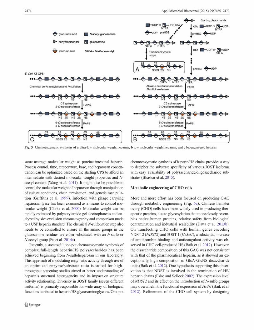
same average molecular weight as porcine intestinal heparin.Process control, time, temperature, base, and heparosan concen-tration can be optimized based on the starting CPS to afford anintermediate with desired molecular weight properties and N-acetyl content (Wang et al. 2011). It might also be possible tocontrol the molecular weight of heparosan throughmanipulationof culture conditions, chain termination, and genetic manipula-tion (Griffiths et al. 1999). Infection with phage carryingheparosan lyase has been examined as a means to control mo-lecular weight (Clarke et al. 2000). Molecular weight can berapidly estimated by polyacrylamide gel electrophoresis and an-alyzed by size exclusion chromatography and comparison madeto a USP heparin standard. The chemicalN-sulfonation step alsoneeds to be controlled to ensure all the amino groups in theglucosamine residues are either substituted with an N-sulfo orN-acetyl group (Fu et al. 2014a).
Recently, a successful one-pot chemoenzymatic synthesis ofcomplex full-length heparin/HS polysaccharides has beenachieved beginning from N-sulfoheparosan in our laboratory.This approach of modulating enzymatic activity through use ofan optimized enzyme/substrate ratio is suited for high-throughput screening studies aimed at better understanding ofheparin’s structural heterogeneity and its impact on structureactivity relationship. Diversity in 3OST family (seven differentisoforms) is primarily responsible for wide array of biologicalfunctions attributed to heparin/HS glycosaminoglycans. One-pot
chemoenzymatic synthesis of heparin/HS chains provides a wayto decipher the substrate specificity of various 3OST isoformswith easy availability of polysaccharide/oligosaccharide sub-strates (Bhaskar et al. 2015).
Metabolic engineering of CHO cells
More and more effort has been focused on producing GAGthrough metabolic engineering (Fig. 6a). Chinese hamsterovary (CHO) cells have been widely used in producing ther-apeutic proteins, due to glycosylation that more closely resem-bles native human proteins, relative safety from biologicalcontamination and industrial scalability (Datta et al. 2013b).On transfecting CHO cells with human genes encodingNDST-2 (NDST2) and 3OST-1 (Hs3st1), a substantial increaseof antithrombin-binding and anticoagulant activity was ob-served in CHO cell-produced HS (Baik et al. 2012). However,the disaccharide composition of this GAG was not consistentwith that of the pharmaceutical heparin, as it showed an ex-ceptionally high composition of GlcA-GlcNS disaccharideunits (Baik et al. 2012). One hypothesis supporting this obser-vation is that NDST is involved in the termination of HS/heparin chains (Esko and Selleck 2002). The expression levelof NDST2 and its effect on the introduction of N-sulfo groupsmay overwhelm the functional expression ofHs3st (Baik et al.2012). Refinement of the CHO cell system by designing
Fig. 5 Chemoenzymatic synthesis of a ultra-low molecular weight heparins; b low molecular weight heparins; and c bioengineered heparin
7474 Appl Microbiol Biotechnol (2015) 99:7465–7479

Golgi-targeted Hs3st1 enhanced the expression of 3OST-1(Datta et al. 2013a). The improvement of anticoagulant activ-ity was further confirmed by the structural signature of theantithrombin-binding site by tetrasaccharide analysis (Dattaet al. 2013a). Moreover, the overexpression of Hs3st1 in theGolgi compartment may also result in the up-regulation of othersulfotransferases natively expressed in CHO cell Golgi. Engi-neering CHO cell HS into a more heparin-like structure willclearly require better control of heparin/HS polymerization, aswell as up-regulation of the genes controlling the introductionof critical 2-O-sulfo and 6-O-sulfo groups (Datta et al. 2013a).
Metabolic engineering of E. coli strains producingchondroitin and heparosan
Capsular polysaccharides produced by pathogenic E. colistrains such as K4 and K5 are an important source of precur-sors for chemoenzymatic synthesis of CS and heparin poly-saccharides. Several studies have been performed to improvethe production and yield of K4 CPS comprised offructosylated chondroitin (Manzoni et al. 1996; Zoppetti andOreste 2004; Cimini et al. 2010a, b; Restaino et al. 2011). Thegrowth of E. coli K4 has been optimized by altering mediumcomposition, including the use of glucose, glycerol, and soyapeptone. Direct feeding of monosaccharide precursors includ-ing GlcA, GalNAc, and fructose results in an increased yieldof CPS (Restaino et al. 2013b). High cell density cultivation,accomplished through microfiltration fermentation to preventacetate accumulation, increased the amount of K4 CPS(Restaino et al. 2011).
In addition to fermentation optimization, increasing attentionhas been focused on genetic modification and the preparation ofrecombinant strains to produce CPS (Zanfardino et al. 2010;
Cimini et al. 2010b; Doherty et al. 2011). More information onthe precise function of biosynthetic genes should be useful forimproving CPS production. Non-pathgenetic production of non-fructosylated chondroitin can be achieved by integrating group I,II, and III genes related to the transportation and biosynthesis ofE. coli K4 capsular polysaccharide into the chromosomes ofE. coli K-12 and Xanthomonas campestris (pv. campestris)(Doherty et al. 2011). Homologous overexpression of kfoC inK4 reportedly increased CPS productivity by 100 %, althoughthe compatibility of the plasmid in wild-type E. coli K4 strainmay cause gene expression stability issues during scale-up(Cimini et al. 2010b). A recent publication suggests that utilizingengineered IS2 transposable elements from K4 fused with kfoCwill result in a more stable overexpression system upon integra-tion into the genome (He et al. 2015). A 2.5-fold increase isreported in large-scale fermenter (He et al. 2015). Moreover, asingle mutation on chondroitin polymerase kfoC (R313Q) po-tentially enhances the affinity of the polymerase to the UDP-GalNAc increasing K4 CPS productivity by 80 % (Zanfardinoet al. 2010). Recently, our group investigated optimizing expres-sion levels for biosynthetic genes of K4 CPS in E. coli BL21Star™ (DE3). By using ePathbrick platform vectors designedfor tunable gene copy number and promoter strength in a singleplasmid, a maximum production of 2.4 g/L was observed inDO-STAT fed-batch fermenter (Cimini et al. 2013). A similarstrategy has also been applied to the production of heparosan.Introduction of the four heparosan biosynthetic genes (KfiA-D)from E. coli K5 into E. coli BL21 resulted in a final yield of1.88 g/L in a DO-STAT fed-batch fermenter (Zhang et al. 2012).
Transcription factors may also play a major role in regulat-ing CPS biosynthesis. Homologous overexpression of rfaH inE. coli K4 resulted in a total CPS yield of 5.3 g/L in a fed-batch experiment, representing the highest reported level of
Fig. 6 Alternate strategies formetabolic engineering ofglycosaminoglycans. a CHO cellengineering to produce heparin,based on manipulation of existingpathway for HS biosynthesis. bAn approach for the E. coli-basedproduction of chondroitin sulfateusing microbialbiotransformation. Three E. colistrains produce components forCS synthesis, includingchondroitin, sulfotransferases,and the sulfate donor PAPS,which are then combined toproduce CS
Appl Microbiol Biotechnol (2015) 99:7465–7479 7475

bacterial chondroitin production. The rfaH gene is a transcrip-tional activator that carries out an anti-termination processduring CPS expression. It binds to the operon polarity sup-pressor element located just upstream ofmany CPS gene startsand controls promoter distal gene expression by preventingthe termination of transcripts and promoting transcription overa long distance (Cimini et al. 2013). Overexpression of thetranscriptional regulator slyA enhances the E. coli K4 CPSproduction by 1.85-fold higher than the wild-type strain (Wuet al. 2013). As a global transcriptional regulator, slyAmay up-regulate region II gene cluster expression for E. coli K4 CPSsynthesis while down-regulating the genes involved in glycol-ysis and citrate cycle pathway (Wu et al. 2013).
Conclusions and future prospects
The small-scale chemoenzymatic synthesis of bioengineeredheparin has been demonstrated and work is proceeding onscaling-up and commercializing its production. It should notbe long before similar studies are successful in thechemoenzymatic synthesis of various chondroitin sulfates.Enzyme engineering should be useful to improve the foldingof the recombinant GAG biosynthetic enzymes increasingtheir production levels in E. coli. Similarly, enzyme engineer-ing or alternative expression systems should be useful in in-creasing the activity and stability of these recombinantcatalysts.
In the future, the metabolic engineering of GAGs may alsobe possible. While some initial progress has been made in themetabolic engineering of CHO cells, the use of CRISPRmight allow the controlled up-regulation and down-regulation of Golgi enzymes to control expression levels re-quired for GAG targets with different fine structures. Meta-bolic engineering of prokaryotes (without a Golgi), such asE. coli, posses even greater challenges (Bhan et al. 2013; Xuet al. 2013; Cress et al. 2015; Jones et al. 2015). One interme-diate approach can be envisioned using currently availabletechnology to couple metabolic engineering with biotransfor-mation. For example, the production of chondroitin CPS fromglucose using an engineered E. coli is currently possible (Heet al. 2015). Moreover, PAPS can be produced from ATP andinorganic sulfate using three E. coli expressed recombinantenzymes, adenosine 5′-triphosphate (ATP) sulfurylase, aden-osine 5′-phosphosulfate (APS) kinase, and pyrophosphatase(Zhou et al. 2011) (Fig. 6b). Finally, recombinant C4ST-1can be expressed in E. coli. A relatively straightforward pro-cess to prepare chondroitin-4-sulfate can be envisioned bycombining chondroitin produced in a first E. coli fermenta-tion, PAPS is produced in a second E. coli fermentation, andrecombinant C4ST-1 is produced in a third E. coli fermenta-tion (Fig. 6). Certainly, it is also possible, but considerably
more challenging, to metabolically engineer a single E. colito biosynthesize CS and shed it into the culture media.
Acknowledgments The authors are grateful for support from the Na-tional Institutes of Health (HL094463, GM102137, HL62244,HL096972) and the National Science Foundation (MCB-1448657).
Conflict of interest The authors declare no competing interests.
References
Adebowale AO, Cox DS, Liang Z, Eddington ND (2000) Analysis ofglucosamine and chondroitin sulfate content in marketed productsand the Caco-2 permeability of chondroitin sulfate raw materials. JAm Nutraceutical Assoc 3:37–44
Baik JY, Gasimli L, Yang B, Datta P, Zhang F, Glass CA, Esko JD,Linhardt RJ, Sharfstein ST (2012) Metabolic engineering ofChinese hamster ovary cells: towards a bioengineered heparin.Metab Eng 14:81–90. doi:10.1016/j.ymben.2012.01.008
Bernfield M, Götte M, Park PW, Reizes O, Fitzgerald ML, Lincecum J,Zako M (1999) Functions of cell surface heparan sulfate proteogly-cans. Annu Rev Biochem 68:729–777. doi:10.1146/annurev.biochem.68.1.729
Berninsone P, Hirschberg C (1998) Heparan sulfate/heparin N-deacetylase/N-sulfotransferase: the N-sulfotransferase activity do-main is at the carboxyl half of the holoenzyme. J Biochem 273:25556–25559
Bhan N, Xu P, Koffas MAG (2013) Pathway and protein engineeringapproaches to produce novel and commodity small molecules.Curr Opin Biotechnol 24:1137–1143. doi:10.1016/j.copbio.2013.02.019
Bhaskar U, Sterner E, Hickey AM, Onishi A, Zhang F, Dordick JS,Linhardt RJ (2012) Engineering of routes to heparin and relatedpolysaccharides. Appl Microbiol Biotechnol 93:1–16. doi:10.1007/s00253-011-3641-4
Bhaskar U, Li G, Fu L, Onishi A, Suflita M, Dordick J, Linhardt RJ(2015) Combinatorial one-pot chemoenzymatic synthesis of hepa-rin. Carbohydr Polymer 122:399–407
Bick RL, Frenkel EP, Walenga J, Fareed J, Hoppensteadt DA (2005)Unfractionated heparin, low molecular weight heparins, and penta-saccharide: basic mechanism of actions, pharmacology, and clinicaluse. Hematol Oncol Clin North Am 19:1–51. doi:10.1016/j.hoc.2004.09.003
Burkart M, Wong C (1999) A continuous assay for the spectrophotomet-ric analysis of sulfotransferases using aryl sulfotransferase IV. AnalBiochem 274:131–137. doi:10.1006/abio.1999.4264
Burkart M, IzumiM, Chapman E, Lin C,Wong C (2000) Regeneration ofPAPS for the enzymatic synthesis of sulfated oligosaccharides. J OrgChem 65:5565–5574
Castelli R, Porro F, Tarsia P (2004) The heparins and cancer: review ofclinical trials and biological properties. Vasc Med 9:205–213. doi:10.1191/1358863x04vm566ra
Chavaroche AAE, Van Den Broek LAM, Boeriu C, Eggink G (2012)Synthesis of heparosan oligosaccharides by Pasteurella multocidaPmHS2 single-action transferases. Appl Microbiol Biotechnol 95:1199–1210. doi:10.1007/s00253-011-3813-2
Chen M, Bridges A, Liu J (2006) Determination of the substrate speci-ficities of N-acetyl-d-glucosaminyltransferase. Biochemistry 45:12358–12365
Chen Y, Li Y, YuH, Sugiarto G, ThonV, Hwang J, Ding L, Hie L, Chen X(2013) Tailored design and synthesis of heparan sulfate oligosaccha-ride analogues using sequential one-pot multienzyme systems.
7476 Appl Microbiol Biotechnol (2015) 99:7465–7479

Angew Chem Int Ed Engl 52:11852–11856. doi:10.1002/anie.201305667
Cimini D, Restaino OF, Catapano A, De Rosa M, Schiraldi C (2010a)Production of capsular polysaccharide from Escherichia coli K4 forbiotechnological applications. Appl Microbiol Biotechnol 85:1779–1787. doi:10.1007/s00253-009-2261-8
Cimini D, De Rosa M, Viggiani A, Restaino OF, Carlino E, Schiraldi C(2010b) Improved fructosylated chondroitin production by kfoCoverexpression in E. coli K4. J Biotechnol 150:324–331. doi:10.1016/j.jbiotec.2010.09.954
Cimini D, De Rosa M, Carlino E, Ruggiero A, Schiraldi C (2013)Homologous overexpression of rfaH in E. coli K4 improves theproduction of chondroitin-like capsular polysaccharide. MicrobCell Factories 12:46. doi:10.1186/1475-2859-12-46
Clarke BR, Esumeh F, Roberts IS (2000) Cloning, expression, and puri-fication of the K5 capsular polysaccharide lyase (KflA) from coli-phage K5A: evidence for two distinct K5 lyase enzymes. J Bacteriol182:3761–3766
Cress BF, Englaender JA, He W, Kasper D, Linhardt RJ, Koffas MAG(2014) Masquerading microbial pathogens: capsular polysaccha-rides mimic host-tissue molecules. FEMS Microbiol Rev 38:660–697. doi:10.1111/1574-6976.12056
Cress BF, Toparlak OD, Guleria S, Lebovich M, Stieglitz JT, EnglaenderJA, Jones JA, Linhardt RJ, Koffas MAG (2015) CRISPathBrick:modular combinatorial assembly of type II-A CRISPR arrays fordCas9-mediated multiplex transcriptional repression in E. coli.ACS Synth Biol. doi:10.1021/acssynbio.5b00012
Datta P, Li G, Yang B, Zhao X, Baik JY, Gemmill TR, Sharfstein ST,Linhardt RJ (2013a) Bioengineered Chinese hamster ovary cellswith Golgi-targeted 3-O-sulfotransferase-1 biosynthesize heparansulfate with an antithrombin-binding site. J Biol Chem 288:37308–37318. doi:10.1074/jbc.M113.519033
Datta P, Linhardt RJ, Sharfstein ST (2013b) An ’omics approach towardsCHO cell engineering. Biotechnol Bioeng 110:1255–1271. doi:10.1002/bit.24841
DeAngelis PL,White CL (2002) Identification andmolecular cloning of aheparosan synthase fromPasteurella multocida type D. J Biol Chem277:7209–7213. doi:10.1074/jbc.M112130200
Deangelis PL, Liu J, Linhardt RJ (2013) Chemoenzymatic synthesis ofglycosaminoglycans: re-creating, re-modeling and re-designing na-ture’s longest or most complex carbohydrate chains. Glycobiology23:764–777. doi:10.1093/glycob/cwt016
Doherty DH, Weaver CA, Miyamoto K, Minamisawa T (2011)Compositions and methods for bacterial production of chondroitin.US Patent# US20110244520 A1
Esko JD, Lindahl U (2001) Molecular diversity of heparan sulfate. J ClinInvest 108:169–173. doi:10.1172/JCI13530
Esko JD, Selleck SB (2002) Order out of chaos: assembly of ligandbinding sites in heparan sulfate. Annu Rev Biochem 71:435–471.doi:10.1146/annurev.biochem.71.110601.135458
Evans TD, Mozen MM (1962) Process for purifying heparin. US Patent#3058884
Fu L, Li G, Yang B, Onishi A, Li L, Sun P, Zhang F, Linhardt R(2013) Structural characterization of pharmaceutical heparinsprepared from different animal tissues. J Pharm Sci 102:1447–1457
Fu L, Li L, Cai C, Li G, Zhang F, Linhardt RJ (2014a) Heparin stabilityby determining unsubstituted amino groups using hydrophilic inter-action chromatography mass spectrometry. Anal Biochem 461:46–48. doi:10.1016/j.ab.2014.05.028
Fu L, Zhang F, Li G, Onishi A, Bhaskar U, Sun P, Linhardt RJ (2014b)Structure and activity of a new low-molecular-weight heparin pro-duced by enzymatic ultrafiltration. J Pharm Sci 10(100):1375–1383.doi:10.1002/jps.23939
Gijsen HJM, Qiao L, Fitz W, Wong C-H (1996) Recent advances in thechemoenzymatic synthesis of carbohydrates and carbohydrate mi-metics. Chem Rev 96:443–474. doi:10.1021/cr950031q
Griffiths G, Barrett B, Cook N, Roberts IS (1999) Biosynthesis of theEscherichia coli K5 capsular polysaccharide. Biochem Soc Trans27:507–512
Guerrini M, Beccati D, Shriver Z, Naggi A, Viswanathan K, Bisio A,Capila I, Lansing JC, Guglieri S, Fraser B, Al-Hakim A, Gunay NS,Zhang Z, Robinson L, Buhse L, Nasr M, Woodcock J, Langer R,Venkataraman G, Linhardt RJ, Casu B, Torri G, Sasisekharan R(2008) Oversulfated chondroitin sulfate is a major contaminant inheparin associated with adverse clinical events. Nat Biotechnol 26:669–675
He W, Fu L, Li G, Jones A, Linhardt RJ, Koffas M (2015) Production ofchondroitin in metabolically engineered E. coli. Metab Eng 27:92–100. doi:10.1016/j.ymben.2014.11.003
Higashi K, Hosoyama S, OhnoA,Masuko S, Yang B, Sterner E,Wang Z,Linhardt R, Toida T (2012) Photochemical preparation of a novelmolecular weight heparin. Carbohydr Polym 87:1737–1743
Higashi K, Takeuchi Y, Mukuno A, Tomitori H, Miya M, Linhardt RJ,Toida T (2015) Composition of glycosaminoglycans in elasmo-branchs including several deep-sea sharks: identification ofchondroitin/dermatan sulfate from the dried fins of Isurusoxyrinchus and Prionace glauca. PLoS One 10, e0120860. doi:10.1371/journal.pone.0120860
Jones JA, Toparlak ÖD, KoffasMA (2015) Metabolic pathway balancingand its role in the production of biofuels and chemicals. Curr OpinBiotechnol 33:52–59. doi:10.1016/j.copbio.2014.11.013
Karst NA, Linhardt RJ (2003) Recent chemical and enzymatic ap-proaches to the synthesis of glycosaminoglycan oligosaccharides |BenthamScience. Curr Med Chem 10:1993–2031
Kato M, Wang H, Bernfield M, Gallagher JT, Turnbull JE (1994) Cellsurface syndecan-1 on distinct cell types differs in fine structure andligand binding of its heparan sulfate chains. J Biol Chem 269:18881–18890
Keire DA, Buhse LF, Al-Hakim A (2013) Characterization of currentlymarketed heparin products: composition analysis by 2D-NMR.AnalMethods 5:2984–2994
Kuberan B, Lech MZ, Beeler DL, Wu ZL, Rosenberg RD (2003)Enzymatic synthesis of antithrombin III-binding heparan sulfatepentasaccharide. Nat Biotechnol 21:1343–1346. doi:10.1038/nbt885
Kusche-Gullberg M, Kjellén L (2003) Sulfotransferases in glycosamino-glycan biosynthesis. Curr Opin Struct Biol 13:605–611. doi:10.1016/j.sbi.2003.08.002
Lassen MR, Dahl OE, Mismetti P, Destrée D, Turpie AG (2009)AVE5026, a new hemisynthetic ultra-low-molecular-weight heparinfor the prevention of venous thromboembolism in patients after totalknee replacement surgery—TREK: a dose-ranging study. J ThrombHaemost 7:566–572. doi:10.1111/j.1538-7836.2009.03301.x
Lauder RM (2009) Chondroitin sulphate: a complex molecule with po-tential impacts on a wide range of biological systems. ComplementTher Med 17:56–62. doi:10.1016/j.ctim.2008.08.004
Li JP, Vlodavsky I (2009) Heparin, heparan sulfate and heparanase ininflammatory reactions. Thromb Haemost 102:823–828. doi:10.1160/TH09-02-0091
Li L, Zhang F, Zaia J, Linhardt RJ (2012) Top-down approach for thedirect characterization of low molecular weight heparins using LC-FT-MS. Anal Chem 84:8822–8829. doi:10.1021/ac302232c
Li G, Li L, Tian F, Zhang L, Xue C, Linhardt RJ (2015)Glycosaminoglycanomics of cultured cells using a rapid and sensi-tive LC-MS/MS approach. ACS Chem Biol. doi:10.1021/acschembio.5b00011
Lindahl U, Kusche-Gullberg M, Kjellen L (1998) Regulated diversity ofheparan sulfate. J Biol Chem 273:24979–24982. doi:10.1074/jbc.273.39.24979
Appl Microbiol Biotechnol (2015) 99:7465–7479 7477
Lindahl U, Li J, Kusche-Gullberg M, Salmivirta M, Alaranta S, VeromaaT, Emeis J, Roberts I, Taylor C, Oreste P, Zopetti G, Naggi A, TorriG, Casu B (2005) Generation of Bneoheparin^ from E. coli K5capsular polysaccharide. J Med Chem 48(2):349–352. doi:10.1021/jm049812m
Linhardt RJ (2003) Heparin: strucuture and activity. J Med Chem 46:2521–2564
Linhardt RJ, Liu J (2012) Synthetic heparin. Curr Opin Pharmacol 12:217–219. doi:10.1016/j.coph.2011.12.002
Linhardt RJ, Ampofo SA, Fareed J, Hoppensteadt D, Folkman J,Mulliken JB (1992) Isolation and characterization of human heparin.Biochemistry 31:12441–12445. doi:10.1021/bi00164a020
Liu J, Shriver Z, Pope RM, Thorp SC, Duncan MB, Copeland RJ, RaskaCS, Yoshida K, Eisenberg RJ, Cohen G, Linhardt RJ, SasisekharanR (2002) Characterization of a heparan sulfate octasaccharide thatbinds to herpes simplex virus type 1 glycoprotein D. J Biol Chem277:33456–33467. doi:10.1074/jbc.M202034200
Liu H, Zhang Z, Linhardt RJ (2009) Lessons learned from the contami-nation of heparin. Nat Prod Rep 26:313–321. doi:10.1039/b819896a
Liu R, Xu Y, Chen M, Weïwer M, Zhou X, Bridges AS, DeAngelis PL,Zhang Q, Linhardt RJ, Liu J (2010) Chemoenzymatic design ofheparan sulfate oligosaccharides. J Biol Chem 285:34240–34249.doi:10.1074/jbc.M110.159152
Liu C, Sheng J, Krahn JM, Perera L, Xu Y, Hsieh PH, Dou W, Liu J,Pedersen LC (2014) Molecular mechanism of substrate specificityfor heparan sulfate 2-O-sulfotransferase. J Biol Chem 289:13407–13418. doi:10.1074/jbc.M113.530535
Loganathan D, Wang HM, Mallis LM, Linhardt RJ (1990) Structural var-iation in the antithrombin III binding site region and its occurrence inheparin from different sources. Biochemistry 29:4362–4368
Manzoni M, Bergomi S, Molinari F, Cavazzoni V (1996) production andpurification of an extracellularly produced K4 polysaccharide fromEscherichia coli. Biotechnol Lett 4:383–386. doi:10.1007/BF00143456
Masuko S, Bera S, Green DE, Weïwer M, Liu J, Deangelis PL, LinhardtRJ (2012) Chemoenzymatic synthesis of uridine diphosphate-GlcNAc and uridine diphosphate-GalNAc analogs for the prepara-tion of unnatural glycosaminoglycans. J Org Chem 77:1449–1456.doi:10.1021/jo202322k
Mikami T, Kitagawa H (2013) Biosynthesis and function of chondroitinsulfate. Biochim Biophys Acta Gen Subj 1830:4719–4733. doi:10.1016/j.bbagen.2013.06.006
MoonAF, Edavettal SC, Krahn JM,Munoz EM, Negishi M, Linhardt RJ,Liu J, Pedersen LC (2004) Structural analysis of the sulfotransferase(3-O-sulfotransferase isoform 3) involved in the biosynthesis of anentry receptor for herpes simplex virus 1. J Biol Chem 279:45185–45193. doi:10.1074/jbc.M405013200
Moon AF, Xu Y,Woody SM, Krahn JM, Linhardt RJ, Liu J, Pedersen LC(2012) Dissecting the substrate recognition of 3-O-sulfotransferasefor the biosynthesis of anticoagulant heparin. Proc Natl Acad Sci109:5265–5270. doi:10.1073/pnas.1117923109
Mousa SA, Fareed J (2001) Overview: from heparin to low molecularweight heparin: beyond anticoagulation. Curr Opin Investig Drugs2:1077–1080
Murugesan S, Xie J, Linhardt RJ (2008) Immobilization of heparin: ap-proaches and applications. Curr Top Med Chem 8:80–100. doi:10.2174/156802608783378891
Naggi A, Torri G, Casu B, Oreste P, Zoppetti G, Li JP, Lindahl U (2001)Toward a biotechnological heparin through combined chemical andenzymatic modification of the Escherichia coli K5 polysaccharide.Semin Thromb Hemost 27:437–443. doi:10.1055/s-2001-17954
Okuyama T, Yoshida K, Sakuraik, Ogurat, Horie K, Tawada A (1975)Method of separating and recovering mucopolysaccharides fromconnective tissues of animals. US Patent #3862003
Orellana A, Hirschberg CB, Wei Z, Swiedler SJ, Ishihara M (1994)Molecular cloning and expression of a glycosaminoglycan N-acetylglucosaminyl N-deacetylase/N-sulfotransferase from aheparin-producing cell line. J Biol Chem 269:2270–2276
Osterman MT, Lichtenstein GR (2007) Current and future anti-TNF ther-apy for inflammatory bowel disease. Curr Treat Options inGastroenterol 10:195–207. doi:10.1007/s11938-007-0013-3
Pacheco B, Maccarana M, Goodlett DR, Malmström A, Malmström L(2009a) Identification of the active site of DS-epimerase 1 and re-quirement of N-glycosylation for enzyme function. J Biol Chem284:1741–1747. doi:10.1074/jbc.M805479200
Pacheco B, Malmström A, Maccarana M (2009b) Two dermatan sulfateepimerases form iduronic acid domains in dermatan sulfate. J BiolChem 284:9788–9795. doi:10.1074/jbc.M809339200
Papoutsaki M, Osório F, Morais P, Torres T, Magina S, Chimenti S,Costanzo A (2013) Infliximab in psoriasis and psoriatic arthritis.BioDrugs 27(Suppl 1):13–23. doi:10.1007/BF03325638
Parish CR, Freeman C, Hulett MD (2001) Heparanase: a key enzymeinvolved in cell invasion. Biochim Biophys Acta 1471:M99–M108
Petitou M, Duchaussoy P, Lederman I, Choay J, Sinaÿ P, Jacquinet J-C,Torri G (1986) Synthesis of heparin fragments. A chemical synthesisof the pentasaccharide O-(2-deoxy-2-sulfamido-6-O-sulfo-alpha-D-glucopyranosyl)-(1–4)-O-(beta-D-glucopyranosyluronic acid)-(1–4)-O-(2-O-sulfo-alpha-L-idopyranosyluronic acid)-(1–4)-2-deoxy-2-sulfamido-6-. Carbohydr Res 147:221–236
Qin Y, Ke J, Gu X, Fang J, Wang W, Cong Q, Li J, Tan J, Brunzelle JS,ZhangC, JiangY,Melcher K, Li J, XuHE, Ding K (2015) Structuraland functional study of D-glucuronyl C5-epimerase. J Biol Chem290:4620–4630. doi:10.1074/jbc.M114.602201
Ramani VC, Purushothaman A, Stewart M, Thompson C, Vlodavsky I,Au J, Sanderson RD (2013) The heparanase/syndecan-1 axis incancer: mechanisms and therapies. FEBS J 280:2294–2306. doi:10.1016/j.biotechadv.2011.08.021
Restaino OF, Cimini D, De Rosa M, Catapano A, Schiraldi C, De RosaM, Catapano A, Schiraldi C, De Rosa M, Catapano A, Schiraldi C(2011) High cell density cultivation of Escherichia coli K4 in amicrofiltration bioreactor: a step towards improvement of chondroi-tin precursor production. Microb Cell Factories 10:1–11. doi:10.1186/1475-2859-10-10
Restaino OF, Bhaskar U, Paul P, Li L, De Rosa M, Dordick JS, LinhardtRJ (2013a) High cell density cultivation of a recombinant E. colistrain expressing a key enzyme in bioengineered heparin produc-tion. Appl Microbiol Biotechnol 97:3893–3900. doi:10.1007/s00253-012-4682-z
Restaino OF, di Lauro I, Cimini D, Carlino E, De Rosa M, Schiraldi C(2013b) Monosaccharide precursors for boosting chondroitin-likecapsular polysaccharide production. Appl Microbiol Biotechnol97:1699–1709. doi:10.1007/s00253-012-4343-2
Sakai S, Otake E, Toida T, Goda Y (2007) Identification of the origin ofchondroitin sulfate in Bhealth foods^. Chem PharmBull (Tokyo) 55:299–303
Saribaş AS, Mobasseri A, Pristatsky P, Chen X, Barthelson R, Hakes D,Wang J (2004) Production of N-sulfated polysaccharides usingyeast-expressed N-deacetylase/N-sulfotransferase-1 (NDST-1).Glycobiology 14:1217–1228. doi:10.1093/glycob/cwh129
Sasisekharan R, Venkataraman G (2000) Heparin and heparan sulfate:biosynthesis, structure and function. Curr Opin Chem Biol 4:626–631. doi:10.1016/S1367-5931(00)00145-9
Sato N, Meijer L, Skaltsounis L, Greengard P, Brivanlou AH (2004)Maintenance of pluripotency in human and mouse embryonic stemcells through activation of Wnt signaling by a pharmacologicalGSK-3-specific inhibitor. Nat Med 10:55–63. doi:10.1038/nm979
Schiraldi C, Cimini D, De Rosa M (2010) Production of chondroitinsulfate and chondroitin. Appl Microbiol Biotechnol 87:1209–1220. doi:10.1007/s00253-010-2677-1
7478 Appl Microbiol Biotechnol (2015) 99:7465–7479
Schonberger L (1998) New variant Creutzfeldt-Jakob disease and bovinespongiform encephalopathy. Infect Dis Clin N Am 12:111–121
Sheng J, Liu R, Xu Y, Liu J (2011) The dominating role of N-deacetylase/N-sulfotransferase 1 in forming domain structures in heparan sul-fate. J Biol Chem 286:19768–19776. doi:10.1074/jbc.M111.224311
Sheng J, Xu Y, Dulaney SB, Huang X, Liu J (2012) Uncovering biphasiccatalytic mode of C5-epimerase in heparan sulfate biosynthesis. JBiol Chem 287:20996–21002. doi:10.1074/jbc.M112.359885
Shi X, Zaia J (2009) Organ-specific heparan sulfate structural phenotypes.J Biol Chem 284:11806–11814. doi:10.1074/jbc.M809637200
Silbert JE, Sugumaran G (2002) Biosynthesis of chondroitin/dermatans u l f a t e . I UBMB L i f e 5 4 : 1 7 7 – 1 8 6 . d o i : 1 0 . 1 0 8 0 /15216540290114450
Sismey-Ragatz AE, Green DE, Otto NJ, Rejzek M, Field RA, DeAngelisPL (2007) Chemoenzymatic synthesis with distinct Pasteurellaheparosan synthases: monodisperse polymers and unnatural struc-tures. J Biol Chem 282:28321–28327. doi:10.1074/jbc.M701599200
Sterner E, Li L, Paul P, Beaudet JM, Liu J, Linhardt RJ, Dordick JS(2014) Assays for determining heparan sulfate and heparin O-sulfotransferase activity and specificity. Anal Bioanal Chem 406:525–536. doi:10.1007/s00216-013-7470-4
Sugiura N, Tawada A, Sugimoto K, Watanabe H (2002) Molecular clon-ing and characterization of chondroitin polymerase fromEscherichia coli strain K4. J Biol Chem 277:21567–21575. doi:10.1074/jbc.M201719200
Sugiura N, Shioiri T, ChibaM, Sato T, Narimatsu H, Kimata K,WatanabeH (2012) Construction of a chondroitin sulfate library with definedstructures and analysis of molecular interactions. J Biol Chem 287:43390–43400. doi:10.1074/jbc.M112.412676
Sun X, Li L, Overdier KH, Ammons LA, Douglas IS, Burlew CC, ZhangF, Schmidt EP, Chi L, Linhardt RJ (2015) Analysis of total humanurinary glycosaminoglycan disaccharides by liquid chromatogra-phy–tandem mass spectrometry. Anal Chem 87:6220–6227. doi:10.1021/acs.analchem.5b00913
Taipale J, Keski-Oja J (1997) Growth factors in the extracellular matrix.FASEB J Off Publ Fed Am Soc Exp Biol 11:51–59
Thelin MA, Bartolini B, Axelsson J, Gustafsson R, Tykesson E, Pera E,Oldberg A, MacCarana M, Malmstrom A (2013) Biological func-tions of iduronic acid in chondroitin/dermatan sulfate. FEBS J 280:2431–2446. doi:10.1111/febs.12214
Toschi V, LettinoM (2007) Fondaparinux: pharmacology and clinical expe-rience in cardiovascular medicine. Mini - Rev Med Chem 7:383–387
Tully SE, Rawat M, Hsieh-Wilson LC (2006) Discovery of a TNF-alphaantagonist using chondroitin sulfate microarrays. J Am Chem Soc128:7740–7741. doi:10.1021/ja061906t
Van Gorp CL, Vosburgh F, Schubert RL (1997) Protein hydrolysate de-rived from mucosa tissue. US Patent #5607840
Vidic H-J (1981) Process for the preparation of heparin. US Patent #4283530.
Viskov C, Just M, Laux V, Mourier P, Lorenz M (2009) Description of thechemical and pharmacological characteristics of a new hemisyntheticultra-low-molecular-weight heparin, AVE5026. J Thromb Haemost 7:1143–1151. doi:10.1111/j.1538-7836.2009.03447.x
Vlodavsky I, Friedmann Y (2001) Heparan sulfate proteoglycans molec-ular properties and involvement of heparanase in cancer metastasisand angiogenesis. J Clin Investig 108:341–347. doi:10.1172/JCI200113662
Wang Z, Yang B, Zhang Z, Mellisa L, Takieddin M, Mousa S, Liu J,Dordick JS, Linhardt RJ (2011) Control of the heparosan N-deacetylation leads to an improved bioengineered heparin. ApplMicrobiol Biotechnol 91:91–99
Wang W, Englaender JA, Xu P, Mehta KK, Suwan J, Dordick JS, ZhangF, Yuan Q, Linhardt RJ, Koffas M (2013) Expression of low endo-toxin 3-o-sulfotransferase in Bacillus subtilis and Bacillusmegaterium. Appl Biochem Biotechnol 171:954–962. doi:10.1007/s12010-013-0415-8
Williams RE (1967) Process for the recovery of heparin. US Patent#3337409
Wu Q, Yang A, Zou W, Duan Z, Liu J, Chen J, Liu L (2013)Transcriptional engineering of Escherichia coliK4 for fructosylatedchondroitin production. Biotechnol Prog 29:1140–1149. doi:10.1002/btpr.1777
Xiong J, Bhaskar U, Li G, Fu L, Li L, Zhang F, Dordick JS, Linhardt RJ(2013) Immobilized enzymes to convert N-sulfo, N-acetylheparosan to a critical intermediate in the production ofbioengineered heparin. J Biotechnol 167:241–247. doi:10.1016/j.jbiotec.2013.06.018
Xu Y, Masuko S, Takieddin M, Xu H, Liu R, Jing J, Mousa SA, LinhardtRJ, Liu J, Xu Y, Masuko S, Takieddin M, Xu H, Liu R, Jing J,Mousa S, Linhardt RJ, Liu J (2011) Chemoenzymatic synthesis ofstructurally homogeneous ultra-low molecular weight heparins.Science 334(80-):498–501. doi:10.1126/science.1207478
Xu P, Bhan N, Koffas MAG (2013) Engineering plant metabolism intomicrobes: from systems biology to synthetic biology. Curr OpinBiotechnol 24:291–299. doi:10.1016/j.copbio.2012.08.010
Xu Y, Cai C, Chandarajoti K, Hsieh P-H, Li L, Pham TQ, SparkenbaughEM, Sheng J, Key NS, Pawlinski R, Harris EN, Linhardt RJ, Liu J(2014) Homogenous low-molecular-weight heparins with reversibleanticoagulant activity. Nat Chem Biol 10:248–250. doi:10.1038/nchembio.1459
Yang B, Chang Y, Weyers AM, Sterner E, Linhardt RJ (2012)Disaccharide analysis of glycosaminoglycan mixtures by ultra-high-performance liquid chromatography-mass spectrometry. JChromatogr A 1225:91–98. doi:10.1016/j.chroma.2011.12.063
Zanfardino A, Restaino OF, Notomista E, Cimini D, Schiraldi C, De RosaM, De Felice M, Varcamonti M (2010) Isolation of an Escherichiacoli K4 kfoC mutant over-producing capsular chondroitin. MicrobCell Factories 9:34. doi:10.1186/1475-2859-9-34
Zhang Z, McCallum SA, Xie J, Nieto L, Corzana F, Jimenez-Barbero J,Chen M, Liu J, Linhardt RJ (2008) Solution structures ofchemoenzymatically synthesized heparin and its precursors. J AmChem Soc 130:12998–13007
Zhang F, Yang B, Mellisa L, Kemal S, Xiao Z, Wang Z, Beaudet JM,Torelli AY, Dordick JS, Linhardt RJ (2011) Structural characteriza-tion of heparins from different commercial sources. Anal BioanalChem 401:2793–2803
Zhang C, Liu L, Teng L, Chen J, Liu J, Li J, Du G, Chen J (2012)Metabolic engineering of Escherichia coli BL21 for biosynthesisof heparosan, a bioengineered heparin precursor. Metab Eng 14:521–527. doi:10.1016/j.ymben.2012.06.005
Zhang J, Suflita M, Fiaschetti CM, Li G, Li L, Zhang F, Dordick JS,Linhardt RJ (2015a) High cell density cultivation of a recombinantEscherichia coli strain expressing a 6-O-sulfotransferase for the pro-duction of bioengineered heparin. J Appl Microbiol 118:92–98. doi:10.1111/jam.12684
Zhang J, Suflita M, Li G, ZhongW, Li L, Dordick JS, Linhardt RJ, ZhangF (2015b) High cell density cultivation of recombinant Escherichiacoli strains expressing 2-O-sulfotransferase and C5-epimerase forthe production of bioengineered heparin. Appl BiochemBiotechnol 175:2986–2995. doi:10.1007/s12010-014-1466-1
Zhou YJ, Hao XF, Tian ZJ, Tong GZ, Yoo D, An TQ, Zhou T, LiGX, Qiu HJ, Wei TC, Yuan XF (2008) Highly virulent por-cine reproductive and respiratory syndrome virus emerged inChina. Transbound Emerg Dis 55:152–164. doi:10.1111/j.1865-1682.2008.01020.x
Zhou X, Chandarajoti K, Pham TQ, Liu R, Liu J (2011) Expression ofheparan sulfate sulfotransferases in Kluyveromyces lactis and prep-aration of 3′-phosphoadenosine-5′-phosphosulfate. Glycobiology21:771–780. doi:10.1093/glycob/cwr001
Zoppetti G, Oreste P (2004) process for the preparation of chondroitinsulfates from K4 polysaccharide and obtained products. US Patent6777398 B2
Appl Microbiol Biotechnol (2015) 99:7465–7479 7479