hericium erinaceus (bull.: fr.) pers., a medicinal ... · fr.) pers., a medicinal mushroom,...
TRANSCRIPT
Seediscussions,stats,andauthorprofilesforthispublicationat:http://www.researchgate.net/publication/265096046
Hericiumerinaceus(Bull.:Fr.)Pers.,amedicinalmushroom,activatesperipheralnerveregeneration
ARTICLEinCHINESEJOURNALOFINTEGRATIVEMEDICINE·AUGUST2014
ImpactFactor:1.4·DOI:10.1007/s11655-014-1624-2·Source:PubMed
CITATION
1
DOWNLOADS
124
VIEWS
187
5AUTHORS,INCLUDING:
WongKahHui
UniversityofMalaya
33PUBLICATIONS192CITATIONS
SEEPROFILE
GowriKanagasabapathy
UniversityofMalaya
14PUBLICATIONS42CITATIONS
SEEPROFILE
PamelaDavid
UniversityofMalaya
27PUBLICATIONS118CITATIONS
SEEPROFILE
VikineswarySabaratnam
UniversityofMalaya
190PUBLICATIONS940CITATIONS
SEEPROFILE
Availablefrom:WongKahHui
Retrievedon:14July2015
• 1 •Chin J Integr Med
Following an injury to a peripheral nerve, it would be important for the peripheral nervous system to rescue its cellular components from cell death and then regenerate the damaged axons. Protein kinase B (Akt) pathway has been suggested to be involved in neuronal survival(1) and mitogen-activated protein kinase (MAPK) pathway in neurite outgrowth during development.(2)
Immediate early genes are known to take part in injury-related cellular mechanisms in many systems.(3) Peripheral sciatic nerve injuries have been previously shown to induce c-Jun expression in dorsal root ganglia (DRG) neurons. Expression of c-Jun and c-Fos are involved in a successful regenerative response. c-Fos is used as a marker of neuronal activity and is expressed in the nuclei of neurons in response to tactile, chemical, noxious and innocuous stimulation.(4)
Injury-conditioned axons showed an increase in content of protein synthesis machinery compared with that of axons extending from naive neurons.(5) Up to 100 different axonally synthesized proteins can be discerned in injury-conditioned DRG axons.(6) As expected, this includes many cytoskeletal proteins that are needed for axonal outgrowth and growth cone motility.
Traditionally, mushroom has been defined as a fleshy, aerial umbrella-shaped, fruitbodies of macrofungi, and has been consumed by Asian people for over two thousand years because of the pleasant flavor and texture.(7) Medicinal properties of Hericium erinaceus (Bull.: Fr) Pers., also known as Lion's Mane, Monkey's Head and Yamabushitake have been well known for hundreds of years in traditional Chinese and Japanese herbal medicine to treat various human diseases. The fruitbodies are composed of numerous constituents such as polysaccharides, proteins, lectins, phenols, hericenones, erinacines and terpenoids.
The most promising activity of H. erinaceus is
the stimulation of nerve growth factor (NGF) synthesis
ORIGINAL ARTICLEHericium Erinaceus (Bull.: Fr.) Pers., A Medicinal Mushroom,
Activates Peripheral Nerve Regeneration
Kah-Hui Wong1,2, Gowri Kanagasabapathy2, Murali Naidu1,2, Pamela David1,2, and Vikineswary Sabaratnam3,2
©The Chinese Journal of Integrated Traditional and Western Medicine Press and Springer-Verlag Berlin Heidelberg 2013Supported by the High Impact Research Grant and Fundamental Research Grant Scheme FP023/2008C of University of Malaya1. Department of Anatomy, Faculty of Medicine, University of Malaya, Kuala Lumpur 50603, Malaysia; 2. Mushroom Research Centre, University of Malaya, Kuala Lumpur 50603, Malaysia; 3. Institute of Biological Sciences, Faculty of Science, University of Malaya, Kuala Lumpur 50603, Malaysia;Correspondence to: Dr. Kah-Hui Wong, Tel: 603-79674729, Fax: 603-79674724, E-mail: [email protected]: 10.1007/s11655-013-1624-2
ABSTRACT Objective: To study the ability of aqueous extract of Hericium erinaceus mushroom in the treatment of nerve injury following peroneal nerve crush in Sprague-Dawley rats. Methods: Aqueous extract of Hericium erinaceus was given by daily oral administration following peroneal nerve crush injury in Sprague-Dawley rats. The expression of protein kinase B (Akt) and mitogen-activated protein kinase (MAPK) signaling pathways; and c-Jun and c-Fos genes were studied in dorsal root ganglia (DRG) whereas the activity of protein synthesis was assessed in peroneal nerves by immunohistochemical method. Results: Peripheral nerve injury leads to changes at the axonal site of injury and remotely located DRG containing cell bodies of sensory afferent neurons. Immunofluorescence studies showed that DRG neurons ipsilateral to the crush injury in rats of treated groups expressed higher immunoreactivities for Akt, MAPK, c-Jun and c-Fos as compared with negative control group (P<0.05). The intensity of nuclear ribonucleoprotein in the distal segments of crushed nerves of treated groups was significantly higher than in the negative control group (P<0.05). Conclusion: H. erinaceus is capable of promoting peripheral nerve regeneration after injury. Potential signaling pathways include Akt, MAPK, c-Jun, and c-Fos, and protein synthesis have been shown to be involved in its action. KEYWORDS Hericium erinaceus, dorsal root ganglia, signaling pathways, gene expression, protein synthesis machinery, peripheral nerve regeneration
• 2 • Chin J Integr Med
by hericenones from fruitbodies and erinacines from mycelium.(8) Extract of fruitbodies exerted neurotrophic action and improved myelination process in mature myelinating fibers of nerve cells in vitro(9) and also promoted normal development of cultivated cerebellar cells and demonstrated a regulatory effect on the process of myelin genesis in vitro.(10) Our studies have shown that aqueous extract of H. erinaceus could stimulate neurite outgrowth of the cultured cells of the neural hybrid clone NG108-15(11) and enhance functional recovery following injury to rodent peroneal nerve.(12) These findings indicate that H. erinaceus may have a potential in stimulation of neurons to regrow in the treatment of senility, Alzheimer's disease, repairing neurological trauma from strokes, improve muscle or motor response pathways and cognitive function.
Research on the medicinal value of H. erinaceus grown in Malaysia is minimal and has not been explored. In this study, we investigated the activity of aqueous extract of H. erinaceus fresh fruitbodies in triggering the expression of Akt, MAPK, c-Jun and c-Fos in the respective DRG of normal and injured peroneal nerves, and axonal protein synthesis after crush injury.
METHODS
Fruibodies of H. Erinaceus and Preparation of Aqueous Extract
Fresh fruitbodies of H. erinaceus were obtained from Vita Agrotech Mushroom Farm in Tanjung Sepat, Selangor, Malaysia. They were boiled in distilled water at a ratio of 1:1 for 30 min with agitation, left covered for 30 min, cooled and filtered. Biologically active substances of H. erinaceus dissolved readily in water and retained its neurotrophic and trophic action for several days at 4 ℃.(9) In this study, the aqueous extract was used within a week after preparation.
Proximate Composition Analysis of H. ErinaceusThe total carbohydrate content in the fruitbodies
was determined by the method of Association of Official Analytical Chemists.(13) The contents of crude ash, crude fat, crude protein and crude fibre were measured using the Approved Methods of the American Association of Cereal Chemists.(14)
Isolation of Crude Polysaccharides and Measurement of β-glucan Content
Preparation of crude hot water polysaccharide
was carried out according to the method of Roy, et al.(15) Fresh fruitbodies of H. erinaceus (500 g) were boiled in 500 mL of distilled water for 8 h. The whole mixture was kept overnight at 4 ℃ and then filtered through linen cloth. The filtrate was centrifuged at 8,000 r/min for 45 min at 4 ℃. Supernatant was collected and precipitated in ethanol at 1:5 (v/v). It was kept overnight at 4 ℃ and again centrifuged as above. The precipitated material (polysaccharide) was washed with ethanol 4 times and then freeze-dried. The freeze-dried material was dissolved in 30 mL of distilled water and dialyzed through dialysis tubing cellulose membrane (Sigma-Aldrich, St. Louis, MO, retaining more than MW 12400) against distilled water for 4 h to remove low molecular weight materials. The aqueous solution was then collected from the dialysis bag and freeze-dried to yield crude polysaccharide of 1.4 g.
Con ten ts o f t o ta l and α -g lucans were determined in the polysaccharide extracts using the mushroom and yeast β-glucan assay procedure K-YBGL 09/2009 (Megazyme International Ireland). The enzyme kit contains exo-1,3-β-glucanase, β-glucosidase, amyloglucosidase, invertase, glucose determination reagent-(GOPOD-glucose oxidase, peroxidase, 4-aminoantipyrine) and glucose standard solution.(16) The β-glucan content was calculated by subtracting the α-glucan from the total glucan content, therefore, β-glucan comprises 0.3% of fresh weight. All values of glucan contents were expressed as g/100 g of fresh weight.
Principle of Animal GroupingThe rat experiment was approved by the Animal
Care and Use Committee of Faculty of Medicine, University of Malaya. Eighteen adult female Sprague-Dawley rats were randomly assigned into three groups of six rats each. Negative control group received daily oral administration of distilled water (10 mL/kg body weight per day), experimental group received aqueous extract of fresh fruitbodies (10 mL/kg body weight/day) and positive control group received mecobalamin (Natural Factors®, batch. 020564, 130 μg/kg body weight per day) for 14 days to function as pre-treatment before injury.
Surgical ProcedureAfter 14 days of pre-treatment, the rats were
anesthetized with an intraperitoneal injection of 3.5% chloral hydrate (10 mL/kg body weight), then shaved
• 3 •Chin J Integr Med
and washed with antiseptic solution before positioning for surgery. The right sciatic nerve and its two major branches were exposed through a gluteal muscle-splitting incision. A crush injury was created using a fine watchmaker forceps No. 4 for 10 s on the peroneal nerve at 10 mm from extensor digitorum longus muscle and complete crush was confirmed by presence of a translucent band across the nerve. All operations were performed on right limb and the left limb served as an uninjured control. After closing the incision with sutures, veterinary wound powder was applied to wounds. Distilled water, aqueous extracts or mecobalamin was continuingly fed for another 7 days. All rats were observed for general well being and had ad libitum access to food and water throughout the study.
Removal and Immunofluorescence Staining of DRG
In our previous study, the recovery of hind limb function started by 7 days after crush injury.(12) Complete functional recovery was observed by 10 to 14 days after injury in the treated groups or 4 to 7 days earlier than negative control group using walking track analysis. Therefore, the expression of Akt, MAPK, c-Jun and c-Fos in the respective DRG of normal and injured peripheral nerves were investigated after 7 days of injury. Rats were perfused transcardially with 4% paraformaldehyde in 0.1 mmol/L phosphate buffer saline. DRGs were traced from their respective peroneal nerves to the intervertebral foramen [vertebral level—fifth lumbar (L5) to third sacral (S3)] and the surrounding bone was removed with fine bone clippers before ganglia removal. L5 DRGs from crushed nerves (ipsilateral) and their uncrushed counterparts (contralateral) were dissected out.
After deparaffinization and antigen retrieval, longitudinal sections (5 μm) were washed in 0.1 mol/L phosphate buffer saline containing 0.3% Triton X-100 (washing buffer). Non-specific binding sites were blocked with 10% normal sheep serum in washing buffer (blocking buffer) for 1 h at room temperature. The sections were then incubated with a mixture of primary antibodies (rabbit anti-Akt antibody), 1:100 dilution, rabbit anti-p44/42 MAPK (ERK1/2), 1:60 dilution, rabbit anti-c-fos antibody, 1:100 dilution, Cell Signaling Technology, Beverly, MA or rabbit anti-c-Jun antibody, 1:100 dilution, Chemicon, Temecula, CA, USA; and mouse anti-neurofilament 200 (NF-200), a marker for myelinated A-fibres, 1:200 dilution, Sigma-Aldrich, St. Louis, MO,
USA in blocking buffer) at 4 ℃ for 20 h in humidity chamber, washed with washing buffer and further reaction with a mixture of secondary antibodies (Cy3-conjugated sheep anti-rabbit IgG, 1:100 dilution and FITC-conjugated sheep anti-mouse IgG, 1:80 dilution, Sigma-Aldrich, St. Louis, MO, USA in blocking buffer) at room temperature for 1 h. After the same washing procedure, sections were coverslipped with antifade reagent with 4',6-diamidino-2-phenylindole (DAPI). DAPI binds strongly to deoxyribonucleic acid (DNA).
Removal and Immunofluorescence Staining of Peroneal Nerve
Righ t peronea l nerves of a l l ra ts were carefully dissected out and their proximal and distal ends were identified after 7 days of surgery. The contralateral peroneal nerves were also obtained. A series of 20 μm-thick longitudinal sections were cut on a cryostat microtome at –20 ℃ and mounted on poly-L-lysine coated slides. The sections were fixed by freezing in acetone for 20 min, then washed 3 times for 5 min each with 0.1 mmol/L phosphate buffer saline containing 0.3% Triton X-100 (washing buffer).(17)
For the assessment of protein synthesis activity, sections were incubated in 10% normal goat serum in washing buffer (blocking buffer) for 1 h at room temperature. The sections were then incubated with mouse anti-nuclear ribonucleoprotein monoclonal antibody (1:20 dilution in blocking buffer, purchased from Chemicon, Temecula, CA, USA) at 4 ℃ for 20 h in humidity chamber, washed with washing buffer and followed by further reaction with the FITC-conjugated secondary antibody goat anti-mouse IgM (1:80 in blocking buffer, purchased from Sigma, St. Louis, MO, USA) at room temperature for 1 h. After the same washing procedure, sections were coverslipped with antifade reagent.
Image AnalysisFour sections of DRG or peroneal nerve were
randomly selected from ipsilateral or contralateral side of each rat and tested for each antibody. Images were analyzed with Nikon Eclipse 80i upright microscope equipped with a digital color camera controller (DS-5Mc-U2), neutral density (ND) 0.1 OD filter and NIS-Elements algorithm (NIS-Elements Advanced Research, Nikon, Japan). DS-5Mc-U2 controls the motorized fluorescence attachment through NIS-
• 4 • Chin J Integr Med
Elements software. The attachment is to minimize photobleaching. Intensity of Cy3 staining regions as a relative figure that demonstrated immunoreactivities for anti-Akt, anti-p44/42 MAPK (ERK1/2), anti c-Jun or anti-c-Fos; or FITC staining regions for protein synthesis was examined under fluorescence illumination.
Statistical AnalysisThe means of data (n=6 animals per group)
were subjected to a one-way analysis of variance and the significance of the difference between means was determined by the Duncan's multiple range tests at 95% least significant difference (P<0.05). Data were expressed as mean ± standard error (P<0.05 compared to negative control for uninjured/injured category).
RESULTSComposition Analysis and Glucan Content of H. Erinaceus
Total carbohydrate, crude protein, crude fat, crude fiber and crude ash contents of H. erinaceus were 45.49%, 27.30%, 0.54% 16.3% and 10.37% on a dry weight basis, respectively. Total glucan content of H. erinaceus was 0.32 g/100 g of fresh weight while α-glucan content was 0.02 g/100 g of fresh weight.
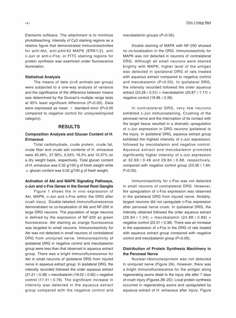
Activation of Akt and MAPK Signaling Pathways, c-Jun and c-Fos Genes in the Dorsal Root Ganglia
Figure 1 shows the in vivo expression of Akt, MAPK, c-Jun and c-Fos within the DRG after crush injury. Double-labeled immunofluorescence demonstrated no co-localization of Akt and NF-200 in large DRG neurons. The population of large neurons is defined by the expression of NF-200 as green fluorescence. Akt staining as orange fluorescence was targeted to small neurons. Immunoreactivity for Akt was not detected in small neurons of contralateral DRG from uninjured nerve. Immunoreactivity of ipsilateral DRG in negative control and mecobalamin group were less than that observed in aqueous extract group. There was a bright immunofluorescence for Akt in small neurons of ipsilateral DRG from injured nerve in aqueous extract group. In ipsilateral DRG, the intensity recorded followed the order aqueous extract (21.21±0.38) > mecobalamin (18.02±0.92) > negative control (17.41±0.79). The significant increase in intensity was detected in the aqueous extract group compared with the negative control and
mecobalamin groups (P<0.05).
Double staining of MAPK with NF-200 showed no co-localization in the DRG. Immunoreactivity for MAPK was not detected in neurons of contralateral DRG. Although all small neurons were stained brightly with MAPK, higher level of the antigen was detected in ipsilateral DRG of rats treated with aqueous extract compared to negative control and mecobalamin (P<0.05). In ipsilateral DRG, the intensity recorded followed the order aqueous extract (23.28±0.31) > mecobalamin (20.87±1.17) > negative control (18.96±0.39).
In contra latera l DRG, very few neurons exhibited c-Jun immunostaining. Crushing of the peroneal nerve and the interruption of its contact with the target tissue resulted in a dramatic upregulation of c-Jun expression in DRG neurons ipsilateral to the injury. In ipsilateral DRG, aqueous extract group exhibited the highest intensity of c-Jun expression, followed by mecobalamin and negative control. Aqueous extract and mecobalamin promoted significantly higher intensity of c-Jun expression at 32.69±0.48 and 29.84±0.88, respectively, compared with negative control group (23.56±1.64, P<0.05).
Immunoreactivity for c-Fos was not detected in small neurons of contralateral DRG. However, the upregulation of c-Fos expression was observed in the ipsilateral DRG from injured nerve. Notably, largest neurons did not upregulate c-Fos expression after peroneal nerve crush. In ipsilateral DRG, the intensity obtained followed the order aqueous extract (26.84±1.04) > mecobalamin (24.88±0.89) > negative control (23.31±0.38). There was an increase in the expression of c-Fos in the DRG of rats treated with aqueous extract group compared with negative control and mecobalamin group (P<0.05).
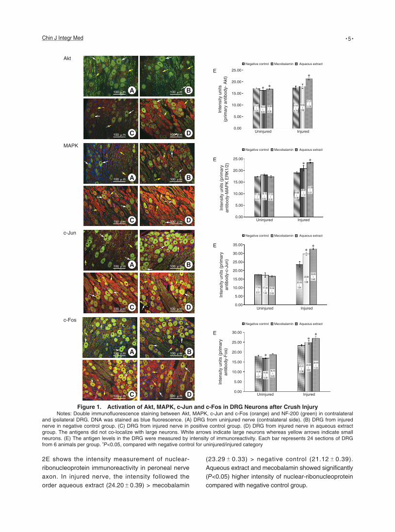
Distribution of Protein Synthesis Machinery in the Peroneal Nerve
Nuclear-ribonucleoprotein was not detected in uninjured nerve (Figure 2A). However, there was a bright immunofluorescence for the antigen along regenerating axons distal to the injury site after 7 days of crush injury (Figures 2B–2D). Local protein synthesis occurred in regenerating axons and upregulated by aqueous extract of H. erinaceus after injury. Figure
• 5 •Chin J Integr Med
Figure 1. Activation of Akt, MAPK, c-Jun and c-Fos in DRG Neurons after Crush InjuryNotes: Double immunofluorescence staining between Akt, MAPK, c-Jun and c-Fos (orange) and NF-200 (green) in contralateral
and ipsilateral DRG. DNA was stained as blue fluorescence. (A) DRG from uninjured nerve (contralateral side). (B) DRG from injured nerve in negative control group. (C) DRG from injured nerve in positive control group. (D) DRG from injured nerve in aqueous extract group. The antigens did not co-localize with large neurons. White arrows indicate large neurons whereas yellow arrows indicate small neurons. (E) The antigen levels in the DRG were measured by intensity of immunoreactivity. Each bar represents 24 sections of DRG from 6 animals per group. P<0.05, compared with negative control for uninjured/injured category
2E shows the intensity measurement of nuclear-ribonucleoprotein immunoreactivity in peroneal nerve axon. In injured nerve, the intensity followed the order aqueous extract (24.20±0.39) > mecobalamin
(23.29±0.33) > negative control (21.12±0.39). Aqueous extract and mecobalamin showed significantly (P<0.05) higher intensity of nuclear-ribonucleoprotein compared with negative control group.
Inte
nsity
uni
ts (p
rimar
y an
tibod
y-Fo
s)
30.00
25.00
20.00
15.00
10.00
5.00
0.00
18.06±
0.27
16.93±
0.29
18.70±
0.48
23.31±
0.38
24.88±
0.89
26.84±
1.04
Uninjured Injured
E
Mecobalamin Aqueous extractNegative control
Uninjured Injured
Inte
nsity
uni
ts (p
rimar
y an
tibod
y-c-
Jun)
35.00
30.00
25.00
20.00
15.00
10.00
5.00
0.00
17.62±
0.57
17.34±
1.22
16.56±
0.58
23.56±
1.64
29.84±
0.88
32.69±
0.48
E
Mecobalamin Aqueous extractNegative control
Uninjured InjuredIn
tens
ity u
nits
(prim
ary
antib
ody-
MA
PK
ER
K1/
2)
25.00
20.00
15.00
10.00
5.00
0.00
17.29±
0.31
17.29±
0.38
18.15±
0.52
18.96±
0.39
20.87±
1.17
23.28±
0.31
E
Mecobalamin Aqueous extractNegative control
Inte
nsity
uni
ts
(prim
ary
antib
ody-
Akt
)
25.00
20.00
15.00
10.00
5.00
0.00Uninjured
17.21±
0.23
16.33±
0.09
16.88±
0.28
17.41±
0.79
18.02±
0.92
21.21±
0.38
Injured
EMecobalamin Aqueous extractNegative control
BA
DC
Akt
BA
DC
MAPK
BA
DC
c-Jun
BA
DC
c-Fos
100 μm
100 μm
100 μm
100 μm
100 μm
100 μm
100 μm
100 μm
100 μm
100 μm
100 μm
100 μm
100 μm
100 μm
100 μm
100 μm
• 6 • Chin J Integr Med
aqueous extract group before peripheral nerve injury.
Functional deterioration following crush injury is not only related to the impact of the crush itself, but also includes other important components such as ischemia of the limb. Studies on crush injury models in peripheral nerves have shown better regeneration and functional recovery when therapies were directed against ischemia-reperfusion injury by antioxidants, lipid peroxidation inhibitors and anti-inflammatory agents.(18) With this in mind, the present study evaluated the peripheral nerve regeneration mechanisms of H. erinaceus that possesses antioxidant activity as an alternative herbal pharmacotherapy for nerve repair.(19)
Mushrooms con ta in b io log ica l l y ac t i ve polysaccharides in fruitbodies, cultured mycelium and culture broth. Water-soluble polysaccharides from the extract of Tremella fuciformis have been reported to stimulate NGF production in the brain and ameliorate memory deficits in rats(20) while glycosaminoglycans promoted neurite formation, nerve regeneration and muscle reinnervation following sciatic nerve injury.(21) A number of β-glucans have immune-stimulating effects and participate in physiological processes related to the metabolism in the human body.(22) Peripherally applied Lentinan (β-glucan from lentinus edodes) has been shown to facilitate the synaptic efficacy of the dentate gyrus neurons in vivo.(23) The activation of immune system by lentinan may contribute to the synaptic efficacy in the central nervous system. Thus the β-glucan component of H. erinaceus may play an important role in its neuroprotective effect on regenerating peripheral nerves.
Studies that use radiolabeling have verified that both small and large fragments of polysaccharides such as β-D-glucan are found in the serum, which indicates they are absorbed from the gastrointestinal tract.(22,24) However, whether these metabolites can cross the blood-brain barrier and send signal for DRG neurons to evoke regenerative responses awaits further study. Polysaccharide components in our locally grown H. erinaceus have been quantified by Choong, et al. (25) High-performance l iquid chromatography analysis showed arabinose as the major component with minor components of glucose, rhamnose, deoxyribose and galactose. Nevertheless, they did not characterize the specific types of polysaccharides present.
Figure 2. Distribution of Protein Synthesis Machinery in Peroneal Nerve Axons after Crush Injury
Notes: Fluorescence imaging staining for nuclear ribonucleoprotein in peroneal nerve axons as green fluorescent patches. DNA was stained as blue fluorescence. (A) Uninjured nerve (contralateral side). (B) Distal to the injury site of injured nerve in negative control group. (C) Distal to the injury site of injured nerve in positive control group. (D) Distal to the injury site of injured nerve in aqueous extract group. Scale bar = 100 μm. (E) Protein synthesis machinery levels in the peroneal nerve as measured by intensity of immunoreactivity. Each bar represents 24 sections of peroneal nerve from 6 animals per group. P<0.05 compared to negative control for uninjured/injured category
Inte
nsity
uni
ts (p
rimar
y an
tibod
y-nu
clea
r rib
onuc
leop
rote
in)
30.00
25.00
20.00
15.00
10.00
5.00
0.00
17.69±
0.35
18.38±
0.18
18.34±
0.29
21.12±
0.39
23.29±
0.33
24.20±
0.62
Uninjured
Negative controlMecobalaminAqueous extract
Injured
E
DISCUSSIONThis study revealed the relationship between
activation of Akt and MAPK signaling pathways, c-Jun and c-Fos genes, protein synthesis machinery, and peripheral nerve regeneration following crush injury in an in vivo experiment by a medicinal mushroom renowned for its neurological effects. Upregulation of Akt, MAPK, c-Jun and c-Fos in ipsilateral DRG neurons and nuclear-ribonucleoprotein in the injured nerves after 7 days of crush injury is consistent with the beginning of functional recovery of injured right hind limb.(12)
Natural products have been traditionally accepted as remedies due to popular belief that they present minor adverse effects. Mushrooms have always been prepared for medicinal use by hot water extraction as in brewing of teas or decoctions in Chinese medicine for the prevention of oxidative stress-related diseases. In our study, pre-treatment with aqueous extract was employed to build up strength and immune system in the
A B
DC
100 μm
100 μm
100 μm
100 μm
• 7 •Chin J Integr Med
Injury to neurons results in complex sequences of molecular responses that play an important role in the successful regenerative response and the eventual recovery of function. Peripheral nerve injury induced peripheral sensitization causing activation of Akt, MAPK, c-Jun and c-Fos in small DRG neurons. These contributed to pain hypersensitivity found at the site of tissue damage and inflammation. In general, DRG neurons can be divided into large (>1200 μm2), medium (600–1200 μm2) and small (<600 μm2) neurons. Small neurons respond to thermal, mechanical and chemical nociceptive stimulations whereas large neurons transmit touch and proprioceptive sensations.(26) Akt, MAPK, c-Jun or c-Fos did not co-localize with NF-200 in large DRG neurons. It is possible that aqueous extract of H. erinaceus could trigger the expression of protein kinases and early genes that regulate nociceptive function and inflammation associated with nerve recovering.
Activation of Akt, MAPK, c-Jun and c-Fos persisted for one week after injury until axonal regeneration occurred.(12) The findings are in accordance with a study by Naidu, et al.(27) In their study, immunoreactivity for phospho-Akt was detected in ipsilateral DRG after 2, 4 and 7 days of sciatic nerve crush by using Western blot analysis whereas clear upregulation of both phospho p44 MAPK and phospho p42 MAPK were detected after 7 days of injury. In a study using in situ hybridisation, Ito, et al(28) reported the enhanced gene expression for PI3K in the hypoglossal motor neurons following axonal crush in the first week after injury. Phospho-Akt was clearly expressed in the normal DRG and nerve, and was upregulated during nerve regeneration in the DRG neurons. The expression of Akt in the normal DRG and nerve may be required to maintain cell survival and an upregulation could be essential to prevent mass cell death as a result of nerve injury.
Activation of MAPK pathway is essential for neurite outgrowth, regeneration, synaptic plasticity and memory functions in mature neurons.(29) Naidu, et al(27) showed that phospho-MAPK was expressed in normal DRG but confirmed its presence in the rat sciatic nerves even after injury. This was probably needed for axonal regeneration. A transient increase of ERK1 mRNA in motor neurons during rat hypoglossal nerve regeneration was reported by Namikawa, et al.(30) which adds further evidence to the claim that MAPK is
essential for neurite outgrowth even in in vivo.
Downstream events influenced by crush injury-activated kinases include upregulation or activation of several transcription factors. Activated Akt and MAPK induces upregulation and phosphorylation of the transcription factors c-Jun and c-Fos into the nucleus, leading to formation of activator protein 1 complexes that activate many downstream genes.(31) The expression of immediate early genes after nerve injury has been shown to be induced according to the nerve and the type of injury.(32,33)
In this study, upregulation of c-Jun and c-Fos occurred in ipsilateral DRG sensory neurons after 7 days of crush injury. DRG sensory neurons contributing to the crushed peroneal nerve in the treated groups displayed more intense c-Jun and c-Fos after staining with anti-c-Jun and anti-c-Fos antibodies, respectively compared to ipsilateral DRG in negative control group or contralateral DRG. Similar to the study on signaling pathways, activation of c-Jun and c-Fos also persisted for 1 week after injury until axonal regeneration takes place.(12)
c-Jun is a protein that may be important for successful axonal regeneration. Studies by Broude, et al(3) on adult rat DRG showed that c-Jun was substantially upregulated in DRG neurons following a peripheral axotomy. However, only 18% of the neurons expressed c-Jun after a central axotomy. Similar strong upregulation of c-Jun in the adult rat DRG following peripheral nerve transection was also reported by Leah, et al.(34) c-Fos expression was also increased in animal models of neuropathic pain including noxious electrical stimulation, formalin, mechanical stimuli and paw inflammation.(35) Therefore, c-Fos is used as a marker for neuronal activation. In a study by Chi, et al(35) c-Fos expression in the lumbar spinal cord increased in the superficial dorsal horn and deep dorsal horn within hours and lasted for at least 4 weeks following sciatic nerve transection in rats.
Local protein synthesis occurred in regenerating axons and upregulated by aqueous extract of H. erinaceus after injury. According to the original dogma, necessary components for axonal growth and regeneration are usually synthesized by cell body and sent along the axons by fast or slow axonal transport to their respective targets, which are usually hundreds
• 8 • Chin J Integr Med
of micrometers away from the cell body.(5) These processes are crucial in facilitating axonal regeneration and subsequently reinnervation of target muscle. Zheng, et al(36) showed that axons isolated from cultured adult rat neurons of peripheral nervous system are capable of synthesizing new proteins. Because the axons are 'regrowing' in culture, this observation suggested that protein synthesis might be reactivated by injury.
The pool of newly synthesised cytoskeletal proteins is likely to act as a source of structural proteins for growth cone reformation and axonal growth,(37) as well as to regulate cytoskeletal dynamics.(38) The injured axon must transition to a growth state, and this transition distinguishes protein synthesis in adult axons from that in developing axons. The old arguments that adult axons do not synthesize proteins were based largely on the absence of polysomes in axons compared with those in dendrites.(39) This scarcity of protein synthesis machinery suggests that the mature axon must recruit (and activate) ribosomes and mRNAs to the injury site. It is not clear what triggers this recruitment of protein synthesis machinery after injury, but increase in axoplasmic [Ca2+] is a likely candidate. Adult vertebrate axons have the capacity to synthesize many different proteins.(38)
Upregulation of Akt, MAPK, c-Jun, c-Fos and protein synthesis machinery are crucial in facilitating axonal regeneration and subsequently reinnervation of target muscle, that brings to the return of hind limb function. From this study, we have shown that aqueous extract of H. erinaceus fresh fruitbodies could promote regeneration of injured peripheral nerve and may provide future benefit in facilitating recovery in patients with crush injuries. Patients who receive H. erinaceus may experience a more expeditious improvement in the quality of life after injury. Moreover, by taking mecobalamin for the treatment of nerve injury gives rise to side effects such as gastrointestinal problem. Future research models will focus on identification and characterization of polysaccharides and other compounds which may trigger signaling cascades in response to axonal injury, and refining strategies to enhance regeneration.
Conflicts of InterestAuthors declared they have no conflicts of interest.
Author ContributionsKH Wong designed and performed experiments,
analysed data and wrote the paper; G. Kanagasabapathy
designed and performed experiments; M. Naidu, P. David and
V. Sabaratnam involved in the design of the study.
REFERENCES1. Crowder RJ, Freeman RS. Phosphatidylinositol 3-kinase
and Akt protein kinase are necessary and sufficient for
the survival of nerve growth factor-dependent sympathetic
neurons. J Neurosci 1998;18:2933-2943.
2. Kaplan DR, Mil ler FD. Signal transduction by the
neurotrophin receptors. Curr Opin Cell Biol 1997;9:213-221.
3. Broude E, McAtee M, Kelley MS, Bregman BS. c-Jun
expression in adult rat dorsal root ganglion neurons:
differential response after central or peripheral axotomy.
Exp Neurol 1997;148:367-377.
4. Tsai YC, So EC, Chen HH, Wang LK, Chien CH. Effect of
intrathecal octreotide on thermal hyperalgesia and evoked
spinal c-Fos expression in rats with sciatic constriction
injury. Pain 2002;99:407-413.
5. Verma P, Chierzi S, Codd AM, Campbell DS, Meyer RL,
Holt CE, et al. Axonal protein synthesis and degradation
are necessary for efficient growth cone regeneration. J
Neurosci 2005;25:331-342.
6. Willis D, Li KW, Zheng JQ, Smit AB, Kelly TK, Merianda
TT, et al. Differential transport and local translation of
cytoskeletal, injury-response, and neurodegeneration
protein mRNAs in axons. J Neurosci 2005;25:778-791.
7. Miles PG, Chang ST. Mushroom biology: concise basics
and current developments. Singapore: World Scientific
Press; 1997:1-9.
8. Kawagishi H, Zhuang C, Yunoki R. Compounds for dementia
from Hericium erinaceum. Drugs Future 2008;33:149-155.
9. Moldavan MG, Grygansky A, Kolotushkina OV, Kirchhoff B,
Skibo GG, Pedarzani P. Neurotropic and trophic action of
lion's mane mushroom Hericium erinaceus (Bull.: Fr) Pers.
(Aphyllophoromycetideae) extracts on nerve cells in vitro.
Int J Med Mush 2007;9:15-28.
10. Kolotushkina EV, Moldavan MG, Voronin KY, Skibo GG.
The influence of Hericium erinaceus extract on myelination
process in vitro. Fiziologicheskii Zhur 2003;49:38-45.
11. Wong KH, Sabaratnam V, Abdullah N, Naidu M, Keynes R.
Activity of aqueous extracts of lion's mane mushroom Hericium
erinaceus (Bull.: Fr.) Pers. (Aphyllophoromycetideae) on the
neural cell line NG108-115. Int J Med Mushr 2007;9:57-65.
12. Wong KH, Naidu M, David P, Abdulla MA, Abdullah N,
Kuppusamy UR, et al. Peripheral nerve regeneration
following crush injury to rat peroneal nerve by aqueous
extract of medicinal mushroom Hericium erinaceus
(Bull.: Fr) Pers. (Aphyllophoromycetideae). Evid Based
Complment Altern Med 2011;doi: 10.1093/ecam/neq062
• 9 •Chin J Integr Med
13. Association of Official Analytical Chemists (AOAC). Official
methods of analysis of the Association of Official Analytical
Chemists. 15th ed. USA: Washington, DC; 1990:751.
14. American Associat ion of Cereal Chemists (AACC
International). Approved methods of analysis–method
08-01.01, 30-10.01, 46-12.01 and 32-10.01. 16th ed. USA:
St. Paul, MN; 1995.
15. Roy SK, Maiti D, Mondal S, Das D, Islam SS. Structural
analysis of a polysaccharide isolated from the aqueous
extract of an edible mushroom, Pleurotus sajor-caju, cultivar
Black Japan. Carbohydrate Res 2008;343:1108-1113.
16. Kozarski M, Klaus A, Niksi M, Jakovljevic D, Helsper JPFG,
van Griensven LJLD. Antioxidative and immunomodulating
activities of polysaccharide extracts of the medicinal
mushrooms Agaricus bisporus, Agaricus brasiliensis,
Ganoderma lucidum and Phellinus linteus. Food Chem
2011:129:1667-1675.
17. Chen B, Song Y, Liu Z. Promotion of nerve regeneration in
peripheral nerve by short-course FK506 after end-to-side
neurorrhaphy. J Surg Res 2009;152:303-310.
18. Algora J, Chen LE, Seaber AV, Wong GH, Urbaniak JR.
Functional effects of lymphotoxin on crushed peripheral
nerve. Microsurgery 1996;17:131-135.
19. Wong KH, Sabaratnam V, Abdullah N, Kuppusamy UR,
Naidu M. Effects of cultivation techniques and processing
on antimicrobial and antioxidant activities of Hericium
erinaceus (Bull.: Fr.) Pers. extracts. Food Tech Biotechnol
2009;47:47-55.
20. Kim JH, Ha HC, Lee MS, Kang JI, Kim HS, Lee SY, et al.
Effect of Tremella fuciformis on the neurite outgrowth of
PC12h cells and the improvement of memory in rats. Biol
Pharm Bull 2007;30:708-714.
21. G o r i o A , L e s m a E , V e r g a n i L , D i G i u l i o A M .
Glycosaminoglycan supplementation promotes nerve
regeneration and muscle reinnervation. Eur J Neurosci
1997;9:1748-1753.
22. Rop O, Mlcek J, Jurikova T. Beta-glucans in higher fungi
and their health effects. Nutr Rev 2009;67:624-631.
23. Edagawa Y, Smriga M, Nishiyama N, Saito H. Systemic
administration of lentinan, a branched beta-glucan,
enhances long-term potentiation in the rat dentate gyrus in
vivo. Neurosci Lett 2001; 314:139-142.
24. Tsukagoshi S, Hashimoto Y, Fujii G, Kobayashi H, Nomoto K,
Orita K. Krestin (PSK). Cancer Treat Rev 1984;11:131-155.
25. Choong YK, Abdul Rashid BA, Young SI, Young SI, Ismail Z.
Quantification and identification of polysaccharide contents
in Hericium erinaceus. Nutr Food Sci 2007;37:260-271.
26. Wen YR, Suter MR, Ji RR, Yeh GC, Wu YS, Wang KC, et
al. Activation of p38 mitogen-activated protein kinase in
spinal microglia contributes to incision-induced mechanical
allodynia. Anesthesiology 2009;1:155-165.
27. Naidu M, David RP, Asher R, Fawcett J. Expression of
Akt and MAPK in the normal and regenerating peripheral
nerves and their dorsal root ganglia. Malaysian J Biochem
Mol Biol 2009;17:16-19.
28. Ito Y, Sakagami H, Kondo H. Enhanced gene expression for
phosphatidylinositol 3-kinase in the hypoglossal motoneurons
following axonal crush. Mol Brain Res 1996;37:329-332.
29. Sweatt JD. The neuronal MAP kinase cascade: a
biochemical signal integration system subserving synaptic
plasticity and memory. J Neurochem 2001;76:1-10.
30. Namikawa K, Honma M, Abe K, Takeda M, Mansur K,
Obata T, et al. Akt/protein kinase B prevents injury-induced
motoneuron death and accelerates axonal regeneration. J
Neurosci 2002;20:2875-2886.
31. Raivich G, Behrens A. Role of the AP-1 transcription factor
c-Jun in developing, adult and injured brain. Prog Neurobiol
2006;78:347-363.
32. Kajander KC, Madsen AM, Iadarola MJ, Draisci G,
Wakisaka S. Fos-like immunoreactivity increases in the
lumbar spinal cord following a chronic constriction injury to
the sciatic nerve of rat. Neurosci Lett 1996;206:9-12.
33. Kenney AM, Kocsis JD. Timing of c-Jun protein induction
in lumbar dorsal root ganglia after sciatic nerve transection
varies with lesion distance. Brain Res 1997;751:90-95.
34. Leah JD, Herdegen T, Bravo R. Selective expression of
Jun proteins following axotomy and axonal transport block
in peripheral nerves in the rat: evidence for a role in the
regeneration process. Brain Res 1991;566:198-207.
35. Chi SI, Levine JD, Basbaum AI. Peripheral and central
contributions to the persistent expression of spinal cord fos-
like immunoreactivity produced by sciatic nerve transection
in the rat. Brain Res 1993;617:225-237.
36. Zheng JQ, Kelly TK, Chang B, Ryanzantsev S, Rajasekaran
AK, Martin KC, et al. A functional role for intra-axonal
protein synthesis during axonal regeneration from adult
sensory neurons. J Neurosci 2001;21:9291-9303.
37. Willis DE, Twiss JL. The evolving roles of axonally
synthesized proteins in regeneration. Curr Opin Neurobiol
2006;16:111-118.
38. Bamburg JR, McGough A, Ono S. Putting a new twist on
actin: ADF/cofilins modulate actin dynamics. Trends Cell
Biol 1999;9:364-370.
39. Giuditta A, Kaplan BB, van Minnen J, Alvarez J, Koenig E.
Axonal and presynaptic protein synthesis: new insights into
the biology of the neuron. Trends Neurosci.
(Received August 27, 2012)Edited by WANG Wei-xia