heterogeneous presynaptic distribution of monoacylglycerol ...real.mtak.hu/10048/1/katona1.pdf ·...
TRANSCRIPT

Heterogeneous presynaptic distribution ofmonoacylglycerol lipase, a multipotent regulator ofnociceptive circuits in the mouse spinal cord
Eszter Horv�ath,1,* Stephen G. Woodhams,1,* Rita Nyilas,1 Christopher M. Henstridge,1 Masanobu Kano,2
Kenji Sakimura,3 Masahiko Watanabe4 and Istv�an Katona11Momentum Laboratory of Molecular Neurobiology, Institute of Experimental Medicine, Hungarian Academy of Sciences,Szigony utca 43., H-1083 Budapest, Hungary2Department of Neurophysiology, Graduate School of Medicine, University of Tokyo, Tokyo, Japan3Department of Cellular Neurobiology, Brain Research Institute, Niigata University, Niigata, Japan4Department of Anatomy, Hokkaido University School of Medicine, Sapporo, Japan
Keywords: endocannabinoid, MAGL, monoglyceride lipase, pain, prostaglandin
Abstract
Monoacylglycerol lipase (MGL) is a multifunctional serine hydrolase, which terminates anti-nociceptive endocannabinoid signalingand promotes pro-nociceptive prostaglandin signaling. Accordingly, both acute nociception and its sensitization in chronic painmodels are prevented by systemic or focal spinal inhibition of MGL activity. Despite its analgesic potential, the neurobiologicalsubstrates of beneficial MGL blockade have remained unexplored. Therefore, we examined the regional, cellular and subcellulardistribution of MGL in spinal circuits involved in nociceptive processing. All immunohistochemical findings obtained with light, con-focal or electron microscopy were validated in MGL-knockout mice. Immunoperoxidase staining revealed a highly concentratedaccumulation of MGL in the dorsal horn, especially in superficial layers. Further electron microscopic analysis uncovered that themajority of MGL-immunolabeling is found in axon terminals forming either asymmetric glutamatergic or symmetric c-aminobutyricacid/glycinergic synapses in laminae I/IIo. In line with this presynaptic localization, analysis of double-immunofluorescence stain-ing by confocal microscopy showed that MGL colocalizes with neurochemical markers of peptidergic and non-peptidergic nocicep-tive terminals, and also with markers of local excitatory or inhibitory interneurons. Interestingly, the ratio of MGL-immunolabelingwas highest in calcitonin gene-related peptide-positive peptidergic primary afferents, and the staining intensity of nociceptive ter-minals was significantly reduced in MGL-knockout mice. These observations highlight the spinal nociceptor synapse as a potentialanatomical site for the analgesic effects of MGL blockade. Moreover, the presence of MGL in additional terminal types raises thepossibility that MGL may play distinct regulatory roles in synaptic endocannabinoid or prostaglandin signaling according to its dif-ferent cellular locations in the dorsal horn pain circuitry.
Introduction
To detect and minimize tissue damage, numerous signaling mecha-nisms operate together in the peripheral, spinal and supraspinal paincircuits (Basbaum et al., 2009). Persistent noxious stimuli evokevarious forms of molecular and cellular adaptations in these signal-ing processes (Sandk€uhler, 2009). Some may last beyond the resolu-tion of tissue injury leading to chronic pain syndromes; a majorconceptual and practical challenge for modern medicine, whichrequires a detailed understanding of how dynamic molecularchanges are integrated into the cellular context of nociceptive pro-cessing in spinal and brain circuits (Kuner, 2010). However, the
tremendous cellular complexity of the neuronal pain-processing cir-cuitry (Todd, 2010) renders this task very difficult.A promising development is the recent delineation of two inter-
related signaling pathways with robust, but generally opposite,effects on nociception. Anti-nociceptive endocannabinoid signalingand pro-nociceptive prostaglandin signaling both regulate nocicep-tive transmission and its plasticity at specific anatomical locations(Reinold et al., 2005; Agarwal et al., 2007; Monory et al., 2007;Vardeh et al., 2009). Moreover, molecular components of thesepathways exhibit region- and cell-type-specific quantitative changesin chronic pain models (Samad et al., 2001; Zeilhofer, 2007; Sagaret al., 2012; Simonetti et al., 2013). Promoting endocannabinoidsignaling or attenuating prostaglandin signaling are approaches gen-erally considered to have analgesic potential (Zeilhofer & Brune,2006; Jhaveri et al., 2007). In fact, medical preparations from thecannabis plant stimulating cannabinoid (CB) receptors, and thosefrom willow bark inhibiting prostaglandin-endoperoxidase synthases
Correspondence: Dr I. Katona, as above.E-mail: [email protected]
*E.H. and S.G.W. contributed equally to this study.
Received 26 September 2013, revised 29 November 2013, accepted 2 December 2013
© 2014 The Authors. European Journal of Neuroscience published by Federation of European Neuroscience Societies and John Wiley & Sons Ltd.This is an open access article under the terms of the Creative Commons Attribution License, which permits use, distribution and reproductionin any medium, provided the original work is properly cited.
European Journal of Neuroscience, Vol. 39, pp. 419–434, 2014 doi:10.1111/ejn.12470
European Journal of Neuroscience

(cyclooxygenases; COXs), are among the most ancient analgesicdrugs used since antiquity.An unexpected recent observation indicates that these two signaling
pathways may be functionally linked by coordinated metabolism inthe nervous system (Nomura et al., 2008). Monoacylglycerol lipase(MGL) was first identified as a serine hydrolase inactivating theendocannabinoid 2-arachidonoylglycerol (2-AG) in the brain (Karls-son et al., 1997; Dinh et al., 2002; Blankman et al., 2007). However,the resultant arachidonic acid pool can be further utilized by COXs toproduce pro-nociceptive prostaglandins (Nomura et al., 2011). Thus,MGL inhibition may have immense analgesic potential by facilitatinganti-nociceptive 2-AG signaling and/or suppressing pro-nociceptiveprostaglandin synthesis (Mulvihill & Nomura, 2013). Indeed, sys-temic administration of the selective MGL inhibitor 4-nitrophenyl-4-[bis(1,3-benzodioxol-5-yl)(hydroxy)methyl]piperidine-1-carboxylate(JZL184) suppresses thermal, visceral and noxious chemical pain(Long et al., 2009; Schlosburg et al., 2010; Busquets-Garcia et al.,2011), and reduces mechanical and cold allodynia in neuropathic andinflammatory chronic pain (Kinsey et al., 2009, 2010; Schlosburget al., 2010; Ghosh et al., 2013). This anti-nociceptive effect mayinvolve the regulation of endocannabinoid and/or prostaglandin sig-naling in the spinal nociceptive circuitry, because neuropathic painincreases spinal 2-AG levels and MGL expression (Wilkerson et al.,2012; Guindon et al., 2013). Moreover, direct spinal administrationof JZL184 also efficiently reduces mechanically evoked or inflamma-tion-induced nociceptive responses (Woodhams et al., 2012).Despite its apparent physiological and pathophysiological signifi-
cance, direct evidence for the presence of MGL in neuronal compo-nents of the pain transmission pathway is lacking. Therefore, weaimed to investigate its regional and cellular distribution as well asits subcellular localization at different synapse types in the spinalnociceptive circuitry.
Materials and methods
Animals
Animal experiments were approved by the Hungarian Committee ofthe Scientific Ethics of Animal Research (license number: XIV-1-001/2332-4/2012), and were carried out according to the HungarianAct of Animal Care and Experimentation (1998, XXVIII, Section243/1998), which are in accordance with the European CommunitiesCouncil Directive of 24 November 1986 (86⁄609⁄EEC; Section 243/1998). Littermate wild-type C57BL/6N mice (MGL+/+) and micedeficient in MGL (MGL�/�) (n = 17 and 18, respectively, 30–33 days old) were used in this study. The mgll gene was inactivatedby deleting exon 3 containing the Ser-122 residue, a component ofthe catalytic triad for hydrolytic activity. Generation, breeding andgenotyping of this line has been described in detail previously(Uchigashima et al., 2011).
Perfusion and preparation of tissue sections
Mice were deeply anesthetized with a mixture of ketamine–xylazine(25 mg/mL ketamine, 5 mg/mL xylazine and 0.1 w/w% prometh-azine in H2O; 1 mL per 100 g, i.p.). Animals were transcardiallyperfused with 0.9% saline for 2 min, followed by 100 mL of a fixa-tive containing 4% paraformaldehyde (PFA) in 0.1 M phosphate buf-fer (PB; pH 7.4) for 20 min. After perfusion, the spinal cord wasremoved from the spinal column and post-fixed for 2 h in 4% PFA,then washed in 0.1 M PB. Fifty-micrometer transverse sections ofthe lumbar spinal cord were cut with a Leica VTS-1000 vibratome
(Leica Microsystems, Wetzlar, Germany). All reagents werepurchased either from Sigma-Aldrich Kft, Merck Kft, Roche Kft orReanal Kft (all in Budapest, Hungary), unless otherwise stated.
Peroxidase-based immunohistochemistry
After slicing and several washing steps in 0.1 M PB, the spinal cordsections for peroxidase-based immunohistochemistry were incubatedin 30% sucrose overnight, followed by freeze–thawing over liquidnitrogen four times to facilitate permeability within the tissue. Sec-tions were washed extensively in 0.1 M PB to remove residualsucrose, and incubated for 10 min in 1% H2O2 in 0.1 M PB to blockendogenous peroxidase activity. After washing in 0.1 M PB, sectionswere processed for immunoperoxidase reaction utilizing a standardprotocol. All further washing steps and dilutions of the antibodieswere performed in 0.05 M Tris-buffered saline (TBS; pH 7.4). Fol-lowing extensive washing in TBS, sections were blocked in 1%human serum albumin (Sigma-Aldrich) for 2 h, then incubated witha polyclonal affinity-purified rabbit anti-MGL primary antibody(1 : 500; approximately 0.5 lg/mL) raised against the N-terminal 35residues of the mouse MGL protein (Uchigashima et al., 2011).Incubation was performed overnight at room temperature, then for afurther 24 h at 4 °C. Specificity of the anti-MGL antibody was con-firmed by the lack of immunostaining in spinal cord sections derivedfrom MGL�/� mice, which were co-incubated within the samereaction wells throughout the entire process. After primary antibodyincubation, the sections were washed in TBS three times and thenincubated in biotinylated goat anti-rabbit IgG (1 : 400; Vector Labo-ratories, Burlingame, CA, USA) for 4 h. After washing, sectionswere kept in TBS at 4 °C overnight followed by an incubation withavidin-biotinylated horseradish peroxidase complex (1 : 500; Elite-ABC, Vector) for 3 h. After washing in TBS and then in Tris buffer(TB; 0.05 M, pH 7.6) twice, sections were incubated in the chromo-gen 3,3′-diaminobenzidine (DAB; 0.05% dissolved in TB) for 15min in the dark. The immunoperoxidase reaction was initiated byaddition of 0.01% H2O2 to the solution, and was terminated afterapproximately 15 min by changing the chromogen solution to TB.Sections were washed first in TB, and then extensively in PB, andfinally stored in PB to await further processing.
Light microscopic analysis
For light microscopy, sections were briefly immersed in chromiumgelatine [0.5% chromium (III) potassium sulfate dodecahydrate;Sigma-Aldrich] and mounted onto slides. After complete drying(approximately 60 min), slides were sequentially washed in Xylol Iand II for 10 min each, covered with DePeX (Serva ElectrophoresisGmbH, Heidelberg, Germany) and coverslipped. MGL-immunostain-ing was analysed with a Nikon Eclipse 80i microscope equippedwith a Nikon DS-U2 digital camera using NIS-Elements Br software(Nikon Instruments). Digital images were processed with AdobePhotoshop CS5 software (Adobe Systems, San Jose, CA, USA).Images of sections from MGL+/+ and MGL�/� spinal cords incu-bated within the same well and mounted onto the same slide weremerged into a single file. All post hoc image processing was per-formed simultaneously and identically for MGL+/+ and MGL�/�images, and no part of an image was modified separately.
Electron microscopic analysis
For electron microscopy, after development of the immunoperoxi-dase reaction, sections were first treated with 1% OsO4 in 0.1 M PB
© 2014 The Authors. European Journal of Neuroscience published by Federation of European Neuroscience Societies and John Wiley & Sons Ltd.European Journal of Neuroscience, 39, 419–434
420 E. Horv�ath et al.

for 10 min in the dark, on ice, and then dehydrated in an ascendingseries of ethanol solutions, followed by acetonitrile. An additionaltreatment with uranyl acetate (1% in 70% ethanol for 10 min in thedark, on ice) was included during the dehydration process. Sectionswere embedded in Durcupan (ACM, Fluka, Buchs, Switzerland).Areas of interest containing the dorsolateral fasciculus (Lissauer’stract) and the superficial laminae were cut from the dorsal horn oflumbar segments of both MGL+/+ and MGL�/� spinal cords, andre-sectioned to produce ultrathin 50-nm thin sections with a LeicaEM UC6 Ultramicrotome (Leica Microsystems). These sectionswere collected on a Formvar-coated single-slot copper grid, con-trasted with lead citrate (Ultrostain2; Leica), and examined with aHitachi 7100 electron microscope (Hitachi High-Technologies,Tokyo, Japan). Electron micrographs at 40 000 9 magnificationwere acquired with a Veleta CCD camera (Olympus Soft ImagingSolutions, Munster, Germany).
Double-immunofluorescence staining
To exclude a vague reported possibility of cross-reaction of the anti-MGL primary antibody with other primary antibodies when used inmultiple immunofluorescence incubations, we followed a sequentialimmunostaining protocol as described earlier (Uchigashima et al.,2011). All washing steps and antibody dilutions were performed in0.05 M TBS (pH 7.4). After slicing and extensive washing, spinalcord sections containing the lumbar segments of MGL+/+ andMGL�/� mice were treated with a blocking solution containing 1%human serum albumin and 0.01% Triton X-100 in TBS (0.05 M,pH 7.4) for 2 h. Sections were then incubated with rabbit anti-MGLantibody (1 : 500) overnight at room temperature, followed by 24 hat 4 °C. Bound primary antibody was then detected by incubationwith an Alexa Fluor 488-conjugated anti-rabbit secondary antibody(1 : 400; Jackson ImmunoResearch Europe, Suffolk, UK) for 5 h atroom temperature. After several washing steps, a second blockingperiod was included with 10% normal rabbit serum (Vector) for2 h. Subsequently, sections were incubated with one of the follow-ing antibodies as neurochemical markers of different axon terminaltypes: vesicular glutamate transporter 2 (vGluT2; polyclonal affinity-purified antibody raised in goat, 1 : 500, 0.4 lg/mL; FrontierScience, Hokkaido, Japan); vesicular inhibitory amino acid trans-porter (VIAAT; monoclonal affinity-purified antibody raised inmouse, 1 : 500, 2 lg/mL; Synaptic Systems, Goettingen, Germany);calcitonin gene-related peptide (CGRP; polyclonal antibody raised insheep, 1 : 1000; Enzo Life Sciences, Farmingdale, NY, USA); orwith the biotin-conjugated glycoprotein isolectin B4 (IB4) fromBandeiraea simplicifolia (L2140, 1 : 1000; Sigma). Primary anti-body incubations were performed overnight at room temperature,followed by 24 h at 4 °C. After washing steps, immunostaining wasvisualized by using Alexa Fluor 594-conjugated species-specific sec-ondary antibody treatment for 5 h at a dilution of 1 : 400, or withAlexa Fluor 594-conjugated streptavidin at a dilution of 1 : 200 forIB4 (all Jackson ImmunoResearch). Sections were then washedand stored in 0.1 M PB overnight, before mounting in Vectashield(Vector).
Antibody specificity
Specificity of the MGL-immunofluorescence staining examined insections from MGL+/+ mice was validated by the almost completeabsence of fluorescent signal in MGL�/� spinal cord sections. Veryweak fluorescent puncta were scarcely detected, but this signal wasnot different to that seen when the primary anti-MGL antibody was
omitted and MGL+/+ spinal cord sections were incubated with thefluorescent secondary antibody alone. Finally, further sequentialstaining experiments were also conducted in which the second pri-mary antibody targeted to the given neurochemical marker wasomitted. The absence of immunolabeling in these experimentsexcluded the possibility that false positive colocalization resultedfrom an interaction between the bound MGL antibody complex andthe second fluorescent antibody, a situation that cannot be ruled outby using MGL�/� samples.Full genetic deletion of vGluT2 produces a lethal phenotype
ex utero, due to a lack of respiratory rhythm generation in the brain-stem (Wall�en-Mackenzie et al., 2006), whilst deletion of VIAATproduces a lethal phenotype in which embryos die between E18.5and birth (Wojcik et al., 2006). Thus, unequivocal establishment ofspecificity of antibodies used to label vGluT2 or VIAAT is not fea-sible in the adult mouse spinal cord until cell-type-specific condi-tional knockout models become available. On the other hand, theseantibodies are widely used to identify specific terminal types in thespinal cord and were used for this purpose in the present study,rather than to make specific observations about the respective pro-teins. Nevertheless, and despite the lack of available knockout con-trols, additional lines of evidence still suggest good target specificityof these antibodies. The goat polyclonal antibody directed againstvGluT2 was generated with the same epitope and by using identicalmethods to an antibody raised in guinea pig (Miyazaki et al., 2003).The two antibodies produce a similar characteristic vGluT2 stainingpattern (Miura et al., 2006), which could be entirely blocked bypre-incubation with the blocking peptide. The monoclonal mouseanti-VIAAT antibody, clone 117G4, has also been tested in numer-ous anatomical studies in combination with markers of other termi-nal types, and results in an entirely non-overlapping pattern to thatseen with glutamatergic markers (Bogen et al., 2006; Tafoya et al.,2006; Baer et al., 2007; Micheva et al., 2010; Fan et al., 2012;Hanson et al., 2013). The sheep polyclonal antibody directed againstCGRP produces the lamina I–IIo staining pattern characteristic ofthe laminar distribution of this neuropeptide in the rodent superficialspinal cord (Todd et al., 2003). This staining pattern was entirelyabsent when the antiserum was pre-incubated with 10 nmol/mLCGRP, but not when incubated with the neuropeptides substance Por galanin. Moreover, this same characteristic CGRP-immunoreac-tivity in the superficial layers of the spinal cord is absent in CGRPknockout mice (Zhang et al., 2001). The binding of the glycoproteinIB4 is generally considered to specifically visualize non-peptidergicprimary afferent fibers in mammals (Silverman & Kruger, 1988;Snider & McMahon, 1998), and the biotin-conjugated form utilizedhere has also been widely used in previous anatomical studies toidentify these neurons (Todd et al., 2003; Zhao et al., 2010; Wrobelet al., 2011).
Confocal microscopic analysis
Images were obtained from the superficial dorsal horn with aNikon A1R confocal laser-scanning system built on a Ti-E invertedmicroscope and operated by NIS-Elements AR 3.5 software. MGArgon Ion Laser (457–514 nm, 40 mW) and MG Yellow DPSSLaser (561 nm, 20 mW) were used as excitation lasers with appro-priate filters for Alexa Fluor 488 and Alexa Fluor 594, respec-tively. Optimal confocal settings (laser power, gain, offset, pixeldwell, pixel size and confocal aperture) were initially determinedon spinal cord sections derived from MGL+/+ mice, and haveremained identical for all subsequent scans and images under eachstaining condition. Images were acquired in a sequential acquisition
© 2014 The Authors. European Journal of Neuroscience published by Federation of European Neuroscience Societies and John Wiley & Sons Ltd.European Journal of Neuroscience, 39, 419–434
MGL distribution in the spinal dorsal horn 421

mode, and special care was taken to ensure that no pixels corre-sponding to any of the target proteins were saturated. Images wereobtained by using a 1.4 NA 60 9 CFI Plan Apochromat VC(Nikon) oil-immersion objective from a region of interest directlyadjacent to the dorsolateral fasciculus (Lissauer’s tract) in the med-ial portion of the dorsal horn. This area, corresponding to laminaeI, IIo and a portion of IIi, comprised a 72 9 72 lm region in thecenter of the field of view (pixel size, 0.07 lm), and were rotatedto lie parallel with the white matter border. Altogether 15 imageswere acquired at a z-separation of 0.15 lm, and starting at a depthof 4 lm from the upper surface of the section. For restoration of3D image stacks, the Classical Maximum Likelihood Estimationalgorithm in the Huygens deconvolution software (Scientific Vol-ume Imaging, Hilversum, the Netherlands) was used. For eachstaining condition, fluorescence intensity threshold limits werethen set to exclude background and maximize visibility of the spe-cific signal. Identical thresholds were applied for each markerwithin each staining condition, and for MGL across all stainingconditions.
Colocalization analysis
To ensure specificity and accuracy of colocalization quantification,all analyses were performed in parallel on both MGL+/+ andMGL�/� spinal cord sections. To measure the staining within andbetween genotypes, overall mean intensity values for the entireregion of interest in the central optical section were determined forMGL, and for each of the markers, by ImageJ software (Mac Bio-photonics, NIH, Bethesda, Maryland, USA). In each deconvolvedoptical stack, the colocalization ratios between the neuropeptideCGRP (a marker of peptidergic nociceptive primary afferents), IB4(a marker of non-peptidergic nociceptive primary afferents), vGluT2(a vesicular glutamate transporter found primarily in axon terminalsof intrinsic excitatory interneurons) or VIAAT [a vesicular c-amin-obutyric acid (GABA)/glycine transporter found in axon terminalsof intrinsic inhibitory interneurons] and MGL were assessed in ran-domly selected terminals. Altogether > 300 terminals of each typewere analysed in three sections per animal, and in three animals pergenotype. Boutons labeled with a terminal type-specific markerwere selected in a single-channel image obtained from the center ofthe optical stack (approximately 5 lm from the surface of the sec-tion), and analysed by Adobe Photoshop CS5. A 6 9 6 grid ofedge length of 12.5 lm was superimposed over each image, andone terminal was randomly selected within each box to ensure equaldistribution of sampling in case of vGluT2- and VIAAT-immunosta-inings, which cover the superficial laminae fairly homogenously.Because CGRP-immunostaining is only present in the upper half ofthe region of interest (LI–IIo), two profiles were selected from eachbox in the top three rows of the grid. Likewise, because IB4-bind-ing terminals are only present in the lower half of the region ofinterest, the central portion of lamina II, two profiles were selectedfrom each box in the bottom three rows of the grid. The MGL con-tent of these randomly selected terminals was subsequently exam-ined in a dual-color 3D projection throughout the entire opticalstack in NIS-Elements. Assessment of the presence of a punctumrepresenting MGL-immunolabeling was performed by visuallyexamining the entire 3D extent of each terminal. Terminals possess-ing one or more MGL-immunofluorescent puncta falling entirelywithin the boundaries of the marker-defined bouton volume wereconsidered to be MGL-positive. Puncta with any portion appearingoutside the boundaries of the terminal were removed from theanalysis sample, as were those falling within the first and last
optical sections of the stack as deconvolution is not reliable at thesepositions. Percentage colocalization values for each marker werethen calculated for each section and each animal, and then aver-aged. Data are expressed as mean � SEM. The intra-terminal inten-sity of MGL staining for each of the 244 MGL-positive puncta wassubsequently measured by ImageJ software. Terminals were selectedfrom the center of unaltered deconvolved optical stacks, and theoptical section in which the MGL-positive puncta were largest andmost intense was identified. A region of interest encompassing theMGL-positive puncta was then selected. The mean intensity ofstaining was measured for each MGL-positive puncta. Data areexpressed as arbitrary units and are the median � interquartilerange (IQR).
Statistical analysis
Potential increases in MGL-immunolabeling in CGRP-containingboutons vs. IB4-, or vGluT2- or VIAAT-positive axon terminalswere assessed by one-tailed Mann–Whitney U-test followed by Bon-ferroni-correction, therefore an a-level of P < 0.016 was consideredstatistically significant. Potential differences in the intensity ofCGRP-, IB4-, vGluT2- and VIAAT-staining between MGL+/+ andMGL�/� dorsal horn tissue were determined by the two-tailedMann–Whitney U-test. An a-level of P < 0.05 was considered sta-tistically significant.
Results
Regional distribution of MGL in the mouse spinal cord
To determine the spinal circuits in which MGL may play a regula-tory role in 2-AG and/or prostaglandin signaling, the regionaldistribution pattern of MGL was first investigated by immunoper-oxidase immunohistochemistry. The localization of MGL in lumbarspinal cord sections was visualized by a rabbit polyclonal antibodydirected against the N-terminal 35 amino acids of MGL (Uchiga-shima et al., 2011). The validity of the pattern of MGL-immuno-reactivity was confirmed by co-incubating spinal cord sectionsfrom wild-type (MGL+/+) and knockout (MGL�/�) mice withinthe same reaction well throughout the entire staining process(Fig. 1A–D).At the light microscopic level, a striking accumulation of dense
MGL-positive immunostaining was detected in the dorsal horn ofspinal cord sections derived from MGL+/+ mice (Fig. 1A). Con-versely, MGL-immunoreactive precipitate was not visible in spinalcord sections derived from MGL�/� mice, confirming the specific-ity of the staining (Fig. 1B). A prominent and specific gradient inthe intensity of MGL-immunostaining towards the superficial lami-nae was clearly visible within the dorsal horn. The strongest labelingwas observed in lamina I–II with sparser labeling in deeper laminae(Fig. 1C–E). The level of MGL-immunostaining did not reachdetection threshold in other regions of the spinal cord, for examplearound the central canal or in the ventral horn. Varying antibodyconcentrations or incubation times resulted in brown precipitatethroughout the spinal cord; however, a similar staining intensity wasobserved in sections derived from MGL+/+ and MGL�/� mice(data not shown). A more sensitive antibody may reveal low levelsof MGL in other spinal regions in the future, but this characteristicimmunostaining pattern apparently predicts that the majority of theMGL enzyme is highly concentrated in neuronal and/or glial ele-ments of the nociceptive circuitry in the superficial laminae of thedorsal horn.
© 2014 The Authors. European Journal of Neuroscience published by Federation of European Neuroscience Societies and John Wiley & Sons Ltd.European Journal of Neuroscience, 39, 419–434
422 E. Horv�ath et al.

Presynaptic localization of MGL in excitatory and inhibitoryaxon terminals of the superficial dorsal horn
At higher magnification using light microscopy, the nature of theimmunostaining pattern was also characteristic with high density of
individual MGL-immunoreactive puncta of varying sizes and inten-sities (Fig. 1E). Larger and more strongly immunostained MGL-positive profiles were much more frequent in the superficial laminaeespecially in laminae I–II (Fig. 1E). This staining pattern was con-sistent across the medial and lateral aspects of the dorsal horn(Fig. 1C). These light microscopic observations indicate that thepresence of MGL in cellular elements is highly compartmentalizedwithin the termination zone of primary afferent neurons, an area inwhich expression of both the synthesizing enzyme of 2-AG, diacyl-glycerol lipase-a (DGL-a), and its major receptor target, the CB1
receptor, are enriched (Nyilas et al., 2009).To elucidate which subcellular profiles MGL expression was con-
centrated in, spinal cord sections derived from MGL+/+ andMGL�/� mice with immunoperoxidase staining for MGL werefurther processed for analysis via electron microscopy. This high-resolution approach revealed that the vast majority of MGL-immu-noreactivity in sections derived from MGL+/+ mice was confinedwithin axon terminals in the superficial laminae of the dorsalhorn (Figs 2 and 3). Importantly, the immunoreactive material wascompletely absent at the ultrastructural level in sections derivedfrom MGL�/� mice (data not shown). In contrast to the predomi-nantly presynaptic expression profile, postsynaptic structures likespine heads, smaller- or larger-sized dendritic shafts and cell bodieswere always MGL-immunonegative (Figs 2 and 3). ConsistentMGL-positive immunolabeling was observed only rarely in small-diameter structures, which may represent passing fibers or glial pro-cesses, structures that are not distinguishable at the ultrastructurallevel.Interestingly, intense accumulation of MGL-immunoreactivity
was primarily observed in axon terminals. However, these termi-nals represented several morphologically different types. The mostprominent labeling was found in boutons forming asymmetricalsynapses with a characteristic postsynaptic density in the postsyn-aptic profile (Fig. 2). These terminals presumably contain theexcitatory neurotransmitter glutamate. Both large and small gluta-matergic terminals were frequently found, which targeted largerdendritic shafts (Fig. 2A1 and A2), but sometimes also smallerspine-like structures (Fig. 2B1 and B2). On the other hand, despitea targeted analysis of synaptic glomeruli (n = 43) of both Type Iand II in lamina II/III, none containing DAB precipitate in thecentral terminal was identified. Several other terminals that formedsymmetrical, presumed GABA/glycinergic inhibitory synapses alsoturned out to be MGL-positive (Fig. 3). These boutons usually tar-geted larger-diameter dendritic shafts or sometimes formed sym-metric synapses on the central terminal of a Type II glomerulus,demonstrating MGL-expression in axo-axonic inhibitory terminals(Fig. 3C).Notably, many axon terminals were MGL-immunonegative
(Fig. 3). These terminals were often found adjacent to MGL-immu-nopositive boutons, and formed both asymmetrical (Fig. 3A1 andA2) as well as symmetrical synapses (Fig. 3B1 and B2). The absenceof immunolabeling may merely indicate that these terminals expressMGL at levels below our detection threshold. However, the highdensity of DAB precipitate within labeled terminals was consis-tently present throughout several serial sections, whereas nearbyMGL-immunonegative terminals consistently lacked DAB throughall investigated sections. This likely suggests that the unlabeled ter-minals do not contain this serine hydrolase.Taken together, these electron microscopic observations revealed
that the majority of MGL is localized presynaptically in a heteroge-nous population of cellular elements within the spinal nociceptivecircuitry.
A B
C D
E
Fig. 1. Specific monoacylglycerol lipase (MGL)-immunoreactivity accumu-lates within the mouse lumbar superficial dorsal horn. (A and C) Lightmicrographs depict the distribution of the MGL protein, which is visualizedvia brown DAB precipitate formation at the site of immunoreaction. The pat-tern of MGL immunoreactivity was remarkably concentrated in the superfi-cial laminae of the dorsal horn in an MGL+/+ mouse. Note that the stainingremained below detection threshold in the ventral horn and around the cen-tral canal. (B and D) Specificity of the immunostaining was confirmed by theabsence of the DAB precipitate in a spinal cord section derived from theMGL�/� knockout mouse. Importantly, the MGL+/+ and MGL�/� sectionsillustrated here were obtained from a littermate pair of animals, and wereprocessed in parallel throughout the entire immunostaining and post hoc con-trasting process. (E) A higher magnification light micrograph of the boxedregion in (C) reveals the punctate nature of MGL-immunoreactivity through-out the superficial laminae, indicating expression in a restricted subset ofsubcellular compartments. Puncta of varying sizes and intensities may indi-cate expression in multiple such compartments. The dorsolateral funiculus(‘df’) contains very few MGL-positive profiles. In contrast, the majority ofMGL-immunoreactivity is concentrated in lamina I (I) and lamina II (II) ofRexed, with staining intensity visibly decreasing towards deeper laminae,e.g. lamina III (III). Scale bars: 200 lm (A and B); 100 lm (C and D);20 lm (E).
© 2014 The Authors. European Journal of Neuroscience published by Federation of European Neuroscience Societies and John Wiley & Sons Ltd.European Journal of Neuroscience, 39, 419–434
MGL distribution in the spinal dorsal horn 423

MGL is localized in CGRP-positive nociceptive primaryafferents
To determine which extrinsic and/or intrinsic afferents of the noci-ceptive circuitry of the superficial dorsal horn carry presynapticMGL, we utilized double-immunofluorescence staining and confocalmicroscopy. Given the earlier findings that direct spinal inhibition ofMGL activity attenuates both acute and chronic forms of nociceptivesignaling (Woodhams et al., 2012), and endocannabinoid signalingmediates long-term depression of excitatory transmission on thespinal terminals of primary nociceptors (Kato et al., 2012), we firstinvestigated the presence of MGL at the initial central synapse ofthe nociceptive pathway. All primary afferent neurons terminatingwithin the superficial dorsal horn are glutamatergic, and a significantproportion of those that carry capsaicin-sensitive nociceptive inputco-express neuropeptides such as CGRP (Rosenfeld et al., 1983;Wiesenfeld-Hallin et al., 1984; Franco-Cereceda et al., 1987). Infact, most nociceptive primary afferents in lamina I and IIo inrodents are thought to contain CGRP (Ju et al., 1987). Thus, havingobserved MGL expression within excitatory axon terminals in theselayers, we assessed the percentage of CGRP-positive profiles pos-sessing MGL puncta within the superficial laminae of the medialdorsal horn. The region investigated comprised lamina I, IIo and amid-portion of II, and thus constitutes the major termination zone ofnociceptive primary afferent neurons (Todd, 2010).
In agreement with numerous previous reports, we observed acharacteristic dense network of CGRP-immunofluorescent fibersespecially in laminae I and IIo (Fig. 4A and B). In line with ourobservations utilizing an immunoperoxidase approach, the nature ofMGL-positive fluorescent immunostaining consisted of a dense,punctate labeling pattern throughout the superficial dorsal horn ofspinal cord sections derived from MGL+/+ mice (Fig. 4A). Thisstaining pattern was almost entirely absent from MGL�/� sections,although a scarce residual background stain was still visible. Hence,colocalization analysis was performed on CGRP-positive terminalprofiles from both MGL+/+ and MGL�/� tissue (n = 312, 3–3 ani-mals). Numerous MGL-positive puncta of varying size and intensitywere observed in CGRP-positive axon terminals in the superficiallaminae of MGL+/+ spinal cord sections. A total of 127 MGL-immunofluorescent puncta was detected in a population of 81 of theassessed profiles, corresponding to an average colocalization ratio of26 � 4% (Fig. 4D). The majority of co-expressing structures con-tained a single MGL-immunopositive puncta, although a small pop-ulation showed multiple intensely fluorescent profiles (Fig. 4C). Allcolocalization events were confirmed in all three dimensions toexclude that adjacent immunolabeling only visible from a singleangle would result in false-positive colocalization values (Fig. 4C).A visible, but weak fluorescent signal was detectable only in anegligible minority of the selected terminals from MGL�/�tissue (3 � 1%), validating the colocalization analysis performed in
A1 A2
B1 B2
Fig. 2. MGL is presynaptically located in excitatory axon terminals in the superficial laminae of the mouse lumbar dorsal horn. (A and B) High-power elec-tron micrographs demonstrate the presence of MGL in boutons (‘b’) forming asymmetric synapses (arrowheads) in the mouse superficial dorsal horn. MGL-immunoreactivity is represented by the black, electron-dense, DAB precipitate filling these terminals. The terminals are likely glutamatergic, based on the largepostsynaptic density that is known to contain ionotropic glutamatergic receptors. Note the complete absence of MGL-immunoreactivity in the postsynapticstructures, which was consistent even when following the same bouton through consecutive sections (A1 and A2, and B1 and B2). Postsynaptic targets ofMGL-positive axon terminals were variable from large-diameter dendritic shafts (A1 and A2) to small-diameter, putative dendritic spine heads (B1 and B2).Scale bars: 0.2 lm.
© 2014 The Authors. European Journal of Neuroscience published by Federation of European Neuroscience Societies and John Wiley & Sons Ltd.European Journal of Neuroscience, 39, 419–434
424 E. Horv�ath et al.

wild-type animals (Fig. 4D). Improved conditions for antibody binding(higher primary antibody concentration, enhanced tissue permeabiliza-tion and comparison of multiple blocking agents) were also testedin an attempt to attenuate the number of potential false-negativeterminals. However, these attempts also reduced labeling specificityin parallel to increasing signal intensity, as determined in controlknockout samples. Therefore, all reported colocalization ratios in thepresent study should be regarded as minimal estimations. Moreover,these observations highlight the mandatory use of knockout controlsfor each specific experiment, and for every subcellular compartment,if one aims to establish specific colocalization values.
Collectively, these experiments provided direct evidence that asignificant population (at least approximately 23%) of peptidergicprimary afferents contain the MGL enzyme in the superficial lami-nae of the dorsal horn.
MGL is expressed in a minor subset of IB4-binding,non-peptidergic primary afferent terminals
To determine whether MGL-immunoreactive profiles seen in laminaII might also correspond to MGL expression in the non-peptidergicpopulation of nociceptive primary afferents, we next examined the
A1 A2
B1 B2
C1C2
C3
Fig. 3. Presynaptic localization of MGL in inhibitory axon terminals forming symmetrical synapses in the superficial laminae of the mouse lumbar dorsal horn.(A and B) High-power electron micrographs acquired from serial sections revealed high-density MGL-immunoreactivity in boutons (b2 and b4) terminating onMGL-immunonegative postsynaptic dendrites with symmetric, putative GABA/glycinergic synapses (black arrowhead). (A1 and A2) An MGL-immunonegativeexcitatory axon bouton (b1) forming an asymmetric synapse (labeled by asterisk) is also shown, which indicates heterogeneity in the type of excitatory terminalscarrying presynaptic MGL enzyme (see Fig. 2). (B1 and B2) Likewise, an MGL-immunonegative bouton (b3) forming a symmetric synapse (white arrowhead)next to an MGL-positive bouton (b4) suggests similar variability among inhibitory terminal types in the mouse superficial dorsal horn. (C1) A further terminaltype is illustrated in an electron micrograph depicting a Type II synaptic glomerulus, comprising a large central D-hair, non-nociceptive putative Ad fiber andits multiple postsynaptic dendritic targets (d1–5, white arrows). Whilst the central glutamatergic terminal is MGL-immunonegative, an adjacent MGL-immuno-positive bouton (b5) forms a putative GABA/glycinergic axo-axonic synapse (black arrow) onto the central D-hair Ad terminal. (C2 and C3) Consecutive sec-tions demonstrate the symmetrical nature of the axo-axonic synapse and confirm the dense accumulation of MGL-associated DAB within the bouton (b5). Thepresence or absence of MGL-immunolabeling in a given pre- or postsynaptic profile was always consistent across all tested consecutive sections. Scale bars:0.2 lm.
© 2014 The Authors. European Journal of Neuroscience published by Federation of European Neuroscience Societies and John Wiley & Sons Ltd.European Journal of Neuroscience, 39, 419–434
MGL distribution in the spinal dorsal horn 425

colocalization of MGL with IB4. The binding of IB4 is generallyconsidered to be a specific marker of non-peptidergic primary affer-ent fibers in mammals, and these neurons project their central termi-nals to the mid-portion of lamina II (Silverman & Kruger, 1988;Snider & McMahon, 1998).Double-immunofluorescence studies revealed a very similar pat-
tern of MGL-immunoreactivity to that observed previously inMGL+/+ tissue (Figs 4A and 5A). We also observed the characteris-tic, restricted pattern of IB4 binding largely confined to the mid-por-tion of lamina II (Fig. 5A). In contrast to the high level ofcolocalization seen between MGL and CGRP, far fewer double IB4-positive and MGL-immunopositive terminals were detected. In arandomly selected population of 318 IB4-positive profiles within themid-portion of lamina II (n = 3 animals), only 28 MGL-immunoflu-orescent puncta were identified corresponding to an average colocal-ization ratio of 9 � 1% (Fig. 5D). Only a single IB4-positiveterminal with multiple MGL puncta was observed, and in generalthe puncta were of moderate intensity (Fig. 5C). In agreement withprevious observations, only one of 323 terminals selected fromMGL�/� tissue (n = 3 animals) contained a fluorescent signal(Fig. 5B), highlighting the specificity of immunostaining in theseexperiments. These observations demonstrate MGL protein expres-sion in a small subset of non-peptidergic, primary afferent terminals,but suggest that the enzyme is much less abundant than in their pep-tidergic counterparts.
MGL is present in a small population of glutamatergicterminals derived from local interneurons in the superficialdorsal horn
Given the frequent occurrence of MGL-positive terminals formingasymmetrical synapses at the electron microscopic level, we nextinvestigated the axon terminals of intrinsic glutamatergic interneu-rons. Spinal cord expression of vGluT2 is restricted to axon termi-nals, and predominantly to those found in lamina I and II of thelumbar dorsal horn (Todd et al., 2003). Although not an exclusivemarker for local glutamatergic interneurons, the few primary affer-ents positive for vGluT2 either contain weaker levels of this neuro-chemical marker than local excitatory terminals or are likely to besmaller in number compared with the high density of strongvGluT2-positive axon terminals (Todd et al., 2003), which are gen-erally considered to originate from local glutamatergic interneurons(Polg�ar et al., 2010; Yasaka et al., 2010).In line with these reports, we observed a vGluT2-immunofluores-
cence labeling pattern consistent with axonal varicosities throughoutthe superficial dorsal horn (Fig. 6A and B). The distribution patternof MGL-immunoreactivity was indistinguishable to that seen in pre-vious experiments (Fig. 6A and B), and overall MGL-positive fluo-rescence intensity did not differ (data not shown). Colocalizationanalysis was performed on 319 vGluT2-positive axon terminals fromMGL+/+ spinal cord sections, and on 323 vGluT2-positive boutons
A B
C D
Fig. 4. Monoacylglycerol lipase (MGL) is present in calcitonin gene-related peptide (CGRP)-containing peptidergic primary afferents in the superficial laminaeof the mouse lumbar dorsal horn. (A and B) Deconvolved confocal microscopy images of double-immunofluorescence staining reveal the distribution of MGL(green) and CGRP (red) in lamina I and II. Images were obtained from the center of 2-lm optical stacks from a region of interest in the medial lumbar dorsalhorn of MGL+/+ (in A) and MGL�/� mice (in B). Note the characteristic lamina I–IIo concentration of CGRP-immunostaining in MGL+/+ mice, which issomewhat reduced in intensity in spinal cord sections derived from MGL�/� mice. In contrast, the dense MGL-immunostaining visible in the dorsal horn ofMGL+/+ mice almost completely disappears in the MGL�/� spinal cord. (C) Magnified (500%) 3D reconstruction of the white boxed region depicted in (A),showing multiple profiles double-labeled for MGL and CGRP (arrowheads). Confirmation of colocalization is provided via the complete 3D optical reconstruc-tions in the panels below and to the right of the main image panel. (D) Quantification of the percentage of CGRP-positive profiles also expressing MGL. Valuesobtained from MGL�/� spinal cord sections are also included as an indication of very low background levels of immunostaining. Scale bars: 10 lm (A andB); 2 lm (C).
© 2014 The Authors. European Journal of Neuroscience published by Federation of European Neuroscience Societies and John Wiley & Sons Ltd.European Journal of Neuroscience, 39, 419–434
426 E. Horv�ath et al.

from MGL�/� sections (n = 3–3 animals). Interestingly, MGL-positive immunofluorescence signal was observed in only a minorityof vGluT2-positive terminals. In total, 37 MGL-positive puncta weredetected across 33 of the 319 terminals analysed, giving an averagecolocalization value of 10 � 2% (Fig. 6D). A minor proportion ofthe selected terminals from MGL�/� sections displayed weak fluo-rescence signal (six out of 323 boutons; 2 � 1%), which illustratesthe very low level of background labeling. The intensity of MGL-immunostaining tended to be weak in vGluT2-containing terminals,and it was extremely rare to detect more than one punctum in a sin-gle terminal (Fig. 6C). In contrast, the few colocalizing MGL-posi-tive terminals contained high levels of vGluT2-immunofluorescentsignal (Fig. 6C). Thus, it appears that MGL is also present in a sub-set of intrinsic excitatory terminals (approximately 10%) in thesuperficial laminae of the dorsal horn.
MGL is expressed within a subset of inhibitory axon terminalsin the superficial dorsal horn
Because terminals forming symmetrical synapses were also stronglylabeled for MGL at the ultrastructural level, we next investigated thecolocalization between MGL and the inhibitory amino acid trans-porter protein VIAAT. This neurochemical marker is a non-selectiveinhibitory amino acid transporter responsible for vesicular loading ofboth GABA and glycine in inhibitory axon terminals (Dumoulinet al., 1999). As such, it can be used to visualize inhibitory axonterminals within neural tissue, including the spinal cord.
In the superficial dorsal horn, VIAAT-immunoreactivity was con-sistent with expression in axonal varicosities. In contrast to vGluT2-immunostaining, which was fairly homogenously distributed, thedensity of VIAAT-positive terminals was more prominent in laminaII than in lamina I (Fig. 7A and B). The distribution of MGL-posi-tive immunofluorescent labeling was indistinguishable from previousexperiments (Fig. 7A and B). Colocalization was assessed in a totalof 321 and 323 terminals in spinal cord sections derived fromMGL+/+ and MGL�/� mice, respectively (n = 3–3 animals). Alto-gether 52 MGL-positive fluorescent puncta were observed in 43 ofthe 321 terminals analysed, giving an average colocalization ratio of13 � 2% in GABA/glycinergic boutons. In contrast, only a negligi-ble number of terminals were labeled in knockout sections (four outof 323 terminals, 1 � 1%). The apparent heterogeneity in the lami-nar distribution of descending supraspinal GABAergic afferents,concentrated around lamina I–IIo (Antal et al., 1996), was notmatched by the homogeneous distribution of MGL-containing VIA-AT-positive boutons observed here. Given that all primary afferentterminals are glutamatergic, if these GABAergic MGL-positive ter-minals are not supraspinal in origin it follows that they must belongto local inhibitory interneurons. Indeed, the vast majority of inhibi-tory lamina II interneurons give rise to dense local axon arbors alsoin lamina II (Yasaka et al., 2010), and the MGL-positive axo-axonicterminals (Fig. 3C) are also supposed to derive from local parvalbu-min-positive GABAergic cells (Hughes et al., 2012). Thus, it seemslikely that a subpopulation (approximately 12%) of local inhibitoryinterneurons express presynaptic MGL in their axon terminals.
A B
C D
Fig. 5. Monoacylglycerol lipase (MGL) is present in a minority of isolectin B4 (IB4)-binding, non-peptidergic primary afferents in the superficial laminae ofthe mouse lumbar dorsal horn. (A and B) Deconvolved confocal microscopy images from sections double-stained for MGL (green) and IB4 (red), a marker ofnon-peptidergic, nociceptive primary afferent neurons, reveal little colocalization in the medial superficial dorsal horn. IB4-positive neurons terminate exclu-sively in the mid-portion of lamina II, thus labeled profiles seen in more superficial regions likely represent the passing axons of these cells. IB4-labeling is ofnotably greater intensity in MGL+/+ tissue (A) compared with MGL�/� tissue (B), although the overall staining pattern is similar. Specificity of MGL-labelingis indicated by the lack of immunofluorescence in MGL�/� tissue. (C) Enlarged magnification and 3D representation of the white boxed region depicted in(A), showing few profiles double-labeled for MGL and IB4 (arrowheads). The majority of IB4-positive profiles lack MGL-immunoreactivity. (D) Quantificationof the percentage of IB4-positive profiles also expressing MGL. Values obtained from MGL�/� spinal cord sections are also included as an indication of theweak background labeling. Scale bars: 10 lm (A and B); 2 lm (C).
© 2014 The Authors. European Journal of Neuroscience published by Federation of European Neuroscience Societies and John Wiley & Sons Ltd.European Journal of Neuroscience, 39, 419–434
MGL distribution in the spinal dorsal horn 427

Taken together, the colocalization experiments revealed that MGLis mainly present in peptidergic primary afferents, but it is alsofound in a smaller subset of non-peptidergic primary afferents, aswell as in intrinsic glutamatergic and GABA/glycinergic terminals.
MGL levels vary within different axon terminal populations
The observation that more CGRP-positive terminals contain MGLthan other terminal types raises the possibility that MGL levels alsovary in a synapse type-specific manner. To this end, MGL-immu-nolabeling in CGRP-positive boutons was compared with the otherthree bouton populations. The majority of terminals of all types con-tained only a single MGL punctum, but a number of both CGRP-and sometimes VIAAT-positive terminals were observed to havemultiple immunofluorescent signals. This was significantly more fre-quent in the CGRP-positive population compared with IB4-positiveterminals (one-tailed Mann–Whitney U-test with Bonferroni correc-tion; **P = 0.002) or in vGluT2-positive terminals (one-tailedMann–Whitney test with Bonferroni correction; *P = 0.004), butnot in VIAAT-positive terminals (one-tailed Mann–Whitney U-testwith Bonferroni correction; P = 0.02). The intensity of MGL-immu-nostaining was highly variable even between terminals of the sametype. Individual intensity values for each MGL-positive puncta weremeasured in CGRP-positive terminals [median � IQR: 449 � 322arbitrary units (AUs)], and revealed a significantly higher MGL-im-munolabeling intensity compared with those measured within
vGluT2-positive terminals (307 � 367 U; one-tailed Mann–WhitneyU-test with Bonferroni correction; *P = 0.006). In contrast, thestaining intensity of MGL-fluorescent puncta was not higher in theCGRP-positive terminals than those positive for IB4 (439 � 308;one-tailed Mann–Whitney U-test with Bonferroni correction;P = 0.3) or VIAAT (494 � 310; one-tailed Mann–Whitney U-testwith Bonferroni correction; P = 0.2).
Genetic deletion of MGL results in the reduction of bothCGRP-immunofluorescent and IB4-positive labeling intensity inthe superficial dorsal horn
Previous findings suggested that MGL regulates axonal growth dur-ing neurodevelopment (Keimpema et al., 2010). In addition, chronicpharmacological and genetic inactivation of MGL induces substan-tial changes in the efficacy of synaptic endocannabinoid signaling(Chanda et al., 2010; Tanimura et al., 2012). These observationsraise the possibility that certain molecular or anatomical plasticitymechanisms shape the nociceptive circuitry in the dorsal horn of theMGL�/� mice, which would indicate a functional link betweenMGL and the activity of a given synapse type. To test this idea,intensity values for all four neurochemical markers were quantifiedfrom unaltered single images collected from the center of each opti-cal stack and compared between the two genotypes (Fig. 8). Inter-estingly, median intensity values for CGRP-immunolabeling weresignificantly lower in spinal cord sections derived from MGL�/�
A B
C D
Fig. 6. Vesicular glutamate transporter 2 (vGluT2)-positive local excitatory terminals rarely contain monoacylglycerol lipase (MGL) in the superficial laminaeof the mouse lumbar dorsal horn. (A) Deconvolved confocal microscopy images of MGL-immunofluorescence labeling (green) show a largely non-overlappingdistribution pattern with vGluT2-immunostaining (red) in lamina I–II in the medial dorsal horn of MGL+/+ wild-type mice. vGluT2-immunoreactivity predomi-nantly labels the axon terminals of intrinsic excitatory interneurons. (B) Whilst alterations in vGluT2-immunostaining were not discernible in spinal cord sec-tions derived from MGL�/� mice, an almost complete absence of MGL-immunofluorescence was evident. (C) High-magnification of the white boxed regionindicated in (A) shows occasional axon terminals, which contain both MGL and vGluT2 (arrowheads) as validated by 3D reconstruction analysis. Note that col-ocalization was observed in intensely labeled vGluT2-positive terminals, which are considered to belong to local excitatory interneurons. (D) Quantification ofthe percentage of vGluT2-immunofluorescent boutons, which are also immunopositive for MGL. Values obtained from MGL�/� sections are included as anindication of the low background levels of staining. Scale bars: 10 lm (A and B); 2 lm (C).
© 2014 The Authors. European Journal of Neuroscience published by Federation of European Neuroscience Societies and John Wiley & Sons Ltd.European Journal of Neuroscience, 39, 419–434
428 E. Horv�ath et al.
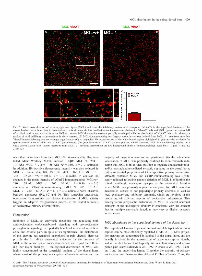
mice than in sections from their MGL+/+ littermates (Fig. 8A; two-tailed Mann–Whitney U-test, median � IQR: MGL+/+, 294 �104 AU; MGL�/�, 249 � 56 AU; *P = 0.03, n = 3–3 animals).In addition, IB4-positive fluorescence intensity was also reduced inMGL�/� tissue (Fig. 8B; MGL+/+, 429 � 166 AU; MGL�/�,332 � 102 AU; **P = 0.006, n = 3–3 animals). In contrast, nochanges in the mean intensity of vGluT2-immunostaining (MGL+/+,280 � 138 AU; MGL�/� 265 � 60 AU; P = 0.86, n = 3–3animals) or VIAAT-immunostaining (MGL+/+, 205 � 75 AU;MGL�/� 220 � 80 AU; P = 1, n = 3–3 animals) were observedbetween genotypes (Fig. 8C and D). This somewhat unexpectedobservation demonstrates that chronic inactivation of MGL activitytriggers an adaptive reorganization process in the central terminalsof nociceptive primary afferent fibers.
Discussion
Inhibition of MGL, an enzymatic metabolic hub regulating bothanti-nociceptive endocannabinoid signaling and pro-nociceptiveprostaglandin signaling, is reportedly beneficial in several models ofacute and chronic pain. In spite of its significance, the distributionof this enzyme has remained unexplored in pain circuits. Here weprovide the first direct anatomical evidence for the presence ofMGL in the mouse spinal nociceptive circuit, and report the follow-ing four major findings: (i) the regional distribution of MGL washighly concentrated in the superficial laminae of the dorsal horn,where most of the primary nociceptive afferents terminate and the
majority of projection neurons are positioned; (ii) the subcellularlocalization of MGL was primarily confined to axon terminals indi-cating that MGL is in an ideal position to regulate endocannabinoid-and/or prostaglandin-mediated synaptic signaling in the dorsal horn;(iii) a substantial proportion of CGRP-positive primary nociceptiveafferents contained MGL, and CGRP-immunostaining was signifi-cantly reduced following genetic deletion of MGL highlighting thespinal peptidergic nociceptor synapse as the anatomical locationwhere MGL may primarily regulate nociception; (iv) MGL was alsodetected in subsets of non-peptidergic primary afferents as well aslocal excitatory and inhibitory terminals, which are involved in theprocessing of different aspects of nociceptive information. Thisheterogeneous presynaptic distribution of MGL in several neuronalelements of the nociceptive circuitry is consistent with the conceptthat its multiple enzymatic functions may vary at distinct synapticlocalizations.
MGL abundance in the superficial laminae of the dorsal horn
The superficial laminae represent an anatomical hotspot where noci-ception can be most efficiently regulated (Todd, 2010). Most projec-tion neurons are concentrated in lamina I of the dorsal horn, and areheavily involved in the transmission of acute nociceptive signals,and in the development of hyperalgesia in inflammatory and neuro-pathic pain states (Mantyh et al., 1997; Nichols et al., 1999). Lam-ina I and the underlying lamina II receive the majority of primarynociceptive and thermoceptive Ad and C fiber afferents. Thus, the
A B
C D
Fig. 7. Weak colocalization of monoacylglycerol lipase (MGL) and vesicular inhibitory amino acid transporter (VIAAT) in the superficial laminae of themouse lumbar dorsal horn. (A) A deconvolved confocal image depicts double-immunofluorescence labeling for VIAAT (red) and MGL (green) in lamina I–IIof a spinal cord section derived from an MGL+/+ mouse. MGL-immunofluorescence partially overlapped with the distribution of VIAAT, which is primarily amarker of local inhibitory axon terminals in these laminae. (B) MGL-immunostaining was largely absent in sections derived from MGL�/� knockout mice, butVIAAT-immunolabeling was not changed significantly. (C) A magnified 3D reconstruction of the white boxed region highlighted in (A) provided evidence forsparse colocalization of MGL and VIAAT (arrowheads). (D) Quantification of VIAAT-positive profiles, which contained MGL-immunolabeling resulted in aweak colocalization ratio. Values measured from MGL�/� sections demonstrate the low background levels of immunostaining. Scale bars: 10 lm (A and B);2 lm (C).
© 2014 The Authors. European Journal of Neuroscience published by Federation of European Neuroscience Societies and John Wiley & Sons Ltd.European Journal of Neuroscience, 39, 419–434
MGL distribution in the spinal dorsal horn 429

striking density gradient of MGL-immunoreactivity observed in lam-ina I and IIo is entirely consistent with the notion that MGL plays aprominent regulatory role in physiological and pathophysiologicalforms of nociception (Guindon & Hohmann, 2009). Notably, topicalapplication of the selective MGL inhibitor JZL184 onto the dorsalhorn potently suppresses both acute and inflammatory nociceptiveactivity in spinal neurons, an effect mediated by the CB1 receptor(Woodhams et al., 2012). The involvement of CB1 receptors, themajor target of the endocannabinoid 2-AG in the nervous system(Katona & Freund, 2008), suggests a regulatory role for MGL innociception via termination of 2-AG signaling. In light of thesefunctional data, it is particularly striking that both DGL-a, the syn-thesizing enzyme of 2-AG, and the CB1 receptor share an exactlyoverlapping regional distribution with that of MGL in the superficialdorsal horn (Nyilas et al., 2009).
Presynaptic MGL at spinal synapses
In contrast to the overlapping regional distribution pattern, the syn-thesizing and degrading enzymes of 2-AG display complementary
synaptic distribution at the subcellular level. DGL-a was foundexclusively in the somatodendritic domain of spinal neurons, mostoften located perisynaptically at the postsynaptic edge of metabo-tropic glutamate receptor 5 (mGluR5)-containing excitatory synapses(Nyilas et al., 2009). Conversely, in the present study the majorityof MGL-immunoreactivity was found presynaptically in axon termi-nals throughout the superficial laminae. This presynaptic localizationof MGL at spinal synapses is consistent with recent reports fromother brain regions (Guly�as et al., 2004; Lud�anyi et al., 2011; Uchi-gashima et al., 2011; Tanimura et al., 2012), and suggests thatMGL plays a conserved role in the regulation of synaptic endocann-abinoid signaling throughout the CNS.Genetic inactivation of DGL-a eliminates several forms of endoc-
annabinoid-dependent synaptic plasticity in the brain, providingunequivocal evidence that 2-AG has a fundamental synaptic function(Gao et al., 2010; Tanimura et al., 2010; Yoshino et al., 2011).Genetic or pharmacological blockade of MGL in the brain has anopposite effect, promoting 2-AG-mediated synaptic signaling pro-cesses (Pan et al., 2009, 2011; Straiker et al., 2009). These forms ofsynaptic plasticity all require presynaptically located CB1 receptors(Kano et al., 2009), which are also frequently found on axon termi-nals in the dorsal horn of the spinal cord (Hegyi et al., 2009; Nyilaset al., 2009). This canonical molecular organization ideally supportsretrograde 2-AG signaling and seems to be a conserved feature ofmany synapses in the CNS, including the spinal cord (Katona &Freund, 2008).This retrograde signaling pathway certainly has significance in
various forms of nociception and nociceptive plasticity. Forexample, spinal 2-AG levels are increased in a non-opioid form ofstress-induced analgesia (Suplita et al., 2006). Stress-induced anti-nociception is facilitated by mGluR5 activation, and requires bothDGL-a and CB1 activity (Nyilas et al., 2009). Notably, direct spinalblockade of MGL has similar beneficial effects, which also requirespinal CB1 receptors (Suplita et al., 2006). Taken together, thesefindings support the view that presynaptic MGL is integrated intothe retrograde 2-AG signaling pathway at spinal synapses, therebyplaying an important regulatory role in nociception.
The spinal nociceptor synapse: a potential anatomical site forthe anti-nociceptive effects of MGL inhibition
Given the primarily anti-nociceptive effect of systemic and spinalMGL inhibition (Woodhams et al., 2012; Mulvihill & Nomura,2013), a major goal of the present study was to reveal the anatomi-cal location(s) where MGL inhibition may suppress pain transmis-sion in the dorsal horn nociceptive circuitry. Because the electronmicroscopic analysis revealed that MGL is mostly located in axonterminals, another set of experiments was dedicated to determinewhich of the major synapse types contain the highest density ofMGL.The highest colocalization ratio was observed in CGRP-positive
primary afferents. These terminals target several cellular elements inthe dorsal horn and, in parallel to their arborization in lamina II,they extensively innervate different types of lamina I projectionneurons giving rise to ascending pathways, for example to the lat-eral parabrachial nucleus (Polg�ar et al., 2010). These projectionneurons are characterized by high expression levels of somatoden-dritic neurokinin 1 receptors, and their activity is facilitated by bothsubstance P and CGRP released from a subset of primary afferents(Seybold, 2009). Thus, the presence of MGL in these nociceptiveafferents supports the notion that MGL inhibition may control noci-ception at these spinal nociceptor synapses. Most importantly, a
A B
C D
Fig. 8. Quantitative analysis of monoacylglycerol lipase (MGL)-immuno-staining and immunofluorescence labeling for neurochemical markers inMGL+/+ wild-type and MGL�/� knockout mice. (A and B) Genetic dele-tion of MGL results in a significantly reduced calcitonin gene-related peptide(CGRP)-immunostaining and isolectin B4 (IB4)-labeling. (C and D) In con-trast, vesicular glutamate transporter 2 (vGluT2)- or vesicular inhibitoryamino acid transporter (VIAAT)-immunolabeling remained unaltered in thesuperficial dorsal horn of MGL�/� mice. Mean intensity values for (A)CGRP, (B) IB4, (C) vGluT2 and (D) VIAAT in the medial superficial dorsalhorn of MGL+/+ vs. MGL�/� mice are presented. *P = 0.03, **P = 0.006,two-tailed Mann–Whitney U-test.
© 2014 The Authors. European Journal of Neuroscience published by Federation of European Neuroscience Societies and John Wiley & Sons Ltd.European Journal of Neuroscience, 39, 419–434
430 E. Horv�ath et al.

similar cell-type-specific knockout approach such as used for CB1
receptor inactivation (Marsicano et al., 2003; Agarwal et al., 2007;Pernia-Andrade et al., 2009) will be necessary to determine howMGL regulates endogenous analgesia at spinal nociceptor synapsesin vivo.Certainly, presynaptic CB1 receptors on these nociceptors may
also be critical for mediating anti-nociceptive 2-AG signaling. Inter-estingly, the ratio of CGRP-positive terminals containing MGL inthe present study was in the same range as that reported recently, interminals that colocalize substance P, CGRP and CB1 receptors(Kato et al., 2012). Furthermore, cell-type-specific deletion of CB1
receptors provided direct evidence that retrograde endocannabinoidsignaling mediates long-term depression of glutamatergic currents atthese very same primary afferent terminals (Kato et al., 2012),which have a surprisingly conserved evolutionary function at pri-mary nociceptor synapses (Yuan & Burrell, 2013). Besides phasicendocannabinoid-mediated long-term depression as a candidateendogenous analgesic mechanism (Kato et al., 2012), the involve-ment of tonic endocannabinoid signaling in the regulation of noci-ception may also be relevant. For example, two studies recentlydescribed downregulation of CB1 receptors following chronic inacti-vation of MGL (Chanda et al., 2010; Schlosburg et al., 2010).Because this perturbation led to a functional tolerance for CB anal-gesic effects, it will therefore be interesting to investigate in thefuture whether such changes are also present in the spinal nocicep-tive circuitry, especially on peptidergic primary afferents.Irrespective of the temporal dynamics of spinal endocannabinoid
signaling, attenuation of glutamate release from nociceptive primaryafferents is generally considered to suppress pain. Accordingly, acti-vation of presynaptic CB1 receptors on primary afferents reducesglutamate release from their terminals and consequently attenuatesnociception (Kelly & Chapman, 2001; Morisset & Urban, 2001).These and many other studies collectively suggest that the 2-AGsignaling pathway forms a tonic and/or phasic endogenous analgesiccircuit by activating presynaptic CB1 receptors at central nociceptorsynapses (Chapman, 1999; Walker & Hohmann, 2005). These CB1
receptors may therefore represent one target of beneficial action ofCB preparations currently licensed to attenuate pain in cancer andmultiple sclerosis (Pertwee, 2012). On the other hand, one alsoneeds to take into consideration that the neurobiological substratefor this effect is certainly more complex (Agarwal et al., 2007). Forexample, inhibition of MGL also contributes to distinct aspects ofanalgesia at supraspinal sites such as the periaqueductal gray matterand/or in the periphery (Hohmann et al., 2005; Spradley et al.,2010; Gregg et al., 2012; Guindon et al., 2013).
Heterogeneous distribution of MGL in various cellularelements of the nociceptive circuitry
The present finding that MGL, a regulator of at least two majorbrain signaling pathways (Mulvihill & Nomura, 2013), is locatedwithin at least four major synapse types supports the idea that thisserine hydrolase may indeed play distinct physiological functions atdifferent circuit locations. Moreover, the partial colocalization withneurochemical markers raises the possibility of further functionalsegregation of MGL into different subpopulations of primary affer-ents, intrinsic excitatory neurons or inhibitory terminals. For exam-ple, it will be crucial to study which of the four functionally distinctmajor inhibitory interneuron types in the dorsal horn contain presyn-aptic MGL (Polg�ar et al., 2013). Our initial observations identifiedsome MGL-positive inhibitory terminals forming axo-axonic syn-apses with Type II synaptic glomeruli, which may originate from
the parvalbumin-positive interneuron population recently implicatedin these connections (Hughes et al., 2012). Interestingly, theseaxo-axonic synapses may represent a location at which MGL inhibi-tion may facilitate tactile allodynia, rather than inducing a generalanalgesic effect. Moreover, postsynaptic target cell-specific quantita-tive differences in endocannabinoid-mediated plasticity are known inother brain regions (Peterfi et al., 2012). Thus, future experimentscould address whether the heterogeneity in endocannabinoid-medi-ated plasticity at spinal nociceptor synapses (Kato et al., 2012)results from spatial segregation of MGL and CB1 receptors into ter-minals innervating different postsynaptic target cell populations.A circuit-specific contribution of MGL to the regulation of prosta-
glandins may also be relevant given the complexity of the molecularcomponents of these pathways (Zeilhofer & Brune, 2006). Althoughpro-nociceptive prostaglandin E receptor 2 is located at specificGABAergic synapses in a cell-type-dependent manner (Ahmadiet al., 2002; Reinold et al., 2005), these receptors may also triggerglutamate release presynaptically (Sang et al., 2005). To furthercomplicate this picture, it is even possible for the same molecularpathway to play opposite roles in nociception, as exemplified by thereported pro-nociceptive role of CB1 receptor signaling at spinalGABAergic synapses (Pernia-Andrade et al., 2009), and the anti-nociceptive role of superficial spinal prostaglandin E receptor 3(Nakamura et al., 2000; Natura et al., 2013), which implies thatMGL inhibition may not even be anti-nociceptive at every circuitlocation.Finally, another indication for the temporally and spatially com-
plex functions of MGL in the spinal cord comes from the observa-tion that both CGRP-immunoreactivity and IB4-labeling aredownregulated in spinal cord sections from MGL�/� mice. Thisimplies that MGL may play an important developmental function inthe dorsal horn in accordance with earlier findings in other neuronalsystems (Keimpema et al., 2010). Activation of CB1 receptors dur-ing axonal path finding in vitro causes growth cone repulsion andcollapse (Berghuis et al., 2007), and hyperactivity of the 2-AG-syn-thesizing enzyme DGL-a results in spherical exclusion of growthcones and diminishes cell–cell contact in vitro (Keimpema et al.,2013). Deletion of MGL leads to a marked elevation in spinal cord2-AG levels (Chanda et al., 2010), and it is possible that this over-spill of 2-AG produces similar effects in vivo. The observed reduc-tion in CGRP-immunoreactivity and IB4-labeling may thus merelyrepresent a developmental defect due to a loss of precise temporaland spatial control of 2-AG signaling during the critical period ofprimary afferent axonal targeting.
Conclusion
The neuronal circuitry of spinal nociceptive processing displaysremarkable cellular and synaptic complexity (Todd, 2010). Contrib-uting signaling pathways, such as the endocannabinoid system,fulfill multiple physiological functions at different circuit locations(Katona & Freund, 2012). The present anatomical findings demon-strating commensurate heterogeneous MGL distribution in the spinaldorsal horn therefore highlight the need for specific pre- and post-synaptic cell-type-specific approaches to elucidate the functional sig-nificance of endocannabinoid and prostaglandin signaling indifferent pain circuit components.
Acknowledgements
This work was primarily supported by a grant from Switzerland through theSwiss Contribution (SH/7/2/18), by the European Research Council Grant
© 2014 The Authors. European Journal of Neuroscience published by Federation of European Neuroscience Societies and John Wiley & Sons Ltd.European Journal of Neuroscience, 39, 419–434
MGL distribution in the spinal dorsal horn 431

243153, by the Wellcome Trust International Senior Research Fellowship toI.K. (090946/Z/09/Z), and by the Momentum Program (LP2013-54/2013) ofthe Hungarian Academy of Sciences. We are grateful to Prof. Hanns UlrichZeilhofer and Carolin von Shoultz for discussions. We are also indebted toErika Tischler, Bal�azs Pint�er and Gy}oz}o Goda for their excellent technicalassistance, and to Barna Dudok for his advice on image processing and dataanalysis. We thank L�aszl�o Barna, the Nikon Microscopy Center at Instituteof Experimental Medicine, Nikon Austria GmbH, and Auro-Science Consult-ing, Ltd, for kindly providing microscopy support. The authors declare nocompeting financial interests.
Abbreviations
2-AG, 2-arachidonoylglycerol; AU, arbitrary units; CB1, cannabinoid recep-tor 1; CGRP, calcitonin gene-related peptide; COX, cyclooxygenase; DAB,3,3′-diaminobenzidine; DGL-a, diacylglycerol lipase-alpha; GABA, c-amin-obutyric acid; IB4, plant isolectin B4; IQR, interquartile range; JZL184, 4-ni-trophenyl-4-[bis(1,3-benzodioxol-5-yl)(hydroxy)methyl]piperidine-1-carboxyl-ate; MGL, monoacylglycerol lipase; mGluR5, metabotropic glutamatereceptor 5; PB, phosphate buffer; PFA, paraformaldehyde; TB, Tris buffer;TBS, Tris-buffered saline; vGluT2, vesicular glutamate transporter 2; VIA-AT, vesicular inhibitory amino acid transporter.
References
Agarwal, N., Pacher, P., Tegeder, I., Amaya, F., Constantin, C.E., Brenner,G.J., Rubino, T., Michalski, C.W., Marsicano, G., Monory, K., Mackie,K., Marian, C., Batkai, S., Parolaro, D., Fischer, M.J., Reeh, P., Kunos,G., Kress, M., Lutz, B., Woolf, C.J. & Kuner, R. (2007) Cannabinoidsmediate analgesia largely via peripheral type 1 cannabinoid receptors innociceptors. Nat. Neurosci., 10, 870–879.
Ahmadi, S., Lippross, S., Neuhuber, W.L. & Zeilhofer, H.U. (2002) PGE(2)selectively blocks inhibitory glycinergic neurotransmission onto rat superfi-cial dorsal horn neurons. Nat. Neurosci., 5, 34–40.
Antal, M., Petk�o, M., Polg�ar, E., Heizmann, C.W. & Storm-Mathisen, J.(1996) Direct evidence of an extensive GABAergic innervation of thespinal dorsal horn by fibres descending from the rostral ventromedialmedulla. Neuroscience, 73, 509–518.
Baer, K., B€urli, T., Huh, K.-H., Wiesner, A., Erb-V€ogtli, S., G€ockeritz-Dujmo-vic, D., Moransard, M., Nishimune, A., Rees, M.I., Henley, J.M., Fritschy,J.-M. & Fuhrer, C. (2007) PICK1 interacts with a7 neuronal nicotinic ace-tylcholine receptors and controls their clustering. Mol. Cell. Neurosci., 35,339–355.
Basbaum, A.I., Bautista, D.M., Scherrer, G. & Julius, D. (2009) Cellular andmolecular mechanisms of pain. Cell, 139, 267–284.
Berghuis, P., Rajnicek, A.M., Morozov, Y.M., Ross, R.A., Mulder, J.,Urb�an, G.M., Monory, K., Marsicano, G., Matteoli, M., Canty, A., Irving,A.J., Katona, I., Yanagawa, Y., Rakic, P., Lutz, B., Mackie, K. & Hark-any, T. (2007) Hardwiring the brain: endocannabinoids shape neuronalconnectivity. Science, 316, 1212–1216.
Blankman, J.L., Simon, G.M. & Cravatt, B.F. (2007) A comprehensive pro-file of brain enzymes that hydrolyze the endocannabinoid 2-arachidonoyl-glycerol. Chem. Biol., 14, 1347–1356.
Bogen, I.L., Boulland, J.-L., Mariussen, E., Wright, M.S., Fonnum, F., Kao,H.-T. & Walaas, S.I. (2006) Absence of synapsin I and II is accompaniedby decreases in vesicular transport of specific neurotransmitters. J. Neuro-chem., 96, 1458–1466.
Busquets-Garcia, A., Puighermanal, E., Pastor, A., de la Torre, R., Maldonado,R. & Ozaita, A. (2011) Differential role of anandamide and 2-arachidonoyl-glycerol in memory and anxiety-like responses. Biol. Psychiat., 70, 479–486.
Chanda, P.K., Gao, Y., Mark, L., Btesh, J., Strassle, B.W., Lu, P., Piesla,M.J., Zhang, M.-Y., Bingham, B., Uveges, A., Kowal, D., Garbe, D., Ko-uranova, E.V., Ring, R.H., Bates, B., Pangalos, M.N., Kennedy, J.D.,Whiteside, G.T. & Samad, T.A. (2010) Monoacylglycerol lipase activity isa critical modulator of the tone and integrity of the endocannabinoid sys-tem. Mol. Pharmacol., 78, 996–1003.
Chapman, V. (1999) The cannabinoid CB1 receptor antagonist, SR141716A,selectively facilitates nociceptive responses of dorsal horn neurones in therat. Brit. J. Pharmacol., 127, 1765–1767.
Dinh, T.P., Carpenter, D., Leslie, F.M., Freund, T.F., Katona, I., Sensi, S.L.,Kathuria, S. & Piomelli, D. (2002) Brain monoglyceride lipase participat-ing in endocannabinoid inactivation. Proc. Natl. Acad. Sci. USA, 99,10819–10824.
Dumoulin, A., Rostaing, P., Bedet, C., Levi, S., Isambert, M.F., Henry, J.P.,Triller, A. & Gasnier, B. (1999) Presence of the vesicular inhibitory aminoacid transporter in GABAergic and glycinergic synaptic terminal boutons.J. Cell Sci., 112, 811–823.
Fan, K.Y., Baufreton, J., Surmeier, D.J., Chan, C.S. & Bevan, M.D. (2012)Proliferation of external globus pallidus-subthalamic nucleus synapses fol-lowing degeneration of midbrain dopamine neurons. J. Neurosci., 32,13718–13728.
Franco-Cereceda, A., Henke, H., Lundberg, J.M., Petermann, J.B., H€okfelt,T. & Fischer, J.A. (1987) Calcitonin gene-related peptide (CGRP) in cap-saicin-sensitive substance P-immunoreactive sensory neurons in animalsand man: distribution and release by capsaicin. Peptides, 8, 399–410.
Gao, Y., Vasilyev, D.V., Goncalves, M.B., Howell, F.V., Hobbs, C., Reisen-berg, M., Shen, R., Zhang, M.-Y., Strassle, B.W., Lu, P., Mark, L., Piesla,M.J., Deng, K., Kouranova, E.V., Ring, R.H., Whiteside, G.T., Bates, B.,Walsh, F.S., Williams, G., Pangalos, M.N., Samad, T.A. & Doherty, P.(2010) Loss of retrograde endocannabinoid signaling and reduced adultneurogenesis in diacylglycerol lipase knock-out mice. J. Neurosci., 30,2017–2024.
Ghosh, S., Wise, L.E., Chen, Y., Gujjar, R., Mahadevan, A., Cravatt, B.F. &Lichtman, A.H. (2013) The monoacylglycerol lipase inhibitor JZL184 sup-presses inflammatory pain in the mouse carrageenan model. Life Sci., 92,498–505.
Gregg, L.C., Jung, K.M., Spradley, J.M., Nyilas, R., Suplita, R.L. II, Zim-mer, A., Watanabe, M., Mackie, K., Katona, I., Piomelli, D. & Hohmann,A.G. (2012) Activation of type 5 metabotropic glutamate receptors anddiacylglycerol lipase-a initiates 2-arachidonoylglycerol formation andendocannabinoid-mediated analgesia. J. Neurosci., 32, 9457–9468.
Guindon, G. & Hohmann, A. (2009) The endocannabinoid system and pain.CNS Neurol. Disord.-Dr., 8, 18.
Guindon, J., Lai, Y., Takacs, S.M., Bradshaw, H.B. & Hohmann, A.G.(2013) Alterations in endocannabinoid tone following chemotherapy-induced peripheral neuropathy: effects of endocannabinoid deactivationinhibitors targeting fatty-acid amide hydrolase and monoacylglycerol lipasein comparison to reference analgesics following cisplatin treatment. Phar-macol. Res., 67, 94–109.
Guly�as, A.I., Cravatt, B.F., Bracey, M.H., Dinh, T.P., Piomelli, D., Boscia,F. & Freund, T.F. (2004) Segregation of two endocannabinoid-hydrolyzingenzymes into pre- and postsynaptic compartments in the rat hippocampus,cerebellum and amygdala. Eur. J. Neurosci., 20, 441–458.
Hanson, J.E., Deng, L., Hackos, D.H., Lo, S.-C., Lauffer, B.E., Steiner, P. &Zhou, Q. (2013) Histone deacetylase 2 cell autonomously suppresses excit-atory and enhances inhibitory synaptic function in CA1 pyramidal neurons.J. Neurosci., 33, 5924–5929.
Hegyi, Z., Kis, G., Holl�o, K., Ledent, C. & Antal, M. (2009) Neuronal andglial localization of the cannabinoid-1 receptor in the superficial spinaldorsal horn of the rodent spinal cord. Eur. J. Neurosci., 30, 251–262.
Hohmann, A.G., Suplita, R.L., Bolton, N.M., Neely, M.H., Fegley, D.,Mangieri, R., Krey, J.F., Walker, J.M., Holmes, P.V., Crystal, J.D., Duran-ti, A., Tontini, A., Mor, M., Tarzia, G. & Piomelli, D. (2005) Anendocannabinoid mechanism for stress-induced analgesia. Nature, 435,1108–1112.
Hughes, D.I., Sikander, S., Kinnon, C.M., Boyle, K.A., Watanabe, M., Call-ister, R.J. & Graham, B.A. (2012) Morphological, neurochemical and elec-trophysiological features of parvalbumin-expressing cells: a likely sourceof axo-axonic inputs in the mouse spinal dorsal horn. J. Physiol., 590,3927–3951.
Jhaveri, M.D., Richardson, D. & Chapman, V. (2007) Endocannabinoidmetabolism and uptake: novel targets for neuropathic and inflammatorypain. Brit. J. Pharmacol., 152, 624–632.
Ju, G., Hokfelt, T., Brodin, E., Fahrenkrug, J., Fischer, J.A., Frey, P., Elde,R.P. & Brown, J.C. (1987) Primary sensory neurons of the rat showing calci-tonin gene-related peptide immunoreactivity and their relation to substanceP-, somatostatin-, galanin-, vasoactive intestinal polypeptide- and cholecys-tokinin-immunoreactive ganglion cells. Cell Tissue Res., 247, 417–431.
Kano, M., Ohno-Shosaku, T., Hashimotodani, Y., Uchigashima, M. &Watanabe, M. (2009) Endocannabinoid-mediated control of synaptic trans-mission. Physiol. Rev., 89, 309–380.
Karlsson, M., Contreras, J.A., Hellman, U., Tornqvist, H. & Holm, C. (1997)cDNA cloning, tissue distribution, and identification of the catalytic triadof monoglyceride lipase. J. Biol. Chem., 272, 27218–27223.
Kato, A., Punnakkal, P., Pernia-Andrade, A.J., von Schoultz, C., Sharopov,S., Nyilas, R., Katona, I. & Zeilhofer, H.U. (2012) Endocannabinoid-dependent plasticity at spinal nociceptor synapses. J. Physiol., 590, 4717–4733.
© 2014 The Authors. European Journal of Neuroscience published by Federation of European Neuroscience Societies and John Wiley & Sons Ltd.European Journal of Neuroscience, 39, 419–434
432 E. Horv�ath et al.

Katona, I. & Freund, T.F. (2008) Endocannabinoid signaling as a synapticcircuit breaker in neurological disease. Nat. Med., 14, 923–930.
Katona, I. & Freund, T. (2012) Multiple functions of endocannabinoid sig-naling in the brain. Annu. Rev. Neurosci., 35, 529–558.
Keimpema, E., Barabas, K., Morozov, Y.M., Tortoriello, G., Torii, M., Cam-eron, G., Yanagawa, Y., Watanabe, M., Mackie, K. & Harkany, T. (2010)Differential subcellular recruitment of monoacylglycerol lipase generatesspatial specificity of 2-arachidonoyl glycerol signaling during axonal path-finding. J. Neurosci., 30, 13992–14007.
Keimpema, E., Alpar, A., Howell, F., Malenczyk, K., Hobbs, C., Hurd, Y.L.,Watanabe, M., Sakimura, K., Kano, M., Doherty, P. & Harkany, T. (2013)Diacylglycerol lipase alpha manipulation reveals developmental roles forintercellular endocannabinoid signaling. Sci. Rep., 3, 2093.
Kelly, S. & Chapman, V. (2001) Selective cannabinoid CB1 receptor activa-tion inhibits spinal nociceptive transmission in vivo. J. Neurophysiol., 86,3061–3064.
Kinsey, S.G., Long, J.Z., O’Neal, S.T., Abdullah, R.A., Poklis, J.L., Boger,D.L., Cravatt, B.F. & Lichtman, A.H. (2009) Blockade of endocannabi-noid-degrading enzymes attenuates neuropathic pain. J. Pharmacol. Exp.Ther., 330, 902–910.
Kinsey, S.G., Long, J.Z., Cravatt, B.F. & Lichtman, A.H. (2010) Fatty acidamide hydrolase and monoacylglycerol lipase inhibitors produce anti-allo-dynic effects in mice through distinct cannabinoid receptor mechanisms.J. Pain, 11, 1420–1428.
Kuner, R. (2010) Central mechanisms of pathological pain. Nat. Med., 16,1258–1266.
Long, J.Z., Li, W.W., Booker, L., Burston, J.J., Kinsey, S.G., Schlosburg,J.E., Pavon, F.J., Serrano, A.M., Selley, D.E., Parsons, L.H., Lichtman,A.H. & Cravatt, B.F. (2009) Selective blockade of 2-arachidonoylglycerolhydrolysis produces cannabinoid behavioral effects. Nat. Chem. Biol., 5,37–44.
Lud�anyi, A., Hu, S.S.J., Yamazaki, M., Tanimura, A., Piomelli, D., Watana-be, M., Kano, M., Sakimura, K., Magl�oczky, Z., Mackie, K., Freund, T.F.& Katona, I. (2011) Complementary synaptic distribution of enzymesresponsible for synthesis and inactivation of the endocannabinoid 2-arachi-donoylglycerol in the human hippocampus. Neuroscience, 174, 50–63.
Mantyh, P.W., Rogers, S.D., Honore, P., Allen, B.J., Ghilardi, J.R., Li, J.,Daughters, R.S., Lappi, D.A., Wiley, R.G. & Simone, D.A. (1997) Inhibi-tion of hyperalgesia by ablation of lamina I spinal neurons expressing thesubstance P receptor. Science, 278, 275–279.
Marsicano, G., Goodenough, S., Monory, K., Hermann, H., Eder, M.,Cannich, A., Azad, S.C., Cascio, M.G., Gutierrez, S.O., van der Stelt, M.,Lopez-Rodriguez, M.L., Casanova, E., Sch€utz, G., Zieglgansberger, W.,Di Marzo, V., Behl., C. & Lutz, B. (2003) CB1 cannabinoid receptors andon-demand defense against excitotoxicity. Science, 302, 84–88.
Micheva, K.D., Busse, B., Weiler, N.C., O’Rourke, N. & Smith, S.J. (2010)Single-synapse analysis of a diverse synapse population: proteomic imag-ing methods and markers. Neuron, 68, 639–653.
Miura, E., Fukaya, M., Sato, T., Sugihara, K., Asano, M., Yoshioka, K. &Watanabe, M. (2006) Expression and distribution of JNK/SAPK-associatedscaffold protein JSAP1 in developing and adult mouse brain. J. Neuro-chem., 97, 1431–1446.
Miyazaki, T., Fukaya, M., Shimizu, H. & Watanabe, M. (2003) Subtypeswitching of vesicular glutamate transporters at parallel fibre–Purkinje cellsynapses in developing mouse cerebellum. Eur. J. Neurosci., 17, 2563–2572.
Monory, K., Blaudzun, H., Massa, F., Kaiser, N., Lemberger, T., Sch€utz, G.,Wotjak, C.T., Lutz, B. & Marsicano, G. (2007) Genetic dissection ofbehavioural and autonomic effects of D9Tetrahydrocannabinol in mice.PLoS Biol., 5, e269.
Morisset, V. & Urban, L. (2001) Cannabinoid-induced presynaptic inhibitionof glutamatergic EPSCs in substantia gelatinosa neurons of the rat spinalcord. J. Neurophysiol., 86, 40–48.
Mulvihill, M.M. & Nomura, D.K. (2013) Therapeutic potential of monoacyl-glycerol lipase inhibitors. Life Sci., 92, 492–497.
Nakamura, K., Kaneko, T., Yamashita, Y., Hasegawa, H., Katoh, H. & Negi-shi, M. (2000) Immunohistochemical localization of prostaglandin EP3receptor in the rat nervous system. J. Comp. Neurol., 421, 543–569.
Natura, G., Bar, K.J., Eitner, A., Boettger, M.K., Richter, F., Hensellek, S.,Ebersberger, A., Leuchtweis, J., Maruyama, T., Hofmann, G.O., Halbhu-ber, K.J. & Schaible, H.G. (2013) Neuronal prostaglandin E2 receptor sub-type EP3 mediates antinociception during inflammation. Proc. Natl. Acad.Sci. USA, 110, 13648–13653.
Nichols, M.L., Allen, B.J., Rogers, S.D., Ghilardi, J.R., Honore, P., Luger,N.M., Finke, M.P., Li, J., Lappi, D.A., Simone, D.A. & Mantyh, P.W.
(1999) Transmission of chronic nociception by spinal neurons expressingthe substance P receptor. Science, 286, 1558–1561.
Nomura, D.K., Hudak, C.S.S., Ward, A.M., Burston, J.J., Issa, R.S., Fisher,K.J., Abood, M.E., Wiley, J.L., Lichtman, A.H. & Casida, J.E. (2008)Monoacylglycerol lipase regulates 2-arachidonoylglycerol action and ara-chidonic acid levels. Bioorg. Med. Chem. Lett., 18, 5875–5878.
Nomura, D.K., Morrison, B.E., Blankman, J.L., Long, J.Z., Kinsey, S.G.,Marcondes, M.C., Ward, A.M., Hahn, Y.K., Lichtman, A.H., Conti, B. &Cravatt, B.F. (2011) Endocannabinoid hydrolysis generates brain prosta-glandins that promote neuroinflammation. Science, 334, 809–813.
Nyilas, R., Gregg, L.C., Mackie, K., Watanabe, M., Zimmer, A., Hohmann,A.G. & Katona, I. (2009) Molecular architecture of endocannabinoid sig-naling at nociceptive synapses mediating analgesia. Eur. J. Neurosci., 29,1964–1978.
Pan, B., Wang, W., Long, J.Z., Sun, D., Hillard, C.J., Cravatt, B.F. & Liu,Q.-S. (2009) Blockade of 2-arachidonoylglycerol hydrolysis by selectivemonoacylglycerol lipase inhibitor 4-nitrophenyl 4-(Dibenzo[d][1,3]dioxol-5-yl(hydroxy)methyl)piperidine-1-carboxylate (JZL184) enhances retro-grade endocannabinoid signaling. J. Pharmacol. Exp. Ther., 331, 591–597.
Pan, B., Wang, W., Zhong, P., Blankman, J.L., Cravatt, B.F. & Liu, Q.-S.(2011) Alterations of endocannabinoid signaling, synaptic plasticity, learn-ing, and memory in monoacylglycerol lipase knock-out mice. J. Neurosci.,31, 13420–13430.
Pernia-Andrade, A.J., Kato, A., Witschi, R., Nyilas, R., Katona, I., Freund,T.F., Watanabe, M., Filitz, J., Koppert, W., Schuttler, J., Ji, G., Neu-gebauer, V., Marsicano, G., Lutz, B., Vanegas, H. & Zeilhofer, H.U.(2009) Spinal endocannabinoids and CB1 receptors mediate C-fiber-induced heterosynaptic pain sensitization. Science, 325, 760–764.
Pertwee, R.G. (2012) Targeting the endocannabinoid system with cannabi-noid receptor agonists: pharmacological strategies and therapeutic possibil-ities. Philos. T. Roy. Soc. B., 367, 3353–3363.
Peterfi, Z., Urban, G.M., Papp, O.I., Nemeth, B., Monyer, H., Szabo, G.,Erdelyi, F., Mackie, K., Freund, T.F., Hajos, N. & Katona, I. (2012) En-docannabinoid-mediated long-term depression of afferent excitatory syn-apses in hippocampal pyramidal cells and GABAergic interneurons.J. Neurosci., 32, 14448–14463.
Polg�ar, E., Al Ghamdi, K.S. & Todd, A.J. (2010) Two populations of neu-rokinin 1 receptor-expressing projection neurons in lamina I of the ratspinal cord that differ in AMPA receptor subunit composition and densityof excitatory synaptic input. Neuroscience, 167, 1192–1204.
Polg�ar, E., Sardella, T.C.P., Tiong, S.Y.X., Locke, S., Watanabe, M. &Todd, A.J. (2013) Functional differences between neurochemically-definedpopulations of inhibitory interneurons in the rat spinal dorsal horn. Pain,154, 2606–2615.
Reinold, H., Ahmadi, S., Depner, U.B., Layh, B., Heindl, C., Hamza, M.,Pahl, A., Brune, K., Narumiya, S., Muller, U. & Zeilhofer, H.U. (2005)Spinal inflammatory hyperalgesia is mediated by prostaglandin E receptorsof the EP2 subtype. J. Clin. Invest., 115, 673–679.
Rosenfeld, M.G., Mermod, J.J., Amara, S.G., Swanson, L.W., Sawchenko,P.E., Rivier, J., Vale, W.W. & Evans, R.M. (1983) Production of a novelneuropeptide encoded by the calcitonin gene via tissue-specific RNA pro-cessing. Nature, 304, 129–135.
Sagar, D.R., Burston, J.J., Woodhams, S.G. & Chapman, V. (2012) Dynamicchanges to the endocannabinoid system in models of chronic pain. Philos.T. Roy. Soc. B., 367, 3300–3311.
Samad, T.A., Moore, K.A., Sapirstein, A., Billet, S., Allchorne, A., Poole,S., Bonventre, J.V. & Woolf, C.J. (2001) Interleukin-1beta-mediatedinduction of Cox-2 in the CNS contributes to inflammatory pain hypersen-sitivity. Nature, 410, 471–475.
Sandk€uhler, J. (2009) Models and mechanisms of hyperalgesia and allodynia.Physiol. Rev., 89, 707–758.
Sang, N., Zhang, J., Marcheselli, V., Bazan, N.G. & Chen, C. (2005) Post-synaptically synthesized prostaglandin E2 (PGE2) modulates hippocampalsynaptic transmission via a presynaptic PGE2 EP2 receptor. J. Neurosci.,25, 9858–9870.
Schlosburg, J.E., Blankman, J.L., Long, J.Z., Nomura, D.K., Pan, B., Kinsey,S.G., Nguyen, P.T., Ramesh, D., Booker, L., Burston, J.J., Thomas, E.A.,Selley, D.E., Sim-Selley, L.J., Liu, Q.S., Lichtman, A.H. & Cravatt, B.F.(2010) Chronic monoacylglycerol lipase blockade causes functional antag-onism of the endocannabinoid system. Nat. Neurosci., 13, 1113–1119.
Seybold, V.S. (2009) The role of peptides in central sensitization. In Can-ning, B.J. & Spina, D. (Eds), Sensory Nerves. Springer, Berlin, Heidel-berg, pp. 451–491.
Silverman, J.D. & Kruger, L. (1988) Lectin and neuropeptide labeling ofseparate populations of dorsal root ganglion neurons and associated
© 2014 The Authors. European Journal of Neuroscience published by Federation of European Neuroscience Societies and John Wiley & Sons Ltd.European Journal of Neuroscience, 39, 419–434
MGL distribution in the spinal dorsal horn 433
“nociceptor” thin axons in rat testis and cornea whole-mount preparations.Somatosens. Res., 5, 259–267.
Simonetti, M., Hagenston, A.M., Vardeh, D., Freitag, H.E., Mauceri, D., Lu,J., Satagopam, V.P., Schneider, R., Costigan, M., Bading, H. & Kuner, R.(2013) Nuclear calcium signaling in spinal neurons drives a genomic pro-gram required for persistent inflammatory pain. Neuron, 77, 43–57.
Snider, W.D. & McMahon, S.B. (1998) Tackling pain at the source: newideas about nociceptors. Neuron, 20, 629–632.
Spradley, J.M., Guindon, J. & Hohmann, A.G. (2010) Inhibitors of monoacly-glycerol lipase, fatty-acid amide hydrolase and endocannabinoid transportdifferentially suppress capsaicin-induced behavioral sensitization throughperipheral endocannabinoid mechanisms. Pharmacol. Res., 62, 249–258.
Straiker, A., Hu, S.S.-J., Long, J.Z., Arnold, A., Wager-Miller, J., Cravatt,B.F. & Mackie, K. (2009) Monoacylglycerol lipase limits the duration ofendocannabinoid-mediated depolarization-induced suppression of excitationin autaptic hippocampal neurons. Mol. Pharmacol., 76, 1220–1227.
Suplita, R.L., Gutierrez, T., Fegley, D., Piomelli, D. & Hohmann, A.G.(2006) Endocannabinoids at the spinal level regulate, but do not mediate,nonopioid stress-induced analgesia. Neuropharmacology, 50, 372–379.
Tafoya, L.C.R., Mameli, M., Miyashita, T., Guzowski, J.F., Valenzuela, C.F.& Wilson, M.C. (2006) Expression and function of SNAP-25 as a universalSNARE component in GABAergic neurons. J. Neurosci., 26, 7826–7838.
Tanimura, A., Yamazaki, M., Hashimotodani, Y., Uchigashima, M., Kawata,S., Abe, M., Kita, Y., Hashimoto, K., Shimizu, T., Watanabe, M., Sakim-ura, K. & Kano, M. (2010) The endocannabinoid 2-arachidonoylglycerolproduced by diacylglycerol lipase a mediates retrograde suppression ofsynaptic transmission. Neuron, 65, 320–327.
Tanimura, A., Uchigashima, M., Yamazaki, M., Uesaka, N., Mikuni, T.,Abe, M., Hashimoto, K., Watanabe, M., Sakimura, K. & Kano, M. (2012)Synapse type-independent degradation of the endocannabinoid 2-arachido-noylglycerol after retrograde synaptic suppression. Proc. Natl. Acad. Sci.USA, 109, 12195–12200.
Todd, A.J. (2010) Neuronal circuitry for pain processing in the dorsal horn.Nat. Rev. Neurosci., 11, 823–836.
Todd, A.J., Hughes, D.I., Polg�ar, E., Nagy, G.G., Mackie, M., Ottersen, O.P.& Maxwell, D.J. (2003) The expression of vesicular glutamate transportersVGLUT1 and VGLUT2 in neurochemically defined axonal populations inthe rat spinal cord with emphasis on the dorsal horn. Eur. J. Neurosci.,17, 13–27.
Uchigashima, M., Yamazaki, M., Yamasaki, M., Tanimura, A., Sakimura,K., Kano, M. & Watanabe, M. (2011) Molecular and morphological con-figuration for 2-arachidonoylglycerol-mediated retrograde signaling atmossy cell & granule cell synapses in the dentate gyrus. J. Neurosci., 31,7700–7714.
Vardeh, D., Wang, D., Costigan, M., Lazarus, M., Saper, C.B., Woolf, C.J.,Fitzgerald, G.A. & Samad, T.A. (2009) COX2 in CNS neural cells medi-ates mechanical inflammatory pain hypersensitivity in mice. J. Clin.Invest., 119, 287–294.
Walker, J.M. & Hohmann, A.G. (2005) Cannabinoid mechanisms of painsuppression. Handb. Exp. Pharmacol., 168, 509–554.
Wall�en-Mackenzie, �A., Gezelius, H., Thoby-Brisson, M., Nyg�ard, A., Enjin,A., Fujiyama, F., Fortin, G. & Kullander, K. (2006) Vesicular glutamatetransporter 2 is required for central respiratory rhythm generation but notfor locomotor central pattern generation. J. Neurosci., 26, 12294–12307.
Wiesenfeld-Hallin, Z., H€okfelt, T., Lundberg, J.M., Forssmann, W.G., Rei-necke, M., Tschopp, F.A. & Fischer, J.A. (1984) Immunoreactive calcito-nin gene-related peptide and substance P coexist in sensory neurons to thespinal cord and interact in spinal behavioral responses of the rat. Neurosci.Lett., 52, 199–204.
Wilkerson, J.L., Gentry, K.R., Dengler, E.C., Wallace, J.A., Kerwin, A.A.,Armijo, L.M., Kuhn, M.N., Thakur, G.A., Makriyannis, A. & Milligan,E.D. (2012) Intrathecal cannabilactone CB2R agonist, AM1710, controlspathological pain and restores basal cytokine levels. Pain, 153, 1091–1106.
Wojcik, S.M., Katsurabayashi, S., Guillemin, I., Friauf, E., Rosenmund, C.,Brose, N. & Rhee, J.-S. (2006) A shared vesicular carrier allows synapticcorelease of GABA and glycine. Neuron, 50, 575–587.
Woodhams, S.G., Wong, A., Barrett, D.A., Bennett, A.J., Chapman, V. &Alexander, S.P.H. (2012) Spinal administration of the monoacylglycerollipase inhibitor JZL184 produces robust inhibitory effects on nociceptiveprocessing and the development of central sensitization in the rat. Brit. J.Pharmacol., 167, 1609–1619.
Wrobel, L., Schorscher-Petcu, A., Dupr�e, A., Yoshida, M., Nishimori, K. &Tribollet, E. (2011) Distribution and identity of neurons expressing theoxytocin receptor in the mouse spinal cord. Neurosci. Lett., 495, 49–54.
Yasaka, T., Tiong, S.Y.X., Hughes, D.I., Riddell, J.S. & Todd, A.J. (2010)Populations of inhibitory and excitatory interneurons in lamina II of theadult rat spinal dorsal horn revealed by a combined electrophysiologicaland anatomical approach. Pain, 151, 475–488.
Yoshino, H., Miyamae, T., Hansen, G., Zambrowicz, B., Flynn, M., Pedi-cord, D., Blat, Y., Westphal, R.S., Zaczek, R., Lewis, D.A. & Gonzalez-Burgos, G. (2011) Postsynaptic diacylglycerol lipase mediates retrogradeendocannabinoid suppression of inhibition in mouse prefrontal cortex.J. Physiol., 589, 4857–4884.
Yuan, S. & Burrell, B.D. (2013) Non-nociceptive afferent activity depressesnocifensive behavior and nociceptive synapses via an endocannabinoid-dependent mechanism. J. Neurophysiol., 110, 2607–2616.
Zeilhofer, H.U. (2007) Prostanoids in nociception and pain. Biochem. Phar-macol., 73, 165–174.
Zeilhofer, H.U. & Brune, K. (2006) Analgesic strategies beyond the inhibi-tion of cyclooxygenases. Trends Pharmacol. Sci., 27, 467–474.
Zhang, L., Hoff, A.O., Wimalawansa, S.J., Cote, G.J., Gagel, R.F. & Westl-und, K.N. (2001) Arthritic calcitonin/alpha calcitonin gene-related peptideknockout mice have reduced nociceptive hypersensitivity. Pain, 89, 265–273.
Zhao, J., Lee, M.-C., Momin, A., Cendan, C.-M., Shepherd, S.T., Baker,M.D., Asante, C., Bee, L., Bethry, A., Perkins, J.R., Nassar, M.A., Abra-hamsen, B., Dickenson, A., Cobb, B.S., Merkenschlager, M. & Wood,J.N. (2010) Small RNAs control sodium channel expression, nociceptorexcitability, and pain thresholds. J. Neurosci., 30, 10860–10871.
© 2014 The Authors. European Journal of Neuroscience published by Federation of European Neuroscience Societies and John Wiley & Sons Ltd.European Journal of Neuroscience, 39, 419–434
434 E. Horv�ath et al.