heterophyid (trematoda) parasites of cats in … · heterophyid (trematoda) parasites of cats in...
TRANSCRIPT

HETEROPHYID (TREMATODA) PARASITES OF CATS IN NORTH THAILAND WITH NOTES ON A HUMAN
CASE FOUND AT NECROPSY
MICHAEL KUKS and TAVIPAN TANTACHAMRDN
Department of Parasitology and Department of Pathology Faculty of Medicine Chiang Mai University Chiang Mai Thailand
INTRODUCTION
Due to their tolerence of a broad range of hosts heterophyid flukes not uncommonly are able to develop to maturity in man Little is known of the life histories of most heteroshyphyids in their snail hosts Most undergo the metacercarial stage in marine and fresh-water fish which are ingested by the definitive hosts a variety of birds and mammals (Yamaguti 1958 Pearson 1964) Human infection can occur wherever fish are eaten raw or partially cooked In Thailand Manning et al (1971) reported finding Haplorchis yokogawai and H taichui adults in several human autopsies in Northeast Thailand The intermediate hosts were not determined There are no other published reports of human heterophyid infections in mainland Southeast Asia alshythough both of the above parasites and Heterophyes heterophyes have been reported in dogs and the latter in cats also (Segal et a 1968) A number of human cases involving both intestinal infections and ectopic deposhysition of heterophyid eggs (and rarely adults) in the spinal cord and heart have been reshyported from the Philippines (Africa and Garcia 1935 Africa et al 1935 a b 1936 1937 Africa 1937) Heterophyes heterophyes Metagonimus yokogawai Haplorchis yokogashywai and H pumilio are known to be endemic in certain areas of Asia and at least six other heterophyids occasionally have been reshycovered from man (Faust et al 1971)
Stellanthchasmus falcatus adults have been recovered from cats dogs mice birds and
man in the Asian Pacific region the Middle East and Australia (Noda 1959 Alicata 1964 Pearson 1964) and were first described from man by Africa and Garcia (1935) in the Philippines and later by Alicata and Schatshyten burg (1938) in Hawaii Ching (1961) examined stools of 1380 persons in Hawaii and found 76 of Filipinos and native Hashywaiians to be infected with S falcatus As the ova of heterophyid flukes superficially resemshyble those of Opisthorchis and ClonorchiS many heterophyid infections have been asshysigned erroneously to the common liver flukes Despite numerous stool surveys S falcatus has not been previously detected in Thailand in man or animals The present paper reports the finding of S falcatus Haplorchis yokogawai and H taichui infection in fish and cats in Northern Thailand and discusses a possible human infection with the latter fluke discovered at necropsy in Chiang Mai
MATERIALS AND METHODS
During extensive helminth surveys in North Thailand (Ratanasritong and Kliks 1972) a number of different metacercariae were recovered from the flesh of local fish Cysts removed from the fins of Dermogenys pusillus were fed to one previously uninfected cat laboratory rats mice and chickens Eggs of a fluke appeared in the faeces of the cat after 13 days and numerous adults were recovered from the ileum at autopsy after 40 days No other flukes were present in the cats and none of the other animals exposed were
Vol 5 No4 December 1974 547
SOUTHEAST ASIAN J TROP MED PUB HLTH
found infected Adult specimens were sent to Je Pearson of the University of Queensshyland who confirmed the diagnosis of Stelshylanthchasmus falcatus More recently a domestic cat was found to be naturally infected with both Haplorchis yokogawai and H taichui Metacercariae of these flukes have been frequently observed in Puntius orphoides P leicanthus and P gonionotus all of which are consumed raw or marinaded by the local population
Surveys of local snails have uncovered two very similar ocelate parapleurolophocercous cercariae resembling those of Opisthorchis These were shed in the morning by 65 of 567 Melanoides tuberculata (115) collected from the same areas as the infected fish Upon exposure in glass finger bowls the cercariae were observed to penetrate readily and encyst in the fins and flesh of either Punt ius species producing H taichui and H yokogawai cysts and in Dermogenys resulting in S falcatus A definitive descripshytion of S falcatus adults was published by Pearson (1964) Vazquez-Golet and Africa (1940) described the metacercariae and Martin (1958) and Noda (1959) described the cercariae Hsu (1951) made studies on larval stages of heterophyids Likewise descriptions of the adults of Haplorchis taichui and H yokogawai have been made by Manning et al (1971) and Pearson (1964)
While examining necropsy sections of ileum from a patient who died suddenly of heart failure following routine appendectomy secshytions of an adult trematode and numerous eggs were noted The patient did not harbor Opisthorchis infection in the liver Further sections from the same block revealed no additional parasite material Autopsy permission was not given and other organs were not available for study A segment of ileum had been removed at necropsy
DESCRIPTION OF THE PARASITES
Stellanthchasmus falcatus (Figs 1 2)
All measurements are given in microns unless otherwise stated
Adult (8 specimens fixed in 10 formalin stained in carmine mounted in permount) Small pyriform body length 313 to 468 (381) forebody narrower than hindshybody maximum width 126 to 162 (150) across testes (Fig 1) Cuticle covered with scaleshylike spines 32 to 40 long curved posteshyriad and diminishing in size and number
---Gtl ---VgS--Ex --0 --Sv --c
Fig I-Diagramatic sketch of Stellanthchasmus Jafcatus adult dorsal view showing spinashytion pattern genital apparatus and major organs Uterus eggs and vitellarica omitted T testes 0 ovary SV seminal vesicle VgS ventrogenital sac Gtl gonotyl Ex expulsor C caeca (Actual size 381 by 150 microns average)
Vol 5 No4 December 1974 548
HETEROPHYDIASIS IN CATS AND MAN
in posterior fourth Oral sucker subterminal 25 to 36 (34) deep by 29 to 40 (33) wide prepharynx 10 to 16 (12) long pharynx 18 to 28 (23) long by 18 to 29 (24) wide oesophagus 90 to 144 (114) long bifurcation of caeca at midbody caeca extending to near posterior edge of testis Testes large ovoid opposite posterior 72 to 90 (80) long by 36 to 61 (47) wide Seminal vesicle thin-walled 63 to 58 (60) long by 30 to 32 wide to left of ovary connected to muscular expulsor lying dorsal to left caecum Ovary submedian to left of ventral sucker 34 in diameter in one specimen Uterus extensive coiled in posterior third of body terminal portion with thickened musshycular wall masses of sperm often seen in coils Vitelline glands large follicular dorsal extending from level of ovary to posterior extremity Eggs (10 uterine) 18 to 22 (19) long by 8 to 14 (11) wide shell smooth thinshywalled rounded and slightly thickened at the abopercular end without a distinct knob tapering from middle to small operCUlum 4 to 5 across without obvious shoulders or neck (10 faecal in 10 formalin) 18 to 216 (212) by 10 to 12 (106) (Fig 2) Gonotyl small unarmed arising from left side of ventroshygenital sac Ventrogenital sac median to submedian on right posterior to bifurcation of caeca lined by thick cuticle contains ventral sucker gonotyl male and female genital pores Ventral sucker indistinct armed with minute spines 1 to 2 long mostly
~
cJ0 cO
Fig 2-Characteristic eggs of S faleatus from cat faeces (X 2000)
Vol 5 No4 December 1974
distributed over rim in two dense raised masses
Host Domestic cat natural infection experimental infection after feeding cysts from fins of Dermogenys pusillus
Habitat Ileum
Metacercaria (10 specimens alive in wet saline preparation) Cysts spherical to ovoid 184 to 259 (221) by 151 to 234 (192) wall double inner 36 to 48 (42) thick outer delicate often missing Organism usually folded once actively moving residual eyespot pigment occasionally seen in anterior region Cuticle heavily spined Distribution of organs similar to adult oral sucker 43 to 54 (48) by 26-61 (41) ventrogenital apparatus 25 to 43 (33) by 18 to 34 (24) ventral sucker possesses minute spines as in adult Excysted metacercaria active 270 to 410 (318) long by 120 to 180 (132) wide
Host Dermogenys pusillus
Habitat Skin of fins
Haplorchis taichui and H yokogawai
Adult The specimens recovered from cats in Chiang Mai were similar in every way to those described by Manning et al (1971)
Metacercaria The precocious metacershycariae of these two species are similar to the adults and the species can be rdtdily distinguished by the presence of very large spines on the ventral sucker of H taichui and minute ones on that of H yokogawai Both species active within the cyst cuticle heavily spined vestiges of eyespot pigment frequently retained discoidal granules seen inside intesshytinal caeca Encysted H taichui measured 180 to 252 (200) long by 108 to 198 (170) excysted specimens were 220 to 306 (263) long by 160 to 216 (145) wide Encysted H yokogawai measured 180 to 234 (210) long by 162 to 198 (175) wide excysted 360 to 410 (385) long by
549
SOUTHEAST ASIAN J TROP MED PUB HLTH
1to to 180 (145) wide Metacercariae of both species appeared to be fully developed as soon as 5 days after exposure of fish to cercariae shed by M tuberculata
Host Natural infections in Puntius leicanthus P gonionotus P orphoides exshyperimental infection in P gonionotus after exposure to cercariae from M tuberculata
Habitat Skin and muscles
The snail Melanoides tuberculata is vivipashyrous however those shedding heterophyid cercariae were noted to have ceased producshytion of young possibly because of parasitic castration
NOTES ON A HUMAN CASE
Clinical History
A 33-year-old Thai male from Chiang Dao a rural district of Chiang Mai Province in Northern Thailand was admitted to the surgical ward of Nakorn Chiang Mai Hospishytal with a complaint of abdominal pain (which began around the umbilicus) and distension of 3 days duration Seven days prior to admission pain which was not relieved by analgesic drugs developed in the left shoulder A history of bloody mucus diarrhoea during the month prior to admisshysion was given Nausea and vomiting were not reported
Physical examination on admission reshyvealed an alert restless weak moderately dehydrated and hyperpneic man A systolic murmur was detected lungs clear abdomen protruding with guarding and rigidity posishytive shifting dullness and decreased bowel sound liver and spleen not palpable Temshyperature 37degC pulse 120min respiration 28min blood pressure 13090 mm Hg Haemoglobin 150 gm haematocrit 46 WBC 20OOOcmm with lymphocytes 13 neutrophils 86 and eosinophils 1 Stool
examination positive for hookworm ova occult blood 4+ numerous white cells and mucus
Exploratory laparotomy revealed a lesion localized in the right iliac fossa terminal ileum covered by mucofibrinous exudate appendix slightly congested 55 cm long by 08 cm in diameter resected No evidence of perforation Diagnosis of acute pancreatitis considered
Post-operatively patient fed by retained nasogastric tube Haemoglobin 125 gm haematocrit 38 WBC 9880cmm with monocytes 3 neutrophils 60 lymphoshycytes 35 eosinophils 2 Patient had fever and oozing of yellow fluid through operative wound due to peritonitis died on second post-operative day Probable cause of death heart failure Permission to autopsy denied Only the appendix and a short segment of inflammed terminal ileum were removed at necropsy and were available for study
Necropsy Findings
Macroscopic A 8 cm long unfixed specimen of terminal ileum serosal surface covered by a patchy layer of purulent exudate lumen opened along mesenteric border appeared normal mild hyperaemia of mucosa no ulcerations or perforations seen
Microscopic Haematoxylin and eosin stained sections of terminal ileum showed hyperplasia of mucosal epithelium pseudoashytrophic macrovilli moderate infiltration of mononuclear cells in lamina propria (Fig 3) cross sections of a fluke and Enterobius vermishycularis observed in crypts Muscularis mucoshysae moderately infiltrated by eosinophils serosal surface covered by thick layer of fibrinopurulent exudate Sections of appendix showed chronic periappendicial inflammashytion no parasites seen
Parasitological Sections of ileum stained in haematoxylin and eosin each contained
Vol 5 No4 December 1974 550
HETEROPHYDIASIS IN CATS AND MAN
Fig 3-Ileum with cross section (I) ofspined fluke in crypt infiltration of lamina propria by monocytes hyperplasia of mucosal epithelium H amp E (X 200)
Fig 4~Heterophyid fluke cross section showing imbricate spines on cuticle (right) elongate eggsin uterusH amp E(X 900)
SOUTHEAST ASIAN J TROP MED PUB HLm
one cross section of an adult fluke deep in the crypts eggs seen inside worm and free in tissues Largest cross-section measured III by 130 in maximum diameter (Fig 4) integument 32 to 48 thick spines scale-like imbricated largest 45 long by 32 wide arranged in well-spaced rows of 40 to 60 spines each those projecting from lateral edge of sections appeared to be true ~pines Parenchyma vacuolar parenchymal nuclei uniform vesicular 7 to 8 in diameter Uterine tube thin-walled filled with elongate embryoshynating eggs 32 to 34 long by 13 to 15 wide containing 6 to 10 pyknotic nuclei andamorshyphous acidophilic material egg shell 10 to 16 thick with salient operculum and ~bopershycular knob operculum set off from body by distinct neck (Fig 5)
Fig 5-Detail of fig) 4 showing elongate egg with neck operculum and abopercular knob in uterus H amp E (X 3500)
Smaller cross section 126 by 126 tissues degenerate cuticle inflated spines clearly visible around entire circumference Uterus visible in 6 sections 24 to 32 in diameter containing eggs with pyknotic nuclei One section each of seminal vesicle 24 by 54 conshytaining sperm and ovary containing vesicular and pyknotic nuclei (Fig 6) Several cross sections of pinworm Enterobius vermicularis were also present in the specimen of ileum
Fig 6-11eum with cross section (II) of spined fluke in crypt sections of uterus ovary and seminal vesicle H amp E (X 1000)
Diagnosis A mature heterophyid fluke probably Haplorchis taichui
DISCUSSION
Infections with heterophyid flukes in man cats and fish-eating animals are known to be widespread throughout Asia but the real prevalence is frequently masked by the simulshytaneous occurrence in the faeces of ova of Opisthorchis and other liver flukes Heteroshyphyid infection in man therefore are demonshystrated more readily at autopsy necropsy or in tissue sections than by stool examination (Africaet aI 1935 a b 1937 Faust et al 1971) Only after long and careful study were middot Manning et al (1971) able to differentiate the ova of Haplorchis yokogawai and H taichui from those of Opisthorchis in human stools in Northeast Thailand
Vol 5 No4 December 1974 552
HETEROPHYDIASIS IN CATS AND MAN
The prevalence of human Opisthorchis infection in villages in the vicinity of Chiang Mai was reported to vary from 8 to 54 (Cunningham et al 1970) At the time of the present survey no other trematode eggs had been discerned in human faeces in North Thailand and no special effort had been made to find them
Collections of more than 13500 Bithynia snails the known intermediate host for Opisshythorchis in this area revealed prevalence of 002 to 03 (Thirachantra and Kllamboonshyruang 1971) Furthermore 115 of Melanoides tuberculata snails examined in the present work were found to shed at least two types of very similar typical heterophyid cercaria while none of several hundred Bithynia shed any cercaria
Virtually all of the species of cyprinoid fish examined from canals fields and streams in the Chiang Mai area were heavily infected with the characteristic precocious heterophyid metacercariae However Opisthorchis-type metacercariae were not found and no adult liver flukes were produced when the cysts were fed to cats
The present study confirmed the presence of Haplorchis taichui H yokogawai and Stelshylanthchasmus falcatus in naturally-infected cats in Northern Thailand Of 48 cats whose faeces were examined in a recent survey for coccidian parasites one was naturally infected with H yokogawai and H taichui (Wimonshywattrawatee 1974) In routine helminth faunal surveys by the Department of Parasishytology from 1971 to 1974 3 of 4 autopsied cats harboured adults of Opisthorchis and one was infected with Stellanthchasmusfalcatus
Given the paucity of Opisthorchis larval stages in the snail and fish intermediate hosts and the prevalence of reported opisthorchiasis in the human and feline popUlation it is possible that a significant proportion of the human cases as diagnosed by stool examina-
Vol 5 No4 December 1974
tion may be attributed to one of several heterophyid flukes Evidence of this derives from frequent finding at autopsy of small spined flukes deep in the intestinal mucosa in virtually all areas of the world where fish are consumed raw (Alicata 1964)
In the present case where both Haporchis species and S falcatus constitute well-estashyblished enzoonoses in wild and domestic piscavores the human population must also be at considerable risk
While it is not possible to positively identify the fluke seen in the necropsy sections of ileum on the basis of size and morphology of the ova it is believed that H taichui is the most likely candidate Differences in fixation and preparation notwithstanding the maximum cross-section diameter of the fluke in the sections of ileum was 130 which is similar to that of stained and mounted specimens of S falcatus (126 to 162) but smaller than H taichui (210 to 340 Pearson 1964) and H yokogawai (195 to 276 Pearson 1964) The ova of S falcatus however are very much smaller both in utero in mounted speshycimens (average 19 by 11) and in formalinshyfixed faeces (212 by 106) than those seen in the sections (32 to 40 by 13 to 15) The ova of both H taichui (24 to 28 by 12 to 15 Pearson 1964 23 to 32 by 10 to 16 Manning et al 1971) and H yokogawai (28 to 30 by 13 to 17 Pearson 1964) are similar in size Ova of the latter species are rather rounded lack a disshytinct shoulder and operculum and have only a slight abopercular thickening of the shell H taichui ova like those seen in sections (Fig 5) possess a distinct abopercular knob and a salient operculum set off from the body by a well-defined neck Unlike the rounded ova of H yokogawai those of both H taichui and those seen in section were elongate appeared lopsided and slightly asymmetric
The patient in the present human case as in many of those recorded by Africa and his
553
SOUTHEAST ASIAN J TROP MED PUB HLTH
associates (1935 a b 1936) apparently sufshyfered from a pathological cardiac condition probably in the mitral valve While Africa was able to demonstrate heterophyid ova in sclerosed mitral valves and in myocardium permission to autopsy in the current case was denied and cardiac involvement with deposhysited heterophyid ova could not be verified It should be noted that while hookworm ova were found in the patients stool no tremashytoda ova were seen Further studies of sedishymented mucosal scrapings in local autopsies particularly in persons positive for trematode ova in the stool or with cardiac symptoms will be required in order to determine the importance of heterophyid flukes as agents of human disease in Northern Thailand
SUMMARY
Adult flukes recovered from the ileum of a cat at autopsy 40 days after being fed with cysts obtained from local fish was identified as Stellanthchasmus falcatus A brief description of adult metacarcaria host and habitat was given Haplorchis taichui and H yokogawai were also recovered from natural and experimentally infected cats and fish Necropsy sections of ileum from a patient revealed a matured heterophyid fluke probably H taichui
ACKNOWLEDGEMENTS
The authors wish to express their gratitude to Prof Prayuth Thitasut Department of Parasitology Faculty of Medicine Chiang Mai University for his continued interest in and support of the basic parasitological and helminth faunal studies without which the observations reported here could not have been made to Dr Prasan Chariyachotilert Department of Surgery who provided the necropsy material and to Prof Wit Menakashynit for kindly granting permission to publish this interesting case Thanks are also due to
Dr JC Pearson University of Queensland for assistance in identifying the flukes and for his many valuable contributions to the knowshyledge of this group
REFERENCES
AFRICA CM (1937) Ova in the spinal cord of man Phil J Sci 62 393
AFRICA CM and GARCIA EY (1935) Heterophyid trematodes of man and dog in the Philippines with descriptions of three new species Phil I Sci 57 253
AFRICA CM DE LEON W and GARCIA EY (1935a) Heterophyidiasis II Ova in sclerosed mitral valves and other chronic lesions in the myocardium I Phil Med Ass 15 583
AFRICA CM GARCIA EY and DE LEON W (1935b) Intestinal heterophyidiasis with cardiac involvement Phil J Pub Hlth 2 1
AFRICA CM DE LEON W and GARCIA EY (1936) Heterophyidiasis IV Lesions found in the myocardium of eleven infested hearts including three cases with valvular involvement Phil J Pub Hlth 3 (1-2)
AFRICA CM DE LEON W and GARCIA EY (1937) Heterophyidiasis VI Two more cases of heart failure associated with the presence of eggs in sclerosed veins I Phil Med Ass 17 605
AUCATA J (1964) Parasitic Infections of Man and Animals in Hawaii Hawaii Agricultural Experiment Station College of Tropical Agriculture University of Hawaii Techn Bull No 61 Honolulu
AUCATA J and SCHATTENBURG OL(1938) A case of intestinal heterophyidiasis of man in Hawaii IAMA 110 1100
CHING HL (1961) Internal parasites of man in Hawaii with special reference to
Vol 5 No4 December 1974 554
HETEROPHYDIASIS IN
heterophyid flukes Hawaii Med J 20 442
CUNNINGHAM CE DOEGE TC and NA BANG XANG H eds (1970) Studies on Health Problems and Health Behavior in Saraphi District North Thailand Pub by Faculty of Medicine Chiang Mai University and University of Illinois Chiang Mai Project
FAusT EC and NISHIGORI M (1926) The life cycles of two new species of Heteroshyphyidae parasitic in mammals and birds J Parasit 13 91
FAUST EC RUSSELL PP and JUNG RC (1971) Clinical Parasitology 8th ed Lea and Febiger Philadelphia
Hsu PK (1951) A comparative study of the early larval stages of heterophyid trematodes belonging to the genera Haplorchis and Procerovum Lingnan Sci J 23 235
MANNING GS LERTPRASERT P WATANAshySIRMKIT K and CHAMROEN C (1971) A description of newly-discovered intesshytinal parasites endemic to Northeastern Thailand J Med Ass Thailand 54 464
MARTIN WE (1957) The life histories of some Hawaiian heterophyid trematodes J Parasit 44 305
NODA K (1959) The larval development of Stellanthchasmus falcatus (Trematoda Heterophyidae) in the first intermediate host J Parasit 45 635
CATS AND MAN
PEARSON JC (1964) A reVlSlon of the subfamily Haplorchinae Looss 1899 (Trematoda Heterophyidae) I The Haplorchis group Parasitology 54 601
RATANASRITONG S and KLIKS M (1972) A survey of the helminth parasites of fresh-water fish in Chiang Mai Province Bull Chiang Mai Med Tee 5 185
SEGAL D HUMPHREY J EDWARDS D and KIRBY M (1968) Parasites of man and domestic animals in Vietnam Thailand Laos and Cambodia Exp Parasit 23 412
THIRACHANTRA S and KHAMBOONRUANG C (1971) A preliminary study on cercaria of Opisthorchis spp in Chiang Mai Province Bull Chiang Mai Med Tee 4 77
VAZQUEZ-COLET A and AFRICA CM (1940) Morphological studies on various Philippine heterophyid metacercaria with notes on the incidence sitemiddot and degree
I of metacercarial infection in three species of marine fish Phil J Sci 72 395
WIMONWATTRAWATEE T (1974) Studies on the occurrence of coccidian parasites in cats with special reference to human toxoplasmosis in Northern Thailand Thesis Chiang Mai University Thaishyland
YAMAGUTI S (1958) Systema Helminthum Vol 1 Digenetic Trematodes Part I Interscience Publishers Inc NY
Vol 5 No4 December 1974 555

SOUTHEAST ASIAN J TROP MED PUB HLTH
found infected Adult specimens were sent to Je Pearson of the University of Queensshyland who confirmed the diagnosis of Stelshylanthchasmus falcatus More recently a domestic cat was found to be naturally infected with both Haplorchis yokogawai and H taichui Metacercariae of these flukes have been frequently observed in Puntius orphoides P leicanthus and P gonionotus all of which are consumed raw or marinaded by the local population
Surveys of local snails have uncovered two very similar ocelate parapleurolophocercous cercariae resembling those of Opisthorchis These were shed in the morning by 65 of 567 Melanoides tuberculata (115) collected from the same areas as the infected fish Upon exposure in glass finger bowls the cercariae were observed to penetrate readily and encyst in the fins and flesh of either Punt ius species producing H taichui and H yokogawai cysts and in Dermogenys resulting in S falcatus A definitive descripshytion of S falcatus adults was published by Pearson (1964) Vazquez-Golet and Africa (1940) described the metacercariae and Martin (1958) and Noda (1959) described the cercariae Hsu (1951) made studies on larval stages of heterophyids Likewise descriptions of the adults of Haplorchis taichui and H yokogawai have been made by Manning et al (1971) and Pearson (1964)
While examining necropsy sections of ileum from a patient who died suddenly of heart failure following routine appendectomy secshytions of an adult trematode and numerous eggs were noted The patient did not harbor Opisthorchis infection in the liver Further sections from the same block revealed no additional parasite material Autopsy permission was not given and other organs were not available for study A segment of ileum had been removed at necropsy
DESCRIPTION OF THE PARASITES
Stellanthchasmus falcatus (Figs 1 2)
All measurements are given in microns unless otherwise stated
Adult (8 specimens fixed in 10 formalin stained in carmine mounted in permount) Small pyriform body length 313 to 468 (381) forebody narrower than hindshybody maximum width 126 to 162 (150) across testes (Fig 1) Cuticle covered with scaleshylike spines 32 to 40 long curved posteshyriad and diminishing in size and number
---Gtl ---VgS--Ex --0 --Sv --c
Fig I-Diagramatic sketch of Stellanthchasmus Jafcatus adult dorsal view showing spinashytion pattern genital apparatus and major organs Uterus eggs and vitellarica omitted T testes 0 ovary SV seminal vesicle VgS ventrogenital sac Gtl gonotyl Ex expulsor C caeca (Actual size 381 by 150 microns average)
Vol 5 No4 December 1974 548
HETEROPHYDIASIS IN CATS AND MAN
in posterior fourth Oral sucker subterminal 25 to 36 (34) deep by 29 to 40 (33) wide prepharynx 10 to 16 (12) long pharynx 18 to 28 (23) long by 18 to 29 (24) wide oesophagus 90 to 144 (114) long bifurcation of caeca at midbody caeca extending to near posterior edge of testis Testes large ovoid opposite posterior 72 to 90 (80) long by 36 to 61 (47) wide Seminal vesicle thin-walled 63 to 58 (60) long by 30 to 32 wide to left of ovary connected to muscular expulsor lying dorsal to left caecum Ovary submedian to left of ventral sucker 34 in diameter in one specimen Uterus extensive coiled in posterior third of body terminal portion with thickened musshycular wall masses of sperm often seen in coils Vitelline glands large follicular dorsal extending from level of ovary to posterior extremity Eggs (10 uterine) 18 to 22 (19) long by 8 to 14 (11) wide shell smooth thinshywalled rounded and slightly thickened at the abopercular end without a distinct knob tapering from middle to small operCUlum 4 to 5 across without obvious shoulders or neck (10 faecal in 10 formalin) 18 to 216 (212) by 10 to 12 (106) (Fig 2) Gonotyl small unarmed arising from left side of ventroshygenital sac Ventrogenital sac median to submedian on right posterior to bifurcation of caeca lined by thick cuticle contains ventral sucker gonotyl male and female genital pores Ventral sucker indistinct armed with minute spines 1 to 2 long mostly
~
cJ0 cO
Fig 2-Characteristic eggs of S faleatus from cat faeces (X 2000)
Vol 5 No4 December 1974
distributed over rim in two dense raised masses
Host Domestic cat natural infection experimental infection after feeding cysts from fins of Dermogenys pusillus
Habitat Ileum
Metacercaria (10 specimens alive in wet saline preparation) Cysts spherical to ovoid 184 to 259 (221) by 151 to 234 (192) wall double inner 36 to 48 (42) thick outer delicate often missing Organism usually folded once actively moving residual eyespot pigment occasionally seen in anterior region Cuticle heavily spined Distribution of organs similar to adult oral sucker 43 to 54 (48) by 26-61 (41) ventrogenital apparatus 25 to 43 (33) by 18 to 34 (24) ventral sucker possesses minute spines as in adult Excysted metacercaria active 270 to 410 (318) long by 120 to 180 (132) wide
Host Dermogenys pusillus
Habitat Skin of fins
Haplorchis taichui and H yokogawai
Adult The specimens recovered from cats in Chiang Mai were similar in every way to those described by Manning et al (1971)
Metacercaria The precocious metacershycariae of these two species are similar to the adults and the species can be rdtdily distinguished by the presence of very large spines on the ventral sucker of H taichui and minute ones on that of H yokogawai Both species active within the cyst cuticle heavily spined vestiges of eyespot pigment frequently retained discoidal granules seen inside intesshytinal caeca Encysted H taichui measured 180 to 252 (200) long by 108 to 198 (170) excysted specimens were 220 to 306 (263) long by 160 to 216 (145) wide Encysted H yokogawai measured 180 to 234 (210) long by 162 to 198 (175) wide excysted 360 to 410 (385) long by
549
SOUTHEAST ASIAN J TROP MED PUB HLTH
1to to 180 (145) wide Metacercariae of both species appeared to be fully developed as soon as 5 days after exposure of fish to cercariae shed by M tuberculata
Host Natural infections in Puntius leicanthus P gonionotus P orphoides exshyperimental infection in P gonionotus after exposure to cercariae from M tuberculata
Habitat Skin and muscles
The snail Melanoides tuberculata is vivipashyrous however those shedding heterophyid cercariae were noted to have ceased producshytion of young possibly because of parasitic castration
NOTES ON A HUMAN CASE
Clinical History
A 33-year-old Thai male from Chiang Dao a rural district of Chiang Mai Province in Northern Thailand was admitted to the surgical ward of Nakorn Chiang Mai Hospishytal with a complaint of abdominal pain (which began around the umbilicus) and distension of 3 days duration Seven days prior to admission pain which was not relieved by analgesic drugs developed in the left shoulder A history of bloody mucus diarrhoea during the month prior to admisshysion was given Nausea and vomiting were not reported
Physical examination on admission reshyvealed an alert restless weak moderately dehydrated and hyperpneic man A systolic murmur was detected lungs clear abdomen protruding with guarding and rigidity posishytive shifting dullness and decreased bowel sound liver and spleen not palpable Temshyperature 37degC pulse 120min respiration 28min blood pressure 13090 mm Hg Haemoglobin 150 gm haematocrit 46 WBC 20OOOcmm with lymphocytes 13 neutrophils 86 and eosinophils 1 Stool
examination positive for hookworm ova occult blood 4+ numerous white cells and mucus
Exploratory laparotomy revealed a lesion localized in the right iliac fossa terminal ileum covered by mucofibrinous exudate appendix slightly congested 55 cm long by 08 cm in diameter resected No evidence of perforation Diagnosis of acute pancreatitis considered
Post-operatively patient fed by retained nasogastric tube Haemoglobin 125 gm haematocrit 38 WBC 9880cmm with monocytes 3 neutrophils 60 lymphoshycytes 35 eosinophils 2 Patient had fever and oozing of yellow fluid through operative wound due to peritonitis died on second post-operative day Probable cause of death heart failure Permission to autopsy denied Only the appendix and a short segment of inflammed terminal ileum were removed at necropsy and were available for study
Necropsy Findings
Macroscopic A 8 cm long unfixed specimen of terminal ileum serosal surface covered by a patchy layer of purulent exudate lumen opened along mesenteric border appeared normal mild hyperaemia of mucosa no ulcerations or perforations seen
Microscopic Haematoxylin and eosin stained sections of terminal ileum showed hyperplasia of mucosal epithelium pseudoashytrophic macrovilli moderate infiltration of mononuclear cells in lamina propria (Fig 3) cross sections of a fluke and Enterobius vermishycularis observed in crypts Muscularis mucoshysae moderately infiltrated by eosinophils serosal surface covered by thick layer of fibrinopurulent exudate Sections of appendix showed chronic periappendicial inflammashytion no parasites seen
Parasitological Sections of ileum stained in haematoxylin and eosin each contained
Vol 5 No4 December 1974 550
HETEROPHYDIASIS IN CATS AND MAN
Fig 3-Ileum with cross section (I) ofspined fluke in crypt infiltration of lamina propria by monocytes hyperplasia of mucosal epithelium H amp E (X 200)
Fig 4~Heterophyid fluke cross section showing imbricate spines on cuticle (right) elongate eggsin uterusH amp E(X 900)
SOUTHEAST ASIAN J TROP MED PUB HLm
one cross section of an adult fluke deep in the crypts eggs seen inside worm and free in tissues Largest cross-section measured III by 130 in maximum diameter (Fig 4) integument 32 to 48 thick spines scale-like imbricated largest 45 long by 32 wide arranged in well-spaced rows of 40 to 60 spines each those projecting from lateral edge of sections appeared to be true ~pines Parenchyma vacuolar parenchymal nuclei uniform vesicular 7 to 8 in diameter Uterine tube thin-walled filled with elongate embryoshynating eggs 32 to 34 long by 13 to 15 wide containing 6 to 10 pyknotic nuclei andamorshyphous acidophilic material egg shell 10 to 16 thick with salient operculum and ~bopershycular knob operculum set off from body by distinct neck (Fig 5)
Fig 5-Detail of fig) 4 showing elongate egg with neck operculum and abopercular knob in uterus H amp E (X 3500)
Smaller cross section 126 by 126 tissues degenerate cuticle inflated spines clearly visible around entire circumference Uterus visible in 6 sections 24 to 32 in diameter containing eggs with pyknotic nuclei One section each of seminal vesicle 24 by 54 conshytaining sperm and ovary containing vesicular and pyknotic nuclei (Fig 6) Several cross sections of pinworm Enterobius vermicularis were also present in the specimen of ileum
Fig 6-11eum with cross section (II) of spined fluke in crypt sections of uterus ovary and seminal vesicle H amp E (X 1000)
Diagnosis A mature heterophyid fluke probably Haplorchis taichui
DISCUSSION
Infections with heterophyid flukes in man cats and fish-eating animals are known to be widespread throughout Asia but the real prevalence is frequently masked by the simulshytaneous occurrence in the faeces of ova of Opisthorchis and other liver flukes Heteroshyphyid infection in man therefore are demonshystrated more readily at autopsy necropsy or in tissue sections than by stool examination (Africaet aI 1935 a b 1937 Faust et al 1971) Only after long and careful study were middot Manning et al (1971) able to differentiate the ova of Haplorchis yokogawai and H taichui from those of Opisthorchis in human stools in Northeast Thailand
Vol 5 No4 December 1974 552
HETEROPHYDIASIS IN CATS AND MAN
The prevalence of human Opisthorchis infection in villages in the vicinity of Chiang Mai was reported to vary from 8 to 54 (Cunningham et al 1970) At the time of the present survey no other trematode eggs had been discerned in human faeces in North Thailand and no special effort had been made to find them
Collections of more than 13500 Bithynia snails the known intermediate host for Opisshythorchis in this area revealed prevalence of 002 to 03 (Thirachantra and Kllamboonshyruang 1971) Furthermore 115 of Melanoides tuberculata snails examined in the present work were found to shed at least two types of very similar typical heterophyid cercaria while none of several hundred Bithynia shed any cercaria
Virtually all of the species of cyprinoid fish examined from canals fields and streams in the Chiang Mai area were heavily infected with the characteristic precocious heterophyid metacercariae However Opisthorchis-type metacercariae were not found and no adult liver flukes were produced when the cysts were fed to cats
The present study confirmed the presence of Haplorchis taichui H yokogawai and Stelshylanthchasmus falcatus in naturally-infected cats in Northern Thailand Of 48 cats whose faeces were examined in a recent survey for coccidian parasites one was naturally infected with H yokogawai and H taichui (Wimonshywattrawatee 1974) In routine helminth faunal surveys by the Department of Parasishytology from 1971 to 1974 3 of 4 autopsied cats harboured adults of Opisthorchis and one was infected with Stellanthchasmusfalcatus
Given the paucity of Opisthorchis larval stages in the snail and fish intermediate hosts and the prevalence of reported opisthorchiasis in the human and feline popUlation it is possible that a significant proportion of the human cases as diagnosed by stool examina-
Vol 5 No4 December 1974
tion may be attributed to one of several heterophyid flukes Evidence of this derives from frequent finding at autopsy of small spined flukes deep in the intestinal mucosa in virtually all areas of the world where fish are consumed raw (Alicata 1964)
In the present case where both Haporchis species and S falcatus constitute well-estashyblished enzoonoses in wild and domestic piscavores the human population must also be at considerable risk
While it is not possible to positively identify the fluke seen in the necropsy sections of ileum on the basis of size and morphology of the ova it is believed that H taichui is the most likely candidate Differences in fixation and preparation notwithstanding the maximum cross-section diameter of the fluke in the sections of ileum was 130 which is similar to that of stained and mounted specimens of S falcatus (126 to 162) but smaller than H taichui (210 to 340 Pearson 1964) and H yokogawai (195 to 276 Pearson 1964) The ova of S falcatus however are very much smaller both in utero in mounted speshycimens (average 19 by 11) and in formalinshyfixed faeces (212 by 106) than those seen in the sections (32 to 40 by 13 to 15) The ova of both H taichui (24 to 28 by 12 to 15 Pearson 1964 23 to 32 by 10 to 16 Manning et al 1971) and H yokogawai (28 to 30 by 13 to 17 Pearson 1964) are similar in size Ova of the latter species are rather rounded lack a disshytinct shoulder and operculum and have only a slight abopercular thickening of the shell H taichui ova like those seen in sections (Fig 5) possess a distinct abopercular knob and a salient operculum set off from the body by a well-defined neck Unlike the rounded ova of H yokogawai those of both H taichui and those seen in section were elongate appeared lopsided and slightly asymmetric
The patient in the present human case as in many of those recorded by Africa and his
553
SOUTHEAST ASIAN J TROP MED PUB HLTH
associates (1935 a b 1936) apparently sufshyfered from a pathological cardiac condition probably in the mitral valve While Africa was able to demonstrate heterophyid ova in sclerosed mitral valves and in myocardium permission to autopsy in the current case was denied and cardiac involvement with deposhysited heterophyid ova could not be verified It should be noted that while hookworm ova were found in the patients stool no tremashytoda ova were seen Further studies of sedishymented mucosal scrapings in local autopsies particularly in persons positive for trematode ova in the stool or with cardiac symptoms will be required in order to determine the importance of heterophyid flukes as agents of human disease in Northern Thailand
SUMMARY
Adult flukes recovered from the ileum of a cat at autopsy 40 days after being fed with cysts obtained from local fish was identified as Stellanthchasmus falcatus A brief description of adult metacarcaria host and habitat was given Haplorchis taichui and H yokogawai were also recovered from natural and experimentally infected cats and fish Necropsy sections of ileum from a patient revealed a matured heterophyid fluke probably H taichui
ACKNOWLEDGEMENTS
The authors wish to express their gratitude to Prof Prayuth Thitasut Department of Parasitology Faculty of Medicine Chiang Mai University for his continued interest in and support of the basic parasitological and helminth faunal studies without which the observations reported here could not have been made to Dr Prasan Chariyachotilert Department of Surgery who provided the necropsy material and to Prof Wit Menakashynit for kindly granting permission to publish this interesting case Thanks are also due to
Dr JC Pearson University of Queensland for assistance in identifying the flukes and for his many valuable contributions to the knowshyledge of this group
REFERENCES
AFRICA CM (1937) Ova in the spinal cord of man Phil J Sci 62 393
AFRICA CM and GARCIA EY (1935) Heterophyid trematodes of man and dog in the Philippines with descriptions of three new species Phil I Sci 57 253
AFRICA CM DE LEON W and GARCIA EY (1935a) Heterophyidiasis II Ova in sclerosed mitral valves and other chronic lesions in the myocardium I Phil Med Ass 15 583
AFRICA CM GARCIA EY and DE LEON W (1935b) Intestinal heterophyidiasis with cardiac involvement Phil J Pub Hlth 2 1
AFRICA CM DE LEON W and GARCIA EY (1936) Heterophyidiasis IV Lesions found in the myocardium of eleven infested hearts including three cases with valvular involvement Phil J Pub Hlth 3 (1-2)
AFRICA CM DE LEON W and GARCIA EY (1937) Heterophyidiasis VI Two more cases of heart failure associated with the presence of eggs in sclerosed veins I Phil Med Ass 17 605
AUCATA J (1964) Parasitic Infections of Man and Animals in Hawaii Hawaii Agricultural Experiment Station College of Tropical Agriculture University of Hawaii Techn Bull No 61 Honolulu
AUCATA J and SCHATTENBURG OL(1938) A case of intestinal heterophyidiasis of man in Hawaii IAMA 110 1100
CHING HL (1961) Internal parasites of man in Hawaii with special reference to
Vol 5 No4 December 1974 554
HETEROPHYDIASIS IN
heterophyid flukes Hawaii Med J 20 442
CUNNINGHAM CE DOEGE TC and NA BANG XANG H eds (1970) Studies on Health Problems and Health Behavior in Saraphi District North Thailand Pub by Faculty of Medicine Chiang Mai University and University of Illinois Chiang Mai Project
FAusT EC and NISHIGORI M (1926) The life cycles of two new species of Heteroshyphyidae parasitic in mammals and birds J Parasit 13 91
FAUST EC RUSSELL PP and JUNG RC (1971) Clinical Parasitology 8th ed Lea and Febiger Philadelphia
Hsu PK (1951) A comparative study of the early larval stages of heterophyid trematodes belonging to the genera Haplorchis and Procerovum Lingnan Sci J 23 235
MANNING GS LERTPRASERT P WATANAshySIRMKIT K and CHAMROEN C (1971) A description of newly-discovered intesshytinal parasites endemic to Northeastern Thailand J Med Ass Thailand 54 464
MARTIN WE (1957) The life histories of some Hawaiian heterophyid trematodes J Parasit 44 305
NODA K (1959) The larval development of Stellanthchasmus falcatus (Trematoda Heterophyidae) in the first intermediate host J Parasit 45 635
CATS AND MAN
PEARSON JC (1964) A reVlSlon of the subfamily Haplorchinae Looss 1899 (Trematoda Heterophyidae) I The Haplorchis group Parasitology 54 601
RATANASRITONG S and KLIKS M (1972) A survey of the helminth parasites of fresh-water fish in Chiang Mai Province Bull Chiang Mai Med Tee 5 185
SEGAL D HUMPHREY J EDWARDS D and KIRBY M (1968) Parasites of man and domestic animals in Vietnam Thailand Laos and Cambodia Exp Parasit 23 412
THIRACHANTRA S and KHAMBOONRUANG C (1971) A preliminary study on cercaria of Opisthorchis spp in Chiang Mai Province Bull Chiang Mai Med Tee 4 77
VAZQUEZ-COLET A and AFRICA CM (1940) Morphological studies on various Philippine heterophyid metacercaria with notes on the incidence sitemiddot and degree
I of metacercarial infection in three species of marine fish Phil J Sci 72 395
WIMONWATTRAWATEE T (1974) Studies on the occurrence of coccidian parasites in cats with special reference to human toxoplasmosis in Northern Thailand Thesis Chiang Mai University Thaishyland
YAMAGUTI S (1958) Systema Helminthum Vol 1 Digenetic Trematodes Part I Interscience Publishers Inc NY
Vol 5 No4 December 1974 555

HETEROPHYDIASIS IN CATS AND MAN
in posterior fourth Oral sucker subterminal 25 to 36 (34) deep by 29 to 40 (33) wide prepharynx 10 to 16 (12) long pharynx 18 to 28 (23) long by 18 to 29 (24) wide oesophagus 90 to 144 (114) long bifurcation of caeca at midbody caeca extending to near posterior edge of testis Testes large ovoid opposite posterior 72 to 90 (80) long by 36 to 61 (47) wide Seminal vesicle thin-walled 63 to 58 (60) long by 30 to 32 wide to left of ovary connected to muscular expulsor lying dorsal to left caecum Ovary submedian to left of ventral sucker 34 in diameter in one specimen Uterus extensive coiled in posterior third of body terminal portion with thickened musshycular wall masses of sperm often seen in coils Vitelline glands large follicular dorsal extending from level of ovary to posterior extremity Eggs (10 uterine) 18 to 22 (19) long by 8 to 14 (11) wide shell smooth thinshywalled rounded and slightly thickened at the abopercular end without a distinct knob tapering from middle to small operCUlum 4 to 5 across without obvious shoulders or neck (10 faecal in 10 formalin) 18 to 216 (212) by 10 to 12 (106) (Fig 2) Gonotyl small unarmed arising from left side of ventroshygenital sac Ventrogenital sac median to submedian on right posterior to bifurcation of caeca lined by thick cuticle contains ventral sucker gonotyl male and female genital pores Ventral sucker indistinct armed with minute spines 1 to 2 long mostly
~
cJ0 cO
Fig 2-Characteristic eggs of S faleatus from cat faeces (X 2000)
Vol 5 No4 December 1974
distributed over rim in two dense raised masses
Host Domestic cat natural infection experimental infection after feeding cysts from fins of Dermogenys pusillus
Habitat Ileum
Metacercaria (10 specimens alive in wet saline preparation) Cysts spherical to ovoid 184 to 259 (221) by 151 to 234 (192) wall double inner 36 to 48 (42) thick outer delicate often missing Organism usually folded once actively moving residual eyespot pigment occasionally seen in anterior region Cuticle heavily spined Distribution of organs similar to adult oral sucker 43 to 54 (48) by 26-61 (41) ventrogenital apparatus 25 to 43 (33) by 18 to 34 (24) ventral sucker possesses minute spines as in adult Excysted metacercaria active 270 to 410 (318) long by 120 to 180 (132) wide
Host Dermogenys pusillus
Habitat Skin of fins
Haplorchis taichui and H yokogawai
Adult The specimens recovered from cats in Chiang Mai were similar in every way to those described by Manning et al (1971)
Metacercaria The precocious metacershycariae of these two species are similar to the adults and the species can be rdtdily distinguished by the presence of very large spines on the ventral sucker of H taichui and minute ones on that of H yokogawai Both species active within the cyst cuticle heavily spined vestiges of eyespot pigment frequently retained discoidal granules seen inside intesshytinal caeca Encysted H taichui measured 180 to 252 (200) long by 108 to 198 (170) excysted specimens were 220 to 306 (263) long by 160 to 216 (145) wide Encysted H yokogawai measured 180 to 234 (210) long by 162 to 198 (175) wide excysted 360 to 410 (385) long by
549
SOUTHEAST ASIAN J TROP MED PUB HLTH
1to to 180 (145) wide Metacercariae of both species appeared to be fully developed as soon as 5 days after exposure of fish to cercariae shed by M tuberculata
Host Natural infections in Puntius leicanthus P gonionotus P orphoides exshyperimental infection in P gonionotus after exposure to cercariae from M tuberculata
Habitat Skin and muscles
The snail Melanoides tuberculata is vivipashyrous however those shedding heterophyid cercariae were noted to have ceased producshytion of young possibly because of parasitic castration
NOTES ON A HUMAN CASE
Clinical History
A 33-year-old Thai male from Chiang Dao a rural district of Chiang Mai Province in Northern Thailand was admitted to the surgical ward of Nakorn Chiang Mai Hospishytal with a complaint of abdominal pain (which began around the umbilicus) and distension of 3 days duration Seven days prior to admission pain which was not relieved by analgesic drugs developed in the left shoulder A history of bloody mucus diarrhoea during the month prior to admisshysion was given Nausea and vomiting were not reported
Physical examination on admission reshyvealed an alert restless weak moderately dehydrated and hyperpneic man A systolic murmur was detected lungs clear abdomen protruding with guarding and rigidity posishytive shifting dullness and decreased bowel sound liver and spleen not palpable Temshyperature 37degC pulse 120min respiration 28min blood pressure 13090 mm Hg Haemoglobin 150 gm haematocrit 46 WBC 20OOOcmm with lymphocytes 13 neutrophils 86 and eosinophils 1 Stool
examination positive for hookworm ova occult blood 4+ numerous white cells and mucus
Exploratory laparotomy revealed a lesion localized in the right iliac fossa terminal ileum covered by mucofibrinous exudate appendix slightly congested 55 cm long by 08 cm in diameter resected No evidence of perforation Diagnosis of acute pancreatitis considered
Post-operatively patient fed by retained nasogastric tube Haemoglobin 125 gm haematocrit 38 WBC 9880cmm with monocytes 3 neutrophils 60 lymphoshycytes 35 eosinophils 2 Patient had fever and oozing of yellow fluid through operative wound due to peritonitis died on second post-operative day Probable cause of death heart failure Permission to autopsy denied Only the appendix and a short segment of inflammed terminal ileum were removed at necropsy and were available for study
Necropsy Findings
Macroscopic A 8 cm long unfixed specimen of terminal ileum serosal surface covered by a patchy layer of purulent exudate lumen opened along mesenteric border appeared normal mild hyperaemia of mucosa no ulcerations or perforations seen
Microscopic Haematoxylin and eosin stained sections of terminal ileum showed hyperplasia of mucosal epithelium pseudoashytrophic macrovilli moderate infiltration of mononuclear cells in lamina propria (Fig 3) cross sections of a fluke and Enterobius vermishycularis observed in crypts Muscularis mucoshysae moderately infiltrated by eosinophils serosal surface covered by thick layer of fibrinopurulent exudate Sections of appendix showed chronic periappendicial inflammashytion no parasites seen
Parasitological Sections of ileum stained in haematoxylin and eosin each contained
Vol 5 No4 December 1974 550
HETEROPHYDIASIS IN CATS AND MAN
Fig 3-Ileum with cross section (I) ofspined fluke in crypt infiltration of lamina propria by monocytes hyperplasia of mucosal epithelium H amp E (X 200)
Fig 4~Heterophyid fluke cross section showing imbricate spines on cuticle (right) elongate eggsin uterusH amp E(X 900)
SOUTHEAST ASIAN J TROP MED PUB HLm
one cross section of an adult fluke deep in the crypts eggs seen inside worm and free in tissues Largest cross-section measured III by 130 in maximum diameter (Fig 4) integument 32 to 48 thick spines scale-like imbricated largest 45 long by 32 wide arranged in well-spaced rows of 40 to 60 spines each those projecting from lateral edge of sections appeared to be true ~pines Parenchyma vacuolar parenchymal nuclei uniform vesicular 7 to 8 in diameter Uterine tube thin-walled filled with elongate embryoshynating eggs 32 to 34 long by 13 to 15 wide containing 6 to 10 pyknotic nuclei andamorshyphous acidophilic material egg shell 10 to 16 thick with salient operculum and ~bopershycular knob operculum set off from body by distinct neck (Fig 5)
Fig 5-Detail of fig) 4 showing elongate egg with neck operculum and abopercular knob in uterus H amp E (X 3500)
Smaller cross section 126 by 126 tissues degenerate cuticle inflated spines clearly visible around entire circumference Uterus visible in 6 sections 24 to 32 in diameter containing eggs with pyknotic nuclei One section each of seminal vesicle 24 by 54 conshytaining sperm and ovary containing vesicular and pyknotic nuclei (Fig 6) Several cross sections of pinworm Enterobius vermicularis were also present in the specimen of ileum
Fig 6-11eum with cross section (II) of spined fluke in crypt sections of uterus ovary and seminal vesicle H amp E (X 1000)
Diagnosis A mature heterophyid fluke probably Haplorchis taichui
DISCUSSION
Infections with heterophyid flukes in man cats and fish-eating animals are known to be widespread throughout Asia but the real prevalence is frequently masked by the simulshytaneous occurrence in the faeces of ova of Opisthorchis and other liver flukes Heteroshyphyid infection in man therefore are demonshystrated more readily at autopsy necropsy or in tissue sections than by stool examination (Africaet aI 1935 a b 1937 Faust et al 1971) Only after long and careful study were middot Manning et al (1971) able to differentiate the ova of Haplorchis yokogawai and H taichui from those of Opisthorchis in human stools in Northeast Thailand
Vol 5 No4 December 1974 552
HETEROPHYDIASIS IN CATS AND MAN
The prevalence of human Opisthorchis infection in villages in the vicinity of Chiang Mai was reported to vary from 8 to 54 (Cunningham et al 1970) At the time of the present survey no other trematode eggs had been discerned in human faeces in North Thailand and no special effort had been made to find them
Collections of more than 13500 Bithynia snails the known intermediate host for Opisshythorchis in this area revealed prevalence of 002 to 03 (Thirachantra and Kllamboonshyruang 1971) Furthermore 115 of Melanoides tuberculata snails examined in the present work were found to shed at least two types of very similar typical heterophyid cercaria while none of several hundred Bithynia shed any cercaria
Virtually all of the species of cyprinoid fish examined from canals fields and streams in the Chiang Mai area were heavily infected with the characteristic precocious heterophyid metacercariae However Opisthorchis-type metacercariae were not found and no adult liver flukes were produced when the cysts were fed to cats
The present study confirmed the presence of Haplorchis taichui H yokogawai and Stelshylanthchasmus falcatus in naturally-infected cats in Northern Thailand Of 48 cats whose faeces were examined in a recent survey for coccidian parasites one was naturally infected with H yokogawai and H taichui (Wimonshywattrawatee 1974) In routine helminth faunal surveys by the Department of Parasishytology from 1971 to 1974 3 of 4 autopsied cats harboured adults of Opisthorchis and one was infected with Stellanthchasmusfalcatus
Given the paucity of Opisthorchis larval stages in the snail and fish intermediate hosts and the prevalence of reported opisthorchiasis in the human and feline popUlation it is possible that a significant proportion of the human cases as diagnosed by stool examina-
Vol 5 No4 December 1974
tion may be attributed to one of several heterophyid flukes Evidence of this derives from frequent finding at autopsy of small spined flukes deep in the intestinal mucosa in virtually all areas of the world where fish are consumed raw (Alicata 1964)
In the present case where both Haporchis species and S falcatus constitute well-estashyblished enzoonoses in wild and domestic piscavores the human population must also be at considerable risk
While it is not possible to positively identify the fluke seen in the necropsy sections of ileum on the basis of size and morphology of the ova it is believed that H taichui is the most likely candidate Differences in fixation and preparation notwithstanding the maximum cross-section diameter of the fluke in the sections of ileum was 130 which is similar to that of stained and mounted specimens of S falcatus (126 to 162) but smaller than H taichui (210 to 340 Pearson 1964) and H yokogawai (195 to 276 Pearson 1964) The ova of S falcatus however are very much smaller both in utero in mounted speshycimens (average 19 by 11) and in formalinshyfixed faeces (212 by 106) than those seen in the sections (32 to 40 by 13 to 15) The ova of both H taichui (24 to 28 by 12 to 15 Pearson 1964 23 to 32 by 10 to 16 Manning et al 1971) and H yokogawai (28 to 30 by 13 to 17 Pearson 1964) are similar in size Ova of the latter species are rather rounded lack a disshytinct shoulder and operculum and have only a slight abopercular thickening of the shell H taichui ova like those seen in sections (Fig 5) possess a distinct abopercular knob and a salient operculum set off from the body by a well-defined neck Unlike the rounded ova of H yokogawai those of both H taichui and those seen in section were elongate appeared lopsided and slightly asymmetric
The patient in the present human case as in many of those recorded by Africa and his
553
SOUTHEAST ASIAN J TROP MED PUB HLTH
associates (1935 a b 1936) apparently sufshyfered from a pathological cardiac condition probably in the mitral valve While Africa was able to demonstrate heterophyid ova in sclerosed mitral valves and in myocardium permission to autopsy in the current case was denied and cardiac involvement with deposhysited heterophyid ova could not be verified It should be noted that while hookworm ova were found in the patients stool no tremashytoda ova were seen Further studies of sedishymented mucosal scrapings in local autopsies particularly in persons positive for trematode ova in the stool or with cardiac symptoms will be required in order to determine the importance of heterophyid flukes as agents of human disease in Northern Thailand
SUMMARY
Adult flukes recovered from the ileum of a cat at autopsy 40 days after being fed with cysts obtained from local fish was identified as Stellanthchasmus falcatus A brief description of adult metacarcaria host and habitat was given Haplorchis taichui and H yokogawai were also recovered from natural and experimentally infected cats and fish Necropsy sections of ileum from a patient revealed a matured heterophyid fluke probably H taichui
ACKNOWLEDGEMENTS
The authors wish to express their gratitude to Prof Prayuth Thitasut Department of Parasitology Faculty of Medicine Chiang Mai University for his continued interest in and support of the basic parasitological and helminth faunal studies without which the observations reported here could not have been made to Dr Prasan Chariyachotilert Department of Surgery who provided the necropsy material and to Prof Wit Menakashynit for kindly granting permission to publish this interesting case Thanks are also due to
Dr JC Pearson University of Queensland for assistance in identifying the flukes and for his many valuable contributions to the knowshyledge of this group
REFERENCES
AFRICA CM (1937) Ova in the spinal cord of man Phil J Sci 62 393
AFRICA CM and GARCIA EY (1935) Heterophyid trematodes of man and dog in the Philippines with descriptions of three new species Phil I Sci 57 253
AFRICA CM DE LEON W and GARCIA EY (1935a) Heterophyidiasis II Ova in sclerosed mitral valves and other chronic lesions in the myocardium I Phil Med Ass 15 583
AFRICA CM GARCIA EY and DE LEON W (1935b) Intestinal heterophyidiasis with cardiac involvement Phil J Pub Hlth 2 1
AFRICA CM DE LEON W and GARCIA EY (1936) Heterophyidiasis IV Lesions found in the myocardium of eleven infested hearts including three cases with valvular involvement Phil J Pub Hlth 3 (1-2)
AFRICA CM DE LEON W and GARCIA EY (1937) Heterophyidiasis VI Two more cases of heart failure associated with the presence of eggs in sclerosed veins I Phil Med Ass 17 605
AUCATA J (1964) Parasitic Infections of Man and Animals in Hawaii Hawaii Agricultural Experiment Station College of Tropical Agriculture University of Hawaii Techn Bull No 61 Honolulu
AUCATA J and SCHATTENBURG OL(1938) A case of intestinal heterophyidiasis of man in Hawaii IAMA 110 1100
CHING HL (1961) Internal parasites of man in Hawaii with special reference to
Vol 5 No4 December 1974 554
HETEROPHYDIASIS IN
heterophyid flukes Hawaii Med J 20 442
CUNNINGHAM CE DOEGE TC and NA BANG XANG H eds (1970) Studies on Health Problems and Health Behavior in Saraphi District North Thailand Pub by Faculty of Medicine Chiang Mai University and University of Illinois Chiang Mai Project
FAusT EC and NISHIGORI M (1926) The life cycles of two new species of Heteroshyphyidae parasitic in mammals and birds J Parasit 13 91
FAUST EC RUSSELL PP and JUNG RC (1971) Clinical Parasitology 8th ed Lea and Febiger Philadelphia
Hsu PK (1951) A comparative study of the early larval stages of heterophyid trematodes belonging to the genera Haplorchis and Procerovum Lingnan Sci J 23 235
MANNING GS LERTPRASERT P WATANAshySIRMKIT K and CHAMROEN C (1971) A description of newly-discovered intesshytinal parasites endemic to Northeastern Thailand J Med Ass Thailand 54 464
MARTIN WE (1957) The life histories of some Hawaiian heterophyid trematodes J Parasit 44 305
NODA K (1959) The larval development of Stellanthchasmus falcatus (Trematoda Heterophyidae) in the first intermediate host J Parasit 45 635
CATS AND MAN
PEARSON JC (1964) A reVlSlon of the subfamily Haplorchinae Looss 1899 (Trematoda Heterophyidae) I The Haplorchis group Parasitology 54 601
RATANASRITONG S and KLIKS M (1972) A survey of the helminth parasites of fresh-water fish in Chiang Mai Province Bull Chiang Mai Med Tee 5 185
SEGAL D HUMPHREY J EDWARDS D and KIRBY M (1968) Parasites of man and domestic animals in Vietnam Thailand Laos and Cambodia Exp Parasit 23 412
THIRACHANTRA S and KHAMBOONRUANG C (1971) A preliminary study on cercaria of Opisthorchis spp in Chiang Mai Province Bull Chiang Mai Med Tee 4 77
VAZQUEZ-COLET A and AFRICA CM (1940) Morphological studies on various Philippine heterophyid metacercaria with notes on the incidence sitemiddot and degree
I of metacercarial infection in three species of marine fish Phil J Sci 72 395
WIMONWATTRAWATEE T (1974) Studies on the occurrence of coccidian parasites in cats with special reference to human toxoplasmosis in Northern Thailand Thesis Chiang Mai University Thaishyland
YAMAGUTI S (1958) Systema Helminthum Vol 1 Digenetic Trematodes Part I Interscience Publishers Inc NY
Vol 5 No4 December 1974 555

SOUTHEAST ASIAN J TROP MED PUB HLTH
1to to 180 (145) wide Metacercariae of both species appeared to be fully developed as soon as 5 days after exposure of fish to cercariae shed by M tuberculata
Host Natural infections in Puntius leicanthus P gonionotus P orphoides exshyperimental infection in P gonionotus after exposure to cercariae from M tuberculata
Habitat Skin and muscles
The snail Melanoides tuberculata is vivipashyrous however those shedding heterophyid cercariae were noted to have ceased producshytion of young possibly because of parasitic castration
NOTES ON A HUMAN CASE
Clinical History
A 33-year-old Thai male from Chiang Dao a rural district of Chiang Mai Province in Northern Thailand was admitted to the surgical ward of Nakorn Chiang Mai Hospishytal with a complaint of abdominal pain (which began around the umbilicus) and distension of 3 days duration Seven days prior to admission pain which was not relieved by analgesic drugs developed in the left shoulder A history of bloody mucus diarrhoea during the month prior to admisshysion was given Nausea and vomiting were not reported
Physical examination on admission reshyvealed an alert restless weak moderately dehydrated and hyperpneic man A systolic murmur was detected lungs clear abdomen protruding with guarding and rigidity posishytive shifting dullness and decreased bowel sound liver and spleen not palpable Temshyperature 37degC pulse 120min respiration 28min blood pressure 13090 mm Hg Haemoglobin 150 gm haematocrit 46 WBC 20OOOcmm with lymphocytes 13 neutrophils 86 and eosinophils 1 Stool
examination positive for hookworm ova occult blood 4+ numerous white cells and mucus
Exploratory laparotomy revealed a lesion localized in the right iliac fossa terminal ileum covered by mucofibrinous exudate appendix slightly congested 55 cm long by 08 cm in diameter resected No evidence of perforation Diagnosis of acute pancreatitis considered
Post-operatively patient fed by retained nasogastric tube Haemoglobin 125 gm haematocrit 38 WBC 9880cmm with monocytes 3 neutrophils 60 lymphoshycytes 35 eosinophils 2 Patient had fever and oozing of yellow fluid through operative wound due to peritonitis died on second post-operative day Probable cause of death heart failure Permission to autopsy denied Only the appendix and a short segment of inflammed terminal ileum were removed at necropsy and were available for study
Necropsy Findings
Macroscopic A 8 cm long unfixed specimen of terminal ileum serosal surface covered by a patchy layer of purulent exudate lumen opened along mesenteric border appeared normal mild hyperaemia of mucosa no ulcerations or perforations seen
Microscopic Haematoxylin and eosin stained sections of terminal ileum showed hyperplasia of mucosal epithelium pseudoashytrophic macrovilli moderate infiltration of mononuclear cells in lamina propria (Fig 3) cross sections of a fluke and Enterobius vermishycularis observed in crypts Muscularis mucoshysae moderately infiltrated by eosinophils serosal surface covered by thick layer of fibrinopurulent exudate Sections of appendix showed chronic periappendicial inflammashytion no parasites seen
Parasitological Sections of ileum stained in haematoxylin and eosin each contained
Vol 5 No4 December 1974 550
HETEROPHYDIASIS IN CATS AND MAN
Fig 3-Ileum with cross section (I) ofspined fluke in crypt infiltration of lamina propria by monocytes hyperplasia of mucosal epithelium H amp E (X 200)
Fig 4~Heterophyid fluke cross section showing imbricate spines on cuticle (right) elongate eggsin uterusH amp E(X 900)
SOUTHEAST ASIAN J TROP MED PUB HLm
one cross section of an adult fluke deep in the crypts eggs seen inside worm and free in tissues Largest cross-section measured III by 130 in maximum diameter (Fig 4) integument 32 to 48 thick spines scale-like imbricated largest 45 long by 32 wide arranged in well-spaced rows of 40 to 60 spines each those projecting from lateral edge of sections appeared to be true ~pines Parenchyma vacuolar parenchymal nuclei uniform vesicular 7 to 8 in diameter Uterine tube thin-walled filled with elongate embryoshynating eggs 32 to 34 long by 13 to 15 wide containing 6 to 10 pyknotic nuclei andamorshyphous acidophilic material egg shell 10 to 16 thick with salient operculum and ~bopershycular knob operculum set off from body by distinct neck (Fig 5)
Fig 5-Detail of fig) 4 showing elongate egg with neck operculum and abopercular knob in uterus H amp E (X 3500)
Smaller cross section 126 by 126 tissues degenerate cuticle inflated spines clearly visible around entire circumference Uterus visible in 6 sections 24 to 32 in diameter containing eggs with pyknotic nuclei One section each of seminal vesicle 24 by 54 conshytaining sperm and ovary containing vesicular and pyknotic nuclei (Fig 6) Several cross sections of pinworm Enterobius vermicularis were also present in the specimen of ileum
Fig 6-11eum with cross section (II) of spined fluke in crypt sections of uterus ovary and seminal vesicle H amp E (X 1000)
Diagnosis A mature heterophyid fluke probably Haplorchis taichui
DISCUSSION
Infections with heterophyid flukes in man cats and fish-eating animals are known to be widespread throughout Asia but the real prevalence is frequently masked by the simulshytaneous occurrence in the faeces of ova of Opisthorchis and other liver flukes Heteroshyphyid infection in man therefore are demonshystrated more readily at autopsy necropsy or in tissue sections than by stool examination (Africaet aI 1935 a b 1937 Faust et al 1971) Only after long and careful study were middot Manning et al (1971) able to differentiate the ova of Haplorchis yokogawai and H taichui from those of Opisthorchis in human stools in Northeast Thailand
Vol 5 No4 December 1974 552
HETEROPHYDIASIS IN CATS AND MAN
The prevalence of human Opisthorchis infection in villages in the vicinity of Chiang Mai was reported to vary from 8 to 54 (Cunningham et al 1970) At the time of the present survey no other trematode eggs had been discerned in human faeces in North Thailand and no special effort had been made to find them
Collections of more than 13500 Bithynia snails the known intermediate host for Opisshythorchis in this area revealed prevalence of 002 to 03 (Thirachantra and Kllamboonshyruang 1971) Furthermore 115 of Melanoides tuberculata snails examined in the present work were found to shed at least two types of very similar typical heterophyid cercaria while none of several hundred Bithynia shed any cercaria
Virtually all of the species of cyprinoid fish examined from canals fields and streams in the Chiang Mai area were heavily infected with the characteristic precocious heterophyid metacercariae However Opisthorchis-type metacercariae were not found and no adult liver flukes were produced when the cysts were fed to cats
The present study confirmed the presence of Haplorchis taichui H yokogawai and Stelshylanthchasmus falcatus in naturally-infected cats in Northern Thailand Of 48 cats whose faeces were examined in a recent survey for coccidian parasites one was naturally infected with H yokogawai and H taichui (Wimonshywattrawatee 1974) In routine helminth faunal surveys by the Department of Parasishytology from 1971 to 1974 3 of 4 autopsied cats harboured adults of Opisthorchis and one was infected with Stellanthchasmusfalcatus
Given the paucity of Opisthorchis larval stages in the snail and fish intermediate hosts and the prevalence of reported opisthorchiasis in the human and feline popUlation it is possible that a significant proportion of the human cases as diagnosed by stool examina-
Vol 5 No4 December 1974
tion may be attributed to one of several heterophyid flukes Evidence of this derives from frequent finding at autopsy of small spined flukes deep in the intestinal mucosa in virtually all areas of the world where fish are consumed raw (Alicata 1964)
In the present case where both Haporchis species and S falcatus constitute well-estashyblished enzoonoses in wild and domestic piscavores the human population must also be at considerable risk
While it is not possible to positively identify the fluke seen in the necropsy sections of ileum on the basis of size and morphology of the ova it is believed that H taichui is the most likely candidate Differences in fixation and preparation notwithstanding the maximum cross-section diameter of the fluke in the sections of ileum was 130 which is similar to that of stained and mounted specimens of S falcatus (126 to 162) but smaller than H taichui (210 to 340 Pearson 1964) and H yokogawai (195 to 276 Pearson 1964) The ova of S falcatus however are very much smaller both in utero in mounted speshycimens (average 19 by 11) and in formalinshyfixed faeces (212 by 106) than those seen in the sections (32 to 40 by 13 to 15) The ova of both H taichui (24 to 28 by 12 to 15 Pearson 1964 23 to 32 by 10 to 16 Manning et al 1971) and H yokogawai (28 to 30 by 13 to 17 Pearson 1964) are similar in size Ova of the latter species are rather rounded lack a disshytinct shoulder and operculum and have only a slight abopercular thickening of the shell H taichui ova like those seen in sections (Fig 5) possess a distinct abopercular knob and a salient operculum set off from the body by a well-defined neck Unlike the rounded ova of H yokogawai those of both H taichui and those seen in section were elongate appeared lopsided and slightly asymmetric
The patient in the present human case as in many of those recorded by Africa and his
553
SOUTHEAST ASIAN J TROP MED PUB HLTH
associates (1935 a b 1936) apparently sufshyfered from a pathological cardiac condition probably in the mitral valve While Africa was able to demonstrate heterophyid ova in sclerosed mitral valves and in myocardium permission to autopsy in the current case was denied and cardiac involvement with deposhysited heterophyid ova could not be verified It should be noted that while hookworm ova were found in the patients stool no tremashytoda ova were seen Further studies of sedishymented mucosal scrapings in local autopsies particularly in persons positive for trematode ova in the stool or with cardiac symptoms will be required in order to determine the importance of heterophyid flukes as agents of human disease in Northern Thailand
SUMMARY
Adult flukes recovered from the ileum of a cat at autopsy 40 days after being fed with cysts obtained from local fish was identified as Stellanthchasmus falcatus A brief description of adult metacarcaria host and habitat was given Haplorchis taichui and H yokogawai were also recovered from natural and experimentally infected cats and fish Necropsy sections of ileum from a patient revealed a matured heterophyid fluke probably H taichui
ACKNOWLEDGEMENTS
The authors wish to express their gratitude to Prof Prayuth Thitasut Department of Parasitology Faculty of Medicine Chiang Mai University for his continued interest in and support of the basic parasitological and helminth faunal studies without which the observations reported here could not have been made to Dr Prasan Chariyachotilert Department of Surgery who provided the necropsy material and to Prof Wit Menakashynit for kindly granting permission to publish this interesting case Thanks are also due to
Dr JC Pearson University of Queensland for assistance in identifying the flukes and for his many valuable contributions to the knowshyledge of this group
REFERENCES
AFRICA CM (1937) Ova in the spinal cord of man Phil J Sci 62 393
AFRICA CM and GARCIA EY (1935) Heterophyid trematodes of man and dog in the Philippines with descriptions of three new species Phil I Sci 57 253
AFRICA CM DE LEON W and GARCIA EY (1935a) Heterophyidiasis II Ova in sclerosed mitral valves and other chronic lesions in the myocardium I Phil Med Ass 15 583
AFRICA CM GARCIA EY and DE LEON W (1935b) Intestinal heterophyidiasis with cardiac involvement Phil J Pub Hlth 2 1
AFRICA CM DE LEON W and GARCIA EY (1936) Heterophyidiasis IV Lesions found in the myocardium of eleven infested hearts including three cases with valvular involvement Phil J Pub Hlth 3 (1-2)
AFRICA CM DE LEON W and GARCIA EY (1937) Heterophyidiasis VI Two more cases of heart failure associated with the presence of eggs in sclerosed veins I Phil Med Ass 17 605
AUCATA J (1964) Parasitic Infections of Man and Animals in Hawaii Hawaii Agricultural Experiment Station College of Tropical Agriculture University of Hawaii Techn Bull No 61 Honolulu
AUCATA J and SCHATTENBURG OL(1938) A case of intestinal heterophyidiasis of man in Hawaii IAMA 110 1100
CHING HL (1961) Internal parasites of man in Hawaii with special reference to
Vol 5 No4 December 1974 554
HETEROPHYDIASIS IN
heterophyid flukes Hawaii Med J 20 442
CUNNINGHAM CE DOEGE TC and NA BANG XANG H eds (1970) Studies on Health Problems and Health Behavior in Saraphi District North Thailand Pub by Faculty of Medicine Chiang Mai University and University of Illinois Chiang Mai Project
FAusT EC and NISHIGORI M (1926) The life cycles of two new species of Heteroshyphyidae parasitic in mammals and birds J Parasit 13 91
FAUST EC RUSSELL PP and JUNG RC (1971) Clinical Parasitology 8th ed Lea and Febiger Philadelphia
Hsu PK (1951) A comparative study of the early larval stages of heterophyid trematodes belonging to the genera Haplorchis and Procerovum Lingnan Sci J 23 235
MANNING GS LERTPRASERT P WATANAshySIRMKIT K and CHAMROEN C (1971) A description of newly-discovered intesshytinal parasites endemic to Northeastern Thailand J Med Ass Thailand 54 464
MARTIN WE (1957) The life histories of some Hawaiian heterophyid trematodes J Parasit 44 305
NODA K (1959) The larval development of Stellanthchasmus falcatus (Trematoda Heterophyidae) in the first intermediate host J Parasit 45 635
CATS AND MAN
PEARSON JC (1964) A reVlSlon of the subfamily Haplorchinae Looss 1899 (Trematoda Heterophyidae) I The Haplorchis group Parasitology 54 601
RATANASRITONG S and KLIKS M (1972) A survey of the helminth parasites of fresh-water fish in Chiang Mai Province Bull Chiang Mai Med Tee 5 185
SEGAL D HUMPHREY J EDWARDS D and KIRBY M (1968) Parasites of man and domestic animals in Vietnam Thailand Laos and Cambodia Exp Parasit 23 412
THIRACHANTRA S and KHAMBOONRUANG C (1971) A preliminary study on cercaria of Opisthorchis spp in Chiang Mai Province Bull Chiang Mai Med Tee 4 77
VAZQUEZ-COLET A and AFRICA CM (1940) Morphological studies on various Philippine heterophyid metacercaria with notes on the incidence sitemiddot and degree
I of metacercarial infection in three species of marine fish Phil J Sci 72 395
WIMONWATTRAWATEE T (1974) Studies on the occurrence of coccidian parasites in cats with special reference to human toxoplasmosis in Northern Thailand Thesis Chiang Mai University Thaishyland
YAMAGUTI S (1958) Systema Helminthum Vol 1 Digenetic Trematodes Part I Interscience Publishers Inc NY
Vol 5 No4 December 1974 555

HETEROPHYDIASIS IN CATS AND MAN
Fig 3-Ileum with cross section (I) ofspined fluke in crypt infiltration of lamina propria by monocytes hyperplasia of mucosal epithelium H amp E (X 200)
Fig 4~Heterophyid fluke cross section showing imbricate spines on cuticle (right) elongate eggsin uterusH amp E(X 900)
SOUTHEAST ASIAN J TROP MED PUB HLm
one cross section of an adult fluke deep in the crypts eggs seen inside worm and free in tissues Largest cross-section measured III by 130 in maximum diameter (Fig 4) integument 32 to 48 thick spines scale-like imbricated largest 45 long by 32 wide arranged in well-spaced rows of 40 to 60 spines each those projecting from lateral edge of sections appeared to be true ~pines Parenchyma vacuolar parenchymal nuclei uniform vesicular 7 to 8 in diameter Uterine tube thin-walled filled with elongate embryoshynating eggs 32 to 34 long by 13 to 15 wide containing 6 to 10 pyknotic nuclei andamorshyphous acidophilic material egg shell 10 to 16 thick with salient operculum and ~bopershycular knob operculum set off from body by distinct neck (Fig 5)
Fig 5-Detail of fig) 4 showing elongate egg with neck operculum and abopercular knob in uterus H amp E (X 3500)
Smaller cross section 126 by 126 tissues degenerate cuticle inflated spines clearly visible around entire circumference Uterus visible in 6 sections 24 to 32 in diameter containing eggs with pyknotic nuclei One section each of seminal vesicle 24 by 54 conshytaining sperm and ovary containing vesicular and pyknotic nuclei (Fig 6) Several cross sections of pinworm Enterobius vermicularis were also present in the specimen of ileum
Fig 6-11eum with cross section (II) of spined fluke in crypt sections of uterus ovary and seminal vesicle H amp E (X 1000)
Diagnosis A mature heterophyid fluke probably Haplorchis taichui
DISCUSSION
Infections with heterophyid flukes in man cats and fish-eating animals are known to be widespread throughout Asia but the real prevalence is frequently masked by the simulshytaneous occurrence in the faeces of ova of Opisthorchis and other liver flukes Heteroshyphyid infection in man therefore are demonshystrated more readily at autopsy necropsy or in tissue sections than by stool examination (Africaet aI 1935 a b 1937 Faust et al 1971) Only after long and careful study were middot Manning et al (1971) able to differentiate the ova of Haplorchis yokogawai and H taichui from those of Opisthorchis in human stools in Northeast Thailand
Vol 5 No4 December 1974 552
HETEROPHYDIASIS IN CATS AND MAN
The prevalence of human Opisthorchis infection in villages in the vicinity of Chiang Mai was reported to vary from 8 to 54 (Cunningham et al 1970) At the time of the present survey no other trematode eggs had been discerned in human faeces in North Thailand and no special effort had been made to find them
Collections of more than 13500 Bithynia snails the known intermediate host for Opisshythorchis in this area revealed prevalence of 002 to 03 (Thirachantra and Kllamboonshyruang 1971) Furthermore 115 of Melanoides tuberculata snails examined in the present work were found to shed at least two types of very similar typical heterophyid cercaria while none of several hundred Bithynia shed any cercaria
Virtually all of the species of cyprinoid fish examined from canals fields and streams in the Chiang Mai area were heavily infected with the characteristic precocious heterophyid metacercariae However Opisthorchis-type metacercariae were not found and no adult liver flukes were produced when the cysts were fed to cats
The present study confirmed the presence of Haplorchis taichui H yokogawai and Stelshylanthchasmus falcatus in naturally-infected cats in Northern Thailand Of 48 cats whose faeces were examined in a recent survey for coccidian parasites one was naturally infected with H yokogawai and H taichui (Wimonshywattrawatee 1974) In routine helminth faunal surveys by the Department of Parasishytology from 1971 to 1974 3 of 4 autopsied cats harboured adults of Opisthorchis and one was infected with Stellanthchasmusfalcatus
Given the paucity of Opisthorchis larval stages in the snail and fish intermediate hosts and the prevalence of reported opisthorchiasis in the human and feline popUlation it is possible that a significant proportion of the human cases as diagnosed by stool examina-
Vol 5 No4 December 1974
tion may be attributed to one of several heterophyid flukes Evidence of this derives from frequent finding at autopsy of small spined flukes deep in the intestinal mucosa in virtually all areas of the world where fish are consumed raw (Alicata 1964)
In the present case where both Haporchis species and S falcatus constitute well-estashyblished enzoonoses in wild and domestic piscavores the human population must also be at considerable risk
While it is not possible to positively identify the fluke seen in the necropsy sections of ileum on the basis of size and morphology of the ova it is believed that H taichui is the most likely candidate Differences in fixation and preparation notwithstanding the maximum cross-section diameter of the fluke in the sections of ileum was 130 which is similar to that of stained and mounted specimens of S falcatus (126 to 162) but smaller than H taichui (210 to 340 Pearson 1964) and H yokogawai (195 to 276 Pearson 1964) The ova of S falcatus however are very much smaller both in utero in mounted speshycimens (average 19 by 11) and in formalinshyfixed faeces (212 by 106) than those seen in the sections (32 to 40 by 13 to 15) The ova of both H taichui (24 to 28 by 12 to 15 Pearson 1964 23 to 32 by 10 to 16 Manning et al 1971) and H yokogawai (28 to 30 by 13 to 17 Pearson 1964) are similar in size Ova of the latter species are rather rounded lack a disshytinct shoulder and operculum and have only a slight abopercular thickening of the shell H taichui ova like those seen in sections (Fig 5) possess a distinct abopercular knob and a salient operculum set off from the body by a well-defined neck Unlike the rounded ova of H yokogawai those of both H taichui and those seen in section were elongate appeared lopsided and slightly asymmetric
The patient in the present human case as in many of those recorded by Africa and his
553
SOUTHEAST ASIAN J TROP MED PUB HLTH
associates (1935 a b 1936) apparently sufshyfered from a pathological cardiac condition probably in the mitral valve While Africa was able to demonstrate heterophyid ova in sclerosed mitral valves and in myocardium permission to autopsy in the current case was denied and cardiac involvement with deposhysited heterophyid ova could not be verified It should be noted that while hookworm ova were found in the patients stool no tremashytoda ova were seen Further studies of sedishymented mucosal scrapings in local autopsies particularly in persons positive for trematode ova in the stool or with cardiac symptoms will be required in order to determine the importance of heterophyid flukes as agents of human disease in Northern Thailand
SUMMARY
Adult flukes recovered from the ileum of a cat at autopsy 40 days after being fed with cysts obtained from local fish was identified as Stellanthchasmus falcatus A brief description of adult metacarcaria host and habitat was given Haplorchis taichui and H yokogawai were also recovered from natural and experimentally infected cats and fish Necropsy sections of ileum from a patient revealed a matured heterophyid fluke probably H taichui
ACKNOWLEDGEMENTS
The authors wish to express their gratitude to Prof Prayuth Thitasut Department of Parasitology Faculty of Medicine Chiang Mai University for his continued interest in and support of the basic parasitological and helminth faunal studies without which the observations reported here could not have been made to Dr Prasan Chariyachotilert Department of Surgery who provided the necropsy material and to Prof Wit Menakashynit for kindly granting permission to publish this interesting case Thanks are also due to
Dr JC Pearson University of Queensland for assistance in identifying the flukes and for his many valuable contributions to the knowshyledge of this group
REFERENCES
AFRICA CM (1937) Ova in the spinal cord of man Phil J Sci 62 393
AFRICA CM and GARCIA EY (1935) Heterophyid trematodes of man and dog in the Philippines with descriptions of three new species Phil I Sci 57 253
AFRICA CM DE LEON W and GARCIA EY (1935a) Heterophyidiasis II Ova in sclerosed mitral valves and other chronic lesions in the myocardium I Phil Med Ass 15 583
AFRICA CM GARCIA EY and DE LEON W (1935b) Intestinal heterophyidiasis with cardiac involvement Phil J Pub Hlth 2 1
AFRICA CM DE LEON W and GARCIA EY (1936) Heterophyidiasis IV Lesions found in the myocardium of eleven infested hearts including three cases with valvular involvement Phil J Pub Hlth 3 (1-2)
AFRICA CM DE LEON W and GARCIA EY (1937) Heterophyidiasis VI Two more cases of heart failure associated with the presence of eggs in sclerosed veins I Phil Med Ass 17 605
AUCATA J (1964) Parasitic Infections of Man and Animals in Hawaii Hawaii Agricultural Experiment Station College of Tropical Agriculture University of Hawaii Techn Bull No 61 Honolulu
AUCATA J and SCHATTENBURG OL(1938) A case of intestinal heterophyidiasis of man in Hawaii IAMA 110 1100
CHING HL (1961) Internal parasites of man in Hawaii with special reference to
Vol 5 No4 December 1974 554
HETEROPHYDIASIS IN
heterophyid flukes Hawaii Med J 20 442
CUNNINGHAM CE DOEGE TC and NA BANG XANG H eds (1970) Studies on Health Problems and Health Behavior in Saraphi District North Thailand Pub by Faculty of Medicine Chiang Mai University and University of Illinois Chiang Mai Project
FAusT EC and NISHIGORI M (1926) The life cycles of two new species of Heteroshyphyidae parasitic in mammals and birds J Parasit 13 91
FAUST EC RUSSELL PP and JUNG RC (1971) Clinical Parasitology 8th ed Lea and Febiger Philadelphia
Hsu PK (1951) A comparative study of the early larval stages of heterophyid trematodes belonging to the genera Haplorchis and Procerovum Lingnan Sci J 23 235
MANNING GS LERTPRASERT P WATANAshySIRMKIT K and CHAMROEN C (1971) A description of newly-discovered intesshytinal parasites endemic to Northeastern Thailand J Med Ass Thailand 54 464
MARTIN WE (1957) The life histories of some Hawaiian heterophyid trematodes J Parasit 44 305
NODA K (1959) The larval development of Stellanthchasmus falcatus (Trematoda Heterophyidae) in the first intermediate host J Parasit 45 635
CATS AND MAN
PEARSON JC (1964) A reVlSlon of the subfamily Haplorchinae Looss 1899 (Trematoda Heterophyidae) I The Haplorchis group Parasitology 54 601
RATANASRITONG S and KLIKS M (1972) A survey of the helminth parasites of fresh-water fish in Chiang Mai Province Bull Chiang Mai Med Tee 5 185
SEGAL D HUMPHREY J EDWARDS D and KIRBY M (1968) Parasites of man and domestic animals in Vietnam Thailand Laos and Cambodia Exp Parasit 23 412
THIRACHANTRA S and KHAMBOONRUANG C (1971) A preliminary study on cercaria of Opisthorchis spp in Chiang Mai Province Bull Chiang Mai Med Tee 4 77
VAZQUEZ-COLET A and AFRICA CM (1940) Morphological studies on various Philippine heterophyid metacercaria with notes on the incidence sitemiddot and degree
I of metacercarial infection in three species of marine fish Phil J Sci 72 395
WIMONWATTRAWATEE T (1974) Studies on the occurrence of coccidian parasites in cats with special reference to human toxoplasmosis in Northern Thailand Thesis Chiang Mai University Thaishyland
YAMAGUTI S (1958) Systema Helminthum Vol 1 Digenetic Trematodes Part I Interscience Publishers Inc NY
Vol 5 No4 December 1974 555
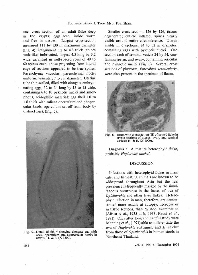
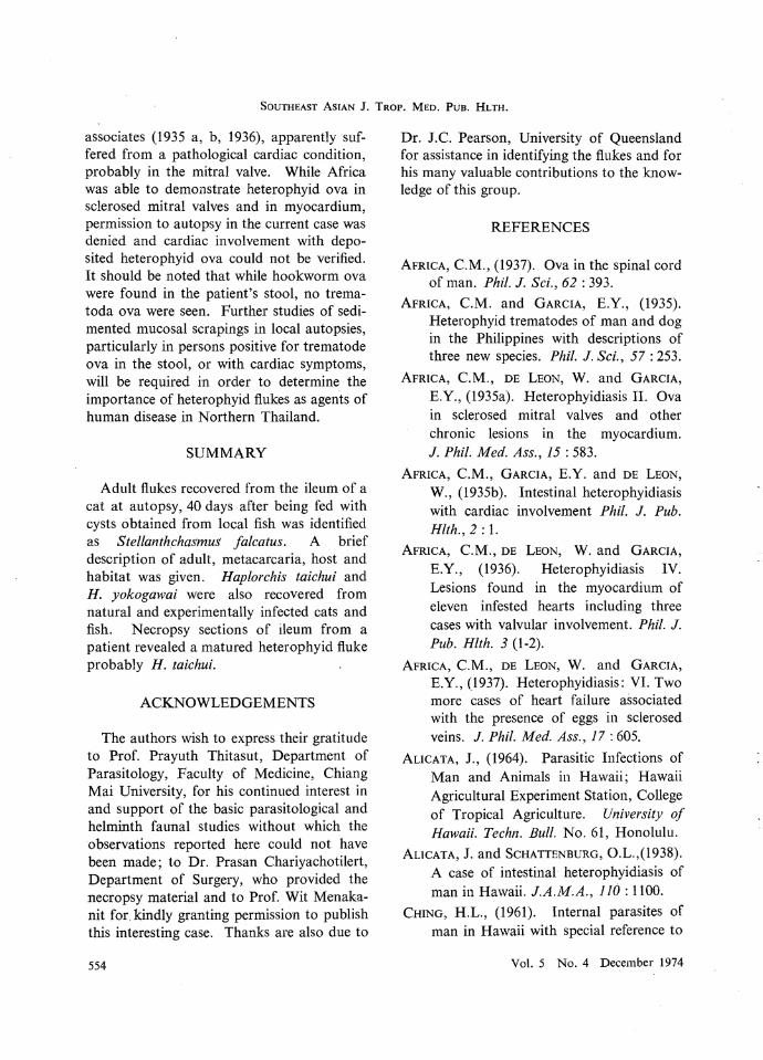