heterotopic ossification following spinal cord injury · heterotopic ossification (ho) is the...
TRANSCRIPT
www.scireproject.com Version 7.0
Heterotopic Ossification Following Spinal Cord Injury
Fatimah Alibrahim MD
Amanda McIntyre RN MSc Pavlina Faltynek MSc
Brooke Benton RDH BSc Swati Mehta PhD
Eldon Loh MD FRCPC Robert Teasell MD FRCPC
Key Points
Anti-inflammatory medications given early post-spinal cord injury reduces development of heterotopic ossification. Warfarin may inhibit the development of heterotopic ossification post-spinal cord injury.
Alendronate does not prevent the development of heterotopic ossification and may cause contractures. Etidronate may be effective for halting the progression of heterotopic ossification when administered early. Pamidronate halts secondary progression of heterotopic ossification post-surgical excision.
Pulse low intensity electromagnetic field therapy is effective in preventing heterotopic ossification post spinal cord injury. Radiotherapy can reduce the progression and recurrence of heterotopic ossification. Surgical resection of heterotopic ossification can improve hip range of motion but it may recur in a large number of individuals. Surgical resection and pamidronate treatment halts secondary heterotopic ossification progression.
This review has been prepared based on the scientific and professional information available in 2018. The SCIRE information (print, CD or web site www.scireproject.com) is provided for informational and educational purposes only. If you have or suspect you have a health problem, you should consult your health care provider. The SCIRE editors, contributors and supporting partners shall not be liable for any damages, claims, liabilities, costs or obligations arising from the use or misuse of this material. Alibrahim F, McIntyre A, Faltynek P, Benton B, Mehta S, Loh E, Teasell RW (2018). Heterotopic Ossification Following Spinal Cord Injury. In Eng JJ, Teasell RW, Miller WC, Wolfe DL, Townson AF, Hsieh JTC, Connolly SJ, Noonan VK, Loh E, McIntyre A, editors. Spinal Cord Injury Rehabilitation Evidence. Version 7.0: p 1-24. www.scireproject.com
Table of Contents
1.0 Executive Summary ..................................................................................................... 1 2.0 Introduction .................................................................................................................. 2 3.0 Pathophysiology of Heterotopic Ossification .................................................................. 2 4.0 Risk Factors and Clinical Presentation ............................................................................ 4 5.0 Diagnosis ............................................................................................................................ 5 6.0 Treatment of Heterotopic Ossification ............................................................................. 6
6.1 Non-Steroidal Anti-Inflammatory Drugs as Prophylaxis .................................................... 7 6.2 Warfarin as Prophylaxis ................................................................................................... 8 6.3 Bisphosphonates .............................................................................................................. 9 5.4 Pulse Low Intensity Electromagnetic Field (PLIMF) Therapy ...........................................12 5.5 Radiation Therapy ...........................................................................................................13 5.6 Surgical Resection ..........................................................................................................15
6.0 Summary ...........................................................................................................................19 7.0 References .........................................................................................................................20 Abbreviations ..........................................................................................................................24
1
Heterotopic Ossification Following Spinal Cord Injury
1.0 Executive Summary Heterotopic ossification (HO) is the formation of pathological bone in muscle or soft tissue (Shehab, Elgazzar & Collier, 2018). The incidence in individuals following a spinal cord injury (SCI) has been reported to vary greatly, ranging from 10% to 78% (Banovac 2001; van Kuijk et al. 2002, Ranganathan et al. 2015). HO occurs most frequently in the first two months after SCI, below the level of paralysis (Banovac et al. 2001) and most commonly forms in the hip but rarely the peripheral joints (Ranganathan et al. 2015). The mechanism underlying HO following spinal cord injury is not fully understood creating challenges in early diagnostic and therapeutic interventions. What are the clinical symptoms of Heterotopic Ossification following injury?
Evaluation of preceding factors, in combination with early intervention and diagnosis, may reduce incidence of HO or improve a patient’s recovery post-operatively (Citak et al. 2012). Clinical symptoms of HO being to appear 3-12 weeks post injury (Schuetz et al. (2005), and include: joint and muscle pain, parasthesias and tissue swelling in the involved region, accompanied by a mild fever (Thomas & Amstutz 1987; Orzel & Rudd 1985; Smith 1998; Shehab et al. 2002), while some SCI patients do not experience any pain. How is Heterotopic Ossification diagnosed?
Triple phase bone scanning: More sensitive, but less specific than plain radiography in detecting early HO (Freed et al. 1982). Plain Radiography: Can detect neurogenic HO 2-6 weeks after diagnosis using triple phase bone scan (Orzel et al. 1985; Freed et al. 1982). Computed tomography (CT) or Magnetic Resonance Imaging (MRI): Better visualization of heterotopic bone (Amendola et al. 1983); useful when considering surgery. Biochemical Markers: Used to evaluated elevations in alkaline phosphatase and creatine phosphokinase, however value has not been validated (Singh et al. 2003; Welch et al. 1973; Rossier et al. 1973). Brooker Classification Scheme: Typically used to diagnose HO in the pelvic region using anteroposterior radiograph of the pelvis which classifies HO into one of five classes (Zychowicz 2013). Has been criticized by some clinicians, as it cannot be used to determine the potential drawbacks of surgery and gives pessimistic results of hip range of motion with some hips classified as III or IV despite no clinical ankylosis found.
Risk Factors for HO (Citak et al. 2012; Krauss et al. 2014)
• Complete neurological deficit
• Spasticity
• Pneumonia
• Thoracic trauma
• Tracheostomy
• Nicotine use
• Urinary tract infection
• Less comorbidities
• Hyper-coagulable states
• Deep vein thrombosis (DVT)
• Pulmonary embolism (PE)
2
What management options are there for Heterotopic Ossification? Non-steroidal anti-inflammatory medications: Indomethacin and Rofecoxib have both been evaluated in the treatment of HO post SCI, however Rofecoxib is no longer available due to cardiovascular side effects (Banovac et al. 2001). Anticoagulants: Warfarin may be useful in the prevention of HO post SCI (Buschbacher et al. 1992). Bisphosphonates: Alendronate had no direct correlation for prevention of HO and may cause contractures (Ploumis et al. 2015). Etidronate was shown in several studies to halt the progression of HO (Garland et al. 1983; Banovac et al. 1993; Banovac et al. 1997; Stover et al. 1987). Pamidronate effectively halts secondary prevention of HO after surgical resection of heterotopic ossification (Schuetz et al. 2005). Pulse Low Intensity Electromagnetic Field Therapy: Effective in preventing HO post-spinal cord injury (Durovic et al. 2009). Radiation Therapy: Although the evidence is limited, studies show that radiotherapy reduces the progression of HO (Sautter-Bihl et al. 2000; Sautter-Bihl et al. 2001). Surgical Resection: Can improve hip range of motion but recurrence and complications are of concern (Genet et al. 2011; Garland & Orwin 1989; Meiners et al. 1997).
GAPS IN THE EVIDENCE
2.0 Introduction
Heterotopic ossification (HO) is the formation of pathological bone in muscle or soft tissue (ref). The incidence in individuals following a spinal cord injury (SCI) has been reported to vary greatly, ranging from 10% to 78% (Banovac 2001; van Kuijk et al. 2002, Ranganathan et al. 2015). HO occurs most frequently in the first two months after SCI, below the level of paralysis (Banovac et al. 2001) and most commonly forms in the hip but rarely in the peripheral joints such as the elbows and knees (Ranganathan et al. 2015).
3.0 Pathophysiology of Heterotopic Ossification
The mechanism underlying HO following spinal cord injury is not fully understood creating challenges in early diagnostic and therapeutic interventions. In the body, bone tissues are maintained by three primary cell types; osteoblasts, osteocytes, and osteoclasts (Findlay, 2018). HO appears to be initiated by metaplasia of mesenchymal cells into bone precursor cells (Schuetz et al. 2005). Mesenchymal stem cells can differentiate into osteogenic cells given the right stimuli and within the right environment (even soft tissue; Chalmers et al. 1975; pape 2004). These mesenchymal stem cells can generate cartilage, muscles, tendons, ligaments or fat, besides bone (Williams et al. 1999) and are thought to play a pivotal role in the development of HO (Pape et al. 2004). After mesenchymal cell differentiation to osteogenic cells, a protein mixture created by bone cells (osteoid) calcifies within a matter of weeks (Pape et al. 2001).
• Mechanism of action of NSAIDs in preventing the occurrence of HO
• The role of NSAIDs once the patient starts to develop symptoms with or without radiographic evidence of HO
• Optimal criteria for decisions regarding excision
3
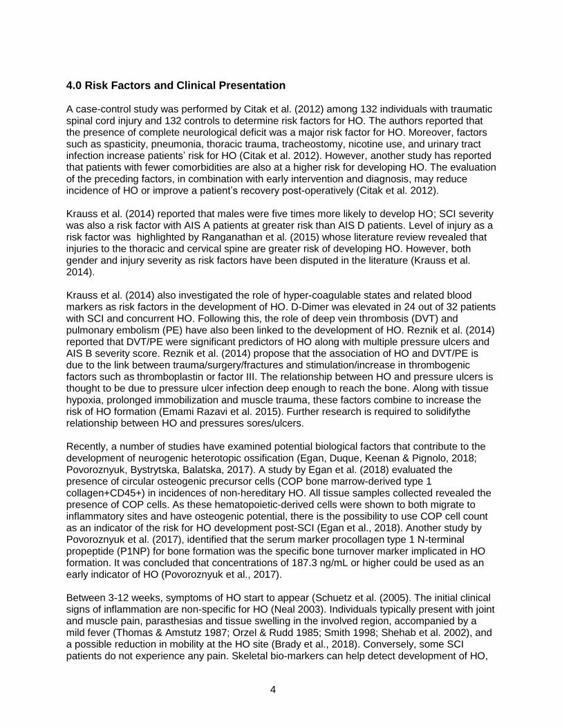
Over the next few months, the calcified osteoid remodels and matures into well-organized trabecular bone (Pape et al. 2001). Months following the initial trauma, patients develop bone formation in muscle and soft tissues in an ectopic location with resultant restriction in range of motion, pain and ankyloses at the affected joint (Banovac & Gonzalez 1997; Garland et al. 1980). The bony lesion has a high metabolic rate, adding new bone at more than three times the rate of normal bone. Osteoclastic (bone removal cells) density is more than twice that found in healthy bone (Puzas et al. 1987). It is suspected there may be a neurogenic factor contributing to HO but the mechanism is poorly understood (Hurvitz et al. 1992; Pape et al. 2001; Pape et al. 2004). Another recent theory for the development of HO has been suggested (Brady, Shultz, McDonald & O’Brien, 2018). As an inflammatory response is triggered by CNS damage, the inflammatory cells involved, such as neutrophils, lymphocytes, and macrophages, are thought to release a variety of growth factors and cytokines (Tannous et al., 2013). These inflammatory lesions are thought to eventually turn into bone through a hypoxic microenvironment ultimately leading to mesenchymal condensations at peripheral injury sites (Wang et al., 2016; Agarwal et a, 2016; Winkler et al., 2015). Figure 1.
Mesenchymal stem cells
Osteoblasts
Soft tissue inflammation
Alkaline Phosphatase Tropocollagen
Calcium Precipitation
Collagen Proteins
Bone Matrix
Mineralization of bone matrix
Ectopic bone
↑prostaglandin
differentiation
produce
Pyrophosphate
polymerizes
form
4
4.0 Risk Factors and Clinical Presentation A case-control study was performed by Citak et al. (2012) among 132 individuals with traumatic spinal cord injury and 132 controls to determine risk factors for HO. The authors reported that the presence of complete neurological deficit was a major risk factor for HO. Moreover, factors such as spasticity, pneumonia, thoracic trauma, tracheostomy, nicotine use, and urinary tract infection increase patients’ risk for HO (Citak et al. 2012). However, another study has reported that patients with fewer comorbidities are also at a higher risk for developing HO. The evaluation of the preceding factors, in combination with early intervention and diagnosis, may reduce incidence of HO or improve a patient’s recovery post-operatively (Citak et al. 2012). Krauss et al. (2014) reported that males were five times more likely to develop HO; SCI severity was also a risk factor with AIS A patients at greater risk than AIS D patients. Level of injury as a risk factor was highlighted by Ranganathan et al. (2015) whose literature review revealed that injuries to the thoracic and cervical spine are greater risk of developing HO. However, both gender and injury severity as risk factors have been disputed in the literature (Krauss et al. 2014). Krauss et al. (2014) also investigated the role of hyper-coagulable states and related blood markers as risk factors in the development of HO. D-Dimer was elevated in 24 out of 32 patients with SCI and concurrent HO. Following this, the role of deep vein thrombosis (DVT) and pulmonary embolism (PE) have also been linked to the development of HO. Reznik et al. (2014) reported that DVT/PE were significant predictors of HO along with multiple pressure ulcers and AIS B severity score. Reznik et al. (2014) propose that the association of HO and DVT/PE is due to the link between trauma/surgery/fractures and stimulation/increase in thrombogenic factors such as thromboplastin or factor III. The relationship between HO and pressure ulcers is thought to be due to pressure ulcer infection deep enough to reach the bone. Along with tissue hypoxia, prolonged immobilization and muscle trauma, these factors combine to increase the risk of HO formation (Emami Razavi et al. 2015). Further research is required to solidifythe relationship between HO and pressures sores/ulcers. Recently, a number of studies have examined potential biological factors that contribute to the development of neurogenic heterotopic ossification (Egan, Duque, Keenan & Pignolo, 2018; Povoroznyuk, Bystrytska, Balatska, 2017). A study by Egan et al. (2018) evaluated the presence of circular osteogenic precursor cells (COP bone marrow-derived type 1 collagen+CD45+) in incidences of non-hereditary HO. All tissue samples collected revealed the presence of COP cells. As these hematopoietic-derived cells were shown to both migrate to inflammatory sites and have osteogenic potential, there is the possibility to use COP cell count as an indicator of the risk for HO development post-SCI (Egan et al., 2018). Another study by Povoroznyuk et al. (2017), identified that the serum marker procollagen type 1 N-terminal propeptide (P1NP) for bone formation was the specific bone turnover marker implicated in HO formation. It was concluded that concentrations of 187.3 ng/mL or higher could be used as an early indicator of HO (Povoroznyuk et al., 2017). Between 3-12 weeks, symptoms of HO start to appear (Schuetz et al. (2005). The initial clinical signs of inflammation are non-specific for HO (Neal 2003). Individuals typically present with joint and muscle pain, parasthesias and tissue swelling in the involved region, accompanied by a mild fever (Thomas & Amstutz 1987; Orzel & Rudd 1985; Smith 1998; Shehab et al. 2002), and a possible reduction in mobility at the HO site (Brady et al., 2018). Conversely, some SCI patients do not experience any pain. Skeletal bio-markers can help detect development of HO,
5
in particular, ALP serum, CPK, C-reactive protein, prostaglandin E2, and erythrocyte sedimentation rate which have been associated with HO after SCI (Ploumis et al. 2015).
5.0 Diagnosis In the early phase of HO, triple phase bone scanning demonstrates increased uptake of osteotropic radionucleotides. Bone scanning has proven to be more sensitive than plain radiography in detecting early HO. Neurogenic HO becomes evident on plain radiography approximately two to six weeks after diagnosis using the triple phase bone scan (Orzel et al. 1985; Freed et al. 1982). However, bone scans have lower specificity than radiography (Freed et al. 1982). Computed tomography (CT) or magnetic resonance imaging (MRI) scanning may be a useful tool when considering surgery as it allows for better visualization of the heterotopic bone (Amendola et al. 1983). A recent study has also shown that ultrasound can be an effective diagnostic tool for early HO detection (Rosteius et al., 2017). Of 217 patients with confirmed HO on CT, signs of HO were correctly noted in 193 (88.9%) patients with SCI three weeks earlier via ultrasound (Rosteius et al., 2017). Some studies have examined diagnosis of HO through elevations in biochemical markers such as alkaline phosphatase (Singh et al. 2003; Tibone et al. 1978) and creatine phosphokinase (Singh et al. 2003; Welch et al. 1973; Rossier et al. 1973). The predictive value of alkaline phosphatase has not been validated (Singh et al. 2003; Welch et al. 1973; Rossier et al. 1973), although there is conflicting evidence of an association with HO and increased serum creatine phosphokinase levels (Singh et al. 2003; Welch et al. 1973). Schurch et al. (1997) studied individuals with acute SCI and found increases in urine 24-hour prostaglandin E2 (PGE2) a valid indicator of early HO formation. The Brooker Classification Scheme is typically used to diagnose HO in the pelvic region (Zychowicz 2013). The system is based on an anteroposterior radiograph of the pelvis which classifies HO into one of five classes. The classes are based on the progression of ossification: Class 0 – no presence of ossification, Class 1 - islands of bone within soft tissue of any size, Class 2 - bone spurs from pelvis or femur with at least one cm between opposing bone surfaces, Class 3 - bone spurs from pelvis and femur reducing space between opposing bone surfaces to less than 1 cm, and Class 4 - complete ankylosis of hip (Zychowicz 2013). The Brooker Classification Scheme has been criticized by some clinicians and adjustments to the traditional classification system have been proposed. Mavrogenis et al (2012) have suggested focusing on the location of the HO formation around the hip joint using the following scheme: Type 1–anterior, Type 2–posterior, Type 3–anteromedial, Type 4–circumferential HO. The adjustments are based on describing the anatomical position of HO, which could permit an estimate of blood loss, transfusion requirements, and recurrence. The new classification system improves ease of use and provides the opportunity for more rapid post-operative planning of surgical approach, evaluation, and prognosis (Mavrogenis et al. 2012). However, Citak et al. (2012) suggested the use of ultrasound, CT or MRI rather than radiographs in order to improve diagnosis and to reduce the use of methods with less sensitivity for early diagnosis. A new classification has been proposed by Arduini et al. (2015) based on the use of a 3D-CT reconstruction scan. The authors propose that this method allows the surgeon to observe the bone bridge in its entirety, can assist in assessing the relationship between neurovascular structures and the HO, and can identify the site of discontinuity, allowing the surgeon to plan the best surgical approach. Arduini et al. (2015) contend that their approach is more advantageous in comparison to the Brooker Classification, which they believe is inadequate at classifying the
6
anatomy of the HO, and Mavrogenis et al. (2012)’s proposal, which the authors argue cannot give precise details regarding the continuity of the bone bridge or the extent to which surrounding muscles and soft-tissues are being affected. Genêt et al. (2015) also highlight that the Brooker Classification cannot be used to determine the potential drawbacks of surgery and gives pessimistic results of hip range of motion post-operatively with some hips classified as III or IV despite no clinical ankylosis found. Arduini et al. (2015) state that their classification system has reproducible high intra- and inter-observer reliability and that previous methods including Garland’s radiological classification and Mavrogenis et al. (2012) CT approach have not been validated. However, it is also important to note that Arduini et al. (2015) used a mixed population of SCI and TBI with SCI accounting for 29% of the sample. Although their approach is of interest, further research is required with an all-SCI, or at least SCI-majority, group of patients.
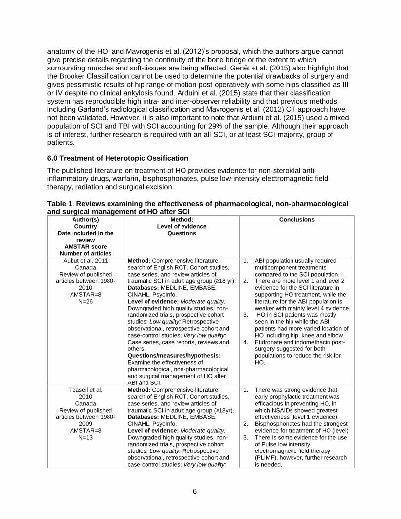
6.0 Treatment of Heterotopic Ossification
The published literature on treatment of HO provides evidence for non-steroidal anti-inflammatory drugs, warfarin, bisphosphonates, pulse low-intensity electromagnetic field therapy, radiation and surgical excision. Table 1. Reviews examining the effectiveness of pharmacological, non-pharmacological and surgical management of HO after SCI
Author(s) Country
Date included in the review
AMSTAR score Number of articles
Method: Level of evidence
Questions
Conclusions
Aubut et al. 2011 Canada
Review of published articles between 1980-
2010 AMSTAR=8
N=26
Method: Comprehensive literature search of English RCT, Cohort studies, case series, and review articles of traumatic SCI in adult age group (≥18 yr). Databases: MEDLINE, EMBASE, CINAHL, PsycInfo. Level of evidence: Moderate quality: Downgraded high quality studies, non-randomized trials, prospective cohort studies; Low quality: Retrospective observational, retrospective cohort and case-control studies; Very low quality: Case series, case reports, reviews and others. Questions/measures/hypothesis: Examine the effectiveness of pharmacological, non-pharmacological and surgical management of HO after ABI and SCI.
1. ABI population usually required multicomponent treatments compared to the SCI population.
2. There are more level 1 and level 2 evidence for the SCI literature in supporting HO treatment, while the literature for the ABI population is weaker with mainly level 4 evidence.
3. HO in SCI patients was mostly seen in the hip while the ABI patients had more varied location of HO including hip, knee and elbow.
4. Etidronate and indomethacin post-surgery suggested for both populations to reduce the risk for HO.
Teasell et al. 2010
Canada Review of published
articles between 1980-2009
AMSTAR=8 N=13
Method: Comprehensive literature search of English RCT, Cohort studies, case series, and review articles of traumatic SCI in adult age group (≥18yr). Databases: MEDLINE, EMBASE, CINAHL, PsycInfo. Level of evidence: Moderate quality: Downgraded high quality studies, non-randomized trials, prospective cohort studies; Low quality: Retrospective observational, retrospective cohort and case-control studies; Very low quality:
1. There was strong evidence that early prophylactic treatment was efficacious in preventing HO, in which NSAIDs showed greatest effectiveness (level 1 evidence).
2. Bisphosphonates had the strongest evidence for treatment of HO (level)
3. There is some evidence for the use of Pulse low intensity electromagnetic field therapy (PLIMF), however, further research is needed.
7
Case series, case reports, reviews and others. Questions/measures/hypothesis: Examine the effectiveness of pharmacological, non-pharmacological and surgical management of HO after SCI.
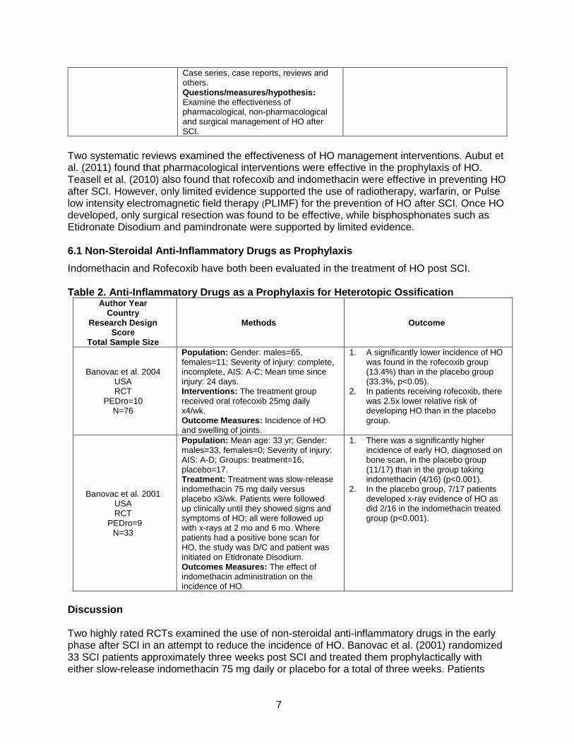
Two systematic reviews examined the effectiveness of HO management interventions. Aubut et al. (2011) found that pharmacological interventions were effective in the prophylaxis of HO. Teasell et al. (2010) also found that rofecoxib and indomethacin were effective in preventing HO after SCI. However, only limited evidence supported the use of radiotherapy, warfarin, or Pulse low intensity electromagnetic field therapy (PLIMF) for the prevention of HO after SCI. Once HO developed, only surgical resection was found to be effective, while bisphosphonates such as Etidronate Disodium and pamindronate were supported by limited evidence. 6.1 Non-Steroidal Anti-Inflammatory Drugs as Prophylaxis
Indomethacin and Rofecoxib have both been evaluated in the treatment of HO post SCI. Table 2. Anti-Inflammatory Drugs as a Prophylaxis for Heterotopic Ossification
Author Year Country
Research Design Score
Total Sample Size
Methods Outcome
Banovac et al. 2004 USA RCT
PEDro=10 N=76
Population: Gender: males=65, females=11; Severity of injury: complete, incomplete, AIS: A-C; Mean time since injury: 24 days. Interventions: The treatment group received oral rofecoxib 25mg daily x4/wk. Outcome Measures: Incidence of HO and swelling of joints.
1. A significantly lower incidence of HO was found in the rofecoxib group (13.4%) than in the placebo group (33.3%, p<0.05).
2. In patients receiving rofecoxib, there was 2.5x lower relative risk of developing HO than in the placebo group.
Banovac et al. 2001 USA RCT
PEDro=9 N=33
Population: Mean age: 33 yr; Gender: males=33, females=0; Severity of injury: AIS: A-D; Groups: treatment=16, placebo=17. Treatment: Treatment was slow-release indomethacin 75 mg daily versus placebo x3/wk. Patients were followed up clinically until they showed signs and symptoms of HO; all were followed up with x-rays at 2 mo and 6 mo. Where patients had a positive bone scan for HO, the study was D/C and patient was initiated on Etidronate Disodium. Outcomes Measures: The effect of indomethacin administration on the incidence of HO.
1. There was a significantly higher incidence of early HO, diagnosed on bone scan, in the placebo group (11/17) than in the group taking indomethacin (4/16) (p<0.001).
2. In the placebo group, 7/17 patients developed x-ray evidence of HO as did 2/16 in the indomethacin treated group (p<0.001).
Discussion Two highly rated RCTs examined the use of non-steroidal anti-inflammatory drugs in the early phase after SCI in an attempt to reduce the incidence of HO. Banovac et al. (2001) randomized 33 SCI patients approximately three weeks post SCI and treated them prophylactically with either slow-release indomethacin 75 mg daily or placebo for a total of three weeks. Patients
8
were carefully followed with regular clinical follow-up and bone scans. There was a significantly higher incidence of HO, diagnosed on bone scan and plain radiographs, in the placebo group when compared with the group receiving indomethacin (p<0.001). Banovac et al. (2004) randomized 76 patients in the early phase post SCI into either the treatment group (25 mg rofecoxib daily for two weeks) or a placebo group. A significantly lower incidence of HO was observed in the rofecoxib group (13.4%) than in the placebo group (33.3%; p<0.05). While both RCTs provided compelling evidence that anti-inflammatory drugs given prophylactically reduce the likelihood of developing HO post-SCI, Rofecoxib is no longer available due to cardiovascular side effects. Conclusions
There is strong Level 1a evidence (from two RCTs; Banovac et al. 2001; Banovac et al. 2004) that non-steroidal anti-inflammatory medications can reduce the incidence of heterotopic ossification when administered early after a spinal cord injury.
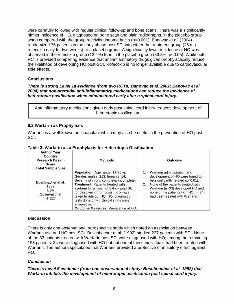
6.2 Warfarin as Prophylaxis
Warfarin is a well-known anticoagulant which may also be useful in the prevention of HO post SCI.
Table 3. Warfarin as a Prophylaxis for Heterotopic Ossification
Author Year Country
Research Design Score
Total Sample Size
Methods Outcome
Buschbacher et al. 1992 USA
Observational N=227
Population: Age range: 17-75 yr; Gender: males=213, females=14; Severity of injury: complete, incomplete. Treatment: Patients treated with warfarin for a mean of 5.4 wk post SCI for deep vein thrombosis; no X-rays taken to rule out HO. HO, diagnostic tests done only if clinical signs were suggestive. Outcome Measures: Prevalence of HO.
1. Warfarin administration and development of HO were found to be significantly related (p<0.01).
2. None of the patients treated with Warfarin (n=33) developed HO and none of the patients with HO (n=34) had been treated with Warfarin.
Discussion There is only one observational retrospective study which noted an association between Warfarin use and HO post SCI. Buschbacher et al. (1992) studied 227 patients with SCI. None of the 33 patients treated with Warfarin post SCI were diagnosed with HO; among the remaining 193 patients, 34 were diagnosed with HO but not one of these individuals had been treated with Warfarin. The authors speculated that Warfarin provided a protective or inhibitory effect against HO.
Conclusion
There is Level 5 evidence (from one observational study; Buschbacher et al. 1992) that Warfarin inhibits the development of heterotopic ossification post spinal cord injury.
Anti-inflammatory medications given early post spinal cord injury reduces development of heterotopic ossification.
9
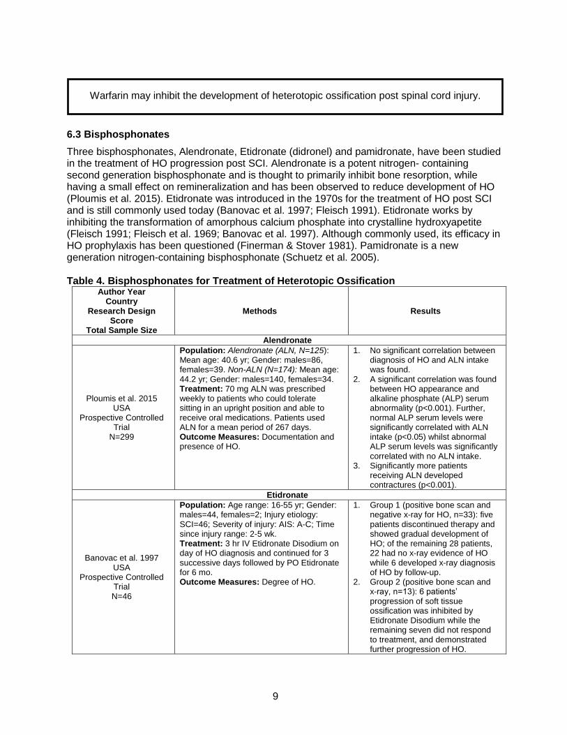
6.3 Bisphosphonates
Three bisphosphonates, Alendronate, Etidronate (didronel) and pamidronate, have been studied in the treatment of HO progression post SCI. Alendronate is a potent nitrogen- containing second generation bisphosphonate and is thought to primarily inhibit bone resorption, while having a small effect on remineralization and has been observed to reduce development of HO (Ploumis et al. 2015). Etidronate was introduced in the 1970s for the treatment of HO post SCI and is still commonly used today (Banovac et al. 1997; Fleisch 1991). Etidronate works by inhibiting the transformation of amorphous calcium phosphate into crystalline hydroxyapetite (Fleisch 1991; Fleisch et al. 1969; Banovac et al. 1997). Although commonly used, its efficacy in HO prophylaxis has been questioned (Finerman & Stover 1981). Pamidronate is a new generation nitrogen-containing bisphosphonate (Schuetz et al. 2005). Table 4. Bisphosphonates for Treatment of Heterotopic Ossification
Author Year Country
Research Design Score
Total Sample Size
Methods Results
Alendronate
Ploumis et al. 2015 USA
Prospective Controlled Trial
N=299
Population: Alendronate (ALN, N=125): Mean age: 40.6 yr; Gender: males=86, females=39. Non-ALN (N=174): Mean age: 44.2 yr; Gender: males=140, females=34. Treatment: 70 mg ALN was prescribed weekly to patients who could tolerate sitting in an upright position and able to receive oral medications. Patients used ALN for a mean period of 267 days. Outcome Measures: Documentation and presence of HO.
1. No significant correlation between diagnosis of HO and ALN intake was found.
2. A significant correlation was found between HO appearance and alkaline phosphate (ALP) serum abnormality (p<0.001). Further, normal ALP serum levels were significantly correlated with ALN intake (p<0.05) whilst abnormal ALP serum levels was significantly correlated with no ALN intake.
3. Significantly more patients receiving ALN developed contractures (p<0.001).
Etidronate
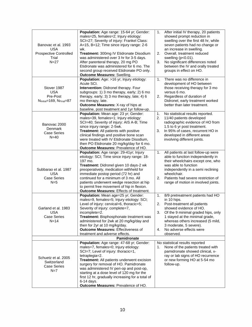
Banovac et al. 1997 USA
Prospective Controlled Trial N=46
Population: Age range: 16-55 yr; Gender: males=44, females=2; Injury etiology: SCI=46; Severity of injury: AIS: A-C; Time since injury range: 2-5 wk. Treatment: 3 hr IV Etidronate Disodium on day of HO diagnosis and continued for 3 successive days followed by PO Etidronate for 6 mo. Outcome Measures: Degree of HO.
1. Group 1 (positive bone scan and negative x-ray for HO, n=33): five patients discontinued therapy and showed gradual development of HO; of the remaining 28 patients, 22 had no x-ray evidence of HO while 6 developed x-ray diagnosis of HO by follow-up.
2. Group 2 (positive bone scan and x-ray, n=13): 6 patients’ progression of soft tissue ossification was inhibited by Etidronate Disodium while the remaining seven did not respond to treatment, and demonstrated further progression of HO.
Warfarin may inhibit the development of heterotopic ossification post spinal cord injury.
10
Banovac et al. 1993 USA
Prospective Controlled Trial N=27
Population: Age range: 15-64 yr; Gender: males=25, females=2; Injury etiology: SCI=27; Severity of injury: Frankel Class: A=15, B=12; Time since injury range: 2-6 wk. Treatment: 300mg IV Etidronate Disodium was administered over 3 hr for 3-5 days. After parenteral therapy, 20 mg PO Etidronate was administered for 6 mo. The second group received Etidronate PO only. Outcome Measures: Swelling.
1. After initial IV therapy, 20 patients showed prompt reduction in swelling over the first 48 hr, while seven patients had no change or an increase in swelling.
2. Overall, treatment reduced swelling (p<0.01).
3. No significant differences noted between the IV and orally treated groups in effect on HO.
Stover 1987
USA Pre-Post
NInitial=169, NFinal=87
Population: Age: >16 yr; Injury etiology: Acute SCI. Intervention: Didronel therapy. Four subgroups: 1) 3 mo therapy, early; 2) 6 mo therapy, early; 3) 3 mo therapy, late; 4) 6 mo therapy, late. Outcome Measures: X-ray of hips at baseline, post treatment and 1yr follow-up.
1. There was no difference in development of HO between those receiving therapy for 3 mo versus 6 mo.
2. Regardless of duration of Didronel, early treatment worked better than later treatment.
Banovac 2000 Denmark
Case Series N=40
Population: Mean age: 23 yr; Gender: males=39, females=1; Injury etiology: SCI=40; Severity of injury: AIS A-B; Time since injury range: 2-5wk. Treatment: All patients with positive clinical findings and positive bone scan were treated with IV Etidronate Disodium, then PO Etidronate 20 mg/kg/day for 6 mo. Outcome Measures: Prevalence of HO.
1. No statistical results reported. 2. 11/40 patients developed
radiographic evidence of HO from 1.5 to 6 yr post treatment.
3. In 95% of cases, recurrent HO in developed in different areas involving different joints.
Subbarao et al. 1987 USA
Case Series N=5
Population: Age range: 29-41yr; Injury etiology: SCI; Time since injury range: 18-197 mo. Treatment: Didronel given 10 days-2 wk preoperatively, medication withheld for immediate postop period (72 hr) and continued for a minimum of 3 mo. All patients underwent wedge resection at hip to permit free movement of hip in flexion. Outcome Measures: Effects of treatment.
1. All patients at last follow-up were able to function independently in their wheelchairs except one, who was able to function independently in a semi reclining wheelchair.
2. Patients had severe restriction of range of motion in involved joints.
Garland et al. 1983 USA
Case Series N=14
Population: Mean age=25 yr; Gender: males=9, females=5; Injury etiology: SCI; Level of injury: cervical=6, thoracic=5; Severity of injury: complete=7, incomplete=2. Treatment: Bisphosphonate treatment was administered for 2wk at 20 mg/kg/day and then for 2yr at 10 mg/kg/day. Outcome Measures: Effectiveness of treatment and adverse effects.
1. 8/9 pretreatment patients had HO in 10 hips.
2. Post-treatment all patients showed evidence of HO.
3. Of the 9 minimal graded hips, only 1 stayed at the minimal grade, whereas others increased (5 mild, 3 moderate, 5 severe).
4. No adverse effects were observed.
Pamidronate
Schuetz et al. 2005 Switzerland Case Series
N=7
Population: Age range: 47-68 yr; Gender: males=7, females=0; Injury etiology: SCI=7; Level of injury: thoracic=1, tetraplegia=2. Treatment: All patients underwent excision surgery for removal of HO. Pamidronate was administered IV peri-op and post-op, starting at a dose level of 120 mg for the first 12 hr, gradually increasing for a total of 6-14 days. Outcome Measures: Prevalence of HO.
No statistical results reported 1. None of the patients treated with
pamidronate showed clinical, x-ray or lab signs of HO recurrence or new forming HO at 5-54 mo follow-up.
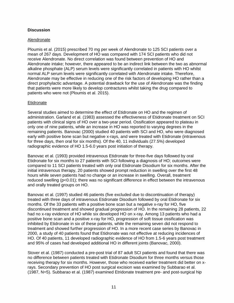
11
Discussion Alendronate Ploumis et al. (2015) prescribed 70 mg per week of Alendronate to 125 SCI patients over a mean of 267 days. Development of HO was compared with 174 SCI patients who did not receive Alendronate. No direct correlation was found between prevention of HO and Alendronate intake; however, there appeared to be an indirect link between the two as abnormal alkaline phosphate (ALP) serum levels were significantly correlated in patients with HO whilst normal ALP serum levels were significantly correlated with Alendronate intake. Therefore, Alendronate may be effective in reducing one of the risk factors of developing HO rather than a direct prophylactic advantage. A potential drawback for the use of Alendronate was the finding that patients were more likely to develop contractures whilst taking the drug compared to patients who were not (Ploumis et al. 2015). Etidronate Several studies aimed to determine the effect of Etidronate on HO and the regimen of administration. Garland et al. (1983) assessed the effectiveness of Etidronate treatment on SCI patients with clinical signs of HO over a two-year period. Ossification appeared to plateau in only one of nine patients, while an increase in HO was reported to varying degrees in the remaining patients. Banovac (2000) studied 40 patients with SCI and HO, who were diagnosed early with positive bone scan but negative x-rays, and were treated with Etidronate (intravenous for three days, then oral for six months). Of the 40, 11 individuals (27.5%) developed radiographic evidence of HO 1.5-6.0 years post initiation of therapy. Banovac et al. (1993) provided intravenous Etidronate for three-five days followed by oral Etidronate for six months to 27 patients with SCI following a diagnosis of HO; outcomes were compared to 11 SCI patients treated with only oral Etidronate Disodium for six months. After the initial intravenous therapy, 20 patients showed prompt reduction in swelling over the first 48 hours while seven patients had no change or an increase in swelling. Overall, treatment reduced swelling (p<0.01); there was no significant difference in effect between the intravenous and orally treated groups on HO. Banovac et al. (1997) studied 46 patients (five excluded due to discontinuation of therapy) treated with three days of intravenous Etidronate Disodium followed by oral Etidronate for six months. Of the 33 patients with a positive bone scan but a negative x-ray for HO, five discontinued treatment and showed gradual progression of HO. In the remaining 28 patients, 22 had no x-ray evidence of HO while six developed HO on x-ray. Among 13 patients who had a positive bone scan and a positive x-ray for HO, progression of soft tissue ossification was inhibited by Etidronate in six of these patients, while the remaining seven did not respond to treatment and showed further progression of HO. In a more recent case series by Banovac in 2000, a study of 40 patients found that Etidronate was not effective at reducing incidences of HO. Of 40 patients, 11 developed radiographic evidence of HO from 1.5-6 years post treatment and 95% of cases had developed additional HO in different joints (Banovac, 2000). Stover et al. (1987) conducted a pre-post trial of 87 adult SCI patients and found that there was no difference between patients treated with Etidronate Disodium for three months versus those receiving therapy for six months. However, those who received earlier treatment did better on x-rays. Secondary prevention of HO post surgical excision was examined by Subbarao et al. (1987, N=5). Subbarao et al. (1987) examined Etridonate treatment pre- and post-surgical hip
12
wedge resection and found that patients still had severe restriction in their range of motion at follow-up. Pamidronate Schuetz et al. (2005) reported that pamidronate was administered pre- and post-surgical removal of HO among individuals with SCI and had no recurrences. It is important to note that sample sizes in both studies were small. The lack of RCTs and variable treatment regimens make it difficult to form definitive conclusions. It appears that Etidronate is able to delay or inhibit HO progression once it is diagnosed and it tends to work better when given earlier after diagnosis.
Conclusions There is Level 2 evidence (from one prospective controlled trial; Ploumis et al. 2015) that Alendronate does not inhibit the development of heterotopic ossification and in fact may contribute to the development of contractures. There is conflicting Level 2 evidence (from two prospective controlled trials; Banovac et al. 1993; Banovac et al. 1997) and Level 4 evidence (from one case series; Branovac 2000) that Etidronate can stop the progression of heterotopic ossification once the diagnosis is made and prevent further HO sites. There is Level 2 evidence (from one prospective controlled trial; Banovac et al. 1997) that Etidronate is not effective once radiographs are positive for HO. There is Level 4 evidence (from one case series; Schuetz et al. 2005) that Pamidronate effectively halts secondary HO progression after surgical resection of HO.
6.4 Pulse Low Intensity Electromagnetic Field (PLIMF) Therapy PLIMF therapy uses magnetic fields to increase oxygen levels and decrease toxic by-products of inflammation by increasing local blood flow (Durovic et al. 2009). Table 5 Pulse Low Intensity for Treatment of Heterotopic Ossification
Alendronate does not prevent the development of heterotopic ossification and may cause contractures.
Etidronate may be effective for halting the progression of heterotopic ossification when
administered early.
Etidronate can halt the progression of heterotopic ossification.
Pamidronate halts secondary progression of heterotopic ossification post-surgical excision.

13
Author Year Country
Research Design Score
Total Sample Size
Methods Results
Durovic et al. 2009 Italy RCT
PEDro=6 N=29
Population: Age range: 18-45 yr. Treatment: Patients were randomly divided into experimental and control groups. Treatment group received 30 min pulse low intensity electromagnetic field therapy (PLIMF) therapy (25 Hz, 10 mT) for 4 wk, approximately 7 wk post SCI. Outcome measures: Incidence of HO, Brooker classification.
1. Significant differences were found in the incidence of HO between the treatment and control groups.
2. 33% of individuals in the control group had incidence of HO; 0 cases of HO in the treatment group.
3. Among control groups individuals with HO post intervention, 2 progressed grade 1, 2 to grade 2, and 1 to grade 3 on the Brooker classification.
4. Significant differences were found in the incidence of HO between the treatment and control group (p<0.04).
Discussion Durovic et al. (2009) randomly assigned 29 SCI patients to a control group or treatment group. Both received range of motion and exercise therapy; however, only the treatment group received PLIMF therapy an average of seven weeks post injury for four weeks. The study showed no incidence of HO in the treatment group yet a 33% incidence in the control group (p<0.04). Conclusion There is Level 1b evidence (from one RCT; Durovic et al. 2009) that Pulse Low Intensity Electromagnetic Field therapy is an effective prophylaxis of HO post SCI
6.5 Radiation Therapy Radiation therapy or radiotherapy, which is the use of ionizing radiation for therapeutic ends, has been proposed as a possible adjunct treatment for HO. Table 6 Radiation Therapy for Treatment of Heterotopic Ossification
Author Year Country
Research Design Score
Total Sample Size
Methods Results
Museler et al., 2017 Germany
Case Series N=244
Population: Mean age: 46.4 yr; Gender: males=207, females=37; Severity of injury: AIS: A=220, B=8, C=12, D=4. Interventions: Single-dose radiation therapy at the hip for HO. Mean time of treatment was 4.9 days. Treatment was administered with either 15 MV or 6 MV.
1. Of the 244 patients, 13 experienced recurrence of HO. All 13 patients initially experienced HO in both hips. Of the 444 initial cases of HO, there were 26 instances of recurrence.
Pulse low intensity electromagnetic field therapy is effective in preventing heterotopic ossification post spinal cord injury.

14
Outcome Measures: HO recurrence, side effect due to radiation.
2. No patients experienced negative side-effects as a result of radiation treatment.
Sautter-Bihl et al. 2001 Germany
Case Series N=52
Population: Mean age: 33 yr; Gender: males=44, females=8; Treatment: Patients received a single dose of radiotherapy 2-10Gy through a linear accelerator at 6-8 MV photons. Outcome Measures: Efficacy, Brooker classification, adverse effects.
1. Prevention of HO was seen in 71% of (41 primarily treated, 9 resected) joints.
2. Radiotherapy treatment did not result in a regression of the Brooker score in any patient.
3. An increase in two Brooker score grades was seen in two joints (1 knee, 1 hip)
4. No adverse effects due to therapy occurred.
5. 16 of 32 hips treated only with radiotherapy (50%) did not show any abnormalities on follow-up.
6. No progression of HO was noted in 30/36 subjects (83%).
7. Re-ossification after therapy which led to a decrease in joint mobility was noted in three subjects.
Sautter-Bihl et al. 2000 Germany
Case Series N=36
Population: Age range: 17-59 yr; Gender: males=32, females=4; Follow-up range: 4-98mo. Treatment: 25/36 subjects received 10 Gy radiotherapy in fraction of 2-2.5 Gy, while four patients received higher doses. In phase 2 seven subjects received a single does of irradiation with 8Gy. In total, 46 joints were irradiated. Outcome Measures: Progression of HO and complications.
1. No statistically significant results were reported.
2. 16 of the 32 hips treated with radiotherapy only did not show any abnormalities on follow-up.
3. No progression of HO was noted in 30/36 subjects.
4. Re-ossification after therapy, which led to a decrease in joint mobility was noted in three subjects.
Discussion Sautter-Bihl et al. (2000) studied 36 patients with HO of whom 27 patients (32 joints) received radiotherapy when ossification was minimal. 11 patients (13 joints) had obvious ossifications, which had to be resected. Post-op radiotherapy was performed 24-36 hours post-operatively. Two patients received radiotherapy both before and after surgery. Mean duration of follow-up was 23.6 months. 30 of the 36 irradiated patients showed no progression of HO. In three patients, reossification after therapy resulted in a moderate decrease in joint mobility. In the follow-up case series by Sautter-Bihl et al. (2001), the authors examined the effectiveness of radiotherapy administered to 52 SCI patients. Radiotherapy effectively prevented primary and secondary HO post-surgical excision in 71% of patients. However, treatment did not result in regression of HO once developed, as measured by the Brooker scale. Two joints increased in Brooker score, although neither of them developed any functional impairment. Another case series by Museler et al. (2017) used either 15 MV or 6 MV radiotherapy to target HO at the hip in 244 patients. Recurrence of HO was found to be very low (5.3%), and of those that experienced recurrence HO was initially present in both hips. A case study by Cramarossa et al. (2013) reported on the use of radiation therapy on a patient who had previously experienced an SCI at the C5-C6 level and had been diagnosed with dysphagia due to HO-induced osteophytes. One day after surgical intervention, which involved anterior cervical decompression and drilling of the osteophytes, the patient received a single treatment of radiation at 8 Gy. At follow-up, the patient reported that they were not experiencing
15
any recurrence of dysphagic symptoms. The authors add that radiation should only be considered for patients at high risk of HO due to the risk of creating a radiotherapy-induced malignancy and that an expansion of the literature is required to better assist treatment decisions. Conclusion There is moderate Level 4 evidence (from three case series studies; Sautter-Bihl et al. 2000; Sautter-Bihl et al. 2001; Museler et al. 2017) that radiotherapy reduces the progression and recurrence of heterotopic ossification.
6.6 Surgical Resection
Surgical resection of HO post SCI is a well-established treatment but is still somewhat controversial.
Table 7 Surgical Resection for Treatment of Heterotopic Ossification
Author Year Country
Research Design Score
Total Sample Size
Methods Outcome
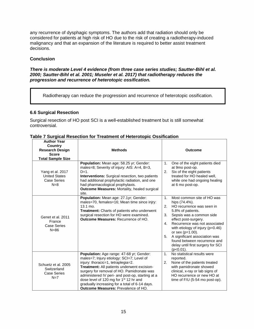
Yang et al. 2017 United States Case Series
N=8
Population: Mean age: 58.25 yr; Gender: males=8; Severity of injury: AIS: A=4, B=3, D=1. Interventions: Surgical resection, two patients had additional prophylactic radiation, and one had pharmacological prophylaxis. Outcome Measures: Mortality, healed surgical site.
1. One of the eight patients died at 9mo post-op.
2. Six of the eight patients treated for HO healed well, while one had ongoing healing at 6 mo post-op.
Genet et al. 2011 France
Case Series N=86
Population: Mean age: 27.1yr; Gender: males=70, females=16; Mean time since injry: 13.1 mo. Treatment: Charts of patients who underwent surgical resection for HO were examined. Outcome Measures: Recurrence of HO.
1. Most common site of HO was hips (74.4%).
2. HO recurrence was seen in 5.8% of patients.
3. Sepsis was a common side effect post-surgery.
4. Recurrence was not associated with etiology of injury (p=0.46) or sex (p=1.00).
5. A significant association was found between recurrence and delay until first surgery for SCI (p<0.01).
Schuetz et al. 2005 Switzerland Case Series
N=7
Population: Age range: 47-68 yr; Gender: males=7; Injury etiology: SCI=7; Level of injury: thoracic=1, tetraplegia=2. Treatment: All patients underwent excision-surgery for removal of HO. Pamidronate was administered IV peri- and post-op, starting at a dose level of 120 mg for 1st 12 hr and gradually increasing for a total of 6-14 days. Outcome Measures: Prevalence of HO.
1. No statistical results were reported.
2. None of the patients treated with pamidronate showed clinical, x-ray or lab signs of HO recurrence or new HO at time of F/U (5-54 mo post-op).
Radiotherapy can reduce the progression and recurrence of heterotopic ossification.
16
Author Year Country
Research Design Score
Total Sample Size
Methods Outcome
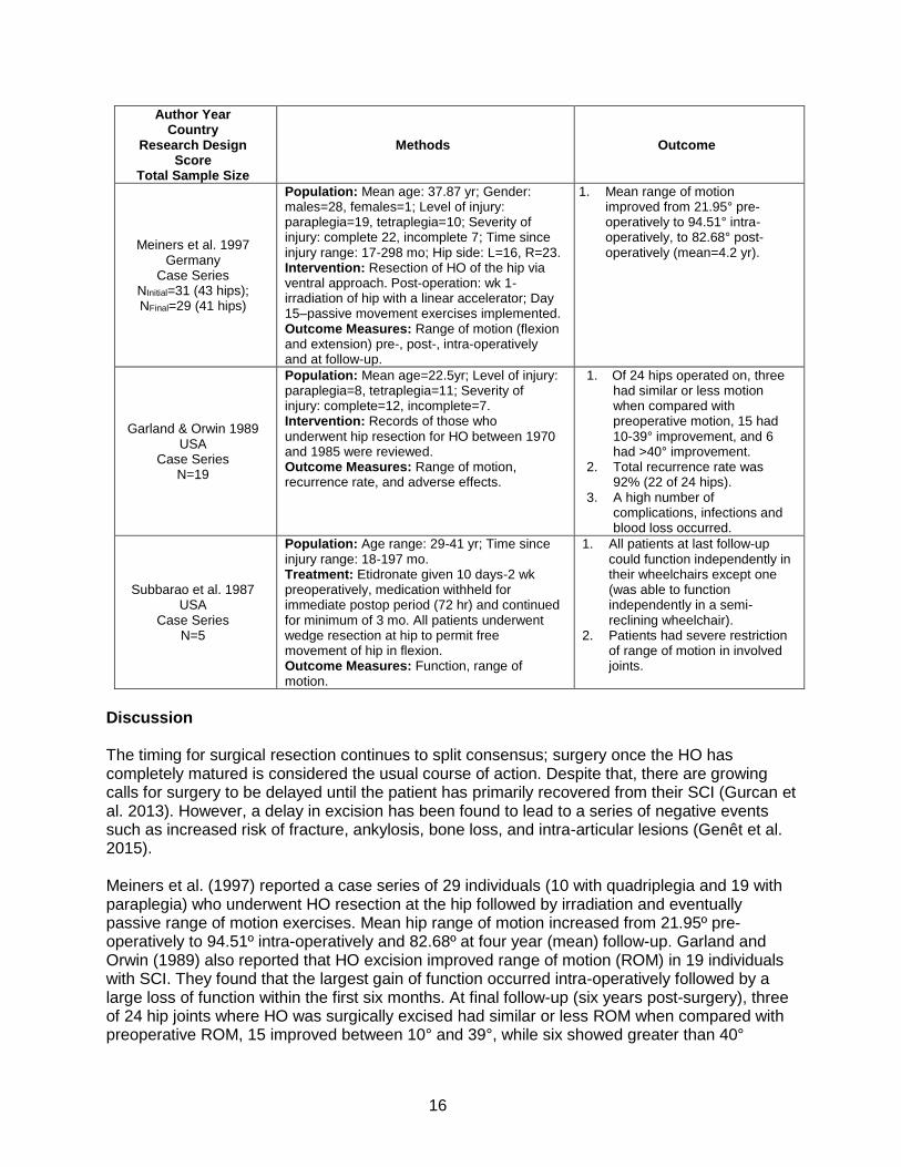
Meiners et al. 1997
Germany Case Series
NInitial=31 (43 hips); NFinal=29 (41 hips)
Population: Mean age: 37.87 yr; Gender: males=28, females=1; Level of injury: paraplegia=19, tetraplegia=10; Severity of injury: complete 22, incomplete 7; Time since injury range: 17-298 mo; Hip side: L=16, R=23. Intervention: Resection of HO of the hip via ventral approach. Post-operation: wk 1-irradiation of hip with a linear accelerator; Day 15–passive movement exercises implemented. Outcome Measures: Range of motion (flexion and extension) pre-, post-, intra-operatively and at follow-up.
1. Mean range of motion improved from 21.95° pre-operatively to 94.51° intra-operatively, to 82.68° post-operatively (mean=4.2 yr).
Garland & Orwin 1989
USA Case Series
N=19
Population: Mean age=22.5yr; Level of injury: paraplegia=8, tetraplegia=11; Severity of injury: complete=12, incomplete=7. Intervention: Records of those who underwent hip resection for HO between 1970 and 1985 were reviewed. Outcome Measures: Range of motion, recurrence rate, and adverse effects.
1. Of 24 hips operated on, three had similar or less motion when compared with preoperative motion, 15 had 10-39° improvement, and 6 had >40° improvement.
2. Total recurrence rate was 92% (22 of 24 hips).
3. A high number of complications, infections and blood loss occurred.
Subbarao et al. 1987 USA
Case Series N=5
Population: Age range: 29-41 yr; Time since injury range: 18-197 mo. Treatment: Etidronate given 10 days-2 wk preoperatively, medication withheld for immediate postop period (72 hr) and continued for minimum of 3 mo. All patients underwent wedge resection at hip to permit free movement of hip in flexion. Outcome Measures: Function, range of motion.
1. All patients at last follow-up could function independently in their wheelchairs except one (was able to function independently in a semi-reclining wheelchair).
2. Patients had severe restriction of range of motion in involved joints.
Discussion The timing for surgical resection continues to split consensus; surgery once the HO has completely matured is considered the usual course of action. Despite that, there are growing calls for surgery to be delayed until the patient has primarily recovered from their SCI (Gurcan et al. 2013). However, a delay in excision has been found to lead to a series of negative events such as increased risk of fracture, ankylosis, bone loss, and intra-articular lesions (Genêt et al. 2015). Meiners et al. (1997) reported a case series of 29 individuals (10 with quadriplegia and 19 with paraplegia) who underwent HO resection at the hip followed by irradiation and eventually passive range of motion exercises. Mean hip range of motion increased from 21.95º pre-operatively to 94.51º intra-operatively and 82.68º at four year (mean) follow-up. Garland and Orwin (1989) also reported that HO excision improved range of motion (ROM) in 19 individuals with SCI. They found that the largest gain of function occurred intra-operatively followed by a large loss of function within the first six months. At final follow-up (six years post-surgery), three of 24 hip joints where HO was surgically excised had similar or less ROM when compared with preoperative ROM, 15 improved between 10° and 39°, while six showed greater than 40°

17
improvement. Yang et al. (2017) found similar rates of improvement following HO resection. In a case series with eight patients that underwent surgical resection, six healed well, one patient had ongoing healing at 6 months due to a post-operative infection, and one patient died (Yang et al., 2017). Some studies stress that surgical resection must be followed by prophylaxis (radiation therapy, NSAID or bisphosphonates) due to high recurrence rates after surgery alone. A case study by Gurcan et al. (2013) investigated the use of surgical resection in a patient with total ankylosis of the right hip following a T8-T9 fracture. Upon excision of the cephalad mass, the patient’s hip could be flexed to 100o and abducted to 30o on the operating table, indicating a successful operation. Post-operatively, the patient completed passive movements of the hip, and was treated with a single dose of radiation (eight Gy) and a prescription for indomethacin with a dosage of 150mg a day. At 12-month follow-up post-surgery, improved range of motion in the hip was preserved with no recurrence of HO or ankylosis. The effectiveness of surgical excision followed by bisphosphonates was examined in two case series (Schuetz et al. 2005; Subbarao et al. 1987). Etidronate treatment post-surgical excision showed that patients were able to function independently in a wheelchair; however, they had severe restrictions in their range of motion (Subbarao et al. (1987). Surgical excision supplemented with pamidronate treatment resulted in no recurrence of HO post-surgery (Schuetz et al. 2005). Genêt et al. (2015) conducted a review of the literature regarding recurrence rates of HO after surgical excision. A finding of concern was the lack of consensus towards the classification of HO and risk of recurrence. While some studies have attempted this, the authors point out that these are based on observation only and are merely descriptive. Moreover, the review was not able to clarify ideal timing for surgical resection, in part due to disparate rates of recurrence post-surgery. A prominent issue of HO recurrence is the definition of recurrence. Genêt et al. (2015) highlight that some patients are not deemed to have had recurring HO post-surgery if the patient’s function is not impaired. Futhermore, pre- and post-surgical care is not standardized; early rehabilitation (including limb mobilization) is often delayed due to inflammation and treated with NSAIDs instead. Conclusion There is level 4 evidence (from four case series; Garland & Orwin 1989; Meiners et al. 1997; Genet et al. 2011; Yang et al. 2017) that resection of HO about the hip post SCI can dramatically improve restricted hip range of motion; however, post-surgical recurrence and complications are a concern for this treatment. There is Level 4 evidence (from one case series; Schuetz et al. 2005) that surgical resection combined with pamidronate treatment effectively halts secondary HO progression. There is level 4 evidence (from one case series; Subbarao et al. 1987) that surgical resection combined with etidronate treatment improves independence with wheelchair use but contributes to reduced range of motion.

18
Surgical resection of heterotopic ossification can improve hip range of motion but it may
reoccur in a large number of individuals.
Surgical resection and pamidronate treatment halts secondary heterotopic ossification progression.
19
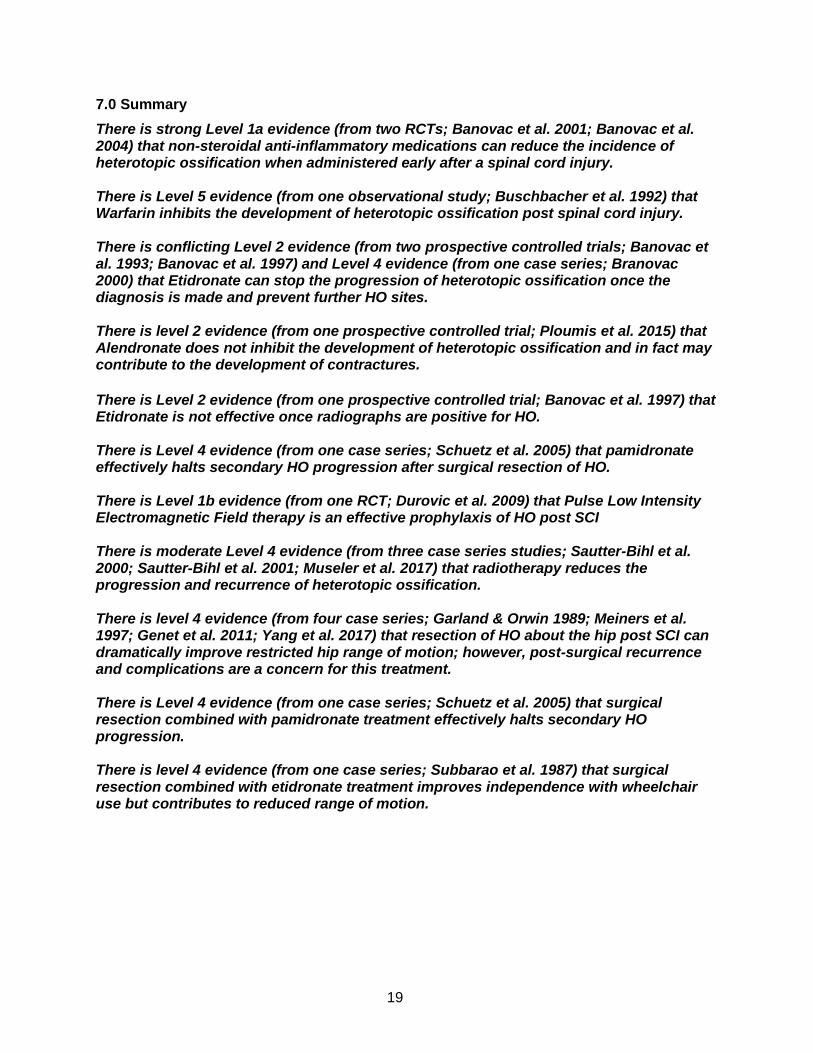
7.0 Summary
There is strong Level 1a evidence (from two RCTs; Banovac et al. 2001; Banovac et al. 2004) that non-steroidal anti-inflammatory medications can reduce the incidence of heterotopic ossification when administered early after a spinal cord injury. There is Level 5 evidence (from one observational study; Buschbacher et al. 1992) that Warfarin inhibits the development of heterotopic ossification post spinal cord injury. There is conflicting Level 2 evidence (from two prospective controlled trials; Banovac et al. 1993; Banovac et al. 1997) and Level 4 evidence (from one case series; Branovac 2000) that Etidronate can stop the progression of heterotopic ossification once the diagnosis is made and prevent further HO sites. There is level 2 evidence (from one prospective controlled trial; Ploumis et al. 2015) that Alendronate does not inhibit the development of heterotopic ossification and in fact may contribute to the development of contractures.
There is Level 2 evidence (from one prospective controlled trial; Banovac et al. 1997) that Etidronate is not effective once radiographs are positive for HO. There is Level 4 evidence (from one case series; Schuetz et al. 2005) that pamidronate effectively halts secondary HO progression after surgical resection of HO. There is Level 1b evidence (from one RCT; Durovic et al. 2009) that Pulse Low Intensity Electromagnetic Field therapy is an effective prophylaxis of HO post SCI There is moderate Level 4 evidence (from three case series studies; Sautter-Bihl et al. 2000; Sautter-Bihl et al. 2001; Museler et al. 2017) that radiotherapy reduces the progression and recurrence of heterotopic ossification. There is level 4 evidence (from four case series; Garland & Orwin 1989; Meiners et al. 1997; Genet et al. 2011; Yang et al. 2017) that resection of HO about the hip post SCI can dramatically improve restricted hip range of motion; however, post-surgical recurrence and complications are a concern for this treatment. There is Level 4 evidence (from one case series; Schuetz et al. 2005) that surgical resection combined with pamidronate treatment effectively halts secondary HO progression. There is level 4 evidence (from one case series; Subbarao et al. 1987) that surgical resection combined with etidronate treatment improves independence with wheelchair use but contributes to reduced range of motion.
20
8.0 References Agarwal S, LoderS, Brownley C, Cholok D, Mangiavini L, Li J, Breuler C Sung HH, Li S,
Ranganathan K, Peterson J, Tompkins R, Herndon D, Xiao W, Jumlongras D, Olsen BR, Davis TA, Mishina Y, Schipani E, Levi B. Inhibition of Hif1α prevents both trauma-induced and genetic heterotopic ossification. PNAS. 2016:133:3:E338-E347.
Amendola MA, Shirazi K, Amendola BE, Kuhns LR, Tisnado J, Yaghmai I. Computed tomography of malignant tumors of the osseous pelvis. Comput.Radiol 1983;7:107-17.
Arduini M, Mancini F, Farsetti P, Piperno A, Ippolito E. A new classification of peri-articular heterotopic ossification of the hip associated with neurological injury: 3D CT scan
assessment and intra-operative findings. Bone Joint J 2015;97B:899-904. Aubut JL, Mehta S, Cullen N, Teasell RW, ERABI Group, the SCIRE research team. A
comparison of heterotopic ossification treatment within the traumatic brain and spinal cord injured population: An evidence based systematic review. Neuro Rehabil. 2011; 28: 151-60.
Banovac K, Gonzalez F, Wade N, Bowker JJ. Intravenous disodium etidronate therapy in spinal cord injury patients with heterotopic ossification. Paraplegia 1993;31:660-6.
Banovac K, Gonzalez F, Evaluation and management of heterotopic ossification in patients with spinal cord injury. Spinal Cord.1997;35:158-62.
Banovac K, Gonzalez F, Renfree KJ. Treatment of heterotopic ossification after spinal cord injury. J Spinal Cord Med .1997;20:60-5
Banovac K, The effect of etidronate on late development of heterotopic ossification after spinal cord injury. The Journal of Spinal Cord Medicine 2000;23:40-4.
Banovac K, Williams JM, Patrick LD, Haniff YM. Prevention of heterotopic ossification after spinal cord injury with indomethacin. Spinal Cord. 2001;39:370-4.
Banovac K, Williams JM, Patrick LD, Levi A. Prevention of heterotopic ossification after spinal cord injury with COX-2 selective inhibitor (rofecoxib). Spinal Cord. 2004;42:707-10.
Brady D, Shultz S, McDonald S, O’Brien T. Neurological heterotopic ossification: Current understanding and future directions. Bone 2018:109:35-42.
Buschbacher R, McKinley W, Buschbacher L, Devaney CW, Coplin B. Warfarin in prevention of heterotopic ossification. Am J Phys Med Rehabil. 1992;71:86-91.
Chalmers J, Gray DH, Rush J. Observations on the induction of bone in soft tissues. J Bone Joint Surg Br. 1975;57:36-45.
Citak M, Suero EM, Backhaus M, Aach M, Godry H, Meindl R, Schildhauer TA. Risk factors for heterotopic ossification in patients with spinal cord injury: A case-control study of 264 patients. Spine. 2012. 37(23), 1953-7.
Durovic A, Miljkovic D, Brdareski Z, Plavsic A, Jevtic M. Pulse low intensity electromagnetic field as prophylaxis of heterotopic ossification in patients with traumatic spinal cord injury. Vojnosanit Pregl 2009; 66: 22–8.
Egan K P, Duque G, Keenan MA, Pignolo R J. Circulating osteogentic precursor cells in non-hereditary heterotopic ossification. Bone. 2018:109, 61-64. Emami Razavi S, Aryan A, Kazemi S, Rostamian A, Jahangiri A, Ghajarzadeh M. Prevalence of
hip ossification and related clinical factors in cases with spinal cord injury. Arch Neurosci
2015;2:e25395. Findlay, D. Biology of bone and the interaction of bone with other organ systems. CISM
International Centre for Mechanical Sciences, Courses and Lectures 2018:578:259-287. Finerman GA, Stover SL. Heterotopic ossification following hip replacement or spinal cord
injury. Two clinical studies with EHPD. Metab Bone Dis Rel Res. 1981;3:337-42. Fleisch H. Biphosphonates. Pharmacology and use in the treatment of tumour-induced
hypercalcaemic and metastatic bone disease. Drugs 1991;42:919-44.
21
Fleisch H, Russell RG, Francis MD. Diphosphonates inhibit hydroxyapatite dissolution in vitro and bone resorption in tissue culture and in vivo. Sci. 1969;165:1262-4.
Freed JH, Hahn H, Menter R, Dillon T. The use of the three-phase bone scan in the early diagnosis of heterotopic ossification (HO) and in the evaluation of didronel therapy. Paraplegia 1982;208-16.
Garland DE, Orwin JF. Resection of heterotopic ossification in patients with spinal cord injuries. Clin Orthop Relat Res 1989; 242: 169-276.
Garland DE, Alday B, Venos KG, and Vogt JC. Diphosphonate treatment for heterotopic ossification in spinal cord injury patients. Clinical orthopaedics and related research 1983;176:197-200.
Garland DE, Thompson R, Waters RL. Musculocutaneous neurectomy for spastic elbow flexion in non-functional upper extremities in adults. J Bone Joint Surg Am. 1980;62:108-12.
Genet F, Jourdan C, Lautridou C, Chehensse C, Minooee K, Denormandie P, Schnitzler A. The Impact of Preoperative Hip Heterotopic Ossification Extent on Recurrence in Patients with Head and Spinal Cord Injury: A Case Control Study. PLoS ONE 2011; 6(8): e23129.
Genêt F, Ruet A, Almangour W, Gatin L, Denormandie P, Schnitzler A. Beliefs relating to recurrence of heterotopic ossification following excision in patients with spinal cord injury: A review. Spinal Cord 2015;53:340-4.
Gurcan S, Ozyurek S, Kose O, Sehirlioglu A. Ankylosing pelvitrochanteric heterotopic
ossification in a patient with spinal cord injury. BMJ Case Rep 2013.Hurvitz EA, Mandac BR, Davidoff G, Johnson JH, Nelson VS. Risk factors for heterotopic ossification in children and adolescents with severe traumatic brain injury. Arch Phys Med Rehabil. 1992;73:459-62.
Krauss H, Maier D, Bühren V, Högel F. Development of heterotopic ossifications, blood markers and outcome after radiation therapy in spinal cord injured patients. Spinal Cord 2015;53:345-8.
Mavrogenis AF, Guerra G, Staals EL, Bianchi G, Ruggieri P. A classification method for neurogenic heterotopic ossification of the hip. J Orthop Traumatol. 2012.13(2), 69-78.
Meiners T, Abel R, Bohm V, Gerner HJ. Resection of heterotopic ossification of the hip in spinal cord injured patients. Spinal Cord .1997;35:443-5.
Museler AC, Grasmucke D, Jansen O, Aach M, Meindl R, Schildhauer TA, Citak M. In-hospital outcomes following single-dose radiation therapy in the treatment of heterotopic ossification of the hip following spinal cord injury-an analysis of 444 cases. Spinal Cord. 2017:55(3), 244-246.
Neal B. Effects of heterotopic bone formation on outcome after hip arthroplasty. A J Surg 2003;73:422-6.
Orzel JA, Rudd TG. Heterotopic bone formation: clinical, laboratory, and imaging correlation. J Nucl Med. 1985;26:125-32.
Pape HC, Lehmann U, van GM, Gansslen A, von GS, Krettek C. Heterotopic ossifications in patients after severe blunt trauma with and without head trauma: incidence and patterns of distribution. J Orthop Trauma. 2001;15:229-37.
Pape HC, Marsh S, Morley JR, Krettek C, Giannoudis PV. Current concepts in the development of heterotopic ossification. J Bone Joint Surg Br. 2004;86:783-7.
Ploumis A, Donovan J, Olurinde M, et al. Association between alendronate, serum alkaline phosphatase level, and heterotopic ossification in individuals with spinal cord injury. J Spin
Cord Med 2015;38:193-8. Povoroznyuk V, Bystrytska M, Balatska N. May P1NP level be the early markers of the
heterotopic ossification in patients with spinal cord injury? Calcified Tissue International. 2017:100(1 Supplement 1).
Puzas JE, Evarts CM, Brand JS. The stimulus for bone formation. Hip 1987;25-38.
22
Ranganathan K, Loder S, Agarwal S, et al. Heterotopic ossification: Basic-science principles
and clinical correlates. J Bone Joint Surg 2015;97:1101-11. Reznik J, Biros E, Marshall R, et al. Prevalence and risk-factors of neurogenic heterotopic
ossification in traumatic spinal cord and traumatic brain injured patients admitted to
specialised units in Australia. J Musculoskel Neuron Interac 2014;14:19-28. Rossier AB, Bussat P, Infante F, Zender R, Courvoisier B, Muhelm G et al. Current facts of
para-osteo-arthropathy (POA). Paraplegia .1973;11(1):38-78. Rosteius T, Suero EM, Grasmucke D, Aach M, Gisevius A, Ohlmeier M, Citak M. The sensitivity
of ultrasound screening examination in detecting heterotopic ossification following spinal cord injury. Spinal Cord. 2017:55(1), 71-73.
Sautter-Bihl ML, Hultenschmidt B, Liebermeister E, Nanassy A Fractionated and single-dose radiotherapy for heterotopic bone formation in patients with spinal cord injury. A phase-I/II study, Strahlenther Onkol 2001; 177: 200-5.
Sautter-Bihl ML, Liebermeister E, Nanassy A. Radiotherapy as a local treatment option for heterotopic ossifications in patients with spinal cord injury. Spinal Cord. 2000;38:33-6.
Schurch B, Capaul M, Vallotton MB, Rossier AB. Prostaglandin E2 measurements: Their value in the early diagnosis of heterotopic ossification in spinal cord injury patients. Arch Phys Med Rehabil. 1997;78(7):687-91
Schuetz P, Mueller B, Christ-Crain M, Dick W, Haas H. Amino-biphosphonates in heterotopic ossification: first experience in five consecutive cases. Spinal Cord. 2005;43:604-10.
Shehab D. Elgazzar AH, Collier BD. Heterotopic ossification. J Nucl Med 2002;43:346-53.Singh RS, Craig MC, Katholi CR, Jackson AB, Mountz JM. The predictive value of creatine phosphokinase and alkaline phosphatase in identification of heterotopic ossification in patients after spinal cord injury. Arch Phys Med Rehabil. 2003;84(11):1584-8.
Smith R. Fibrodysplasia (myositis) ossificans progressive. Clinical lessons learned from a rare disease. Clin Orthop. 1998;346:7-14.
Stover SL. Didtonel in the prevention of heterotopic ossification following spinal cord injury: Determination of an optimal treatment schedule. Rehabil R D Prog Rep. 1987;25:110-1.
Subbarao JV, Nemchausky BA, Gratzer M, Hines E. Resection of heterotopic ossification and didronel therapy – regaining wheelchair independence in the spinal cord injured patient. J AM Paraplegia Soc. 1987;10:3-7.
Tannous O, Stall A, Griffith C, Donaldson C, Castellani R, Pellegrini V. Heterotopic bone formation about the hip undergoes endochondral ossification: A Rabbit model. Clinical Orthopardics and Related Research. 2013:471:5:1584-1592.
Teasell RW, Mehta S, Aubut JL, Ashe MC, Sequeira K, Macaluso S, Tu L, SCIRE research team. A systematic review of the therapeutic interventions for heterotopic ossification after spinal cord injury. Spinal Cord. 2010; 48: 512-21.
Thomas BJ, Amstutz HC. Prevention of heterotopic bone formation: clinical experience with diphosphonates. Hip .1987;59-69.
Tibone J, Sakimura I, Nickel VL, Hsu JD. Heterotopic ossification around the hip in spinal cord-injured patients. A long-term follow-up study. J Bone Joint Surg Am. 1978;60(6):769-75.
van Kuijk AA, Geurts AC, van Kuppevelt HJ. Neurogenic ossification in spinal cord injury. Spinal Cord. 2002;40:313-26.
Wang H, Lindborg C, Lounev V, Kim JH, McCarrick-Walmsley R, Xu M, Mangiavini L, Groppe J, Shore E, Schipani E, Kaplan F, Pignolo R. Cellular hypoxia promotes heterotopic ossification by amplifying BMP signaling. Journal of Bone and Mineral Research. 2016:31:9:1647-1651.
Welch K, Goldberg D. Serum creatine phosphokinase in motor neuron disease. Neurol . 1973;22:697-701.
23
Williams JT, Southerland SS, Souza J, Calcutt AF, Cartledge RG. Cells isolated from adult human skeletal muscle capable of differentiating into multiple mesodermal phenotypes. Am Surg . 1999;65:22-6.
Winkler S, Niedermair T, Fuchtmeier B, Grifka J, Grassel S, Anders S, Heers G. The impact of hypoxia on mesenchymal progenitor cells of human skeletal tissue in the pathogenesis of heterotopic ossification. International Orthopaedics. 2015:12:2495-2501.
Yang K, Graf A, Sanger J. Pressure ulcer reconstruction in patients with heterotopic ossification after spinal cord injury: A case series and review of literature. J Plast Reconstr Aesthet Surg. 2017:70(4), 518-528.
Zychowicz ME. Pathophysiology of heterotopic ossification. Orthop Nurs 2013;32:173-7.
24
Abbreviations ALP Alkaline Phosphate Level COP Circular Osteogenic Precursor Cells CPK Creatine Phosphokinase CT Computed Tomography HO Heterotopic Ossification MRI Magnetic Resonance Imaging PLIMF Pulse Low Intensity Electromagnetic Field SCI Spinal Cord Injury