heterozygous variants of clpb are a cause of severe
TRANSCRIPT
American Society of Hematology2021 L Street NW, Suite 900,Washington, DC 20036Phone: 202-776-0544 | Fax [email protected]
Heterozygous Variants of CLPB are a Cause of Severe Congenital NeutropeniaTracking no: BLD-2021-010762R2
Julia Warren (Washington University, United States) Ryan Cupo (University of Pennsylvania, UnitedStates) Peeradol Wattanasirakul (Washington University, United States) David Spencer (WashingtonUniversity School of Medicine, United States) Adam Locke (Regeneron Genetics Center, United States)Vahagn Makaryan (University of Washington, United States) Audrey Anna Bolyard (University of Washington,United States) Meredith Kelley (University of Washington, United States) Natalie Kingston (WashingtonUniversity, United States) James Shorter (University of Pennsylvia, United States) Christine Bellanné-Chantelot (Pitié-Salpétrière Hospital, APHP, France) Jean Donadieu (Hopital Trousseau, France) DavidDale (University of Washington School of Medicine, United States) Daniel Link (Washington UniversitySchool of Medicine, United States)
Abstract:Severe congenital neutropenia (SCN) is an inborn disorder of granulopoiesis. Approximately one-third ofcases do not have a known genetic cause. Exome sequencing of 104 persons with congenital neutropeniaidentified heterozygous missense variants of CLPB (caseinolytic peptidase B) in 5 SCN cases, with 5 morecases identified through additional sequencing efforts or clinical sequencing. CLPB encodes an adenosinetriphosphatase (ATPase) implicated in protein folding and mitochondrial function. Prior studies showedthat biallelic mutations of CLPB are associated with a syndrome of 3-methylglutaconic aciduria,cataracts, neurologic disease, and variable neutropenia. However, 3-methylglutaconic aciduria was notobserved and, other than neutropenia, these clinical features were uncommon in our series. Moreover, theCLPB variants are distinct, consisting of heterozygous variants that cluster near the ATP-bindingpocket. Both genetic loss of CLPB and expression of CLPB variants results in impaired granulocyticdifferentiation of human hematopoietic progenitors and increased apoptosis. These CLPB variantsassociate with wildtype CLPB and inhibit its ATPase and disaggregase activity in a dominant-negativefashion. Finally, expression of CLPB variants is associated with impaired mitochondrial function butdoes not render cells more sensitive to endoplasmic reticulum stress. Together, these data show thatheterozygous CLPB variants are a new and relatively common cause of congenital neutropenia and should beconsidered in the evaluation of patients with congenital neutropenia.
Conflict of interest: COI declared - see note
COI notes: All authors except JS declare no conflicts of interests. JS is a consultant for DewpointTherapeutics and Maze Therapeutics
Preprint server: No;
Author contributions and disclosures: Author Contributions DDCL and DCD conceived and jointly supervisedthe study. JTW, RRC, JS, DCD and DCL designed the experiments; JTW, RRC, PW, and NLK performed theexperiments. DHS and AEL performed sequence alignment and variant annotation; JTW, PW and DCL analyzedSCNIR exome data and performed filtering. DCD, VM, MLK, and AAB provided patient samples and clinicalinformation from the SCNIR; CBC and JD provided sequencing and clinical information from the French SCNRegistry. JTW and DCL wrote the manuscript. All authors reviewed and contributed to the final version ofthe manuscript.
Non-author contributions and disclosures: No;
Agreement to Share Publication-Related Data and Data Sharing Statement: All data will be made availableby email to the corresponding authors.
Clinical trial registration information (if any):
Dow
nloaded from http://ashpublications.org/blood/article-pdf/doi/10.1182/blood.2021010762/1809413/blood.2021010762.pdf by W
ASHIN
GTO
N U
NIVER
SITY SCH
OO
L, Julia Warren on 11 June 2021

1
Title: Heterozygous Variants of CLPB are a Cause of Severe Congenital Neutropenia
Running Title: Heterozygous CLPB ATP-Binding Pocket Variants Cause SCN
Julia T. Warren1, Ryan R. Cupo2, Peeradol Wattanasirakul3, David H. Spencer3, Adam E.
Locke3, Vahagn Makaryan4, Audrey A. Bolyard4, Meredith L. Kelley4, Natalie L. Kingston5,
James Shorter2, Christine Bellanné-Chantelot6, Jean Donadieu7, David C. Dale4, and Daniel C.
Link3
1Division of Hematology-Oncology, Department of Pediatrics, Washington University School of
Medicine, Saint Louis, MO, USA. 2Department of Biochemistry and Biophysics, Pharmacology
Graduate Group, Perelman School of Medicine at the University of Pennsylvania, Philadelphia,
Pennsylvania, USA. 3Division of Oncology, Department of Medicine, Washington University
School of Medicine, Saint Louis, MO, USA. 4Department of Medicine, University of Washington,
Seattle, WA, USA. 5Medical Scientist Training Program, Washington University School of
Medicine, Saint Louis, MO, USA. 6Département de génétique, Assistance Publique-Hôpitaux de
Paris (AP-HP) Hôpital Pitié Salpêtrière, Sorbonne Université, Paris, France. 7Sorbonne
Université, Inserm, AP-HP, Registre français des neutropénies chroniques, Centre de référence
des Neutrpénies chroniques, Hôpital Trousseau, Service Hémato oncologie Pédiatrique, F-
75012 Paris, France.
Corresponding Authors:
Daniel C. Link, M.D. David C. Dale, M.D.
Department of Medicine Department of Medicine
660 South Euclid Avenue, Box 8007 University of Washington, Box 356422
St. Louis, MO 63110 Seattle, WA 98195
Phone: (314) 362-8871 206-543-7215
E-mail: [email protected] [email protected]
Word count (text and abstract): Text 4,336; Abstract 215; Figure/table count: 6 Figures /1 Table
Reference count: 39
Scientific category: Granulopoiesis
Dow
nloaded from http://ashpublications.org/blood/article-pdf/doi/10.1182/blood.2021010762/1809413/blood.2021010762.pdf by W
ASHIN
GTO
N U
NIVER
SITY SCH
OO
L, Julia Warren on 11 June 2021

2
Key Points:
Heterozygous variants in CLPB, clustered around the ATP-binding pocket, are a newly described and common cause of SCN
ATP-binding pocket CLPB mutants act in a dominant-negative fashion to impair mitochondrial function and disrupt granulocytic differentiation
Dow
nloaded from http://ashpublications.org/blood/article-pdf/doi/10.1182/blood.2021010762/1809413/blood.2021010762.pdf by W
ASHIN
GTO
N U
NIVER
SITY SCH
OO
L, Julia Warren on 11 June 2021

3
ABSTRACT
Severe congenital neutropenia (SCN) is an inborn disorder of granulopoiesis. Approximately
one-third of cases do not have a known genetic cause. Exome sequencing of 104 persons with
congenital neutropenia identified heterozygous missense variants of CLPB (caseinolytic
peptidase B) in 5 SCN cases, with 5 more cases identified through additional sequencing efforts
or clinical sequencing. CLPB encodes an adenosine triphosphatase (ATPase) implicated in
protein folding and mitochondrial function. Prior studies showed that biallelic mutations of
CLPB are associated with a syndrome of 3-methylglutaconic aciduria, cataracts, neurologic
disease, and variable neutropenia. However, 3-methylglutaconic aciduria was not observed and,
other than neutropenia, these clinical features were uncommon in our series. Moreover, the
CLPB variants are distinct, consisting of heterozygous variants that cluster near the ATP-
binding pocket. Both genetic loss of CLPB and expression of CLPB variants results in impaired
granulocytic differentiation of human hematopoietic progenitors and increased apoptosis.
These CLPB variants associate with wildtype CLPB and inhibit its ATPase and disaggregase
activity in a dominant-negative fashion. Finally, expression of CLPB variants is associated with
impaired mitochondrial function but does not render cells more sensitive to endoplasmic
reticulum stress. Together, these data show that heterozygous CLPB variants are a new and
relatively common cause of congenital neutropenia and should be considered in the evaluation
of patients with congenital neutropenia.
Dow
nloaded from http://ashpublications.org/blood/article-pdf/doi/10.1182/blood.2021010762/1809413/blood.2021010762.pdf by W
ASHIN
GTO
N U
NIVER
SITY SCH
OO
L, Julia Warren on 11 June 2021

4
INTRODUCTION
SCN is a rare bone marrow failure syndrome characterized by severe chronic
neutropenia, an arrest of granulocytic differentiation at the promyelocyte or myelocyte stage,
and a marked propensity to develop myeloid malignancies1. It has an estimated prevalence of 5
cases per million individuals. SCN demonstrates multiple modes of inheritance including
autosomal recessive, autosomal dominant, X-linked, and sporadic patterns. The most frequently
mutated gene in SCN is ELANE, which accounts for approximately 60% of SCN cases. ELANE
mutations are also found in the majority of cases of cyclic neutropenia, a related disorder of
granulopoiesis characterized by recurrent episodes of neutropenia with a 14-35 day periodicity.
The genetic cause of approximately 30% of cases of SCN remains unknown.
Several groups recently reported that biallelic mutations of CLPB are associated with a
syndrome characterized by 3-methylglutaconic aciduria (3-MGA), cataracts, neurologic disease,
and neutropenia2–5
. Neutrophils counts are variable, ranging from normal to chronic severe
neutropenia. The CLPB mutations are distributed across the entire gene and include frequent
nonsense or frameshift mutations6, suggesting a loss-of-function mechanism of disease
pathogenesis. CLPB encodes for caseinolytic peptidase B homolog, a member of the Clp/heat
shock protein-100 family of ATPases7. Prokaryotic ClpB has been shown to catalyze protein
unfolding and disaggregation in the setting of cellular stress8–10
. This function is dependent on
ATP hydrolysis and the formation of a hexameric ring through which substrate proteins are
driven. Though the ATPase and disaggregase function of human CLPB has been confirmed11,12
and it does appear to form higher order multimers12, the exact structural basis for its function
may differ as vertebrate CLPB only shares homology to the prokaryotic C-terminal ATPase
domain and has a unique N-terminal region2. CLPB localizes to mitochondria2,11,13,14
, and it has
been shown to regulate mitochondrial function14
.
Dow
nloaded from http://ashpublications.org/blood/article-pdf/doi/10.1182/blood.2021010762/1809413/blood.2021010762.pdf by W
ASHIN
GTO
N U
NIVER
SITY SCH
OO
L, Julia Warren on 11 June 2021
5
Here, we report exome sequencing results of a large cohort of persons with congenital
neutropenia. We identified heterozygous variants in CLPB that cluster in the ATP-binding
pocket in 10 unrelated individuals. Expression of CLPB variants results in impaired granulocytic
differentiation of human hematopoietic stem/progenitor cells (HSPCs) and is associated with
reduced mitochondrial function. These data show that heterozygous ATP-binding pocket
variants in CLPB are a new and relatively common cause of congenital neutropenia.
METHODS Congenital neutropenia samples
Patients with congenital neutropenia were enrolled in the Severe Chronic Neutropenia
International Registry (SCNIR) or the French chronic neutropenia registry, or identified through
clinical sequencing. A total of 85 patients with congenital neutropenia were selected from the
SCNIR based on prior ELANE genotyping, with the majority (70) having no ELANE mutation.
Exome sequencing for the French registry cohort was performed on trios (proband-parents)
after excluding the genes classically involved in SCN by targeted high throughput
sequencing. All patients gave informed consent for these studies under protocols approved by
local institutional review boards. DNA was extracted from blood, bone marrow or saliva
samples.
Exome sequencing
Library preparation, sequencing, and data analysis details are provided in Supplementary
Methods. Data were aligned to genome build GRCh38, and variants passing quality filters and
present in the Exome Aggregation Consortium (ExAC) database15
at a frequency <1% were
identified. Mean gene expression was derived from data using 3 healthy donors with
populations defined as previously reported16
. The following criteria were used to identify
Dow
nloaded from http://ashpublications.org/blood/article-pdf/doi/10.1182/blood.2021010762/1809413/blood.2021010762.pdf by W
ASHIN
GTO
N U
NIVER
SITY SCH
OO
L, Julia Warren on 11 June 2021

6
potentially pathogenic variants: 1) variants that altered amino acid sequence, including
missense, nonsense, or splice site variants; 2) missense variants predicted to be deleterious
based on a combined annotation dependent depletion (CADD) score ≥ 15; 3) variants with a
frequency of < 0.0025 in the ExAC database15
; and 4) variants in genes that are highly
expressed in granulocyte precursors (TPM > 2). Variants of interest were further narrowed to
those that had agreement between in silico predictions algorithms as further outlined in the
supplemental methods. Copy number variation (CNV) analysis was performed with cnvkit17
using default parameters to generate a reference copy number profile across all samples,
followed by the ‘batch’ command for identification of CNVs in each sample.
Human HSPC Isolation and Culture
Human umbilical cord blood was obtained from the Saint Louis Cord Blood Bank. Mononuclear
cells were enriched using Lymphoprep (StemCell Technologies). CD34+ cells were isolated
using biotin anti-CD34 (Biolegend) antibody followed by enrichment with anti-biotin microbeads
(Miltenyi). CD34+ cells were resuspended in StemSpan SFEMII with 100ng/mL of human stem
cell factor (SCF), human thrombopoietin (TPO), and human Fms-like tyrosine kinase 3-ligand
(FLT3-L) (PeproTech). Granulocytic differentiation was assessed by culturing cells in
StemSpan SFEMII media with 3ng/mL of granulocyte-colony stimulating factor (G-CSF),
10ng/mL of SCF, and 10% fetal calf serum for 10-14 days. Colony forming unit-granulocyte
(CFU-G) assays were performed using Methocult H4230 (StemCell Technologies)
supplemented with G-CSF and SCF.
CRISPR/Cas9 knockout and Lentiviral Overexpression
A lentiviral vector was constructed based on the MND promoter for high expression in human
hematopoietic cells18,19
. Human CLPB encoding isoform 2 (the highest expressed isoform in
Dow
nloaded from http://ashpublications.org/blood/article-pdf/doi/10.1182/blood.2021010762/1809413/blood.2021010762.pdf by W
ASHIN
GTO
N U
NIVER
SITY SCH
OO
L, Julia Warren on 11 June 2021

7
HSPCs and myeloid lineage cells) with a C-terminal hemagglutinin (HA) or c-Myc epitope tag
was followed by an internal ribosomal entry site (IRES) linked to green fluorescent protein
(GFP) or blue fluorescent protein (BFP). Following HSPC enrichment, CD34+ cells were
transduced with lentivirus at a multiplicity of infection (MOI) of 15. To assess the degree of
CLPB overexpression, RNA was isolated from day 7 cultures using a spin-column purification kit
(Machery-Nagel) and cDNA was prepared using a reverse synthesis kit (Bio-rad). An exon-
spanning probe was used to detect all isoforms of CLPB, and quantitative real-time PCR
reactions was performed using a TaqMan Universal PCR Mastermix kit (Agilent Biosystems).
For generation of knock-out cells using the clustered regularly interspaced short palindromic
repeats (CRISPR)/Cas9 system, single guide RNA (sgRNA) were designed using the Broad
Institute Genetic Pertubation Platform CRISPRko web-based tool
(https://portals.broadinstitute.org/gpp/public/analysis-tools/sgrna-design). sgRNA were ordered
from Synthego with 2’-O-metyl 3’ phosphorothioate modifications in the capping nucleotides for
increased stability. Recombinant Cas9 protein (IDT) was mixed with sgRNA and incubated at
room temperature to generate ribonuclear protein complexes, followed by nucleofection in to
HSPCs using the Neon system as previously described20
. Insertion/deletion status was
assessed by next generation sequencing (NGS) on the Illumina MiSeq platform, and analyzed
using Crispresso221
.
Flow cytometry
Cells were collected on the indicated day of culture and resuspended in phosphate-buffered
saline (PBS) containing 0.1% bovine serum albumin (BSA) with human FC-block reagent
(Trustain FcX, Biolegend) followed by incubation with antibodies on ice for 20 minutes. After
washing, cells were resuspended in Sytox green for live/dead discrimination (Invitrogen).
Granulocyte precursors were defined as: CD33+, CD14-, CD11b+/-, CD16-; and mature
Dow
nloaded from http://ashpublications.org/blood/article-pdf/doi/10.1182/blood.2021010762/1809413/blood.2021010762.pdf by W
ASHIN
GTO
N U
NIVER
SITY SCH
OO
L, Julia Warren on 11 June 2021
8
neutrophils (PMN) were defined as: CD33+, CD14-, CD11b+, CD16+. Data was collected on an
Attune NxT flow cytometer and analyzed using FlowJo v10. For apoptosis assays on primary
human HSPCs, cells were placed in serum-free media for 16 hours then stained with neutrophil
differentiation markers followed by fixation with BD Fix/Perm solution (BD Biosciences). Cells
were stained with anti-cleaved caspase3 antibody (Biolegend). Apoptosis assays in the myeloid
cell line MOLM-13 followed 24 hours of treatment with control dimethylsulfoxide (DMSO) or the
glycosylation inhibiting drug tunicamycin (Sigma, St. Louis, MO) which was resuspended in
DMSO and used at a final concentration of 1 uM. Cells were washed in EDTA-free media and
resuspended in Annexin V-PE label according to the manufacturer’s instructions or were fixed
and stained for cleaved caspase-3 as above. For cell cycle analysis, following fixation cells were
then stained with anti-Ki67 antibody, washed, and resuspended in FxCycle Violet (Invitrogen).
ATPase and Disaggregase Assays
Human CLPB protein containing amino acids 127-707, representing the ankyrin-rich repeat
domains, ATPase domain, and C-terminal domain but lacking the N-terminal mitochondrial
localization sequence and PARL-cleaved regulatory domains, was purified as previously
described11,22. ATPase activity was assessed via detection of inorganic phosphate release using
a malachite green detection assay (Expedeon) measured on a Tecan Infinite M1000 or Safire2
plate reader, as previously described11,23. Values represent background (time zero) subtracted
from end-point colorimetric change. Luciferase disaggregation was performed using urea-
denatured firefly luciferase, as previously described11. Detection of recovered luminescence
was monitored using the above plate readers. For the mixing studies, WT CLPB was mixed
either WT or variant CLPB at equimolar ratios for 10 minutes at 25oC (total protein concentration
4uM).
Confocal Imaging
Dow
nloaded from http://ashpublications.org/blood/article-pdf/doi/10.1182/blood.2021010762/1809413/blood.2021010762.pdf by W
ASHIN
GTO
N U
NIVER
SITY SCH
OO
L, Julia Warren on 11 June 2021

9
Following lentiviral transduction, MOLM-13 cells were cytospun onto glass slides, fixed using
the BD Perm/Fix reagent, permeabilized with 0.5% Triton X-100 (Sigma), and stained with
antibodies to detect the C-terminal HA epitope tag on CLPB (Cell Signaling) or the mitochondrial
marker TOM-20 (Cell Signaling) followed by incubation with secondary antibody (anti-rabbit
Alexa Fluor 594 or anti-mouse Alexa Flour 647). Nuclei were counterstained with Sytox green,
and slides were mounted in ProLong glass antifade (Thermo Fisher) then imaged on an LSM
700 confocal microscope (Carl Zeiss Microscopy) using a 63X objective. Images were
processed using the Zeiss Zen software.
Mitochondrial Assays
MOLM-13 cells were lentivirally transduced as above using a BFP reporter plasmid, and sorted
on BFP+ cells. The mitochondrial stress test was performed according to manufacturer
instructions and analyzed on a XF96e Seahorse (Agilent). Cells were then stained with Hoescht
live cell nuclear stain (Sigma) and imaged on a Cytation5 analyzer (Biotek). Seahorse data were
normalized to cell number and processed using the Wave software. To estimate mitochondrial
mass, cells were washed with pre-warmed PBS and stained with Mitotracker green (Invitrogen)
for 30 minutes in a 37C incubator. Cells were then seeded on a CelTak coated 96-well black
walled plate and imaged on a Cytation5 analyzer. To estimate mitochondrial membrane
potential, cells were washed with pre-warmed PBS and adhered to a CelTak coated 96-well
black walled plate using centrifugation then stained with the mitochondrial membrane potential
sensitive dye tetramethyl rhodamine methylester (TMRM, Sigma) for 20 minutes in a 37C
incubator. Cells were imaged on a Cytation5 analyzer. Data were processed using the Gen
software (Biotek) to create a cell-size mask, within which mean fluorescence intensity was
calculated.
Dow
nloaded from http://ashpublications.org/blood/article-pdf/doi/10.1182/blood.2021010762/1809413/blood.2021010762.pdf by W
ASHIN
GTO
N U
NIVER
SITY SCH
OO
L, Julia Warren on 11 June 2021
10
Statistical Analysis
Significance was determined using Prism v8.1.2 (GraphPad, San Diego, CA, USA). For single
parameter analysis, unpaired t-test were used to assess statistical significance. For multiple
parameter data, statistical significance was calculated using one-way or two-way analysis of
variance (ANOVA). P values less than 0.05 were considered significant.
RESULTS
Heterozygous CLPB variants in congenital neutropenia
In an effort to identify novel genetic causes of SCN, we performed exome sequencing on
85 persons with congenital neutropenia recruited through the Severe Chronic Neutropenia
International Registry (SCNIR); prior ELANE genotyping identified variants in 15 of 85 persons
(Table S1). Using our filtering strategy defined in the methods section, we identified all known
cases of ELANE-mutated congenital neutropenia and 8 cases of SCN with variants in
established SCN-associated genes (Fig. 1A). In the remaining 62 cases of congenital
neutropenia with no known genetic cause, we identified four unique heterozygous missense
variants in CLPB in 5 persons with SCN. One additional SCN patient was initially identified
through clinical sequencing and subsequently underwent exome sequencing confirming their
heterozygous CLPB variant and the absence of other known SCN causative gene variants.
Independently, exome sequencing was performed on 19 persons (and their parents) with
chronic neutropenia without a known genetic cause enrolled in the French Chronic Neutropenia
Registry (Fig. 1B). This identified one SCN patient with a de novo heterozygous CLPB variant.
An additional three SCN cases with heterozygous CLPB variants were identified through
targeted sequencing of 355 chronic neutropenia cases (Fig. 1C). Altogether, we identified 6
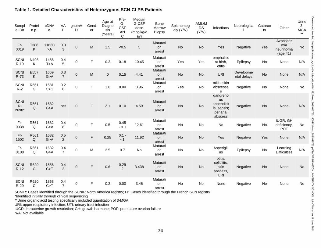
unique CLPB variants within 10 unrelated SCN patients (Table 1). Most of these variants were
confirmed by Sanger sequencing (Fig. S1). For two of these patients, family studies were
available and indicate de novo inheritance pattern (Fig. 1D). We also identified heterozygous
CLPB variants in 2 cases of cyclic neutropenia in the SCNIR cohort, with a third case identified
Dow
nloaded from http://ashpublications.org/blood/article-pdf/doi/10.1182/blood.2021010762/1809413/blood.2021010762.pdf by W
ASHIN
GTO
N U
NIVER
SITY SCH
OO
L, Julia Warren on 11 June 2021

11
in the French Neutropenia Registry; interestingly, all three cases carried the CLPB R628C
variant (Table S2). No copy number alterations of CLPB were detected. Of note, we identified
two additional CLPB variants that passed our filtering strategy (R327W and R603H); however,
both were present in an asymptomatic parent, indicating they are likely benign and are therefore
not included in Table 1.
Prior studies showed that biallelic variants of CLPB are associated with a syndrome
(CLPB syndrome) characterized by 3-methylglutaconic aciduria (3-MGA), cataracts, neurologic
disease, and variable neutropenia2–5
. However, the variants seen in CLPB syndrome and
CLPB-SCN are distinct. In patients with CLPB syndrome, the CLPB variants are always biallelic
and are found scattered throughout the protein6, with half of patients having at least one frame
shift or nonsense variant (Fig. 1E). In contrast, the variants observed in our series are
heterozygous, missense, and the localize to the C-terminal ATP-binding domain. We generated
a structural model of human CLPB by threading the primary amino acid sequence onto that of
Thermus thermophilus ClpB24
. All 6 of the heterozygous CLPB-SCN variants are predicted to
contribute to the ATP-binding pocket (Fig. 1E). Of note, these variants are in evolutionarily
conserved residues and, by homology, most are predicted to be crucial residues for nucleotide
binding and hydrolysis24–26
(Fig. S2).
The clinical characteristics of the 10 patients with SCN carrying ATP-binding pocket
CLPB variants is summarized in Table 1. All of the CLPB-SCN patients were diagnosed under
the age of 5 and most received G-CSF therapy, with a median dose of 5.62 mcg/kg/day (a
typical dose for SCN treatment). All demonstrated a myeloid maturation arrest and most had
documented severe infections prior to G-CSF therapy. One patient developed a myeloid
malignancy and is the only deceased patient from the cohort. In contrast to CLPB syndrome, in
which 3-methylglutaconic aciduria is universal, none of the 5 patients in our series with available
urine samples had 3-methylglutaconic aciduria6. Likewise, whereas cataracts and neurologic
Dow
nloaded from http://ashpublications.org/blood/article-pdf/doi/10.1182/blood.2021010762/1809413/blood.2021010762.pdf by W
ASHIN
GTO
N U
NIVER
SITY SCH
OO
L, Julia Warren on 11 June 2021

12
abnormalities were present in more than 90% of cases of CLPB syndrome (very often co-
occurring), they were uncommon in our CLPB-SCN cohort (2 non-overlapping patients each
with cataracts or epilepsy, and 1 with developmental concerns). No case had more than one
non-neutropenia CLPB syndrome feature. Of note, nearly 20% of patients with CLPB syndrome
did not have neutropenia. The different clinical and molecular features of CLPB syndrome and
CLPB-SCN suggest that these are distinct but related disorders with potentially unique
mechanisms of disease pathogenesis.
Loss of CLPB results in impaired granulocytic differentiation
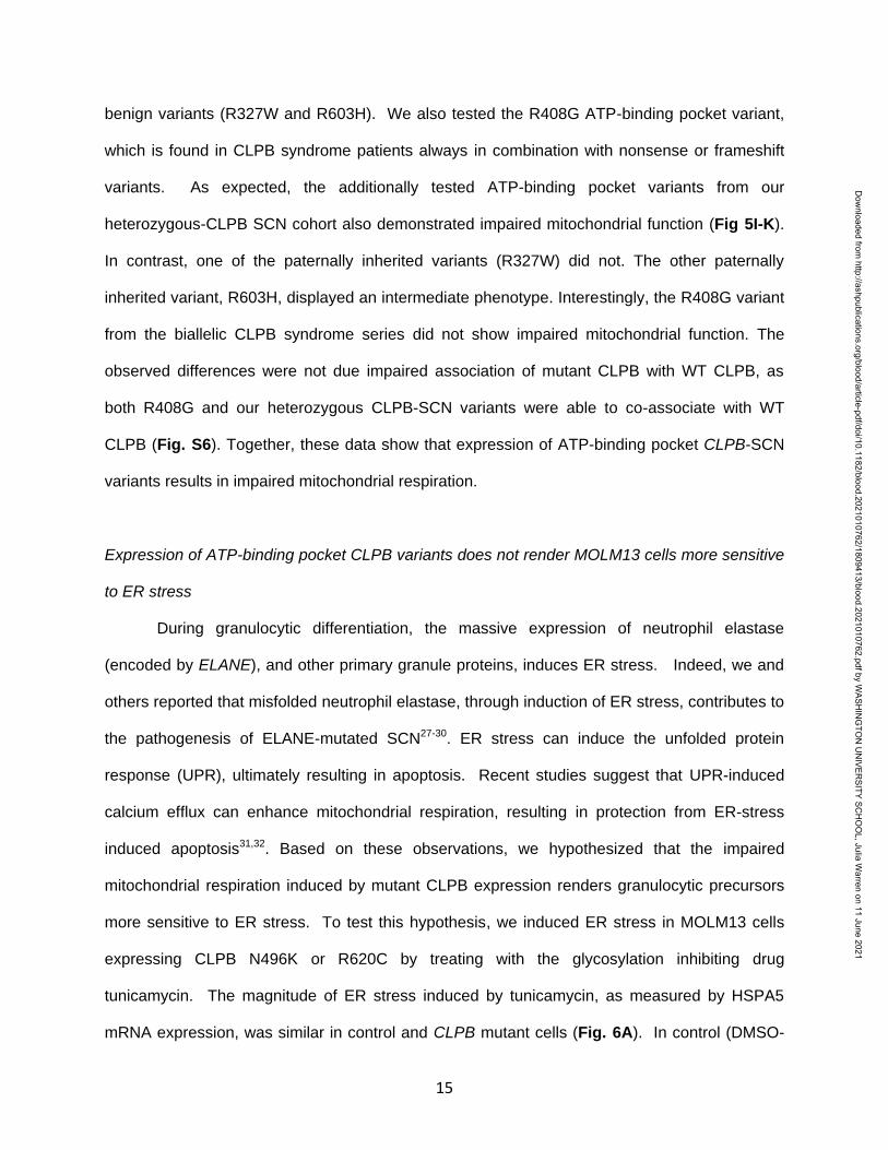
To assess the contribution of CLPB to granulopoiesis, we first used CRISPR-Cas9 gene
editing to generate null mutations in CLPB in human cord blood CD34+ hematopoietic
stem/progenitor cells (HSPCs) (Fig. S3A). We were able to achieve greater than 80% editing
efficiency, with a concordant decrease in protein expression (Fig. S3B-D). The gene-edited
HSPCs were cultured in the presence of G-CSF and stem cell factor (SCF), and differentiation
was assessed on day 14 by flow cytometry or by histomorphometry (Fig. 2A-B). Compared
with HSPCs transduced with control sgRNA, significantly fewer mature neutrophils and an
increase in granulocytic precursors were observed in cells transduced with two independent
sgRNAs targeting CLPB. A significant decrease in CFU-G also was observed (Fig. 2C). Gene
editing with the CRISPR-Cas9 system predominantly generates small insertion/deletions, some
of which are in-frame and may yield functional, intact CLPB protein. Next-generation
sequencing of cells from day 14 cultures showed a significant enrichment for non-targeted and
in-frame CLPB sequences in neutrophils but not granulocytic precursors, consistent with a
selective loss of CLPB-deficient cells during terminal granulocytic differentiation (Fig. 2D).
Impaired granulocytic differentiation was at least in part due to increased apoptosis of early
granulocytic precursors (Fig. 2E), with no change in cell cycle status observed (Fig. 2F and Fig.
S4). Together, these data show that CLPB is required for normal granulocytic differentiation.
Dow
nloaded from http://ashpublications.org/blood/article-pdf/doi/10.1182/blood.2021010762/1809413/blood.2021010762.pdf by W
ASHIN
GTO
N U
NIVER
SITY SCH
OO
L, Julia Warren on 11 June 2021

13
Expression of ATP-binding pocket CLPB variants results in impaired granulocytic differentiation
To assess the impact of heterozygous CLPB variants on granulopoiesis, we transduced
cord blood CD34+ cells with lentivirus expressing each of the four CLPB variants from our
SCNIR exome sequencing cohort (Fig. 3A). We included the likely benign, paternally inherited,
non-ATP binding cleft R603H variant as a negative control. Of note, since CLPB expression was
increased 4-10 fold compared to vector-alone controls (Fig. 3B and Fig. S5), we included a
wildtype CLPB lentiviral cohort to control for the effect of CLPB overexpression (Fig. 3B). A
significant decrease in CFU-G was observed for the four CLPB variants that localize to the ATP-
binding pocket (N496K, E557K, R561G, and R620C) but not the R603H variant (Fig. 3C and
1E). Fewer mature neutrophils and an increase in granulocytic precursors was present in
cultures of HSPCs transduced with the ATP-binding pocket CLPB variants but not the R603H
variant (Fig. 3D-E). Expression of ATP-binding pocket variants resulted in an increase in
apoptosis of granulocytic precursors but no change in cell cycle status (Fig. 3F-G). Collectively,
these data show that heterozygous ATP-binding pocket variants of CLPB cause impaired
granulocyte differentiation.
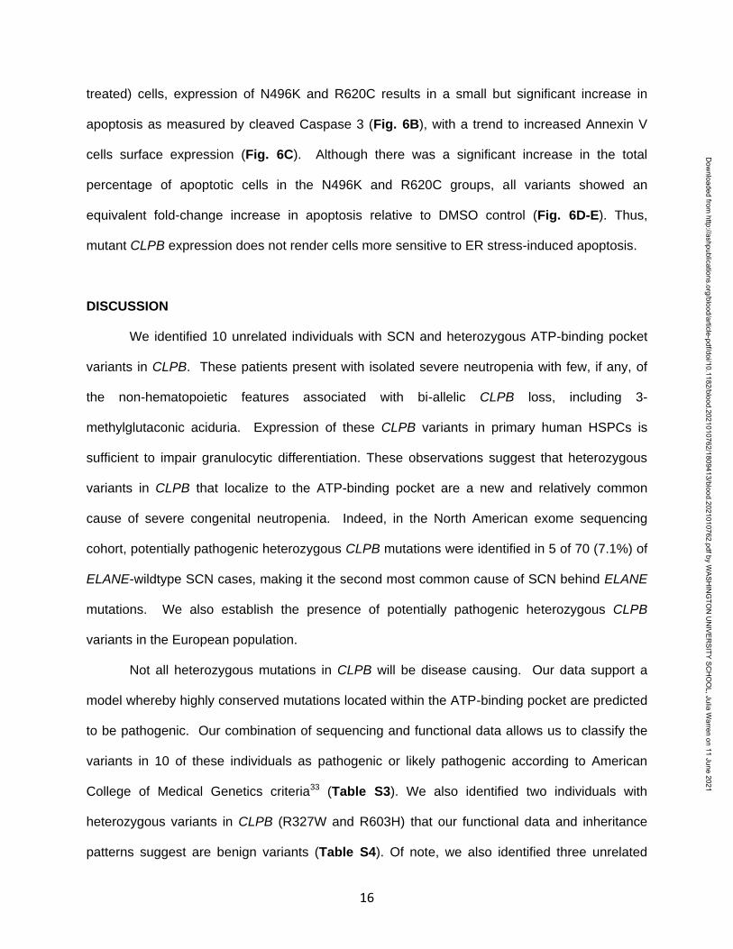
CLPB heterozygous variants have impaired ATPase and disaggregase activity, and exert a
dominant-negative effect on WT CLPB.
Human CLPB is an ATPase with potent disaggregase activity11,12. Biallelic CLPB
variants that impair ATP cleavage also show impaired disaggregase activity, though there also
appear to be ATPase-intact variants with impaired disaggregase activity that occurs through
alternative mechanisms11. The heterozygous nature of our CLPB-SCN variants suggests a
dominant negative effect on wildtype CLPB. To test this hypothesis, we performed mixing
studies with recombinant WT and mutant CLPB proteins. As a control, we included the R408G
CLPB variant that is found in biallelic CLPB syndrome in conjunction with a second CLPB
Dow
nloaded from http://ashpublications.org/blood/article-pdf/doi/10.1182/blood.2021010762/1809413/blood.2021010762.pdf by W
ASHIN
GTO
N U
NIVER
SITY SCH
OO
L, Julia Warren on 11 June 2021

14
variant. Of note, parents with heterozygous R408G CLPB are asymptomatic2,6, even though
purified R408G protein has impaired ATPase activity2. We also included the likely benign
variant, R603H. We show that all of the CLPB variants (except the R603H control) have
impaired ATPase and disaggregase activity when tested in isolation (Fig. 4A-B). When mixed
with WT CLPB, all of the heterozygous CLPB-SCN variants demonstrate greater than 50%
reduction in both ATPase (reaching significance for E557K and R561G) and disaggregase
activity (reaching significance for all CLPB-SCN variants) (Fig. 4C-D). In contrast, the R408G
variant, while having reduced ATPase and disaggregase activity in isolation (Fig. 4A-B), did not
suppress the activity of WT CLPB (Fig. 4C-D). As expected, the R603H variant had no effect.
Together, these data suggest that heterozygous CLPB-SCN variants act in a dominant-negative
fashion.
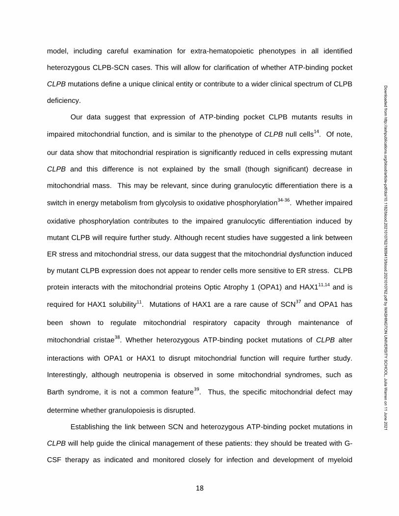
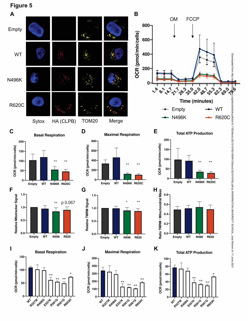
Expression of ATP-binding pocket CLPB variants results in impaired mitochondrial function
CLPB contains a mitochondrial localization sequence and has previously been shown to
localize to the mitochondria in a human myeloid progenitor cell line, MOLM-1314, and absence
of CLPB causes impaired mitochondrial protein solubility11. Therefore, we hypothesized that
CLPB-SCN heterozygous variants may affect granulocyte progenitors by impacting
mitochondrial function. To test this hypothesis, we generated MOLM-13 cells expressing
wildtype or mutant CLPB. We initially focused on CLPB N496K and R620C. Both wildtype and
mutant CLPB localize to mitochondria (Fig 5A). CLPB mutant cells had impaired mitochondrial
respiration, with significantly reduced basal and maximal respiratory capacity and a
corresponding decrease in ATP production (Fig. 5B-E). A small but significant decrease in
mitochondrial mass was observed in CLPB-N496K overexpressing cells, with a corresponding
decrease in mitochondrial membrane potential, as measured using TMRM (Fig. 5F-H).
We next expanded our analysis to include other ATP-binding pocket variants from our
heterozygous CLPB-SCN cohort (E557K, R561G, R561Q) and the two parentally inherited likely
Dow
nloaded from http://ashpublications.org/blood/article-pdf/doi/10.1182/blood.2021010762/1809413/blood.2021010762.pdf by W
ASHIN
GTO
N U
NIVER
SITY SCH
OO
L, Julia Warren on 11 June 2021
15
benign variants (R327W and R603H). We also tested the R408G ATP-binding pocket variant,
which is found in CLPB syndrome patients always in combination with nonsense or frameshift
variants. As expected, the additionally tested ATP-binding pocket variants from our
heterozygous-CLPB SCN cohort also demonstrated impaired mitochondrial function (Fig 5I-K).
In contrast, one of the paternally inherited variants (R327W) did not. The other paternally
inherited variant, R603H, displayed an intermediate phenotype. Interestingly, the R408G variant
from the biallelic CLPB syndrome series did not show impaired mitochondrial function. The
observed differences were not due impaired association of mutant CLPB with WT CLPB, as
both R408G and our heterozygous CLPB-SCN variants were able to co-associate with WT
CLPB (Fig. S6). Together, these data show that expression of ATP-binding pocket CLPB-SCN
variants results in impaired mitochondrial respiration.
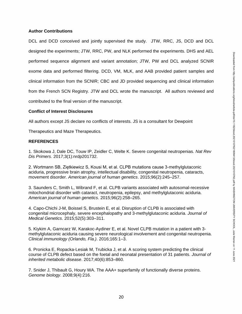
Expression of ATP-binding pocket CLPB variants does not render MOLM13 cells more sensitive
to ER stress
During granulocytic differentiation, the massive expression of neutrophil elastase
(encoded by ELANE), and other primary granule proteins, induces ER stress. Indeed, we and
others reported that misfolded neutrophil elastase, through induction of ER stress, contributes to
the pathogenesis of ELANE-mutated SCN27-30. ER stress can induce the unfolded protein
response (UPR), ultimately resulting in apoptosis. Recent studies suggest that UPR-induced
calcium efflux can enhance mitochondrial respiration, resulting in protection from ER-stress
induced apoptosis31,32. Based on these observations, we hypothesized that the impaired
mitochondrial respiration induced by mutant CLPB expression renders granulocytic precursors
more sensitive to ER stress. To test this hypothesis, we induced ER stress in MOLM13 cells
expressing CLPB N496K or R620C by treating with the glycosylation inhibiting drug
tunicamycin. The magnitude of ER stress induced by tunicamycin, as measured by HSPA5
mRNA expression, was similar in control and CLPB mutant cells (Fig. 6A). In control (DMSO-
Dow
nloaded from http://ashpublications.org/blood/article-pdf/doi/10.1182/blood.2021010762/1809413/blood.2021010762.pdf by W
ASHIN
GTO
N U
NIVER
SITY SCH
OO
L, Julia Warren on 11 June 2021
16
treated) cells, expression of N496K and R620C results in a small but significant increase in
apoptosis as measured by cleaved Caspase 3 (Fig. 6B), with a trend to increased Annexin V
cells surface expression (Fig. 6C). Although there was a significant increase in the total
percentage of apoptotic cells in the N496K and R620C groups, all variants showed an
equivalent fold-change increase in apoptosis relative to DMSO control (Fig. 6D-E). Thus,
mutant CLPB expression does not render cells more sensitive to ER stress-induced apoptosis.
DISCUSSION
We identified 10 unrelated individuals with SCN and heterozygous ATP-binding pocket
variants in CLPB. These patients present with isolated severe neutropenia with few, if any, of
the non-hematopoietic features associated with bi-allelic CLPB loss, including 3-
methylglutaconic aciduria. Expression of these CLPB variants in primary human HSPCs is
sufficient to impair granulocytic differentiation. These observations suggest that heterozygous
variants in CLPB that localize to the ATP-binding pocket are a new and relatively common
cause of severe congenital neutropenia. Indeed, in the North American exome sequencing
cohort, potentially pathogenic heterozygous CLPB mutations were identified in 5 of 70 (7.1%) of
ELANE-wildtype SCN cases, making it the second most common cause of SCN behind ELANE
mutations. We also establish the presence of potentially pathogenic heterozygous CLPB
variants in the European population.
Not all heterozygous mutations in CLPB will be disease causing. Our data support a
model whereby highly conserved mutations located within the ATP-binding pocket are predicted
to be pathogenic. Our combination of sequencing and functional data allows us to classify the
variants in 10 of these individuals as pathogenic or likely pathogenic according to American
College of Medical Genetics criteria33 (Table S3). We also identified two individuals with
heterozygous variants in CLPB (R327W and R603H) that our functional data and inheritance
patterns suggest are benign variants (Table S4). Of note, we also identified three unrelated
Dow
nloaded from http://ashpublications.org/blood/article-pdf/doi/10.1182/blood.2021010762/1809413/blood.2021010762.pdf by W
ASHIN
GTO
N U
NIVER
SITY SCH
OO
L, Julia Warren on 11 June 2021

17
families with cyclic neutropenia carrying the CLPB variant R628C. Although not functionally
validated, these data raise the possibility that this heterozygous CLPB variant may be a rare
cause of cyclic neutropenia. Together, these observations indicate that genetic testing for
CLPB should be included in the work-up of patients presenting with congenital neutropenia and
importantly that heterozygous mutations in the ATP-binding pocket should be considered
potentially pathogenic.
The mechanisms by which heterozygous ATP-binding pocket CLPB variants impair
granulocytic differentiation are not clear. Genetic and biochemical evidence support a
dominant-negative mechanism. Parents of patients with CLPB syndrome carrying heterozygous
null CLPB mutations are asymptomatic2-6. Additionally, we show that ATP-binding pocket CLPB
mutants co-immunoprecipitate with wildtype CLPB and inhibit the ATPase and disaggregase
activity of wildtype CLPB in mixing studies. On the other hand, a simple dominant-negative
mechanism does not account for all of the phenotypic differences between persons with
heterozygous CLPB mutations and CLPB syndrome. In particular, 3-MGA was not detected in
the urine of all evaluable CLPB-SCN cases (5 out of 10). In Pronicka’s review of 31 patients
with CLPB6, urine 3-MGA was detected in 29 of 29 evaluable cases, whereas it was not
detected in any of the 5 cases of CLPB-SCN where urine was available (P < 0.0001 compared
to 0/5 CLPB-SCN cases, by Fisher’s exact test). These observations suggest the possibility
that, while the ATPase activity of CLPB is needed to maintain mitochondrial functions required
for granulocytic differentiation, an (as yet undefined) ATPase-independent activity of CLPB
helps maintain mitochondrial functions required to suppress 3-methylglutagonic production. In
this model, the biallelic CLPB mutations in CLPB syndrome patients results in a loss (or partial
loss) of all CLPB functions, resulting in both neutropenia, 3-methylglutagonic aciduria, and other
nonhematopoietic phenotypes. In contrast, the heterozygous ATP-binding pocket mutations of
CLPB found in SCN cases only impair the ATPase-dependent functions of CLPB, leading to
severe neutropenia without 3-methylglutaconic aciduria. Further study is needed to test this
Dow
nloaded from http://ashpublications.org/blood/article-pdf/doi/10.1182/blood.2021010762/1809413/blood.2021010762.pdf by W
ASHIN
GTO
N U
NIVER
SITY SCH
OO
L, Julia Warren on 11 June 2021
18
model, including careful examination for extra-hematopoietic phenotypes in all identified
heterozygous CLPB-SCN cases. This will allow for clarification of whether ATP-binding pocket
CLPB mutations define a unique clinical entity or contribute to a wider clinical spectrum of CLPB
deficiency.
Our data suggest that expression of ATP-binding pocket CLPB mutants results in
impaired mitochondrial function, and is similar to the phenotype of CLPB null cells14
. Of note,
our data show that mitochondrial respiration is significantly reduced in cells expressing mutant
CLPB and this difference is not explained by the small (though significant) decrease in
mitochondrial mass. This may be relevant, since during granulocytic differentiation there is a
switch in energy metabolism from glycolysis to oxidative phosphorylation34-36
. Whether impaired
oxidative phosphorylation contributes to the impaired granulocytic differentiation induced by
mutant CLPB will require further study. Although recent studies have suggested a link between
ER stress and mitochondrial stress, our data suggest that the mitochondrial dysfunction induced
by mutant CLPB expression does not appear to render cells more sensitive to ER stress. CLPB
protein interacts with the mitochondrial proteins Optic Atrophy 1 (OPA1) and HAX111,14 and is
required for HAX1 solubility11. Mutations of HAX1 are a rare cause of SCN37
and OPA1 has
been shown to regulate mitochondrial respiratory capacity through maintenance of
mitochondrial cristae38
. Whether heterozygous ATP-binding pocket mutations of CLPB alter
interactions with OPA1 or HAX1 to disrupt mitochondrial function will require further study.
Interestingly, although neutropenia is observed in some mitochondrial syndromes, such as
Barth syndrome, it is not a common feature39
. Thus, the specific mitochondrial defect may
determine whether granulopoiesis is disrupted.
Establishing the link between SCN and heterozygous ATP-binding pocket mutations in
CLPB will help guide the clinical management of these patients: they should be treated with G-
CSF therapy as indicated and monitored closely for infection and development of myeloid
Dow
nloaded from http://ashpublications.org/blood/article-pdf/doi/10.1182/blood.2021010762/1809413/blood.2021010762.pdf by W
ASHIN
GTO
N U
NIVER
SITY SCH
OO
L, Julia Warren on 11 June 2021

19
malignancy. Our findings also establish an important cell-intrinsic role for CLPB in normal
human granulopoiesis. Future studies will be aimed at understanding the link between
mitochondrial dysfunction and impaired granulocytic differentiation.
Acknowledgements
We are grateful to the persons who contributed samples and clinical information for this study.
The authors wish to thank the Alvin J. Siteman Cancer Center at Washington University School
of Medicine and Barnes-Jewish Hospital in St. Louis, MO., for the use of the Siteman Flow
Cytometry Core, which provided cell sorting expertise; Jessica Hoisington-Lopez and MariaLynn
Cosby from the DNA Sequencing Innovation Laboratory at the Edison Family center for
Genome Sciences and Systems Biology for expertise with DNA sequencing; Sridhar
Nonavinkere Srivatsan for bioinformatics support; Severine Clauin for sequencing support;
Julien Buratti for bioinformatics analysis support; James Huang for providing comments on the
manuscript; and Paul Coppo, Mohamed Hamidou, and Amelie Servettaz for clinical expertise.
This work was supported by T32 Institutional NRSA Training Program in Developmental
Hematology HD007499-19 and Training of the Pediatric Physician-Scientist HD043010 (JTW),
American Society of Hematology Scholar Award (JTW), Children’s Discovery Institute
Fellowship MC-F-2020-871 (JTW), NIH/NIA F31AG060672 (RRC), NIH/NIGMS T32GM008275
(RRC), Blavatnik Family Foundation Fellowship (RRC), the National Cancer Institute
K08CA190815 (DHS), The G. Harold and Leila Y. Mathers Foundation (JS), NIH/NIGMS
R01GM099836 (JS), Inserm ITMO sante publique, X4 Pharma, Prolong Pharma and Chugai SA
(French SCN Registry), Foundation for Rare Diseases (AO9102LS), 111 Les Arts, the RMHE,
Association Barth France, and the Association Sportive de Saint Quentin Fallavier (CBC and
JD), NIH/NIAID 2R 24 AI 049393-Severe Chronic Neutropenia International Registry (DCD),
Department of Defense grant BM130173 (DCL), and R01HL152632-01 (DCL).
Dow
nloaded from http://ashpublications.org/blood/article-pdf/doi/10.1182/blood.2021010762/1809413/blood.2021010762.pdf by W
ASHIN
GTO
N U
NIVER
SITY SCH
OO
L, Julia Warren on 11 June 2021
20
Author Contributions
DCL and DCD conceived and jointly supervised the study. JTW, RRC, JS, DCD and DCL
designed the experiments; JTW, RRC, PW, and NLK performed the experiments. DHS and AEL
performed sequence alignment and variant annotation; JTW, PW and DCL analyzed SCNIR
exome data and performed filtering. DCD, VM, MLK, and AAB provided patient samples and
clinical information from the SCNIR; CBC and JD provided sequencing and clinical information
from the French SCN Registry. JTW and DCL wrote the manuscript. All authors reviewed and
contributed to the final version of the manuscript.
Conflict of Interest Disclosures
All authors except JS declare no conflicts of interests. JS is a consultant for Dewpoint
Therapeutics and Maze Therapeutics.
REFERENCES 1. Skokowa J, Dale DC, Touw IP, Zeidler C, Welte K. Severe congenital neutropenias. Nat Rev Dis Primers. 2017;3(1):nrdp201732.
2. Wortmann SB, Ziętkiewicz S, Kousi M, et al. CLPB mutations cause 3-methylglutaconic aciduria, progressive brain atrophy, intellectual disability, congenital neutropenia, cataracts, movement disorder. American journal of human genetics. 2015;96(2):245–257.
3. Saunders C, Smith L, Wibrand F, et al. CLPB variants associated with autosomal-recessive mitochondrial disorder with cataract, neutropenia, epilepsy, and methylglutaconic aciduria. American journal of human genetics. 2015;96(2):258–265.
4. Capo-Chichi J-M, Boissel S, Brustein E, et al. Disruption of CLPB is associated with congenital microcephaly, severe encephalopathy and 3-methylglutaconic aciduria. Journal of Medical Genetics. 2015;52(5):303–311.
5. Kiykim A, Garncarz W, Karakoc-Aydiner E, et al. Novel CLPB mutation in a patient with 3-methylglutaconic aciduria causing severe neurological involvement and congenital neutropenia. Clinical immunology (Orlando, Fla.). 2016;165:1–3.
6. Pronicka E, Ropacka-Lesiak M, Trubicka J, et al. A scoring system predicting the clinical course of CLPB defect based on the foetal and neonatal presentation of 31 patients. Journal of inherited metabolic disease. 2017;40(6):853–860.
7. Snider J, Thibault G, Houry WA. The AAA+ superfamily of functionally diverse proteins. Genome biology. 2008;9(4):216.
Dow
nloaded from http://ashpublications.org/blood/article-pdf/doi/10.1182/blood.2021010762/1809413/blood.2021010762.pdf by W
ASHIN
GTO
N U
NIVER
SITY SCH
OO
L, Julia Warren on 11 June 2021
21
8. Haslberger T, Zdanowicz A, Brand I, et al. Protein disaggregation by the AAA+ chaperone ClpB involves partial threading of looped polypeptide segments. Nature structural & molecular biology. 2008;15(6):641–650.
9. Deville C, Carroni M, Franke KB, et al. Structural pathway of regulated substrate transfer and threading through an Hsp100 disaggregase. Science advances. 2017;3(8):e1701726.
10. Rizo AN, Lin J, Gates SN, et al. Structural basis for substrate gripping and translocation by the ClpB AAA+ disaggregase. Nature communications. 2019;10(1):2393.
11. Cupo, RR, Shorter, J. Skd3 (human ClpB) is a potent mitochondrial protein disaggregase that is inactivated by 3-methylglutaconic aciduria-linked mutations. eLife. 2020;9:e55279.
12. Mroz, D, Wyszkowski, H, Szablewski, T, Zawieracz, K, Dutkiewicz, R, Bury, K, Wortmann, S, Wevers, R, Zietkiewicz, S. CLPB (caseinolytic peptidase B homolog), the first mitochondrial protein refoldase associated with human disease. Biochem Biophys Acta Gen Subj. 2020;1864(4):129512.
13. Yoshinaka T, Kosako H, Yoshizumi T, et al. Structural basis of mitochondrial scaffolds by prohibitin complexes: Insight into a role of the coiled-coil region. Iscience. 2019;19:1065–1078.
14. Chen X, Glytsou C, Zhou H, et al. Targeting Mitochondrial Structure Sensitizes Acute Myeloid Leukemia to Venetoclax Treatment. Cancer Discov. 2019;9(7):890–909.
15. Lek M, Karczewski KJ, Minikel EV, et al. Analysis of protein-coding genetic variation in 60,706 humans. Nature. 2016;536(7616):285–291.
16. Network CGAR, Ley TJ, Miller C, et al. Genomic and epigenomic landscapes of adult de novo acute myeloid leukemia. The New England journal of medicine. 2013;368(22):2059–2074.
17. Talevich E, Shain AH, Botton T, Bastian BC. CNVkit: Genome-Wide Copy Number Detection and Visualization from Targeted DNA Sequencing. Plos Comput Biol. 2016;12(4):e1004873.
18. Halene S, Wang L, Cooper RM, et al. Improved Expression in Hematopoietic and Lymphoid Cells in Mice After Transplantation of Bone Marrow Transduced With a Modified Retroviral Vector. Blood. 1999;94(10):3349–3357.
19. Robbins PB, Skelton DC, Yu XJ, et al. Consistent, persistent expression from modified retroviral vectors in murine hematopoietic stem cells. Proceedings of the National Academy of Sciences of the United States of America. 1998;95(17):10182–10187.
20. Brunetti L, Gundry MC, Kitano A, Nakada D, Goodell MA. Highly Efficient Gene Disruption of Murine and Human Hematopoietic Progenitor Cells by CRISPR/Cas9. Journal of visualized experiments : JoVE. 2018;(134):e57278–e57278.
21. Clement K, Rees H, Canver MC, et al. CRISPResso2 provides accurate and rapid genome editing sequence analysis. Nature biotechnology. 2019;37(3):224–226.
Dow
nloaded from http://ashpublications.org/blood/article-pdf/doi/10.1182/blood.2021010762/1809413/blood.2021010762.pdf by W
ASHIN
GTO
N U
NIVER
SITY SCH
OO
L, Julia Warren on 11 June 2021
22
22. Cupo RR, Shorter J. Expression and Purification of Recombinant Skd3 (Human ClpB) Protein and Tobacco Etch Virus (TEV) Protease from Escherichia coli. Bio Protoc. 2020; 10(23):e3858.
23. DeSantis ME, Leung EH, Sweeny EA, Jackrel ME, Cushman-Nick M, Neuhaus-Follini A, Vashist S, Sochor MA, Knight MN, Shorter J. Operational plasticity enables hsp104 to disaggregate diverse amyloid and nonamyloid clients. Cell. 2012;141(4):778-793.
24. Lee S, Sowa ME, Watanabe Y, et al. The structure of ClpB: a molecular chaperone that rescues proteins from an aggregated state. Cell. 2003;115(2):229–240.
25. Zeymer C, Fischer S, Reinstein J. trans-Acting arginine residues in the AAA+ chaperone ClpB allosterically regulate the activity through inter- and intradomain communication. Journal of Biological Chemistry. 2014;289(47):32965–32976.
26. Wendler P, Ciniawsky S, Kock M, Kube S. Structure and function of the AAA+ nucleotide binding pocket. Biochim Biophys Acta. 2011;1823(1):2–14.
27. Grenda, D, Murakami, M, Ghatak, J, Xia, J, Boxer, L, Dale, D, Dinauer, M, Link, D. Mutations of the ELA2 gene found in patients with severe congenital neutropenia induce the unfolded protein response and cellular apoptosis. Blood. 2007;110(13): 4179 - 4187.
28. Nustede, R, Klimiankou, M, Klimenkova, O, Kuznetsova, I, Zeidler, C, Welte, K, Skokowa, J . ELANE mutant-specific activation of different UPR pathways in congenital neutropenia. British J Haemat. 2016;172(2):219 - 227.
29. Köllner, I, Sodeik, B, Schreek, S, Heyn, H, Neuhoff, N, Germeshausen, M, Zeidler, C, Krüger, M, Schlegelberger, B, Welte, K, Beger, CMutations in neutrophil elastase causing congenital neutropenia lead to cytoplasmic protein accumulation and induction of the unfolded protein response. Blood. 2006;108(2):493 - 500.
30. Nanua, S, Murakami, M, Xia, J, Grenda, D, Woloszynek, J, Strand, M, Link, D. Activation of the unfolded protein response is associated with impaired granulopoiesis in transgenic mice expressing mutant Elane. Blood. 2011;117(13):3539 - 3547.
31. Bravo, R, Vicencio, JM, Parra, V, Troncoso, R, Munoz, JP, Bui, M, Quiroga, C, Rodriguez, AE, Verdejo, HE, Ferreira, J, Iglewski, M, Chiong, M, Simmen, T, Zorzano, A, Hill, JA, Rothermel, BA, Szabadkai, G, Lavendero, S. Increased ER-mitochondrial coupling promotes mitochondrial respiration and bioenergetics during early phases of ER stress. J Cell Sci. 2011;124(Pt13):2143-52.
32. Knupp, J, Arvan, P, Chang, A. Increased mitochondrial respiration promotes purvival from endoplasmic reticulum stress. Cell Death Differ. 2019;26:487-501.
33. Richards, S, Aziz, N, Bale, S, Bick, D, Das, S, Fastier-Foster, J, Grody, W, Hegde, M, Lyon, E, Spector, E, Voelkerding, K, Rehm, H, on behalf of the ACMG Laboratory Quality Assurance Committee. Standards and Guidelines for the Interpretation of Sequence Variants: a Joint Consensus Recommendation of the American College of Medical Genetics and Genomics and the Association for Molecular Pathology. Genetics in Medicine. 2015;17: 405-423.
Dow
nloaded from http://ashpublications.org/blood/article-pdf/doi/10.1182/blood.2021010762/1809413/blood.2021010762.pdf by W
ASHIN
GTO
N U
NIVER
SITY SCH
OO
L, Julia Warren on 11 June 2021

23
34. Vannini N, Girotra M, Naveiras O, et al. Specification of haematopoietic stem cell fate via modulation of mitochondrial activity. Nat Commun. 2016;7(1):13125.
35. Six E, Lagresle-Peyrou C, Susini S, et al. AK2 deficiency compromises the mitochondrial energy metabolism required for differentiation of human neutrophil and lymphoid lineages. Cell Death Dis. 2015;6(8):e1856–e1856.
36. Rice CM, Davies LC, Subleski JJ, et al. Tumour-elicited neutrophils engage mitochondrial metabolism to circumvent nutrient limitations and maintain immune suppression. Nat Commun. 2018;9(1):5099.
37. Klein C, Grudzien M, Appaswamy G, et al. HAX1 deficiency causes autosomal recessive severe congenital neutropenia (Kostmann disease). Nature genetics. 2007;39(1):86–92.
38. Mishra P, Carelli V, Manfredi G, Chan DC. Proteolytic Cleavage of Opa1 Stimulates Mitochondrial Inner Membrane Fusion and Couples Fusion to Oxidative Phosphorylation. Cell Metab. 2014;19(4):630–641.
39. Finsterer J. Hematological manifestations of primary mitochondrial disorders. Acta haematologica. 2007;118(2):88–98.
Dow
nloaded from http://ashpublications.org/blood/article-pdf/doi/10.1182/blood.2021010762/1809413/blood.2021010762.pdf by W
ASHIN
GTO
N U
NIVER
SITY SCH
OO
L, Julia Warren on 11 June 2021
24
Table 1. Detailed Characteristics of Heterozygous SCN-CLPB Patients
Sample ID#
Protein p.
cDNA c.
VAF
gnomAD
Gender
Age at Diagno
sis (Years)
Pre-G-
CSF ANC
Median G-CSF dose
(mcg/kg/day)
Bone Marrow Biopsy
Splenomegaly (Y/N)
AML/MDS
(Y/N) Infections
Neurological
Cataracts
Other
Urine 3-
MGA**
Fr-0019
T388K
1163C>A
0.33
0 M 1.5 <0.5 5 Maturati
on arrest
No No Yes Negative Yes
Azoospermia
neurinoma (age 41)
No
SCNIR-19
N496K
1488 T>A
0.45
0 F 0.2 0.18 10.45 Maturati
on arrest
Yes Yes omphalitis
at birth, otitis
Epilepsy No None N/A
SCNIR-73
E557K
1669 G>A
0.37
0 M 0 0.15 4.41 Maturati
on arrest
No No URI Developmental delays
No None N/A
SCNIR-2
R561G
1681 C>G
0.26
0 F 1.6 0.00 3.96 Maturati
on arrest
Yes No otitis, skin abscesse
s Negative No None No
SCNIR-
2698*
R561Q
1682 G>A
het 0 F 2.1 0.10 4.59 Maturati
on arrest
No No
gangrenous
appendicitis, sepsis; perianal abscess
Negative No None N/A
Fr-0038
R561Q
1682 G>A
0.48
0 F 0.5 0.45 - < 1
12.61 Maturati
on arrest
No No No Negative No IUGR, GH deficiency,
POF No
Fr-1502
R561Q
1682 G>A
0.52
0 F 0.25 0.1 - <1
11.92 Maturati
on arrest
No No Yes Negative Yes None N/A
Fr-0108
R561Q
1682 G>A
0.47
0 M 2.5 0.7 No Maturati
on arrest
No No Asperigill
us Epilepsy No
Learning Difficulties
N/A
SCNIR-12
R620C
1858 C>T
0.43
0 F 0.6 0.29
2 3.438
Maturation
arrest No No
otitis, cellulitis,
skin abscess,
URI
Negative No None No
SCNIR-29
R620C
1858 C>T
0.47
0 F 0.2 0.00 3.45 Maturati
on arrest
No No None Negative No None No
SCNIR: Cases identified through the SCNIR North America registry; Fr: Cases identified through the French SCN registry *Identified initially through clinical sequencing **Urine organic acid testing specifically included quantitation of 3-MGA URI: upper respiratory infection; UTI: urinary tract infection IUGR: intrauterine growth restriction; GH: growth hormone; POF: premature ovarian failure N/A: Not available
Dow
nloaded from http://ashpublications.org/blood/article-pdf/doi/10.1182/blood.2021010762/1809413/blood.2021010762.pdf by W
ASHIN
GTO
N U
NIVER
SITY SCH
OO
L, Julia Warren on 11 June 2021
25
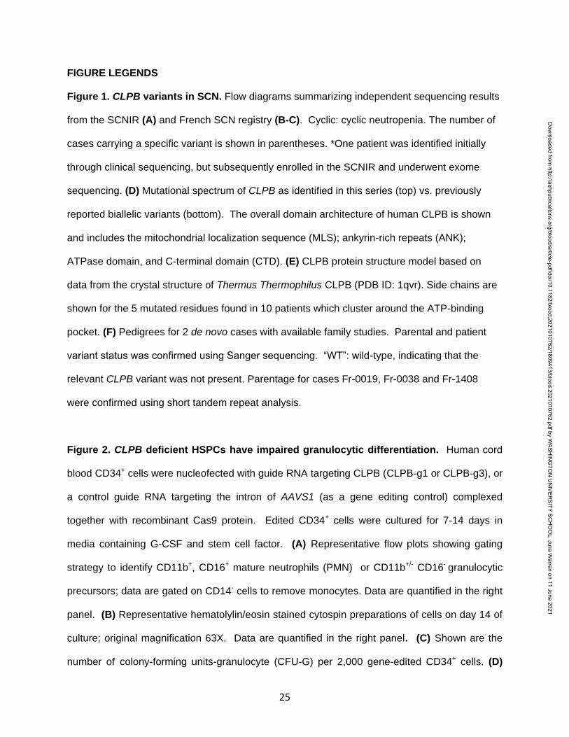
FIGURE LEGENDS
Figure 1. CLPB variants in SCN. Flow diagrams summarizing independent sequencing results
from the SCNIR (A) and French SCN registry (B-C). Cyclic: cyclic neutropenia. The number of
cases carrying a specific variant is shown in parentheses. *One patient was identified initially
through clinical sequencing, but subsequently enrolled in the SCNIR and underwent exome
sequencing. (D) Mutational spectrum of CLPB as identified in this series (top) vs. previously
reported biallelic variants (bottom). The overall domain architecture of human CLPB is shown
and includes the mitochondrial localization sequence (MLS); ankyrin-rich repeats (ANK);
ATPase domain, and C-terminal domain (CTD). (E) CLPB protein structure model based on
data from the crystal structure of Thermus Thermophilus CLPB (PDB ID: 1qvr). Side chains are
shown for the 5 mutated residues found in 10 patients which cluster around the ATP-binding
pocket. (F) Pedigrees for 2 de novo cases with available family studies. Parental and patient
variant status was confirmed using Sanger sequencing. “WT”: wild-type, indicating that the
relevant CLPB variant was not present. Parentage for cases Fr-0019, Fr-0038 and Fr-1408
were confirmed using short tandem repeat analysis.
Figure 2. CLPB deficient HSPCs have impaired granulocytic differentiation. Human cord
blood CD34+ cells were nucleofected with guide RNA targeting CLPB (CLPB-g1 or CLPB-g3), or
a control guide RNA targeting the intron of AAVS1 (as a gene editing control) complexed
together with recombinant Cas9 protein. Edited CD34+ cells were cultured for 7-14 days in
media containing G-CSF and stem cell factor. (A) Representative flow plots showing gating
strategy to identify CD11b+, CD16+ mature neutrophils (PMN) or CD11b+/- CD16- granulocytic
precursors; data are gated on CD14- cells to remove monocytes. Data are quantified in the right
panel. (B) Representative hematolylin/eosin stained cytospin preparations of cells on day 14 of
culture; original magnification 63X. Data are quantified in the right panel. (C) Shown are the
number of colony-forming units-granulocyte (CFU-G) per 2,000 gene-edited CD34+ cells. (D)
Dow
nloaded from http://ashpublications.org/blood/article-pdf/doi/10.1182/blood.2021010762/1809413/blood.2021010762.pdf by W
ASHIN
GTO
N U
NIVER
SITY SCH
OO
L, Julia Warren on 11 June 2021

26
Fold change (from day 0) in edited (for CLPB, edited out-of-frame) or unedited cells (for CLPB,
unedited plus edited in-frame) in PMNs or granulocytic precursors sorted on day 14. (E)
Percentage of caspase-3+ granulocytic precursors on day 7 of culture. (F) Cells were cultured
for 7 days and cell cycle was assessed by flow cytometry. Data represent 3-5 independent
experiments. *p<0.05, **p<0.01, ***p<0.005. Statistical significance was determined using
repeated measures one-way ANOVA.
Figure 3. Expression of ATP-binding pocket CLPB mutants impairs granulocytic
differentiation. Cord blood CD34+ cells were transduced with lentivirus expressing the
indicated CLPB cDNA (or empty vector control). GFP+ cells were sorted at 48 hours, then
seeded into media containing G-CSF and stem factor or methylcellulose containing G-CSF. (A)
Lentiviral vector. LTR; long terminal repeat; HA; hemagglutinin epitope tag; IRES: internal
ribosomal entry site; GFP: green fluorescent protein; WPRE: woodchuck promoter responsive
element. (B) RNA expression of CLPB relative to -actin mRNA. (C) CFU-G per 2,000 GFP+
CD34+ cells. (D-E) The percentage of mature neutrophils (D) and granulocytic precursors (E)
on day 14 of culture is shown; data are gated on CD14- cells to exclude monocytes. (F)
Percentage of caspase-3+ granulocytic precursors on day 7 of culture. (G) Cells were cultured
for 7 days and cell cycle was assessed by flow cytometry. Data represent 3-5 independent
experiments. *p<0.05,**p<0.01,***p<0.005. Statistical significance was determined using
repeated measures one-way ANOVA.
Figure 4. CLPB mutants show impaired ATPase and disaggregase activity, and exhibit a
dominant negative effect on WT CLPB. Purified CLPB protein was used to measure (A)
ATPase activity of the indicated CLPB variants tested in isolation; (B) Disaggregase activity of
the indicated CLPB variants tested in isolation; (C) ATPase activity of WT CLPB when mixed
1:1 with WT CLPB or CLPB variants; and (D) Disaggregase activity of WT CLPB when mixed
Dow
nloaded from http://ashpublications.org/blood/article-pdf/doi/10.1182/blood.2021010762/1809413/blood.2021010762.pdf by W
ASHIN
GTO
N U
NIVER
SITY SCH
OO
L, Julia Warren on 11 June 2021

27
1:1 with WT CLPB or CLPB variants. For (C-D), the dashed line represents 50% of WT CLPB
activity. Data represent 3 independent experiments. For (A-B), comparison was made to WT
CLPB activity. For (C-D), comparison was made to values representing 50% of WT activity.
*p<0.05, ** p<0.01, ***p<0.001, ****p<0.0001. Statistical significance was determined using one-
way ANOVA.
Figure 5. Expression of CLPB mutants impairs mitochondrial respiration without
affecting membrane potential. MOLM-13 cells were transduced with a lentiviral vector
expressing WT CLPB, CLPB-N496K, or CLPB-R620C. (A) Representative photomicrographs of
cells stained with anti-HA antibody to detect CLPB (red) and anti-TOM20 (yellow) to label
mitochondria. Nuclei were counterstained with Sytox (blue); original magnification 63X. (B-E)
The mitochondrial stress test was performed using the Seahorse XF96e analyzer. (B)
Representative graph showing the oxygen consumption rate (OCR) at baseline and after
treatment with the ATP synthase inhibitor oligomycin (OM) and then the uncoupling agent
carbonyl cyanide-4 phenylhydrazone (FCCP). Parameters of mitochondrial respiration include
(C) Basal respiration; (D) Maximal respiration and (E) total ATP production. (F-H) Cells were
labeled with tetramethylrhodamine (TMRM) or mitotracker green (MG). (F) Mitotracker green
signal normalized to empty vector control. (G) TMRM signal normalized to empty vector control.
(H) Ratio of TMRM signal to mitotracker green signal is shown. Mitochondrial respiration
parameters for additional CLPB variants: (I) Basal respiration; (J) Maximal respiration and (K)
total ATP production. *p<0.05,**p<0.01. Statistical significance was determined using repeated
measures one-way ANOVA.
Figure 6. Induction of UPR Does Not Cause Increased Fold-change in Apoptosis in CLPB
Variant-Expressing Myeloid Cells. The myeloid cell line MOLM-13 was transduced with CLPB
variants or empty vector control and cells were sorted based on GFP-positivity. Cells were
Dow
nloaded from http://ashpublications.org/blood/article-pdf/doi/10.1182/blood.2021010762/1809413/blood.2021010762.pdf by W
ASHIN
GTO
N U
NIVER
SITY SCH
OO
L, Julia Warren on 11 June 2021

28
treated with DMSO (D) or the glycosylation-inhibitor tunicamycin (TM) for 24 hours. (A)
Induction of the UPR was confirmed by assessing expression of HSPA5 by qPCR. The
percentage of cleaved caspase-3+ (B) or Annexin V+ (C) cells was assessed by flow cytometry.
Fold-change representation of cleaved caspase-3+ (D) or the Annexin V+ (E) cells expressed
as the ratio of tunicamycin divided by DMSO within each sample. Data represent 3 independent
experiments. *p<0.05,**p<0.01,***p<0.001. Statistical significance was determined using
repeated measures one-way ANOVA.
Dow
nloaded from http://ashpublications.org/blood/article-pdf/doi/10.1182/blood.2021010762/1809413/blood.2021010762.pdf by W
ASHIN
GTO
N U
NIVER
SITY SCH
OO
L, Julia Warren on 11 June 2021

Dow
nloaded from http://ashpublications.org/blood/article-pdf/doi/10.1182/blood.2021010762/1809413/blood.2021010762.pdf by W
ASHIN
GTO
N U
NIVER
SITY SCH
OO
L, Julia Warren on 11 June 2021

Contr
ol
CLPB
-g1
0
1
2
3
Fo
ld-c
han
ge (
Rela
tive t
o D
ay 0
)
PMN
Contr
ol
CLPB
-g1
Granulocytic Precursors
Unedited
Edited
0
10
20
30
%C
asp
ase +
Contr
ol
CLPB
-g1
Contr
ol
CLPB
-g1
0
50
100P
erc
en
t
G0
G1
S
G2/M
Contr
ol
CLPB
-g1
CLPB
-g3
0
20
40
60
CF
U-G
Contr
ol
CLPB
g1
CLPB
g3
0
20
40
60
80
100
Cells/1
00
PMN
Contr
ol
CLPB
g1
CLPB
g3
Granulocytic Precursors
Contr
ol
CLPB
-g1
CLPB
-g3
0
20
40
60
80
100
Perc
en
t o
f C
D14-
PMN
Contr
ol
CLPB
-g1
CLPB
-g3
Granulocytic Precursors
Dow
nloaded from http://ashpublications.org/blood/article-pdf/doi/10.1182/blood.2021010762/1809413/blood.2021010762.pdf by W
ASHIN
GTO
N U
NIVER
SITY SCH
OO
L, Julia Warren on 11 June 2021

Dow
nloaded from http://ashpublications.org/blood/article-pdf/doi/10.1182/blood.2021010762/1809413/blood.2021010762.pdf by W
ASHIN
GTO
N U
NIVER
SITY SCH
OO
L, Julia Warren on 11 June 2021

WT
R40
8G
N49
6K
E557K
R56
1G
R60
3H
R62
0C
0
10
20
30
40
50
ATPase Activity
AT
Pase (
min
-1)
WT:
WT
WT:
R40
8G
WT:
N49
6K
WT:
E557K
WT:
R56
1G
WT:
R60
3H
WT:
R62
0C
0
10
20
30
ATPase (Mixing)
AT
Pase (
min
-1)
WT
R40
8G
N49
6K
E557K
R56
1G
R60
3H
R62
0C
0.0
0.5
1.0
1.5
No
rmalized
Lu
cif
era
se
Reacti
vati
on
Disaggregation
WT:
WT
WT:
R40
8G
WT:
N49
6K
WT:
E557K
WT:
R56
1G
WT:
R60
3H
WT:
R62
0C
0.0
0.5
1.0
1.5
Disaggregation (mixing)
No
rmalized
Lu
cif
era
se
Reacti
vati
on
Dow
nloaded from http://ashpublications.org/blood/article-pdf/doi/10.1182/blood.2021010762/1809413/blood.2021010762.pdf by W
ASHIN
GTO
N U
NIVER
SITY SCH
OO
L, Julia Warren on 11 June 2021
Empty WT N496 R6200.0
0.5
1.0
1.5
** p 0.067
Rela
tive M
ito
tracker
Sig
nal
Empty WT N496 R6200.0
0.5
1.0
1.5
***
Rela
tive T
MR
M S
ign
al
Empty WT N496K R620C0
50
100
150
200
Total ATP Production
OC
R (
pm
ol/m
in/c
ells)
** **
Empty WT N496K R620C0
50
100
150
200
OC
R (
pm
ol/m
in/c
ells)
Basal Respiration
** **
( )
Empty WT N496K R620C0
200
400
600
800
Maximal Respiration
OC
R (
pm
ol/m
in/c
ells)
** **
Empty WT N496 R6200.0
0.2
0.4
0.6
0.8
Rati
o T
MR
M / M
ito
ch
on
dri
al M
ass
1.4
8.1
14.7
21.7
28.3
35.0
42.0
48.7
55.3
62.3
69.0
75.6
0
200
400
600
800
Time (minutes)
OC
R (
pm
ol/m
in/c
ells)
Empty WT
N496K R620C
OM FCCP
WT
R32
7W
R40
8G
E557K
R56
1G
R56
1Q
R60
3H
0
50
100
150
Basal Respiration
OC
R (
pm
ol/m
in/c
ells)
WT
R32
7W
R40
8G
E557K
R56
1G
R56
1Q
R60
3H
0
100
200
300
400
500
Maximal Respiration
OC
R (
pm
ol/m
in/c
ells)
WT
R32
7W
R40
8G
E557K
R56
1G
R56
1Q
R60
3H
0
20
40
60
80
100
Total ATP Production
OC
R (
pm
ol/m
in/c
ells)
Dow
nloaded from http://ashpublications.org/blood/article-pdf/doi/10.1182/blood.2021010762/1809413/blood.2021010762.pdf by W
ASHIN
GTO
N U
NIVER
SITY SCH
OO
L, Julia Warren on 11 June 2021

O M O M O M O M
0
5
10
15
20
HSPA5
Rela
tive E
xp
ressio
n(N
orm
alized
to
GA
PD
H)
0
5
10
15
20
%A
nn
exin
V+
Annexin V+
0
5
10
15
20
Caspase3+
Casp
ase 3
+
Empty W
T
N49
6K
R62
0C
0
2
4
6
8
10
Fo
ld-c
han
ge A
nn
exin
V+
(Rela
tive t
o D
MS
O)
Annexin V+
Empty W
T
N49
6K
R62
0C
0
2
4
6
8
10
Fo
ld-c
han
ge %
Casp
ase3+
(Rela
tive t
o D
MS
O)
Caspase3+ fold-change
Dow
nloaded from http://ashpublications.org/blood/article-pdf/doi/10.1182/blood.2021010762/1809413/blood.2021010762.pdf by W
ASHIN
GTO
N U
NIVER
SITY SCH
OO
L, Julia Warren on 11 June 2021