high-energy neutron therapy for radioresistant cancers › documents › lennoxpaper.pdf ·...
TRANSCRIPT

High-Energy Neutron Therapy for Radioresistant Cancers
A.J. Lennox
Northern Illinois University Institute for Neutron Therapy at FermilabFermi National Accelerator Laboratory, P.O. Box 500 Mail Stop 301, Batavia, Illinois USA
Abstract
Randomized prospective clinical trials have demonstrated that high energy neutron therapy is superior tolow dE/dx radiation such as photons or protons for certain cancers, including salivary gland tumors,melanoma, sarcoma and locally advanced prostate cancer. Neutrons exhibit a biological advantagebecause their interactions in tissue are quite different from those of photons and protons. For best clinicalresults, proton linacs or cyclotrons are used to generate an ~70 MeV proton beam with average intensity~200 microamperes. The proton beam strikes a beryllium target to produce a high energy neutron beamthat, unlike reactor-generated neutron beams, is well collimated and can be used for conformal neutrontherapy or intensity modulated neutron therapy. This paper discusses the radiobiology of neutron-tissueinteractions, presents clinical results of national/international trials, and describes some issues related todesigning a modern high-energy neutron therapy facility, including the possibility of generating medicallyuseful radioisotopes.
1. IntroductionInternational clinical trials with fast neutrons were enthusiastically embraced from the mid-1970’s
through the mid-1980’s, only to be nearly abandoned in the late 1980’s as clinicians observed unacceptableside effects. The problem was analyzed by Griffin et al in a 1986 publication showing that neutrongenerators using primary deuteron beams with energies below 50 MeV produced beams with tissue-penetration properties that made it difficult to achieve good clinical results [1]. By the late 1980’s only oneof the facilities described by Griffin continued to operate, namely the p(66)Be(49) beam at the FermiNational Accelerator Laboratory (Fermilab). In 1984 a new p(50)Be(25) beam was commissioned at theUniversity of Washington in Seattle, Washington. These two high-energy beams continue to operate asclinical beams because the clinicians recognize the value of fast neutron therapy for otherwise intractablecases. In addition, a fast neutron beam modeled on the Fermilab beam continues to operate at iThembaLaboratory in South Africa and a lower-energy fast neutron beam is used to treat salivary gland tumors atthe University Clinic in Essen, Germany. The next section describes the unique biological properties of fastneutron therapy.
2. Radiobiological Aspects of Neutron Therapy
Extensive research leading to an understanding of the radiobiology of neutrons was conducted in Englandduring the 1970’s. A summary of the most important findings is given in reference [2]. Specifically
• Neutrons are more effective per unit dose than x-rays• Cell survival curves for neutrons are more nearly exponential than those of x-rays• The modifying effect of hypoxia is smaller for neutrons than for photons• Cell sensitivity to neutrons is much less dependent on cell growth stage than cell
sensitivity to photons

These phenomena can be explained by comparing the interactions of neutrons and x-rays (photons) intissue. Because protons interact with tissue in much the same way as x-rays, the limitations mentionedabove for x-rays also apply to protons.
2.1 Neutron Interactions with TissueWith conventional photon or proton therapy, the radiation beam interacts with atoms in human tissue
primarily via electromagnetic interactions that may disturb the molecular bonding in cellular DNA. Or thephotons or protons may interact with water molecules in the body to form the OH- radical, that, in turndamages DNA. The presence of oxygen in the cell facilitates the formation of OH-; this explains whysmaller, well-oxygenated tumors are more likely to respond to photon or proton irradiation while largerhypoxic tumors are less affected by these types of radiation. Because the amount of energy transferred tothe cell is relatively small in a single interaction, these interactions are called low Linear-Energy-Transfer(LET) reactions. Low LET damage to tumor cells is often repaired, and the tumor continues to grow.
In contrast, neutrons interact primarily via (n,p) or spallation interactions, depositing a large amount ofenergy, (high LET), and often transforming the atom in the DNA strand into a completely different atom.A tumor cell whose DNA is damaged to this extent cannot repair itself and will ultimately die. Thisinability for the tumor to repair is one factor accounting for the higher relative biological effectiveness(RBE) of neutron therapy and for the differences in the shapes of cell-survival curves. Many of the earlyradiobiology textbooks hypothesized that the number of double-strand breaks in DNA was greater for highLET neutrons than for low LET radiation. It was believed that the greater number of double strand breakswas responsible for the smaller amount of repair observed with high LET radiation. In fact, as severalradiobiological experiments at Fermilab have shown, the number of single and double strand breaks isabout the same for high and low LET radiation. The smaller incidence of repair with high LET radiation isdue to the more extensive nature of the damage at an interaction site rather than a larger number ofinteraction sites. The high RBE associated with high LET radiation has also been attributed to the fact thatfor high LET radiation, cell killing is relatively independent of cell growth-cycle stage [3].
With any type of radiation some healthy tissue will also experience a sub-lethal dose during theprocess of treating a tumor. In cases where a damaged healthy cell cannot repair, other healthy cells in theneighborhood of a damaged healthy cell counteract the damage by generating new cells (repopulation).Thus, as will be seen in the following section, healthy tissue that has received a sublethal dose of neutronswill recover, just as healthy tissue damaged by sublethal doses of photons or protons will recover. In bothcases the extent of long-lasting damage (late side effects) depends on the given dose. A detailedcomparison of the interactions of photons and neutrons in tissue is given in reference [4].
2.2 Illustrative Radiobiological Results
Figure 1 shows a simple example of the difference between neutron and photon cell-killing propertiesfor two different human prostate cancer cell lines, both of which are classified as being radioresistant.DU145 and PC3 human prostate cancer cells were irradiated to a single 3 Gy dose of either p(66)Be(49)neutrons at Fermilab or photons from a cesium source at Rush-Presbyterian-St. Luke’s Medical Center [5].Two characteristics are immediately noticed. First, neutrons were more effective than photons at killingboth types of tumor cells, with the small difference in survival not being statistically significant. Second,there is a considerable difference in survival in the two cell lines when photons are used. The cause of thisdifference was not examined, but it is consistent with earlier observations that with photons the amount ofcell-kill can vary with factors such as cell growth-cycle stage and oxygen levels.
In a more realistic follow-up experiment, the DU145 cells were irradiated with either photons orneutrons using a multiple-fraction schedule corresponding to the multiple fraction schedule typically usedto treat prostate cancer patients in the clinic. Using 0.001 survival as an endpoint, Figure 2 shows that 7 Gyof neutrons are equivalent to 28 Gy of photons, corresponding to an RBE of 4 for this prostate cancer cellline [6]. These results are consistent with the RBE value obtained in a randomized clinical trial involvingprostate cancer patients [7].

Survival curves for two human prostate cancer cell lines.
Figure 1: Simple example of survival of two radioresistant human prostate cell lines invitro following a single 3-Gray dose of either photons or neutrons.
Figure 2: Using a survival endpoint of 0.001 neutrons exhibit an RBE of 4 for this humanprostate cancer cell line. Seven Gray of neutrons exhibit the same “killing power” as 28Gray of photons.
Survival of prostate cancer cells after one exposure to photon or neutron radiation
0%
10%
20%
30%
40%
50%
60%
70%
80%
90%
100%
0 0.5 1 1.5 2 2.5 3 3.5
Dose in Gray
Perc
ent
Sur
viva
l
DU145 - photons
PC3 - photons
DU145 - neutrons
PC3 - neutrons
Survival of Clonogenic DU 145 Prostate Cancer Cells
0.000001
0.00001
0.0001
0.001
0.01
0.1
1
0 5 10 15 20 25 30
Dose in Gray
Photons in 2.00 Gyfractions
Neutrons in 1.75 GyFractions

Experiments with other human cancer cell lines exhibit the same trend seen with prostate cells. Forexample, Figure 3 shows results for human brain tumor cells. These irradiations were performed using theneutron beam at Fermilab and a cesium source at Northern Illinois University [8]. As with the prostatecells, survival curves for brain tumor cells harvested from two different patients vary for photon irradiationbut are the same for neutron irradiation. This reinforces earlier observations that neutron cell-kill is lessdependent on cell stage and oxygen levels than photon (or proton) cell-kill.
Finally, Figure 4 shows results of an experiment using a γH2AX biomarker to study the developmentof double strand breaks for photons and neutrons. The vertical axis shows the number of double strandbreaks identified by the biomarker at various times after the end of radiation. For both types of radiationdouble-strand breaks continued to develop for about ten minutes after irradiation, but after that period thenumber of neutron-induced breaks remained constant, while the photon-induced breaks continued to repair.Further experiments are needed to determine the time at which the photon-induced damage is no longerrepaired [8]. These results support the interpretation that neutrons do not produce more double strand DNAbreaks compared to low LET radiation, but that there is no repair of neutron-induced double strand breaks.
Survival of human brain tumor cancer cells afterexposure to photon or neutron radiation
Figure 3. Different human brain tumor cell lines exposed to neutron therapy experiencethe similar probabilities of being killed. Those exposed to photon therapy havediffering cell-kill probabilities.
0 2 4 6 8 10 120.0001
0.0010
0.0100
0.1000
1.0000
Dose (Gy)
Su
rviv
ing
Fra
ctio
n
U87
U251
U87
U251
nγ
γ
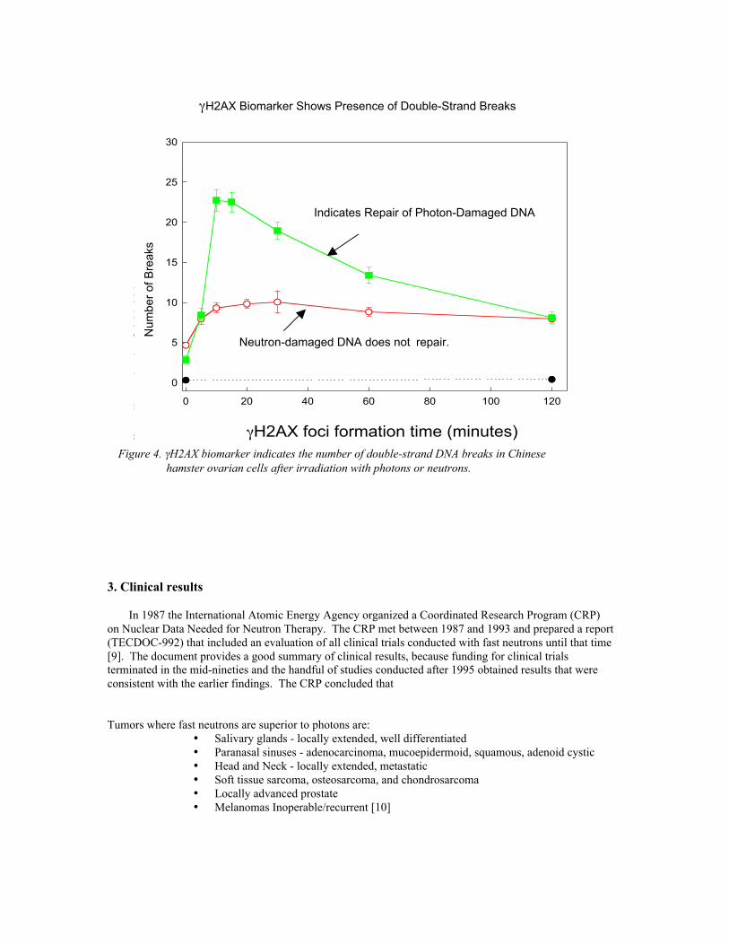
Figure 4. γH2AX biomarker indicates number of double-strand DNA breaks in Chinesehamster ovarian cells irradiated with photons or neutrons
Figure 4. γH2AX biomarker indicates the number of double-strand DNA breaks in Chinesehamster ovarian cells after irradiation with photons or neutrons.
3. Clinical results
In 1987 the International Atomic Energy Agency organized a Coordinated Research Program (CRP)on Nuclear Data Needed for Neutron Therapy. The CRP met between 1987 and 1993 and prepared a report(TECDOC-992) that included an evaluation of all clinical trials conducted with fast neutrons until that time[9]. The document provides a good summary of clinical results, because funding for clinical trialsterminated in the mid-nineties and the handful of studies conducted after 1995 obtained results that wereconsistent with the earlier findings. The CRP concluded that
Tumors where fast neutrons are superior to photons are:• Salivary glands - locally extended, well differentiated• Paranasal sinuses - adenocarcinoma, mucoepidermoid, squamous, adenoid cystic• Head and Neck - locally extended, metastatic• Soft tissue sarcoma, osteosarcoma, and chondrosarcoma• Locally advanced prostate• Melanomas Inoperable/recurrent [10]
0 20 40 60 80 100 120
0
5
10
15
20
25
30
γH2AX foci formation time (minutes)
Average #
γH2AX
foci per cell
Indicates Repair of photon-damaged DNA
Neutron-damaged DNA does not repair.
Num
ber
of B
reak
s
γH2AX Biomarker Shows Presence of Double-Strand Breaks
Indicates Repair of Photon-Damaged DNA

Tumors for which more research is needed are:• Inoperable Pancreatic• Bladder• Esophagus• Recurrent or inoperable rectal• Locally advanced uterine cervix• Neutron boost for brain tumors [11]
Most significantly they concluded that: “The proportion of patients suitable for neutrons ranges from 10-20%, but this is probably a lower limit …with high energy modern cyclotrons neutron therapy will beuseful for a larger proportion of patients.” [12] In fact, most of the neutron patients today are being treatedwith a high-energy proton linac.
Additional clinical results are summarized below.
3.1 Salivary Gland tumors
The best statistics are available for salivary gland tumors, probably because even those acceleratorswhose energy was too low to treat deep-seated tumors could be used to treat salivary gland tumors, whichtend to be fairly superficial. Table 1 summarizes international data collected in the TECDOC-992 report.The term “local control” means that the tumor completely disappeared in the treated volume and did notrecur in or near that volume. It does not address the issue of distant metastasis. A person could achievelocal control of the tumor and still have cancer elsewhere in his/her body.
Table 2 has fewer statistics than Table 1 but is interesting because it was terminated early based onethical considerations. After two years follow-up the results with neutrons were so much better than thosewith photons that it was considered unethical to randomize patients to the photon arm [13]. Figure 5 showsan example of a salivary gland tumor treated with fast neutrons at Fermilab. Note that the tumor isinoperable and is large, implying that it may be hypoxic and may have cells in the resting stage.
Table 1: Loco-regional control of malignant salivary gland tumors.International clinical trials 1972-1990.
Number ofFacilities
Number ofPatients
Local Control(p = 3.7E-25)
Neutrons 10 310 67%Photons 10 254 24%
Table2: Loco-regional control of inoperable salivary gland tumors: an internationalprospective randomized trial.
Photons NeutronsNumber of Patients 12 13Loco-regional control
at 1 yearat 2 years (p=0.01)
17 ± 11%17 ± 11%
67 ± 14%67 ± 14%
Survivalat 1 yearat 2 years (p=0.06)
67 ± 12%24 ± 14%
77 ± 12%62 ± 14%

Figure 5: An inoperable salivary gland tumor before and after treatment with fast neutron therapyat Fermilab.
3.2 Prostate cancer
Figure 6 shows results of multi-institutional clinical trials showing that locally advanced stage prostatecancer patients treated with a combination of neutrons and photons had better long-term survival than thosetreated with photons only. [14] A similar study involving over 700 early stage prostate cancer patientsshowed that the order in which patients receive the radiation influences disease-free survival. Disease freesurvival was 93% for those receiving neutrons before photons and 73% for those receiving photons beforeneutrons [15].
Figure 6. Actuarial survival curves for advanced stage prostate cancer patients treated withphotons only or a combination of neutrons and photons. National clinical trial RTOG7704.
Survival Curves for Advanced Prostate Cancer Patients

3.3 Sarcoma
Figure 7 shows a soft tissue sarcoma located on the buttock, before treatment at Fermilab, just aftertreatment and at the two-month follow-up. The term “sarcoma” refers to a malignant tumor arising inconnective tissue, bone, cartilage or muscle. This figure is an example of a large tumor whose progressionwas controlled by fast neutron therapy. The photo taken at the end of treatment shows damage to healthytissue, but the third photo shows that healthy tissue can repair sublethal damage from neutron therapy.Tables 3 – 5 present results of prospective randomized clinical trials for various types of sarcoma [16].
Table 3: Loco-regional control of unresected osteosarcoma – 2-year actuarialdata - International prospective randomized clinical trials.
Number ofFacilities
Number ofPatients
Local Control(p = 9.1 E-6)
Neutrons 8 97 54%Photons 3 73 21%
Table 4: Loco-regional control of unresected chondrosarcoma – 2-yearactuarial data - International prospective randomized clinical trials.
Number ofFacilities
Number ofPatients
Local Control(p = 0.28)
Neutrons 7 25 49%Photons 2 10 33%
Table 5: Loco-regional control of unresected or partially resected soft-tissue sarcoma - 2-year actuarial data - International prospectiverandomized clinical trials.
Number ofFacilities
Number ofPatients
Local Control(p = 0.047)
Neutrons 11 297 53%Photons 5 49 38%

Figure 7a. Large soft tissue sarcoma on thehip. This tumor is radioresistant.It contains hypoxic cells andcells in various stages ofdevelopment.
Figure 7b. Same patient after being treatedwith fast neutrons at Fermilab.Note residual skin reddening justat the end of treatment. This ischaracteristic of any radiation if ahigh dose is given.
Figure 7c. Same patient at two-monthfollow-up. Note repair ofreddening, but presence oflong-term scar tissue.

4.0 Design of a Modern Fast Neutron Therapy Facility
In designing a modern fast neutron therapy facility, two issues must be considered: theaccelerator/beam transport design and the collimation and beam shaping system. Design parameters for theaccelerator are well understood and at least two options are available for accelerating the proton beamneeded to generate the fast neutrons. The collimation and beam shaping system still requires some researchand development, the primary issue being the problem of providing good beam shaping using materials thathave minimal radioactivation properties and do not allow neutrons to penetrate cracks in the collimators.
4.1 Accelerator/Beam Transport Parameters
Clinical trials have shown that clinically useful fast neutron beams are best generated by an ~70 MeVproton beam impinging on a water-cooled semi-thick cylindrical beryllium target. There are no clinical datafor beams greater than 70 MeV, though it is expected that such beams would have deeper penetrationcapabilities at the expense of RBE. Deeper penetration would be advantageous for tumors such as prostatetumors, but disadvantageous for head and neck tumors because of increased exit dose. Typical proton beamcurrents at existing neutron facilities range from 20 – 70 microamperes, but to be competitive with doserates available at photon facilities, the proton current in a new accelerator should be at least 200microamperes. Target-to-patient distance for existing facilities ranges from 150 – 190 cm. At Fermilab, theoriginal patient position was 153 cm, but the distance was increased to 190 cm to allow easier access for thetherapists to set up the patient. Table 6 summarizes accelerator/beam transport requirements for anoptimized fast neutron facility.
Table 6: Accelerator/beam transport requirements for a modernfast neutron facility.
Accelerated Ion H+ or H-
Beam extraction energy ~ 70 MeVProduction target Beryllium cylinder, 2 cm
diameter, 2 cm lengthAverage ion current ~ 200 microamperesTarget-to-patient distance 190 cm
Logistics for installing a neutron therapy facility at a hospital are still difficult because no commercialfirm is manufacturing neutron therapy systems. As will be described in the next section, some research anddevelopment is still needed to optimize a clinic. Hence it is still appropriate for a neutron facility to belocated at or be affiliated with a large physics laboratory where there is access to engineers and specialists inneutron shielding and collimation. One particularly cost-effective way to generate neutrons is to divert aproton beam from the linac injector used at large research proton synchrotron facilities. This approach isused at Fermilab, where 66 MeV beam is diverted from the drift tube linac to treat patients, Figure 8. Undercomputer control the dipole switching magnet ramps on or off in less than 1/15 second, allowing therapybeam when it is on and high-energy physics beam when it is off.
Both cyclotrons and proton drift-tube linacs have been used to generate clinical 70 MeV beams. Aproton linac optimized for clinic-based fast neutron therapy and capable of producing medical radioisotopesis described in reference 17.

Figure 8: At Fermilab a curved dipole magnet located between linac drift-tubetanks switches beam between high energy physics research (straightahead) and neutron therapy (toward the right). One-fifteenth of a secondis required to switch between modes.
4.2 Beam Collimation and Shaping Systems
Neutrons produced by the p(66)Be(49) interaction are nearly isotropic, with neutrons generated in everydirection. As shown in Figure 9, only those neutrons traveling in a small cone centered on the forwarddirection are clinically useful.
Figure 9: Forward moving neutrons are collimated to fit the size of the tumor.Remaining neutrons are stopped by a heavy concrete shielding wall.Removable collimators made of concrete and polyethylene havevarious sized openings to fit large or small tumors.
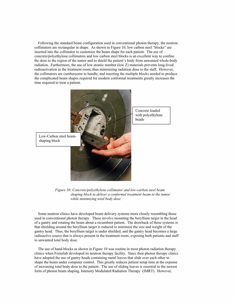
Following the standard beam configuration used in conventional photon therapy, the neutroncollimators are rectangular in shape. As shown in Figure 10, low carbon steel “blocks” areinserted into the collimator to customize the beam shape for each patient. The use ofconcrete/polyethylene collimators and low carbon steel blocks is an excellent way to confinethe dose to the region of the tumor and to shield the patient’s body from unwanted whole-bodyradiation. Furthermore, the use of low atomic number (low Z) materials prevents long-livedradioactivation in the treatment room, thus minimizing radiation dose to the staff. However,the collimators are cumbersome to handle, and inserting the multiple blocks needed to producethe complicated beam shapes required for modern conformal treatments greatly increases thetime required to treat a patient.
Figure 10: Concrete/polyethylene collimator and low-carbon steel beamshaping block to deliver a conformal treatment beam to the tumorwhile minimizing total body dose.
Some neutron clinics have developed beam delivery systems more closely resembling thoseused in conventional photon therapy. These involve mounting the beryllium target in the headof a gantry and rotating the beam about a recumbent patient. The drawback of these systems isthat shielding around the beryllium target is reduced to minimize the size and weight of thegantry head. Thus, the beryllium target is under shielded, and the gantry head becomes a largeradioactive source that is always present in the treatment room, exposing both patients and staffto unwanted total body dose.
The use of hand-blocks as shown in Figure 10 was routine in most photon radiation therapyclinics when Fermilab developed its neutron therapy facility. Since then photon therapy clinicshave adopted the use of gantry heads containing metal leaves that slide over each other toshape the beam under computer control. This greatly reduces patient setup time at the expenseof increasing total body dose to the patient. The use of sliding leaves is essential to the newestform of photon beam shaping, Intensity Modulated Radiation Therapy (IMRT). However,
Concrete loadedwith polyethylenebeads
Low-Carbon steel beam-shaping block

even with photons, whose ability to traverse cracks in the shielding leaves is much less thanthat of neutrons, there is concern that the additional total body dose may lead to thedevelopment of new cancers in IMRT patients [18-19]. A neutron therapy facility that used aphoton-like gantry head along with sliding leaves to shape the beam would deliver more totalbody dose to both the patient and the staff than the system currently in use at Fermilab. Forexample, Rosenberg et al measured the therapist dose to average of 5 microsieverts pertreatment [20] at Fermilab. Risler measured an average of 6 microsieverts per field at theUniversity of Washington, which has a gantry head containing moveable leaves [21].Assuming an average of three fields per treatment, the comparable dose to staff per treatment is18/5 or 3.6 times greater when the gantry is used.
The technological challenge is to devise a system where the patient could be treated in arecumbent position and still keep the total body dose comparable to that achieved using a fixedhorizontal beam. Choice of shielding materials is important. The high-Z materials used forshielding photons are not appropriate for a neutron therapy facility. New materials and bettermanufacturing techniques have been developed since Fermilab’s clinic was built in 1974-76.These materials and techniques are well understood by specialists at large physics laboratories.
5.0 Production of Medically Useful Isotopes
The use of medical radioisotopes for both diagnosis and treatment of disease is becomingincreasingly important. Many of these isotopes have production thresholds of 50-70 MeV andare best produced in this energy range to avoid contamination with isotopes whose productionthreshold is above 70 MeV. Any accelerator that can provide a clinically useful fast neutronbeam can also provide protons to generate these isotopes. Because most of the time the patientspends in the treatment room is used for setup rather than actual treatment, the accelerator isidle about two-thirds of the time during the treatment day. The setup time between treatmentsas well as the evening and night shifts could be used to generate isotopes. Tables 7 and 8 listmedical isotopes that could be produced using a proton beam designed for fast neutron therapy.
Table 7: Short-lived Isotopes with Medical Applications. These isotopes are easily producedusing the same accelerator that provides a clinical fast neutron therapy beam.
Isotope Production Reaction Application52Fe(8.2h) 55Mn(p,4n) Bone Marrow Scanning62Zn(9.1h)→62Cu(9 min)
63Cu(p,2n)Positron emission tomography
of heart or brain
67Ga(78h)67Zn(p,n)68Zn(p,2n)
Detection of cancer, infection orinflammation
111In(67.2h)
112Cd(p,2n)113Cd(p,3n)115Cd(p,4n)116Cd(p,6n)
Molecular labeling
123(13.3h)127I(p,5n)→123Xe→123I
123Te(p,n)123IThyroid Scan
210Tl(74h)203Tl(p,3n)205Tl(p,5n) Heart Imaging

Table 8: Long-lived Isotopes with Medical Applications. These isotopes are easilyproduced using the same accelerator that provides a clinical fast neutron therapybeam.
Isotope Production Reaction Application
68Ge(280d)→68Ga(68min)
69Ga(p,2n)68Ge71Ga(p,4n)68Ge
Calibrate positronemission tomography
imaging devices82Sr(25d)→82Rb(1.5min)
85Rb(p,4n)82SrCardiac perfusion
studies
103Pd(17d) 103Rh(p,n)103PdProstate Cancertreatment seeds
127Xe(36.4d) 133Cs(p,2p,5n)127XeLung Ventilation
Studies
6.0 Summary and Discussion
International, prospective, randomized clinical trials have shown fast neutrons to be superiorto photons for treating radioresistant tumors such as salivary gland tumors, sarcomas, melanomasand locally advanced prostate tumors. More research is needed to evaluate neutron therapy fortumor types that were not adequately investigated during the clinical trials. The higher efficacyof neutrons is due to the fact that their interactions with tissue are significantly different fromphoton (or proton) interactions with tissue. More research is needed to understand the biologicalresponse to these different interactions.
Because there are only a few clinical fast neutron beams in the world only a limited amountof research is being conducted. In addition, there are engineering challenges to be addressed withrespect to neutron beam collimation and shaping before neutron therapy becomes practical andefficient for more extensive clinical use. National laboratories that are already developingaccelerator and beam line technology are well qualified to address the engineering challengesassociated with making neutron therapy more widely available.
7.0 References
[1] T. Griffin, T. Pajak, G.Laramore, and L. Davis, “Analysis of neutron radiotherapy treatmentcomplications,” Bull. Cancer (Paris) 73,5:582-586 (1986).
[2] M. Catterall and D. Bewley, Fast Neutrons in the Treatment of Cancer, (Academic Press London, 1979)pp. 82-114.
[3] E.J. Hall and A.J. Giaccia, Radiobiology for the Radiologist (Lippincott Williams & Wilkins,Philadelphia, 2006) pp. 56-58
[4] Ibid. pp. 5-15[5] E.R. Blazek, J. Urbon, B. Pientak, T.K. Kroc, A.J. Lennox (unpublished)[6] Ibid.[7] J.D. Forman, P.G. Kocheril, K. Hart, P. Chuba, T. Washington, C. Orton, A.T. Porter, “Estimating the
RBE for Pelvic Neutron Irradiation in the Patients Treated for Carcincoma of the Prostate,” Journal ofBrachytherapy Int’l, 13:29-34 (1997).
[8] L. Yasui, T.K. Kroc, A.J. Lennox (unpublished)[9] International Atomic Energy Agency, Nuclear data for neutron therapy: Status and future needs,
(IAEA, Vienna, Austria, 1997)

[10] Ibid. p 23[11] Ibid. pp 13-22[12] Ibid. p 24[13] T. W. Griffin, T.F. Pajak, G.E. Laramore, W. Duncan, M.P. Richter, F. R. Hendrickson, and M.H.
Maor, “Neutron Vs Photon Irradiation of Inoperable Salivary Gland Tumors: Results of an RTOG-MRC Cooperative Randomized Study,” Int J Radiat Oncol Biol Phys 15:1085-1090 (1988)
[14] K.J. Russell, G.E. Laramore, J.M. Krall, F.J. Thomas, M.H. Maor, F.R. Hendrickson, J.N. Krieger, andT.W. Griffin, “Eight Years Experience With Neutron Radiotherapy in the Treatment of Stages C and DProstate Cancer: Updated Results of the RTOG 7704 Randomized Clinical Trial,” The Prostate,11:183-193 (1987)
[15] J.D. Forman, M.Yudelev, S. Bolton, S. Tekyi-Mensah, R. Maughan, “Fast Neutron Irradiation forProstate Cancer,” Cancer and Metastasis Reviews 21:131-135, 2002.
[16] L. Cohen, presented at the Neutron Therapy Symposium in Cape Town, 1995, (unpublished).[17] A.J. Lennox and R.W. Hamm,“ A Compact Proton Linac for Fast Neutron Cancer Therapy,” text of
invited paper presented at a meeting of the American Nuclear Society in Long Beach, California Nov.14-18, 1999, Proceedings of the Third International Topical Meeting on Nuclear Applications of
Accelerator Technology, American Nuclear Society, LaGrange Park, Illinois, 1999, pp. 33-36.[18] E. Glatstein, “Intensity modulated radiation therapy: The inverse, the converse and the perverse,”
Semin Radiat Oncol 12:272-281 (2002).[19] E. Glatstein, “The Return of the Snake Oil Salesmen,” Int. J. Radiation Oncology Biol. Phys. Vol 55,
No. 3 pp 561-562 (2003).[20] I. Rosenberg, “Analysis of Personnel Exposures in Neutron Therapy Facilities,” Health Physics, Vol
46 No. 2 pp. 407-412 (1984)[21] R. Risler, Proceedings of the International Workshop on Clinical High-Energy Neutron Dosimetry,
15-17 April 2004, Fermilab, Batavia, Illinois (unpublished).