high resolution mapping of nonlinear mhz ultrasonic fields ... filepeter kaczkowski* and bryan...
TRANSCRIPT
Abstract — We show that a 2 MHz HIFU
field can be measured in water and with high
spatial resolution by placing a small scatterer in
the field and sensing the scattered wave. We use a
bistatic configuration placing a hydrophone at a
safe distance. Glass and steel tips are fabricated
using simple techniques and evaluated. Tapered tip
geometry proves to be mechanically robust and
not prone to internal resonance or scattering
from other sites than the tip. Glass proves to be
superior to polished carbide steel in its ability t o
delay the initiation of cavitation, and relatively
easy to manufacture using common pipette
drawing technology. In our tests, a highly
nonlinear 2 MHz HIFU field is mapped in
degassed water with a 0.1 mm glass tip. The
fundamental, 2n d, 3rd, and 4th harmonics are
mapped with at least 25 dB dynamic range and are
comparable with numerical results. Numerical
simulations for nonlinear fields in water are
obtained using a well established KZK equation
based calculation.
I. INTRODUCTION
Conventional mapping of high intensity fields
produced by focused transducers is performed by
mechanically scanning a hydrophone through the
field. Unfortunately, repeated measurements at
typical medical HIFU (High Intensity Focused
Ultrasound) levels of thousands of Watts per
square centimeter risks damaging the hydrophone
by heating or by cavitation, even when very short
pulses are used. Furthermore, the resolution of the
field map is limited to the size of the active area
of the hydrophone, typically on the order of 0.5
mm and large compared to many wavelengths of
interest. For highly focused and intense acoustic
fields, power can be significant at the third or
fourth harmonic. For example, at frequencies of 2
or 3 MHz which are commonly used in HIFU
therapy, field feature sizes at the focus can be on
the order of 0.1 mm or smaller. Such features are
not present at low intensities most often used for
field mapping measurements, because the field is
much more linear at such intensities.
Field maps are essential for transducer
development and testing. High power densities
used in HIFU result in severe electrical and
thermal stresses and can lead to failure. Focal gain
is sensitive to imperfections, and partial
delamination between the matching layer and the
active element can produce significant reductions
in focal intensity. At low powers, this problem is
often discovered by measuring the beam
dimensions in the focal region. Government
regulatory compliance testing (primarily for
imaging devices) requires extensive field mapping.
While HIFU therapeutic devices are not yet
approved, field maps will likely be required of any
manufacturer to verify focal characteristics as
well as pre-focal fields to ensure the absence of
“hot spots” where they are not desired. Finally,
the need to compare theory with experiment and
numerical models with data motivates the
development of in situ techniques for measuring
HIFU fields.
II. APPROACH
The idea of measuring a strong field indirectly
by placing a known scatterer within it and
HIGH RESOLUTION MAPPING OF NONLINEAR MHZ ULTRASONIC FIELDS
USING A SCANNED SCATTERER
Peter Kaczkowski* and Bryan CunitzCenter for Industrial and Medical Ultrasound
Applied Physics Laboratory, University of WashingtonSeattle, WA 98105, USA
Vera Khokhlova, Oleg SapozhnikovMoscow State University
Moscow, Russia

measuring the scattered field using a receiver
safely outside the primary source field is not a
new one. Long used by practitioners in ultrasound
laboratories [1-5], the technique has mostly been
applied in a monostatic configuration, that is, one
in which the receiver is in the same location as
the source, and indeed is often the very same
transducer. We sought to measure harmonics of
the field to which the source transducer was not
sensitive due to the rather careful electrical and
mechanical impedance matching done near the
fundamental frequency to optimize HIFU
efficiency (e.g., to minimize transducer heating).
Thus, we chose to use a different transducer as
receiver.
Geometrical arrangement
The receiver we chose to test the approach was
a general purpose hydrophone manufactured by
Sonic Concepts, Woodinville, Washington, USA,
used for a wide range of measurements in our lab.
The transducer is made of PZT with a center
frequency of 5 MHz and has two matching layers,
thus providing a wide sensitivity bandwidth (here
we recorded in the 2 – 8 MHz band). The
transducer is a spherical cap with a diameter of
0.95 cm and a radius of curvature of 7.5 cm. The
setup geometry is diagrammed in Figure 1.
Figure 1. General arrangement of HIFU transducer,scattering tip, and receiver. A rigid frame holding bothscatterer and receiver is essential; the scatterer andreceiver must move as one. The calibration standardhydrophone is not shown; it is positioned at the samelocations as the scattering tip and detects the fielddirectly at safe amplitude settings.
Scatterer properties
To be effective at probing small spatial scales,
the scatterer must be both small and rigidly
supported. Generally, the scatterer must be
smaller than a wavelength; ideally, it behaves as a
Rayleigh scatterer. Consequently, it will scatter
broadly in angle and the choice of incident and
scattered field angles is not particularly
important. Furthermore, the scattered power is
proportional to (frequency)4 and this dependence
helps mitigate the decrease in power with
increasing harmonic number in a nonlinear field,
and can also be used to offset receiver sensitivity
falloff above its central frequency. A tapered tip
provides excellent mechanical support as well as
very little scattering from the taper for a
moderate range of angles. No appreciable pulse
lengthening was observed in the scattered signal
(for a short HIFU pulse) that would be due t o
scattering from points other than the tip.
A rigid frame ensures that the Green’s function
between scatterer and receiver is constant during a
scan, permitting absolute calibration of the
system using a reference hydrophone, or a
standard calibrated source, for each frequency of
interest. The frequency dependence of the system
is highly scatterer and receiver dependent; no
attempt was made to compute it in advance. The
rigid frame holds two hydrophones: the 1 cm
diameter 5 MHz focused PZT element with its
center of curvature centered on the scatterer (and
located outside the source field), and one 0.6 mm
active diameter PVDF needle hydrophone (NTR,
Seattle, WA). The latter is used for calibration by
replacement and is mounted facing the HIFU
source but with sufficient offset from the
scattering tip to keep it out of the HIFU field
during scattering measurements. The PVDF
hydrophone is only used at low intensities.
Tip fabrication techniques
We fabricated tapered rods of steel and glass
with tip diameters between 0.5 mm and 0.01 mm.
Carbide tool rod stock was sharpened by hand
using a grinder and successively finer emery paper.
Though the tip was relatively easy to make, we
were not able to prevent cavitation from
initiating with a few acoustic cycles, even at

moderate HIFU pressures. Glass has different
surface properties than steel, and inhibits
cavitation long enough (at least 10 cycles) t o
provide purely tip-scattered signals. We only
describe the manufacture of glass tips in detail.
Figure 2. A pipette puller is used to draw glass tubesinto extremely small diameter needles for use inbiological experiments. Here, a 5 mm diameter glass rodis held vertically by two chucks. A coiled heating elementis driven by a current source and heats it to a brightorange glow, softening the rod. The upper chuck is fixed,and the bottom one can be lowered by hand or byattaching a fixed weight. Several heat, draw, cool andcleave cycles are repeated to reach the desired tip size.
Glass can easily be drawn into extremely thin
fibers or tubes when heated. Using standard
laboratory borosilicate glass rod and a pipette
puller we fabricated tapered glass tips with face
diameters ranging from 0.01 mm to 1 mm.
Excellent results are achieved with tip face
diameter on the order of 0.1 mm. Figure 2 is a
photograph of a pipette puller.
The glass is heated, drawn, cooled and cleaved
several times to reach the desired tip diameter.
This process leads to tips such as the one depicted
in Figure 3. Cleaving is preferred to melting the
tip which creates a tiny ball; the ball tends t o
resonate. Tips are surprisingly robust; they are
not often damaged by cavitation, or, at least
calibration results do not change measurably. It is
possible that erosion of such small tips would not
cause much change in scattering properties as long
as the tip preserved its diameter. If the latter is
much smaller than the acoustic wavelength details
of the face shape do not measurably affect
scattering properties.
III. FIELD MAPPING PROCEDURE
Field mapping is done over a broad frequency band
by processing the scattered pressure waveform at
each spatial location of the scatterer. The
scattered waveform is collected by amplifying and
digitizing the receive hydrophone voltage during
the initial dozen cycles or so. At high intensities,
cavitation modifies the scattered signal
dramatically. Bubbles are not usually visible at the
tip; rather the waveform becomes completely
unstable. It is interesting to observe that the
instability typically appears near the same time
after the HIFU pulse begins, as long as adequate
time is given for bubbles to re-dissolve in the
highly degassed water. We use a 2 MHz HIFU
burst of 10 cycles at a repetition rate of 10 Hz.
The voltage waveform collected over a 2
microsecond window is processed by FFT, and
harmonic amplitudes are stored for each spatial
location.
Figure 3. Photographs of the same tip at variousmagnifications. Top: no magnification, scale is in cm.Middle: Tip magnified to X5. Bottom: tip magnified toX40; each minor division is 0.01mm.

Calibration of the system is performed by direct
comparison of field maps collected by the
scatterer system and by a conventional calibrated
PVDF needle hydrophone. This approach suffers
from the limited resolution of our PVDF needle,
but there are now hydrophones available with
active areas on the order of 0.1 mm (it would not
be prudent to use these in a HIFU field, however).
The calibration need only be done once for any
given tip and frame setting; we do not yet have a
sense for how rapidly tips erode and how often re-
calibration is needed.
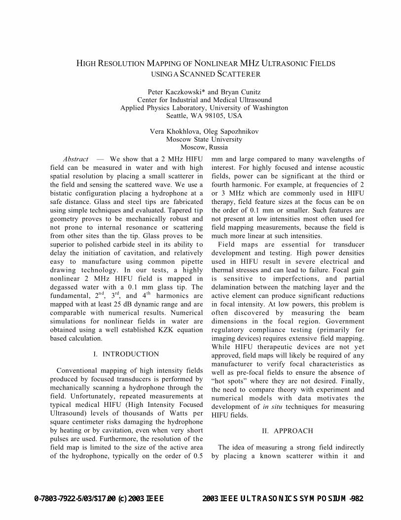
IV. RESULTS AND CONCLUSIONS
We conducted a field map for a 2 MHz HIFU
source emitting a total acoustic power of 60 W
(Sonic Concepts model SU-101, RoC = 55 mm,
Diam = 35 mm). The tip diameter was about 0.1
mm, and the field map step size was set to 0.05
mm. Experimental field maps of the fundamental,
2nd, 3rd, and 4th harmonic fields plotted on a
logarithmic color scale are presented in Figure 5:
the speckle (noise) in the 4th harmonic data is at
25 dB below the peak. On a linear scale, the data
plots are nearly indistinguishable from numerical
simulations (labeled “Theory”) of the
experimental conditions using a KZK equation
method [6].
A simple method using a scanned tapered tip
scatterer can be used to measure MHz range HIFU
fields (including harmonics) at full power, in water
or other fluids without risk of damage to the
hydrophone which is placed outside the direct
HIFU field. Tapered glass tips are effective and
inexpensive to manufacture, and tend to suppress
cavitation due to their surface properties.
V. REFERENCES
[1] Bernier, C.A., L. Huntsman, and R. Martin, "Apractical approach to measuring an intravascularultrasonographic imaging system beam pattern", JUltrasound Med, 14(5): p. 367-73, May 1995.
[2] Parker, K.J., "The thermal pulse decay technique formeasuring ultrasonic absorption coefficients",JASA, 74(5): p. 1356-1361, 1983.
[3] Raum, K. and W.D. O'Brien, Jr., "Pulse-echo fielddistribution measurement technique for high-frequency ultrasound sources", IEEE Trans. UFFC,44(4): p. 810-815, 1997.
[4] Lizzi, F., "Private communication." 2002.[5] Szabo, T., "Private communication." 2003.[6] Khokhlova, V.A., et al., "Numerical modeling of
finite-amplitude sound beams: Shock formation inthe near field of a cw plane piston source", J.Acoust. Soc. Am., 110(1): p. 95-108, Jul 2001.
Figure 4. Top: Scanned scatterer data using a 0.1 mm tip, and a 2 MHz nonlinear HIFU field.Bottom: KZK simulations for the experimental conditions of the scan above.Spatial units are mm; the noise, visible as speckle in the 8 MHz data is 25 dB below peak.