high risk infant · 2016-04-07 · factors –to define high risk infant a) ... live born baby...
TRANSCRIPT
IAP Teaching Slides 2015-2016
HIGH RISK INFANT
1
IAP Teaching Slides 2015-2016 2
INTRODUCTION
• A newborn, regardless of gestational age or birth
weight, who has a greater than average chance of
morbidity or mortality because of conditions or
circumstances superimposed on the normal course
of events associated with birth and the adjustment
to extra uterine existence.
2
IAP Teaching Slides 2015-2016
FACTORS –TO DEFINE HIGH RISK INFANT
A) Demographic social factors:
Maternal age <16 or >40, unmarried, physical stress, Socio‐
economic status
B) Past medical history:
Diabetes Mellitus, genetic disorders, hypertension
C) Previous pregnancy :
Intrauterine death, neonatal death, IUGR, IEMs, congenital
malformations
3
IAP Teaching Slides 2015-2016
D) Present Pregnancy:
Vaginal bleeding, PROM, multiple gestation, preeclampsia,
abnormal USG findings
E)Labor: and delivery:
Obstructed labor, fetal distress, forceps delivery, meconium
stained liquor
F)Neonate:
birth weight <2000 or >4000,gestation <37 or >42
SGA, Respiratory distress, congenital malformation4
FACTORS –TO DEFINE HIGH RISK INFANT
IAP Teaching Slides 2015-2016
DEFINITIONS
Low Birth Weight Infant:
Live born baby weighing 2500 gram or less at birth.
(VLBW: <1500 gm, ELBW:<1000 gm). ‐
Preterm:
When the infant is born before term. i.e.: before 38 weeks
of gestation.
Premature:
When the infant is born before 37 weeks of gestation.5
IAP Teaching Slides 2015-2016
Full term:
When the infant is born between 38 – 42 weeks of
gestation.
Post term:
When the infant is born after 42 weeks of gestation.
6
DEFINITIONS
IAP Teaching Slides 2015-2016
HYPOTHERMIA
7
IAP Teaching Slides 2015-2016
DEFINITION AND CLASSIFICATION
It is a condition characterized by lowering of body
temperature than 36°C.
Types of Hypothermia:
Could be classified according to Causes and
according to Severity.
8
IAP Teaching Slides 2015-2016
CLASSIFICATION BASED ON CAUSE
• Primary Hypothermia: – Seen immediately after delivery– Normal term infant delivered into a warm environment may drop its rectal temperature by 1 – 2°C shortly after birth and may not achieve a normal stable body temperature until the age of 4 – 8 hours.
– In low birth weight infants, the decrease of body temperature may be much greater and more rapid unless special precautions are taken immediately after birth. (loss at least 0.25 °C/ min.) (careful dryness).
9
IAP Teaching Slides 2015-2016
• Secondary Hypothermia:
– This occurs due to factors other than those
immediately associated with delivery.
– Important contributory factors are:
e.g.: Acute infection especially Septicemia.
10
CLASSIFICATION BASED ON CAUSE
IAP Teaching Slides 2015-2016
CLASSIFICATION BASED ON SEVERITY
II) According to Severity:
(1) Mild Hypothermia: < 36°C
(2) Moderate Hypothermia: < 35.5°C.
(3) Severe Hypothermia: < 35°C.
11
IAP Teaching Slides 2015-2016
CLINICAL FEATURES
• Decrease in body temperature measurement
• Cold skin on trunk and extremities
• Poor feeding in the form of poor suckling
• Shallow respiration
• Cyanosis
• Decrease activity, e.g. Weak cry
12
IAP Teaching Slides 2015-2016
1. Evaporation:Heat loss that resulted from expenditure of internal thermal energy to convert liquid on an exposed surface to gases, e.g.: amniotic fluid, sweat.
Prevention: Carefully dry the infant after delivery or after bathing.
2. Radiation:It occurred from body surface to relatively distant objects that are cooler than skin temperature.
FOUR MODALITIES OF HEAT LOSS IN NEONATES
13
IAP Teaching Slides 2015-2016
3. Conduction:Heat loss occurred from direct contact between body surface and cooler solid object.
Prevention:Warm all objects before the infant comes into contact with them.
4. Convection: Heat loss is resulted from exposure of an infant to direct source of air draft.
Prevention:· Keep infant out of drafts.· Close one end of heat shield in incubator to reduce velocity of air.
14
IAP Teaching Slides 2015-2016
MANAGEMENT• Infant should be warmed quickly by wrapping in a warm
towel.
• Uses extra clothes or blankets to keep the baby warm.
• If the infant is in incubator, increase the incubator’s
temperature.
• Avoid exposure to direct source of air drafts.
• Check body temperature frequently.
• Give antibiotic if infection is present. 15
IAP Teaching Slides 2015-2016
HYPERTHERMIA
16
IAP Teaching Slides 2015-2016
It is a condition characterized by an elevation in body
temperature more than 37.5°C.
Causes:
1. Disturbance in Heat Regulating Center caused by
intracranial hemorrhage, or intracranial edema.
2. Incubator temperature is set too high.
DEFINITION
17
IAP Teaching Slides 2015-2016
MANAGEMENT
• Undress the infant. If at home; keep light cloths, cover that
containing light sheet, Or only a diaper if the infant is inside
an incubator.
• Reduction of incubator temperature.
• Provide Tepid sponge bath.
• If available; fill the water mattress with tape water, and
keep it in contact with the infant’s skin.
• Increase fluid intake in the form of 5cc of Glucose 5%
between feeds to prevent dehydration.18
IAP Teaching Slides 2015-2016
HYPOGLYCEMIA
19
IAP Teaching Slides 2015-2016
DEFINITION:
Neonatal hypoglycemia is usually defined as a serum
glucose value of < 40‐45 mg/dl.
For the preterm infant a value of < 30 mg/dl is
considered abnormal (hypoglycemia).
20
IAP Teaching Slides 2015-2016
1- The main cause may become maternal malnutrition during pregnancy which leads to fetal malnutrition and of course a low birth weight.
2‐ Those infants whom are Small for gestational age infants (SGA), that manifested by decrease in their birth weight and subcutaneous fat and hepatic glycogen. 3‐ Those infants’ of diabetic mothers (IDM) or those named as large for gestational age (LGA).
21
NEONATES AT RISK FOR DEVELOPING HYPOGLYCEMIA:
IAP Teaching Slides 2015-2016
4‐ Those whom placentas were abnormal,
e.g. placenta Previa.
5‐ Those whom their mothers had toxemia during
pregnancy, e.g. eclampsia or pre‐eclampsia, induction of
labor preterm infant.
6‐ Those very ill or stressed neonates whom their
metabolic needs were increased due to hypothermia,
infection, respiratory distress syndrome, or cardiac failure.
22
IAP Teaching Slides 2015-2016
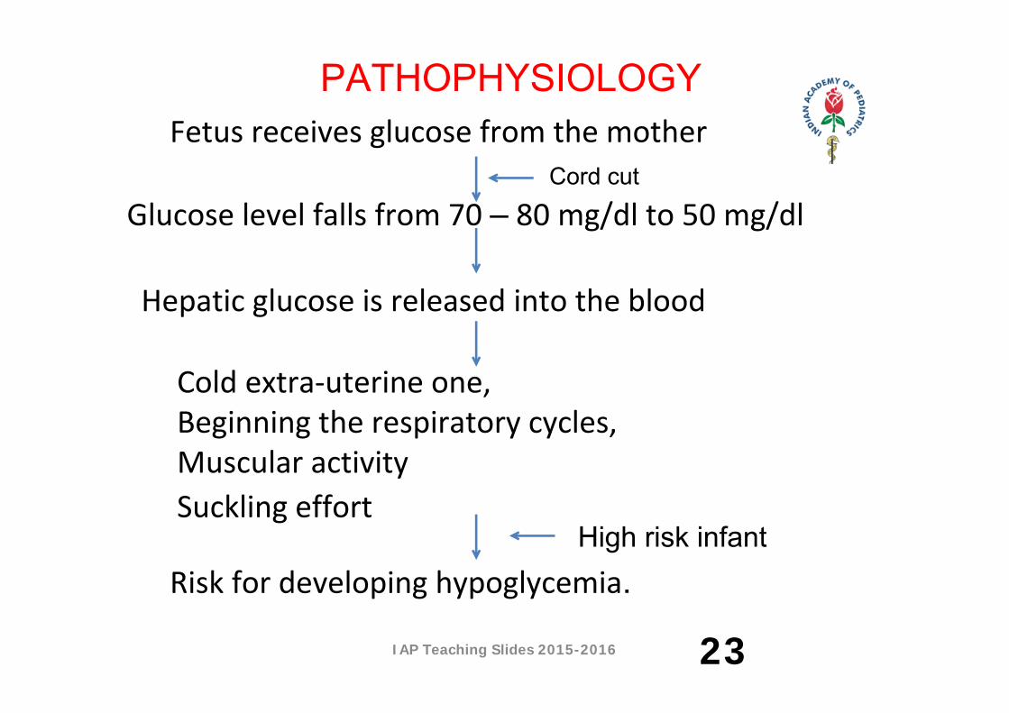
Glucose level falls from 70 – 80 mg/dl to 50 mg/dl
Fetus receives glucose from the mother
Cold extra‐uterine one, Beginning the respiratory cycles,Muscular activitySuckling effort
Hepatic glucose is released into the blood
Risk for developing hypoglycemia.
Cord cut
High risk infant
PATHOPHYSIOLOGY
23
IAP Teaching Slides 2015-2016
CLINICAL MANIFESTATIONS:
1‐ Hypotonia
2‐ Feeding poorly after feeding well
3‐ Tremors
4‐ Cyanotic spells
5‐ Lethargy
6‐ Seizures
24
IAP Teaching Slides 2015-2016
CLINICAL MANIFESTATIONS:
7‐ Hypothermia.
8‐ Irregular respiratory pattern (Apnea).
9‐ Irritability.
10‐ High pitched cry followed by weak cry.
11‐ poor reflexes, especially sucking reflex.
25
IAP Teaching Slides 2015-2016
MANAGEMENT OF THE NEONATE AT RISK:
Prevention:
First of all, providing a warm environment.
Early enteral feeding is the single most important
preventive measure.
If enteral feeding is to be started, breast or artificial milk
should be used if the infant is able to tolerate nipple or
nasogastric tube feeding.
26
IAP Teaching Slides 2015-2016
These infants should have glucose values monitored until they are taking full feedings and have three normal pre‐feeding readings above 40‐45 mg/dl.
Care must be taken to ensure that breastfeeding mothers are providing an adequate intake. If the infant at risk for hypoglycemia is unable to tolerate nipple or tube feeding, maintenance IV therapy with 10% glucose should be initiated and glucose levels monitored.
MANAGEMENT (CONT…)
27
IAP Teaching Slides 2015-2016
Infants who develop hypoglycemia should immediately
be given 2 cc/kg of 10% dextrose over 5 minutes,
repeated as needed.
28
MANAGEMENT OF THE NEONATE WITH HYPOGLYCEMIA:
IAP Teaching Slides 2015-2016
A continuous infusion of 10% glucose at a rate of 8‐10
mg/kg/min should be started to keep glucose values
normal (NOTE: 10 mg/kg/min of 10 % dextrose = 144
cc/kg/day). Frequent bedside glucose monitoring is
necessary.
When feedings are tolerated and frequent bedside glucose
monitoring values are normal, the infusion can be tapered
gradually.
29
IAP Teaching Slides 2015-2016
INFANT OF DIABETIC MOTHER
30

IAP Teaching Slides 2015-2016
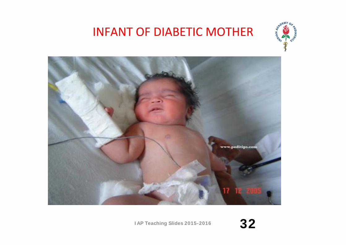
INFANT OF DIABETIC MOTHER
31
IAP Teaching Slides 2015-2016
INFANT OF DIABETIC MOTHER
32
IAP Teaching Slides 2015-2016
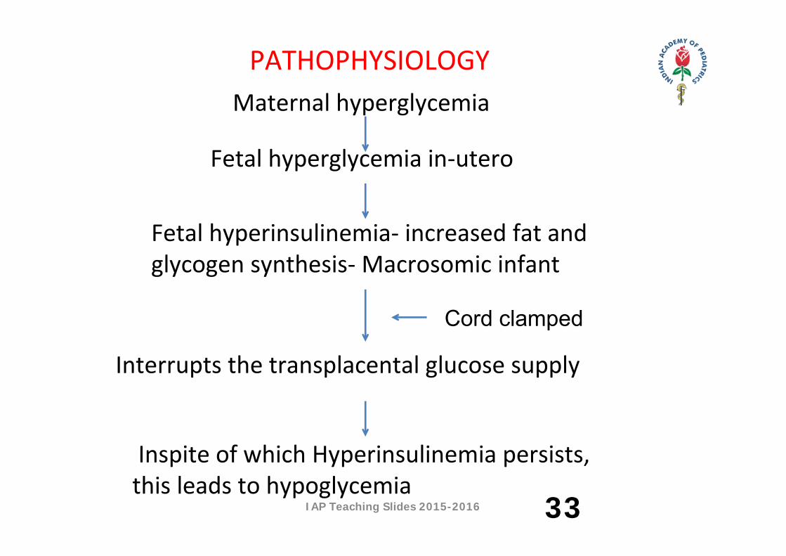
Fetal hyperglycemia in‐utero
Maternal hyperglycemia
Interrupts the transplacental glucose supply
Fetal hyperinsulinemia‐ increased fat and glycogen synthesis‐ Macrosomic infant
Inspite of which Hyperinsulinemia persists, this leads to hypoglycemia
Cord clamped
PATHOPHYSIOLOGY
33
IAP Teaching Slides 2015-2016
DISORDERS IN INFANTS OF DIABETIC MOTHERS
• Hypoglycemia.• Hypocalcemia.• Hypomagnesemia.• Cardio‐respiratory disorders• Hyperbilirubinemia (Unconjugated)• Birth injuries• Congenital malformations
34
IAP Teaching Slides 2015-2016
MANAGEMENT:
• For the mother: Good antenatal care for proper control of maternal diabetes
• For an infant:All IDMs should receive continuous observation and intensive care. Serum glucose levels should be checked at birth and at half an hour, 1, 2, 4, 8, 12, 24, 36 and 48 hours of age:
• 35
IAP Teaching Slides 2015-2016
• If clinically well and normoglycemia; oral or gavage feeding should be started and continued within 2 hours intervals.
• If hypoglycemic; give 2 – 4 ml/kg of 10% dextrose over 5 minutes, repeated as needed.
• A continuous infusion of 10% glucose at a rate of 8‐10 mg/kg/min. Start enteral feeding as soon as possible. Give Corticosteroids in persistent hypoglycemia.
• Oxygen therapy for RDS, Calcium gluconate 10% for hypocalcemia, phototherapy for hyperbilirubinemia
36
MANAGEMENT:
IAP Teaching Slides 2015-2016
NEONATAL SEPSIS
37
IAP Teaching Slides 2015-2016
INTRODUCTION
• The newborn infant is uniquely susceptible to acquire
infection, whether bacterial, viral or fungal.
• Bacterial sepsis and meningitis continue to be major
causes of morbidity and mortality in the newborn.
• The mortality rate due to sepsis ranges from 20% to as
high as 80% among neonates. Surviving infants can have
significant neurologic squeal because of CNS
involvement.38
IAP Teaching Slides 2015-2016
DEFINITION
Neonatal sepsis is a disease of neonates (who
are younger than 1 month) in which they are
clinically ill and have a positive blood culture.
39
IAP Teaching Slides 2015-2016
RISK FACTORS
• Maternal risk factors:
‐ e.g.: Premature rupture of membrane.
• Neonatal risk factors:
‐ e.g.: Prematurity (less immunologic ability to resist
infection + more liable to penetrate their defensive
barriers).
40
IAP Teaching Slides 2015-2016
ROUTES OF TRANSMISSION
• Through the maternal blood through placenta as rubella, toxoplasma, and syphilis.
• From the vagina or cervix, as groups B streptococci. • The newborn may be come contact as it passes through the birth canal as gram negative organisms.
• The newborn may come in contact in its environment after birth (Coagulate positive or negative staphylococci).
• When a susceptible host acquires the pathogenic organism, and the organism proliferates and overcomes the host defense, infection results.
41
IAP Teaching Slides 2015-2016
CLASSIFICATION OF NEONATAL SEPSIS• Early onset Sepsis• Late onset Sepsis Newborns with early‐onset infection present within 24 hours till 72 hours. Early‐onset sepsis is associated with acquisition of microorganisms from the mother during pregnancy (transplacental infection), or during labor (an ascending infection from the cervix). Late‐onset sepsis occurs beyond the first 72 hours of life (most common after the 3rd day till the 7th day after birth) and is acquired from the care giving environment (Nosocomial infection).
42
IAP Teaching Slides 2015-2016
CLINICAL FEATURES
• Decreased activity
• Excessive crying
• Apnea
• Jaundice
• Hypothermia
• Bulging or full fontanel
• Seizures
• Hypotonia 43
IAP Teaching Slides 2015-2016
LAB FINDINGS
• Raised Total leukocyte count (WBC count)
• Raised C – reactive Protein (CRP)
• Increased Erythrocyte Sedimentation Rate (ESR)
• Cultures positive
44
IAP Teaching Slides 2015-2016
MANAGEMENT
• Prevention: through proper application to infection
control practices.
• Early onset sepsis; give intrapartum antimicrobial
prophylaxis (IAP) to the mother.
45
IAP Teaching Slides 2015-2016
MANAGEMENT (CONT..)• Neonates with clinically suspected sepsis:
– Culture should be obtained first.– The recommended antibiotics are ampicillin and gentamicin.
– Third generation cephalosporins (Cefotaxime) may replace gentamicin if meningitis is clinically suspected
• • Late onset neonatal sepsis:
– Vancomycin in combination with either gentamicin or cephalosporins should be considered in penicillin resistant cases.
46
IAP Teaching Slides 2015-2016
PREVENTION • Demonstrate the effect of hand washing upon the prevention of the
nosocomial infections.
• Standard precautions should be applied in the nursery for infection
prevention.
• Instillation of antibiotics into newborn’s eye 1‐2 hours after birth is
done to prevent the infection.
• Skin care should be done using worm water & may use mild soup for
removal of blood or meconium & avoid the removal of vernix caseosa.
• Cord care should be cared out regularly using alcohol or an
antimicrobial agent. 47
IAP Teaching Slides 2015-2016
CURATIVE
•Encourage breast feeding from the mother.
• Adequate fluid and caloric intake should be administered by
gavage feeding or intravenous fluid as ordered.
• Extra‐measure for hypothermia or hyperthermia that may take
place to the newborn.
• Administering medications as doctor order.
• Follow the isolation precautions.
• Monitoring intravenous infusion rate and antibiotics are the
nurse responsibility. 48
IAP Teaching Slides 2015-2016
•Administer the medication in the prescribed dose, route, and
time within hour after it is prepared to avoid the loss of drug
stability.
•Care must be taken in suctioning secretions from the newborn as
it may be infected.
•Isolation procedures are implemented according to the isolation
protocols of the hospital.
•Observe for the complication e.g. meningitis and septic shock.
•Encourage in‐service programs and continuing education of
nurses regarding the infection control precautions. 49
CURATIVE
IAP Teaching Slides 2015-2016
HYPERBILIRUBINEMIA
50
IAP Teaching Slides 2015-2016
DEFINITION
• Hyperbilirubinemia is an elevation in the neonatal serum
bilirubin ≥ 12.9 mg/dl in Full‐term, Formula feed infant OR ≥
15 mg/dl in Preterm, Breast feed infant
• Characterized by JAUNDICE, which is defined as “yellowish
discoloration of skin and mucous membranes”.
• In the neonate clinical jaundice is diagnosed if the total
serum bilirubin is ≥ 7 mg/dl.
51
IAP Teaching Slides 2015-2016
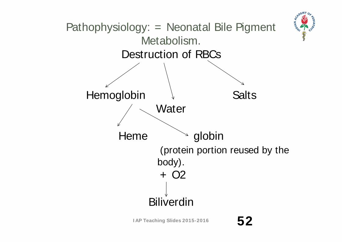
Pathophysiology: = Neonatal Bile Pigment Metabolism.
Destruction of RBCs
Hemoglobin Salts Water
Heme globin
(protein portion reused by the body). + O2
Biliverdin
52

IAP Teaching Slides 2015-2016
More O2
Unconjugated Bilirubin +
Plasma protein Liver : Which released from plasma protein inside the liver and connected with Glucuronic acid and Glucuronyl Transfarese Enzyme (in the presence of normal Ph, O2, and normal body temperature) to become Conjugated Bilirubin, that has 3
pathways: Bile duct Kidney Gastrointestinal tractTo digest fat Urobilin Urobilinogen Stercobilin Stercobilinogen to obtain normal color to obtain normal color
of urine. of stool. 53
IAP Teaching Slides 2015-2016 54
IAP Teaching Slides 2015-2016 55
IAP Teaching Slides 2015-2016
The following are possible causes of hyperbilirubinemia
in the newly born infants:
1. Over production of bilirubin.
2. Under excretion of bilirubin.
3. Combined over production and under excretion.
4. Physiological jaundice.
5. Breast milk associated jaundice.
56
CAUSES
IAP Teaching Slides 2015-2016
COMPLICATION:
The most common complication of hyperbilirubinemia is
Kernicterus (Bilirubin Encephalopathy), which usually
occurs when the unconjugated serum bilirubin level
exceeds than 20 mg/dl. In small, sick preterm infants,
even a bilirubin level in a low range may cause
Kernicterus.
57
IAP Teaching Slides 2015-2016
Clinical Presentation:
Kernicterus progresses through 4 stages:
Stage I: Poor Moro reflex, poor feeding, vomiting, high‐pitched cry,
decreased tone and lethargy.
Stage II: Spasticity, seizures, fever. Neonatal mortality is high at this
stage (80%).
Stage III: A symptomatic (Spasticity decreases and all remaining
clinical signs and symptoms may disappear).
Stage IV: Appears after the neonatal period. Long‐term sequelae can
include: spasticity quadriplegia, deafness and mental retardation (for
the 20%).
58
IAP Teaching Slides 2015-2016
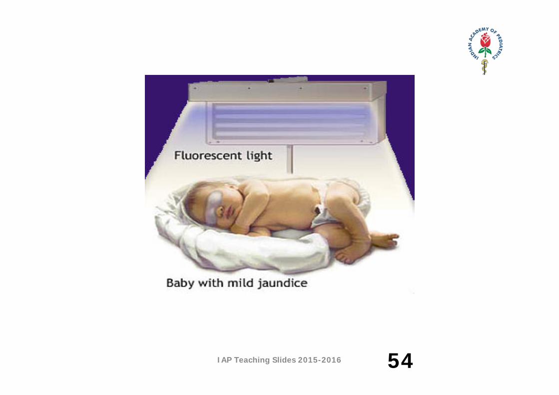
PHOTOTHERAPY:
1.Cover the infant’s eyes and genital organs.
2.The infant must be turned frequently to expose all body
surface areas to the light.
3.Serum bilirubin level /4 – 12 hours.
4.Each shift, eyes are checked for evidence of discharge or
excessive pressure on the lids and eye care should be
done using warm water, then apply eye drops or ointment.
59
IAP Teaching Slides 2015-2016
5. Eye cover should be removed during feeding, and this
opportunity is taken to provide visual and sensory stimuli.
6. Avoid oily lubricants or lotion on the infant’s exposed
skin, because this can act as a barrier that prevent
penetration of light through the skin.
7. Increase feeds in volume and calories. Add 20%
additional fluid volume to compensate for insensible and
intestinal water loss.
8. Intake and output chart. 60
PHOTOTHERAPY:
IAP Teaching Slides 2015-2016
NEONATAL RESPIRATORY DISORDERS
61
IAP Teaching Slides 2015-2016
MANAGEMENT OF RDS:
A) General:
* Basic support including thermal regulation and
parenteral nutrition and medications (antibiotics).
* Oxygen administration, preferably heated and
humidified
B) Specific:
Surfactant replacement therapy through ET tube.
62

IAP Teaching Slides 2015-2016
TRANSIENT TACHYPNEA OF THE NEWBORN (TTN)
• TTN is a benign disease of near‐term or term infants who
display respiratory distress shortly after delivery.
• It occurs when the infant fails to clear the airway of lung
fluid or mucus or has excess fluid in the lungs, this limit
the amount of alveolar surface available for gas
exchange, leading to respiratory rate and depth to
better use of the surface available.
63
IAP Teaching Slides 2015-2016
CLINICAL FEATURES
• The infant is usually near‐term or term.
Exhibits tachypnea (> 80 breaths/min) shortly after
delivery.
• The infant may also display mild grunting, nasal flaring,
intercostal retraction, and cyanosis.
• Spontaneous improvement of the neonate, which
considered as the most important marker of TTN
64
IAP Teaching Slides 2015-2016
MANAGEMENT
• Oxygenation.
• Fluid restriction.
• Start feeding as tachypnea improves.
• Outcome and prognosis:
– Peaks intensity reached at 36 hours of infant’s life.
– The disease is self‐limited (respiratory symptoms
improve as intrapulmonary fluid is naturally absorbed
or artificially mobilized using diuresis).65
IAP Teaching Slides 2015-2016
MECONIUM ASPIRATION SYNDROME (MAS).
• This respiratory disorder is caused by meconium
aspiration by the fetus in utero or by the newborn
during labor and delivery.
• MAS is often a sign that the neonate has suffered
asphyxia before or during birth. The mortality rate can
be as high as 50% and survivors may suffer long‐term
sequelae related to neurological damage
66
IAP Teaching Slides 2015-2016
CAUSES AND PATHOPHYSIOLOGY:
• Fetal hypoxia; e.g. cord prolapse that comes around
the neck of the fetus many days before delivery. • Babies born breech presentation.In both cases; intrauterine hypoxia Or breech presentation vagal nerve stimulation relaxation of the sphincter muscle releasing of the first stool (meconium) in the intrauterine life and becomes mixed with the amniotic fluid, with the first breath the baby can inhale meconium.
67
IAP Teaching Slides 2015-2016
MANGEMENT
• Suctioning of the oropharynx by obstetricians before
delivery of the shoulders.
• Immediate insertion of an ET tube and tracheal
suctioning before ambu bagging (Maintain a neutral
thermal environment).
Gastric lavage, and emptying of the stomach contents
to avoid further aspiration
68
IAP Teaching Slides 2015-2016
MANAGEMENT
• Postural drainage and chest vibration followed by
frequent suctioning.
• Pulmonary toilet to remove residual meconium if
intubated.
• Antibiotic coverage (Ampicillin & Gentamicin).
Oxygenation ( maintain a high saturation > 95%)
• Mechanical ventilation to avoid hypercapnia &
respiratory acidosis69
IAP Teaching Slides 2015-2016
APNEA
70
IAP Teaching Slides 2015-2016
DEFINITION AND CLASSIFICATION
• Apnea is the cessation of respiration accompanied by bradycardia
and/or cyanosis for more than 20 second.
• Types
– Pathological apnea:
Apnea within 24 hours of delivery is usually pathological in
origin.
– Physiological apnea:
Apnea developing after the first three days of life and not
associated with other pathologies, may be classified as apnea
of prematurity 71
IAP Teaching Slides 2015-2016
MANAGEMENT
• Monitor at‐risk neonates of less than 32 weeks of gestation
• Begin with tactile stimulation; gentle shaking or prick the sole
of the foot often stimulate the infant to breath again
• If no response to tactile stimulation, bag and mask ventilation
should be used during the spell.
• CPAP or ventilatory support in recurrent and prolonged apnea
• Pharmacological therapy: Theophylline.
Treat the cause, if identified, e.g., Sepsis, Hypoglycemia,
Anemia
72
IAP Teaching Slides 2015-2016
FOLLOW UP OF HIGH RISK INFANT
73
IAP Teaching Slides 2015-2016
PRE DISCHARGE
• Active surveillance
– Medical examination
– Neurobehavioral and Neurological examination
– Neuroimaging
– ROP screening
– Hearing screening
– Screening for congenital hypothyroidism
– Screening for metabolic disorders
74
IAP Teaching Slides 2015-2016
CATEGORIZE‐ FOR FOLLOW UP• High Risk:
– Babies with <1000g birth weight and/or gestation <28 weeks
– Major morbidities such as chronic lung disease, intraventricular hemorrhage and periventricular leucomalacia
– Perinatal asphyxia ‐ Apgar score 3 or less at 5 min and/or hypoxic ischemic encephalopathy
– Surgical conditions like Diaphragmatic hernia, Tracheoesophageal fistula 5. Small for date (<3rd centile) and large for date (>97th centile)
– Mechanical ventilation for more than 24 hours75
IAP Teaching Slides 2015-2016
– Persistent prolonged hypoglycemia and hypocalcemia
– Seizures, meningitis– Shock requiring inotropic/vasopressor support– Infants born to HIV‐positive mothers– Twin to twin transfusion– Neonatal bilirubin encephalopathy– Inborn errors of metabolism / other genetic disorders
– Abnormal neurological examination at discharge
76
CATEGORIZE ‐ FOR FOLLOW UP
IAP Teaching Slides 2015-2016
MODERATE RISK: – Babies with weight – 1000 g‐ 1500g or gestation < 33 weeks
– Twins/triplets– Moderate Neonatal HIE– Hypoglycemia, Blood sugar<25 m/dl – Neonatal sepsis– Hyperbilirubinemia > 20mg/dL or requirement of exchange transfusion
– IVH grade 2– Suboptimal home environment
77
IAP Teaching Slides 2015-2016
• MILD RISK
– Preterm,
– Weight 1500 g ‐ 2500g
– HIE grade I
– Transient hypoglycemia
– Suspect sepsis
– Neonatal jaundice needing PT
– IVH grade 1 78
IAP Teaching Slides 2015-2016
FOLLOW UP
Low risk: Follow up with pediatrician / primary care
provider with objective to screen for deviation in growth and development.
79
Moderate risk: Follow up with neonatologist and developmental pediatrician: screen for developmental delay, manage intercurrent illnesses with
– Developmental pediatrician ,– Radiologist, Audiologist, Ophthalmologist – Social worker, Dietician, Physiotherapist
IAP Teaching Slides 2015-2016
High risk babies
Neurodevelopmental delay: supervise & screen for developmental
delay with Neonatologist and with
Team as for Moderate risk and
– Pediatric neurologist
– Geneticist
– Occupational therapist
– Speech therapist
– Endocrinologist
– Pediatric surgeon 80
FOLLOW UP
IAP Teaching Slides 2015-2016
THANK YOU
81