histamine h2 receptor-mediated suppression of intestinal ... · histamine h2 receptor-mediated...
TRANSCRIPT

Histamine H2 Receptor-Mediated Suppression of IntestinalInflammation by Probiotic Lactobacillus reuteri
Chunxu Gao,a,b,d Angela Major,d David Rendon,c Monica Lugo,b,d Vanessa Jackson,b,d Zhongcheng Shi,b,d Yuko Mori-Akiyama,b,d
James Versalovica,b,d
Departments of Molecular Virology and Microbiology,a Pathology and Immunology,b and Obstetrics and Gynecology,c Baylor College of Medicine, Houston, Texas, USA;Department of Pathology, Texas Children’s Hospital, Houston, Texas, USAd
ABSTRACT Probiotics and commensal intestinal microbes suppress mammalian cytokine production and intestinal inflamma-tion in various experimental model systems. Limited information exists regarding potential mechanisms of probiotic-mediatedimmunomodulation in vivo. In this report, we demonstrate that specific probiotic strains of Lactobacillus reuteri suppress intes-tinal inflammation in a trinitrobenzene sulfonic acid (TNBS)-induced mouse colitis model. Only strains that possess the hdcgene cluster, including the histidine decarboxylase and histidine-histamine antiporter genes, can suppress colitis and mucosalcytokine (interleukin-6 [IL-6] and IL-1� in the colon) gene expression. Suppression of acute colitis in mice was documented bydiminished weight loss, colonic injury, serum amyloid A (SAA) protein concentrations, and reduced uptake of [18F]fluorodeoxy-glucose ([18F]FDG) in the colon by positron emission tomography (PET). The ability of probiotic L. reuteri to suppress colitisdepends on the presence of a bacterial histidine decarboxylase gene(s) in the intestinal microbiome, consumption of a histidine-containing diet, and signaling via the histamine H2 receptor (H2R). Collectively, luminal conversion of L-histidine to histamineby hdc� L. reuteri activates H2R, and H2R signaling results in suppression of acute inflammation within the mouse colon.
IMPORTANCE Probiotics are microorganisms that when administered in adequate amounts confer beneficial effects on the host.Supplementation with probiotic strains was shown to suppress intestinal inflammation in patients with inflammatory boweldisease and in rodent colitis models. However, the mechanisms of probiosis are not clear. Our current studies suggest that sup-plementation with hdc� L. reuteri, which can convert L-histidine to histamine in the gut, resulted in suppression of colonic in-flammation. These findings link luminal conversion of dietary components (amino acid metabolism) by gut microbes andprobiotic-mediated suppression of colonic inflammation. The effective combination of diet, gut bacteria, and host receptor-mediated signaling may result in opportunities for therapeutic microbiology and provide clues for discovery and development ofnext-generation probiotics.
Received 6 August 2015 Accepted 9 November 2015 Published 15 December 2015
Citation Gao C, Major A, Rendon D, Lugo M, Jackson V, Shi Z, Mori-Akiyama Y, Versalovic J. 2015. Histamine H2 receptor-mediated suppression of intestinal inflammation byprobiotic Lactobacillus reuteri. mBio 6(6):e01358-15. doi:10.1128/mBio.01358-15.
Editor Margaret J. McFall-Ngai, University of Hawaii
Copyright © 2015 Gao et al. This is an open-access article distributed under the terms of the Creative Commons Attribution-Noncommercial-ShareAlike 3.0 Unported license,which permits unrestricted noncommercial use, distribution, and reproduction in any medium, provided the original author and source are credited.
Address correspondence to James Versalovic, [email protected].
The incidence and prevalence of pediatric and adult inflamma-tory bowel diseases (IBDs) have steadily increased with time
during recent decades (1–3). Immunomodulatory treatmentstrategies, including corticosteroids, immunosuppressants, andanti-tumor necrosis factor (anti-TNF) medications continue todominate the approach to amelioration of chronic intestinal in-flammation. Established pharmacological agents target activationof immune cells and cytokine-mediated signaling pathways in hu-man cells. As one example, the proinflammatory cytokine TNFhas been a primary target in IBD therapeutics for more than adecade, and delineation of druggable targets in the innate immunesystem has resulted in dramatic improvements in induction andmaintenance therapy of these chronic inflammatory diseases.However, insufficient attention to targets within intestinal mi-crobes may limit progress in IBD therapeutics and may limit ourunderstanding of the systems biology of chronic inflammation.
The intestinal microbiota are dominated by the phyla Firmic-utes and Bacteroidetes, and differences in bacterial composition
among the Firmicutes have been described in patients with IBD(4). Relative deficiencies of specific intestinal microbes may con-tribute to lack of microbe-derived anti-inflammatory factors inhumans. For example, one commensal bacterial species, Faecali-bacterium prausnitzii, which can suppress human cytokine pro-duction, is present in reduced amounts and often undetectable inpatients with Crohn’s disease (5). In addition, manipulation of thegut microbiome might result in new therapeutic strategies. Forexample, probiotic combinations including lactobacilli and bifi-dobacteria can suppress inflammation when administered to pa-tients with acute and chronic pouchitis (6). The question remainswhether and how host intestinal microbes (e.g., probiotics) con-tribute to chronic intestinal inflammation.
Lactobacillus reuteri is a commensal intestinal firmicute andprobiotic that is widely prevalent in the gastrointestinal tracts ofdiverse avian and mammalian species (7). L. reuteri is generallyrecognized as safe (GRAS) and is considered to be a beneficialmicrobe that has been used globally as a probiotic for approxi-
RESEARCH ARTICLE crossmark
November/December 2015 Volume 6 Issue 6 e01358-15 ® mbio.asm.org 1
on March 9, 2020 by guest
http://mbio.asm
.org/D
ownloaded from

mately 2 decades. L. reuteri has been reported to suppress proin-flammatory cytokines in intestinal epithelial cells (8) and mono-cytes (9) and intestinal inflammation in different rodent models(8, 10–13). However, the underlying mechanisms are still notclear. A recent pangenomic study showed that human-derivedclade II L. reuteri strains contained a complete chromosomal hdcgene cluster (genes hdcA, hdcB, and hdcP) and the genetic capacityto convert L-histidine to histamine, and this clade could also sup-press human TNF production in vitro. In contrast, clade VIstrains, which lacked the hdc gene cluster in their bacterial chro-mosomes, failed to suppress human TNF production in vitro inthe absence of histamine generation (14). Histamine is consideredto be a primary candidate immunomodulin, or immunomodula-tory compound, produced by this Lactobacillus species. Inactiva-tion of histidine-to-histamine converting capacity by mutagenesisof the histidine decarboxylase gene (hdcA) diminished the abilityof hdc-positive L. reuteri strains to suppress production of humanTNF in vitro (9). The question whether histidine metabolism, par-ticularly the production of histamine by hdc� L. reuteri, may con-tribute to the anti-inflammatory effects of this species in vivo de-serves investigation as a possible gateway to deepening ourunderstanding of microbiome-mediated intestinal immuno-modulation (15, 16).
In the present study, we investigated the mechanisms of intes-tinal immunomodulation by probiotics in a mammalian host.Clade II L. reuteri strains can serve as model microbes of the hu-man gut microbiome, and we explored the relative importance ofL-histidine metabolism by hdc� L. reuteri and whether histaminecould represent a key signal modulating intestinal immune re-sponses. We demonstrated that microbiome supplementationwith hdc� L. reuteri in a trinitrobenzene sulfonic acid (TNBS)-induced mouse model of colitis resulted in improvement of over-all health status, reduction of colonic inflammation, and suppres-sion of proinflammatory cytokine production. Both the histidine-to-histamine converting enzyme, histidine decarboxylase, anddietary L-histidine must be present for probiotic L. reuteri to ame-liorate colitis in this mouse model. Luminal conversion of theamino acid L-histidine to histamine and signaling via histamineH2 receptor (H2R) are required for maximal suppression of colitisby L. reuteri.
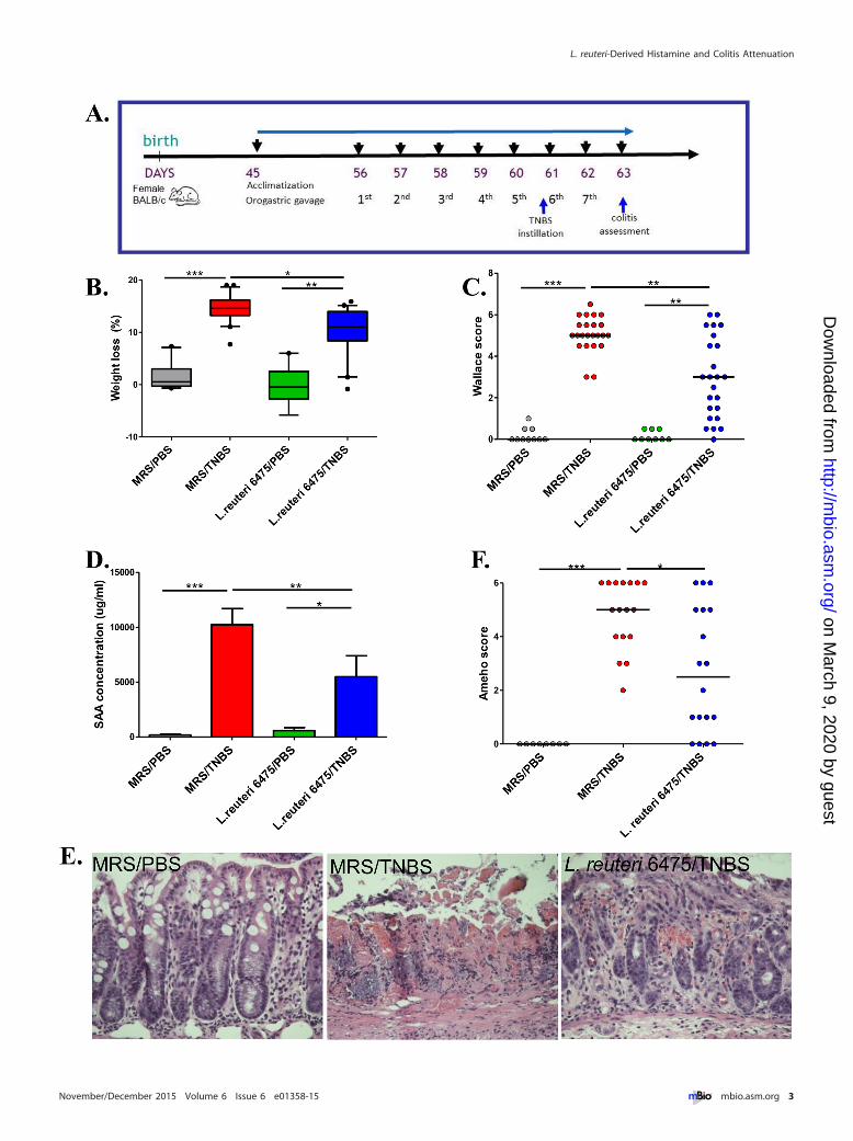
RESULTShdc� L. reuteri attenuates colonic inflammation in vivo. L. reu-teri clade II strain 6475 (L. reuteri 6475) was isolated from a Finn-ish mother’s breast milk sample (8) and has been used commer-cially as a probiotic. This hdc-positive strain suppresses humanTNF production by myeloid cells (9). The TNBS-triggered acutecolitis mouse model (17) was selected for evaluation of colitissuppression by intestinal lactobacilli. Adult (8-week-old) femaleBALB/c mice were fed L. reuteri 6475 (see Fig. S1A in the supple-mental material) by daily orogastric gavage following acclimatiza-tion and at least 5 days prior to TNBS instillation (Fig. 1A). Theseverity of colitis was evaluated 2 days after TNBS instillation byweight loss (overall health status), Wallace and Ameho scores(macroscopic and microscopic colonic injury) (18–20) (seeFig. S1B), and serum amyloid A (SAA) protein concentrations(biomarker of mucosal inflammation) (21). In colitis-negativecontrol mice that received phosphate-buffered saline (PBS) only,L. reuteri 6475 maintained a healthy baseline without any evidenceof colitis. In colitis-positive control mice that were challenged
with TNBS in the absence of L. reuteri, expected results with in-creased weight loss, Wallace scores, and serum SAA concentra-tions were observed (Fig. 1B to D), consistent with acute colitis. Incontrast, administration of L. reuteri 6475 in late-exponential-phase growth attenuated colitis as indicated by significant reduc-tions in weight loss, Wallace scores, and SAA concentrations com-pared with colitis-positive controls lacking L. reuteri 6475 (Fig. 1Bto D).
Ameho scores included assessment of histologic inflammationso that a comprehensive evaluation of colitis could be performed.TNBS instillation (sans L. reuteri) caused necrosis extendingdeeply into the muscularis propria, whereas L. reuteri 6475 admin-istration in TNBS-treated mice yielded mild or prominent muco-sal or submucosal inflammation with preservation of intact mus-cularis mucosae and muscularis propria (Fig. 1E and F). Theseresults were consistent with previous findings (19). Other mor-phological comparisons yielded reduced colon lengths in micechallenged with TNBS compared with L. reuteri 6475-treated mice(see Fig. S1C and D in the supplemental material) and healthycontrol mice receiving PBS only, consistent with previous findings(22, 23).
To extend the findings related to colitis suppression by hdc�
L. reuteri, we introduced a second clade II hdc� L. reuteri strain,ATCC PTA 4659 (L. reuteri 4659), into the same mouse colitismodel. Similarly, L. reuteri 4659 diminished the weight loss phe-notype, reduced Wallace scores, and decreased SAA concentra-tions, compared with control mice receiving MRS medium only(see Fig. S2 in the supplemental material).
PET imaging demonstrates the ability of L. reuteri 6475 tosuppress intestinal inflammation. In order to visualize anti-inflammatory effects of L. reuteri 6475, combined computed to-mography (CT)-positron emission tomography (PET) imagingwas applied to the TNBS colitis model. Healthy control mice,colitic mice, and L. reuteri-treated mice were subjected to live-animal imaging prior to euthanasia (Fig. 2A). [18F]fluorodeoxyg-lucose ([18F]FDG) has been applied to mouse colitis studies andwas used as the tracer compound because immune cells have beenshown to increase glucose uptake and phosphorylation after im-mune activation (24). During colitis, FDG uptake by activatedlymphocytes and other immune cells in the colon can be measuredby comparing relative intensities of tracer signals by anatomiclocation. In healthy control mice, FDG signals were mostly de-tected in the mouse bladder and upper chest region. These siteswere proposed to be body sites where increased glucose uptakeand metabolism in healthy states have been documented (25).Trace amounts of FDG signals were shown in pericolonic areas inthe abdomen, indicating low glucose uptake in the colons ofhealthy mice (Fig. 2B). In colitic control mice, FDG intensities inthe mouse abdomen adjacent to the colon were significantly in-creased (Fig. 2B), indicating increased glucose uptake surround-ing or within the colonic mucosa during colitis. L. reuteri 6475administration lowered FDG intensity in the mouse colon com-pared to colitic controls (Fig. 2B). FDG intensity was also shown inthree-dimensional images (see Videos S1 to S3 in the supplemen-tal material). FDG signal changes in different mouse groups werefurther confirmed by the FDG standardized uptake value (SUV)quantified blindly in the region of interest (ROI) (colon) in eachmouse using Inveon Research Workplace software (Fig. 2C), in-dicating attenuation of colonic inflammation by L. reuteri.
Gao et al.
2 ® mbio.asm.org November/December 2015 Volume 6 Issue 6 e01358-15
on March 9, 2020 by guest
http://mbio.asm
.org/D
ownloaded from
L. reuteri-Derived Histamine and Colitis Attenuation
November/December 2015 Volume 6 Issue 6 e01358-15 ® mbio.asm.org 3
on March 9, 2020 by guest
http://mbio.asm
.org/D
ownloaded from

L. reuteri administration increased microbial hdc gene ex-pression in the intestine. In order to explore whether hdc genescontribute to the anti-inflammatory effects of L. reuteri in vivo,the relative abundances of hdc genes and mRNA were determinedby quantitative PCR (qPCR) in mice gavaged with L. reuteri 6475.Our results demonstrated that L. reuteri 6475 administration sig-nificantly increased the relative abundance of bacterial hdcA genesin the mouse gut microbiome (Fig. 3A). In terms of mRNA, bothhdcA and hdcP gene expression was significantly increased in co-lonic luminal contents of mice receiving L. reuteri (Fig. 3B and C).These studies indicate a correlation between elevated hdc geneexpression via L. reuteri administration and the effect of colitisattenuation. By enhancing the ability of the intestinal microbiometo convert L-histidine to histamine, the gut microbiome was capa-ble of suppressing intestinal inflammation.
Inactivation of the L. reuteri histidine decarboxylase genediminished its ability to suppress intestinal inflammation. Tofurther investigate the importance of the bacterial histidine decar-boxylase gene with respect to intestinal immunomodulation, weexplored the anti-inflammatory effects of hdcA within an isogenicmutant and compared with wild-type L. reuteri 6475 using thesame TNBS colitis model. The hdcA gene encodes histidine decar-boxylase, and the hdcA mutant derived from L. reuteri 6475 doesnot generate histamine from L-histidine (9). The L. reuteri 6475hdcA mutant strain was shown to be deficient in terms of colitissuppression in the TNBS colitis mouse model. The hdcA mutantyielded diminished effects in terms of weight loss, Wallace scores,and SAA concentrations (Fig. 4), suggesting that bacterial histi-dine decarboxylase mediates anti-inflammatory effects via hista-mine generation. To exclude the possibility that TNBS may ad-versely affect the function of L. reuteri in the mouse intestine, weperformed additional in vitro experiments and found that TNBSdid not affect the survival or proliferation of the L. reuteri (wild-type or mutant) strain (see Fig. S3A and B in the supplementalmaterial).
Dietary L-histidine enables hdc� L. reuteri to suppress intes-tinal inflammation. In addition to experiments with differentL. reuteri strains, the relative importance of dietary L-histidine asthe substrate for histamine generation was evaluated by exposingmice to different diets. BALB/c mice were randomly divided intothree feeding groups: regular diet containing approximately 0.4%histidine from intact protein sources; defined diet with all essentialamino acids, including 0.4% histidine; and a histidine-free dietderived from defined diet with all amino acids except histidine(see Table S1 in the supplemental material).
Mice in each feeding group were gavaged with the sameamount of L. reuteri 6475 or MRS medium only as the controlgroup. Mice receiving L. reuteri 6475 and dietary L-histidine (inregular mouse chow or amino acid defined diet) demonstrated theexpected amelioration of colitis by reduced Wallace scores andserum SAA concentrations (Fig. 5A and B). In contrast, when themice received a histidine-free diet, L. reuteri 6475 yielded dimin-ished effects on colitis attenuation, suggesting that histidine intake
is important for the anti-inflammatory activity of L. reuteri 6475.Dietary deficiency of L-histidine results in weight loss in mice (seeFig. S4A in the supplemental material), as other studies haveshown (26), so weight loss was not used as a primary parameter toevaluate colitis severity in this study. To exclude the possibilitythat the diets per se contributed to colitis development, groups ofmice were fed one of the three diets, and none of these micedeveloped colitis without TNBS instillation (see Fig. S4B). Insummary, L-histidine provides the substrate for intestinalL. reuteri to generate histamine in the presence of active histi-dine decarboxylase.
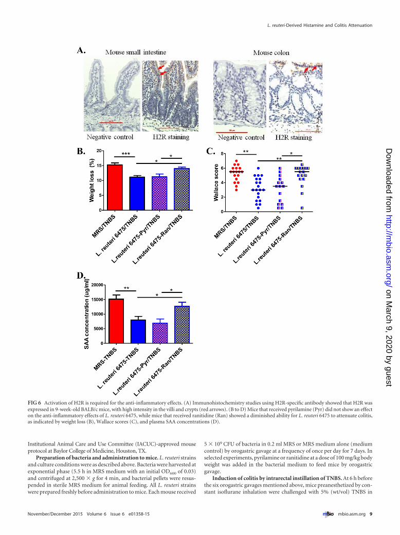
Activation of H2R is required for anti-inflammatory effectsof microbial histamine. Histamine is a biogenic amine that exertsvarious pathophysiological functions via four receptors (H1R,H2R, H3R, and H4R) (27). The predominant histamine receptorsin the intestinal epithelium are H1R and H2R (28). H2R wasshown to be expressed in the intestinal epithelium of humans,simians, and mice (28, 29). Immunohistochemical studies showH2R expression in the BALB/c mouse small intestine and colon,with relatively high intensities in the villi and crypts (Fig. 6A). Ithas been reported previously that H1R activation results in pro-inflammatory effects such as interferon (IFN) production andTh1 cell proliferation, while H2R activation appears to suppressinflammation (27, 30, 31). So, we hypothesized that microbe-derived histamine binds and activates H2R in the intestinal epi-thelium, thereby mediating its anti-inflammatory effects.
To determine whether H2R activation was required for L. reu-teri’s capacity to attenuate colitis in the TNBS model, ranitidine,an H2R-specific antagonist, was used to block H2R activation inthe mouse gut. In the TNBS colitis experiment, addition of rani-tidine (100 mg/kg of body weight) to mice receiving hdc� L. reuteriby orogastric gavage diminished the anti-inflammatory effects ofL. reuteri 6475, whereas blocking H1R with its specific antagonistpyrilamine lacked such effects (Fig. 6B to D). These findings sup-port the proposition that L. reuteri 6475 attenuates colitis via anH2R-dependent signaling mechanism. To exclude the possibilitythat ranitidine or pyrilamine may have adversely affected thefunction of L. reuteri, in vitro assays were performed by addingranitidine or pyrilamine to bacterial cultures, and neither com-pound affected the survival or proliferation of L. reuteri 6475 (seeFig. S3C and D in the supplemental material).
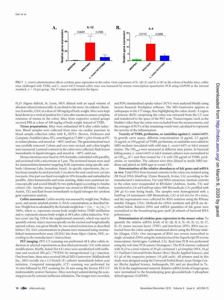
L. reuteri administration affects cytokine gene expression inthe colon. To investigate the consequences of H2R-mediated anti-inflammatory effects by hdc� L. reuteri, relative patterns of muco-sal gene expression of selected cytokines in the colons of colitis-negative, colitis-positive, and L. reuteri 6475-treated mice wereevaluated by qPCR using glyceraldehyde-3-phosphate dehydro-genase (GAPDH) as the internal standard (Fig. 7). TNBS instilla-tion significantly increased expression of genes for proinflamma-tory cytokines interleukin-1� (IL-1�) and IL-6 compared tohealthy mice. L. reuteri 6475 treatment of TNBS-challenged micereduced the relative amounts of mucosal IL-1� and IL-6 geneexpression. Other cytokine genes (TNF, IL-10, IL-12, IFN, IL-17,
FIG 1 hdc� L. reuteri attenuates colonic inflammation in vivo. (A) Time line of the mouse experiments. After 10 days of acclimatization, 8-week-old femaleBALB/c mice received 5 � 109 CFU of bacteria in MRS or MRS medium only by orogastric gavage daily for 7 days. Acute colitis was induced by intrarectalinstillation of TNBS-ethanol before the sixth gavage, and colitis severity was evaluated in 2 days. (B to D) Weight loss (B), Wallace scores (C), and SAAconcentrations (D) of mice challenged with or without TNBS and gavaged with or without L. reuteri 6475. (E and F) Representative microscopic colonic images(hematoxylin and eosin stained) (E) and Ameho scores (F) from mice in the healthy control group (MRS/PBS), the colitis control group (MRS/TNBS), and theL. reuteri-treated group (L. reuteri 6475/TNBS).
Gao et al.
4 ® mbio.asm.org November/December 2015 Volume 6 Issue 6 e01358-15
on March 9, 2020 by guest
http://mbio.asm
.org/D
ownloaded from

and IL-23) were examined, but no significant differences betweencolitic mice and L. reuteri-treated mice were detected.
DISCUSSION
Probiotic lactobacilli modulate intestinal immune responses byluminal conversion of dietary amino acids into bioactive com-
pounds such as histamine. In the current study, hdc� L. reuteristrains protected BALB/c mice in a TNBS-induced colitis model,as indicated by improvement in overall health status and amelio-ration of the colitis phenotype. The colitis phenotype was evalu-ated by macroscopic and microscopic evaluation of colonic tissue,serum biomarker quantitation, mucosal cytokine gene expres-
FIG 2 Detection of colitis attenuation by PET imaging. (A) Time line of the PET imaging experiments. Fasted (for 6 h) mice were anesthetized with isofluraneand received 200 �Ci [18F]FDG by intraperitoneal (IP) injection. One hour later, these mice received 200 �l MD-Gastroview rectally immediately before scaninitiation. Computed tomography (CT) scanning was performed for 10 min followed by PET scanning for 20 min using the Inveon PET-CT multimodalitysystem. Mice were kept sedated during the scanning process by constant isoflurane inhalation. The images were recorded, and FDG standardized uptake values(SUVs) were analyzed blindly using Inveon Research Workplace software. (B) Representative mouse images captured by PET-CT scanning in each group. Thecolor code bar represents FDG signal intensity. (C) Quantification of FDG signals in mouse colon using SUVs in different groups.
L. reuteri-Derived Histamine and Colitis Attenuation
November/December 2015 Volume 6 Issue 6 e01358-15 ® mbio.asm.org 5
on March 9, 2020 by guest
http://mbio.asm
.org/D
ownloaded from

sion, and [18F]FDG live-animal PET imaging. The importance ofdietary histidine and the bacterial enzyme histidine decarboxylasewas established by mouse model studies. In the absence of dietaryL-histidine or a gut microbiome lacking histidine decarboxylase,colitis suppression by probiotic lactobacilli was reduced signifi-cantly. Both the substrate amino acid and the enzymatic machin-ery, histidine decarboxylase, must be present in the intestinal mi-crobiome in order to generate histamine as the bioactivecompound. Histamine H2 receptor signaling in the intestinal ep-ithelium is required for probiotic L. reuteri-mediated immuno-modulation and colitis suppression. Previously, L. reuteri wasshown to suppress H2R-mediated signaling by increasing cyclicAMP (cAMP) production, protein kinase A (PKA) activation, andsuppression of extracellular signal-regulated kinase (ERK)(mitogen-activated protein [MAP] kinase) signaling (9). H2R-mediated signaling via cAMP production and protein kinase A(PKA) activation, followed by inhibition of c-Raf and MEK/ERKMAPK signaling, was described in previous studies (9, 32–34).
The same microbe-derived biochemical compound (hista-mine) can yield different effects in the host depending on thespecific type of histamine receptor. Four different G-protein-coupled histamine receptors have been described, and these re-ceptors differ based on downstream signaling pathways and cell
type distributions (27). Histamine receptors are widely distrib-uted in the body, and the histamine type 2 receptor (H2R) appearsto be enriched in the mammalian gastrointestinal tract (28, 29).H2R was first characterized in the human stomach as an impor-tant target for H2R blockers for treatment of peptic ulcer diseaseby reduction of acid (HCl) secretion (35). In addition to the im-portance of H2R in gastric physiology, it is becoming apparentthat histamine may have an important immunoregulatory role inthe intestine. H2R activation results in cAMP-mediated blockadeof c-Raf and suppression of MAP kinase signaling by inhibition ofERK phosphorylation (9, 27). Activation of H2R by histaminesuppressed IL-12 production by monocytes (36), IFN-� produc-tion by macrophages (37), TNF secretion by mast cells (38), andIL-12 release by immature dendritic cells (39). In vivo studiesshowed that histamine suppressed both Th1- and Th2-type re-sponses by H2R (30). Lactobacillus rhamnosus, which secretes his-tamine significantly, suppressed Peyer patch IL-2, IL-4, IL-5, IL-12, TNF-�, and granulocyte-macrophage colony-stimulatingfactor (GM-CSF) secretion in wild-type but not H2R-deficientmice (31).
These results indicate that the net effect of luminal histaminemay be immunosuppressive and anti-inflammatory in the mam-malian gastrointestinal tract. Prior studies of the human metag-
FIG 3 L. reuteri administration increases hdc gene expression in vivo. (A) Relative abundance of hdcA gene in mouse gut microbiome was significantly increasedin mice gavaged with L. reuteri 6475 compared to control mice gavaged with MRS. The relative abundance was determined by qPCR and normalized to thebacterial housekeeping gene rpoB. (B and C) Both hdcA (B) and hdcP (C) gene expression levels were significantly increased in colonic luminal contents of micereceiving L. reuteri compared to control mice receiving MRS. The relative gene expression was determined by qPCR and normalized to the bacterial housekeepinggene rpoB. n � 6 per group. ****, P � 0.0001.
Gao et al.
6 ® mbio.asm.org November/December 2015 Volume 6 Issue 6 e01358-15
on March 9, 2020 by guest
http://mbio.asm
.org/D
ownloaded from

enome reported that pathways of histidine biosynthesis (hista-mine precursor) were diminished in patients with IBD relative tohealthy controls (40). These studies highlight the potential impor-tance of microbiome-mediated histidine metabolism and hista-mine generation as a microbial mechanism for intestinal immu-nomodulation. Recent evidence suggests that blocking H2Rsignaling pathways in humans may result in adverse effects andsevere intestinal inflammation. Retrospective and prospectiveclinical studies of newborns have documented significantly in-creased incidence and mortality from necrotizing enterocolitis(NEC) following exposure to H2R antagonists (41–43). In addi-tion to NEC, studies have reported an increased risk of exacerba-tions in Crohn’s disease secondary to H2R blocker exposure (44).The histamine receptor H2R appears to be the key receptor on theintestinal epithelium, involved in signaling and immunomodula-tion after binding microbiome-derived histamine.
The consumption of hdc gene cluster-positive probiotics in thepresence of dietary histidine may maximize the potential benefits
by resulting in histamine generation only in intestinal regions en-riched for histamine H2 receptors. Oral administration of hista-mine may cause adverse outcomes, but the careful selection ofmicrobes that colonize specific areas of the small or large intestinemay maximize histamine H2 receptor signaling in the intestinalepithelium. Conversely, provision of L-histidine in the diet en-ables luminal conversion and luminal histamine generation byhdc gene cluster-positive microbes. Another consideration is therelative instability of histamine in vivo. Histamine is unstable invivo and could be quickly metabolized by histamineN-methyltransferase or diamine oxidase (45). Lower concentra-tions of histamine might be protective, whereas higher concentra-tions might be detrimental to epithelial protection from infection(27). The continuous production of small amounts of histamineby the gut microbiome may result in suppression of intestinalinflammation. With respect to bacterial genetics of histamine pro-duction, the current study did not include an hdcA complemen-tation strain because antibiotic consumption by mice to maintain
FIG 4 Inactivation of the L. reuteri histidine decarboxylase gene diminishes its ability to suppress intestinal inflammation. The anti-inflammatory effects of hdcAwithin an isogenic mutant which does not produce histamine were compared with those of wild-type L. reuteri 6475 using the TNBS colitis model. The wild-typestrain attenuated colitis compared with the medium-control group (MRS/TNBS), whereas the hdcA mutant yielded diminished effects in terms of weight loss (A),Wallace scores (B), and SAA concentrations (C).
L. reuteri-Derived Histamine and Colitis Attenuation
November/December 2015 Volume 6 Issue 6 e01358-15 ® mbio.asm.org 7
on March 9, 2020 by guest
http://mbio.asm
.org/D
ownloaded from

antibiotic resistance plasmids may profoundly affect gut micro-bial composition. Future generation of hdcA complementationstrains by recombineering (46) may remove the requirement forantibiotic selection. Such future genetic strategies would be help-ful to confirm whether histidine decarboxylase is essential for sup-pression of intestinal inflammation by probiotic L. reuteri.
Adverse effects of histamine are likely due to the preponder-ance of the H1 receptor in the airways (47) and upper gastrointes-tinal tract. When ingested orally, histamine may cause adversereactions and symptoms such as pruritus, bronchoconstriction,airway inflammation, and allergic symptoms (48). For this reason,the food and beverage industry has developed active programs toscreen for histamine in foodstuffs and contamination byhistamine-generating bacteria. H1R is coupled to Gq/11 familyproteins, triggering downstream calcium mobilization with pro-inflammatory effects. In addition to H1R, H4R also appears tocontribute to respiratory disease symptoms. Classical antihista-
mines relieve respiratory tract symptoms by antagonizing H1Rand, more recently, H4R (49). H3R is primarily neuronal, high-lighting histamine’s role as a potential neurotransmitter. Thecharacterization of the relative distribution of histamine receptorsin the gastrointestinal tract and other body sites will facilitate amore complete understanding of the biology of histamine in vivo.
This report highlights the potential importance of luminalconversion and amino acid metabolism in the biology of the in-testinal microbiome and host-microbe mutualism. Luminal con-version of amino acids and different classes of nutrients effectivelylinks diet, the gut bacteria, and mammalian intestinal biology.Bacterial amino acid decarboxylases have been reported to con-vert glutamate to the neurotransmitter gamma-aminobutyric acid(GABA) (50) and tyrosine to tyramine and phenylalanine to theneuromodulatory compound phenethylamine (51). Differencesin the relative abundances of pathways involved in amino acidmetabolism present in the gut microbiome may contribute to dif-ferent disease phenotypes in individuals genetically predisposedto IBD or other immune-mediated conditions. Dietary amino ac-ids are potential substrates for a variety of microbial amino aciddecarboxylases, and diverse compounds, including biogenicamines, may be produced. These compounds, such as histamine,may have important consequences for mucosal immunity orfunctioning of the nervous system. Our study indicates that lumi-nal conversion of an amino acid, L-histidine, to histamine by hdc�
L. reuteri activates H2R and yields anti-inflammatory effects in themouse colon. This study combined specific cellular elements ofthe intestinal microbiome, the genes and enzymatic machineryinvolved in luminal conversion, and the specific receptors in-volved in receiving microbial signals. These studies could fosterthe development of new probiotic therapies by facilitating the se-lection of natural hdc gene cluster-positive strains (or strains withany defined genetic feature contributing to immunomodulation)combined with dietary elements (e.g., amino acids) or enablinggenetic engineering of next-generation probiotics by defining spe-cific microbial genes involved in mitigation of intestinal inflam-mation. By defining mechanisms of microbiome-mediated im-munomodulation in the mammalian intestine, bacterial strainsand microbial gene databases can be leveraged to identify next-generation probiotics and microbe-derived medicinal com-pounds for the treatment of chronic inflammatory diseases.
MATERIALS AND METHODSBacterial strains and microbiological culture conditions. L. reuteriATCC PTA 6475 and its hdcA mutant as described previously (9) wereused to colonize the mice. L. reuteri ATCC PTA 4659, isolated from thebreast milk of healthy Finnish women, was a gift from BioGaia AB (Stock-holm, Sweden). All L. reuteri strains were cultured at 37°C in deMan,Rogosa, Sharpe (MRS) medium (Difco, Franklin Lakes, NJ) in an anaer-obic workstation (MACS MG-500; Microbiology International, Freder-ick, MD) supplied with a mixture of 10% CO2, 10% H2, and 80% N2.Quantitative analysis of bacteria was performed by counting bacterialCFU on an MRS agar plate per milliliter of bacterial culture relative tooptical density at 600 nm (OD600) measured by a SmartSpec Plus spectro-photometer (Bio-Rad Laboratories, CA).
Animals. Female BALB/c mice (45 days old) were purchased from Har-land Laboratories (Houston, TX) and maintained under specific-pathogen-free (SPF) conditions. Mice were kept under filter-top cages (5 mice per cage)and had free access to distilled water and Harlan rodent chow 2918 (defaultdiet) or other diets as described in Table S1 in the supplemental material. Allmouse experiments were performed in an SPF animal facility according to an
FIG 5 L-histidine deficiency diminishes anti-inflammatory effects. The anti-inflammatory effects of L. reuteri 6475 were compared in mice fed with differ-ent diets using the TNBS colitis model. Mice fed a regular diet or an amino aciddefined diet showed decreased Wallace scores (A) and plasma SAA concentra-tions (B) when receiving L. reuteri 6475 compared to the MRS medium con-trol. When mice were fed an L-histidine-deficient diet, L. reuteri 6475 showeddiminished anti-inflammatory effects in terms of Wallace scores and SAAconcentrations.
Gao et al.
8 ® mbio.asm.org November/December 2015 Volume 6 Issue 6 e01358-15
on March 9, 2020 by guest
http://mbio.asm
.org/D
ownloaded from
Institutional Animal Care and Use Committee (IACUC)-approved mouseprotocol at Baylor College of Medicine, Houston, TX.
Preparation of bacteria and administration to mice. L. reuteri strainsand culture conditions were as described above. Bacteria were harvested atexponential phase (5.5 h in MRS medium with an initial OD600 of 0.03)and centrifuged at 2,500 � g for 4 min, and bacterial pellets were resus-pended in sterile MRS medium for animal feeding. All L. reuteri strainswere prepared freshly before administration to mice. Each mouse received
5 � 109 CFU of bacteria in 0.2 ml MRS or MRS medium alone (mediumcontrol) by orogastric gavage at a frequency of once per day for 7 days. Inselected experiments, pyrilamine or ranitidine at a dose of 100 mg/kg bodyweight was added in the bacterial medium to feed mice by orogastricgavage.
Induction of colitis by intrarectal instillation of TNBS. At 6 h beforethe six orogastric gavages mentioned above, mice preanesthetized by con-stant isoflurane inhalation were challenged with 5% (wt/vol) TNBS in
FIG 6 Activation of H2R is required for the anti-inflammatory effects. (A) Immunohistochemistry studies using H2R-specific antibody showed that H2R wasexpressed in 9-week-old BALB/c mice, with high intensity in the villi and crypts (red arrows). (B to D) Mice that received pyrilamine (Pyr) did not show an effecton the anti-inflammatory effects of L. reuteri 6475, while mice that received ranitidine (Ran) showed a diminished ability for L. reuteri 6475 to attenuate colitis,as indicated by weight loss (B), Wallace scores (C), and plasma SAA concentrations (D).
L. reuteri-Derived Histamine and Colitis Attenuation
November/December 2015 Volume 6 Issue 6 e01358-15 ® mbio.asm.org 9
on March 9, 2020 by guest
http://mbio.asm
.org/D
ownloaded from
H2O (Sigma-Aldrich, St. Louis, MO) diluted with an equal volume ofabsolute ethanol intrarectally (4 cm distal to the anus) via catheter (Brain-tree Scientific, USA) at a dose of 100 mg/kg of body weight. Mice were kepthead down in a vertical position for 2 min after enema to ensure completeretention of enema in the colon. Mice from respective control groupsreceived PBS at a dose of 100 mg/kg of body weight instead of TNBS.
Tissue preparations. Mice were euthanized 48 h after colitis induc-tion. Blood samples were collected from mice via cardiac puncture inblood sample collection tubes with K2-EDTA (Becton, Dickinson andCompany, Franklin Lakes, NJ), centrifuged at 17,000 � g for 10 min at 4°Cto isolate plasma, and stored at �80°C until use. The gastrointestinal tractwas carefully removed. Colons and ceca were excised, and colon lengthswere measured. Luminal contents in the colon were collected, flash frozenimmediately in liquid nitrogen, and stored at �80°C until use.
Mouse intestines were fixed in 10% formalin, embedded with paraffin,and sectioned with a microtome at 5 �m. The sectioned tissues were usedfor immunohistochemistry targeting H2R expression using specific anti-body (Alomone Labs, Jerusalem, Israel). In specific experiments, the co-lon tissue samples located precisely 2 cm above the anal canal were cut intotwo parts. One part was fixed overnight in 10% formalin and embedded inparaffin. After hematoxylin and eosin staining, slides were examined mi-croscopically and scored blindly by two pathologists according to Amehocriteria (20). Another tissue fragment was stored in RNAlater (Ambion,Austin, TX) and flash frozen immediately in liquid nitrogen for cytokinegene expression analysis.
Colitis assessment. Colitis severity was assessed by weight loss, Wallacescore, and serum amyloid protein A (SAA) concentrations as described be-low. Weight loss is calculated by the formula weight loss � [(w1 � w2)/w1] �100%, where w1 represents mouse body weight before TNBS instillationand w2 represents mouse body weight at 48 h after colitis induction. Wal-lace score (see Fig. S1B in the supplemental material), which was used toquantify colonic injury macroscopically on the excised and longitudinallyopened colons, was given blindly by two trained technicians as describedbefore (18). SAA concentrations in plasma were measured using enzyme-linked immunosorbent assay (ELISA) kits from Alpco (Salem, NH), ac-cording to the manufacturer’s instructions.
PET imaging. PET-CT scanning was performed 48 h after colitis in-duction in selected experiments as described previously (24) with minormodifications. Briefly, fasted (for 6 h) mice were anesthetized with isoflu-rane and received 200 �Ci [18F]FDG by intraperitoneal (i.p.) injection.One hour later, these mice received 200 �l MD-Gastroview (MallinckrodtInc., MO) rectally via a 3.5 French (F) catheter immediately before scaninitiation. Computed tomography (CT) scanning was performed for10 min followed by PET scanning for 20 min using the Inveon PET-CTmultimodality system (Siemens). Mice were kept sedated during the scan-ning process by constant isoflurane inhalation. The images were recorded,
and FDG standardized uptake values (SUVs) were analyzed blindly usingInveon Research Workplace software. The MD-Gastroview appears asradiopaque in the CT image, thus highlighting the colon clearly. A regionof interest (ROI) comprising the colon was extracted from the CT scanand transferred to the space of the PET scan. Tissues/organs (such as thebladder) other than the colon were excluded from the measurements, andthe averages of SUVs of the remaining voxels were calculated to representthe severity of the inflammation.
Toxicity of TNBS, pyrilamine, or ranitidine against L. reuteri 6475.In growth curve assays, different concentrations (0 �g/ml, 2.5 �g/ml,25 �g/ml, or 250 �g/ml) of TNBS, pyrilamine, or ranitidine were added toMRS medium inoculated with wild-type L. reuteri 6475 or hdcA mutantstrains. The OD600s were measured at different time points. In bacterialkilling assays, L. reuteri 6475 or hdcA mutant cultures were normalized toan OD600 of 1 and then treated for 1 h with 250 �g/ml of TNBS, pyril-amine, or ranitidine. The cultures were then diluted in sterile MRS me-dium and plated on MRS agar to count CFU.
Determination of the relative abundances of hdc genes and mRNAin vivo. Total DNA from luminal contents in the colon was isolated usingZR Fecal DNA MiniPrep (Zymo Research, Irvine, CA) according to themanufacturer’s instructions. For total RNA extraction, luminal contentsin the colon were resuspended in RNAlater (Ambion, Austin, TX) andtransferred to 2.0-ml FastPrep tubes (MP Biomedicals, CA) prefilled with200 �l 0.1-mm lysing beads. The samples were homogenized with aFastPrep-24 instrument (MP Biomedicals, CA) at 4.0 m/s for 20 s twice,and the supernatants were collected for RNA isolation using the RNeasyminikit (Qiagen, USA). Methods for cDNA synthesis and qPCR are de-scribed below. Relative DNA and mRNA quantities of hdc genes werenormalized to the housekeeping gene rpoB (� subunit of bacterial RNApolymerase).
Determination of cytokine gene expression in the mouse colon. Toquantify the relative mRNA levels of IL-1�, IL-6, IL-10, IL-12, IL-17,IL-23, tumor necrosis factor (TNF), and interferon (IFN), RNA was ex-tracted from the colon samples mentioned above using the RNeasy mini-kit (Qiagen, USA). One microgram of RNA was reverse transcribed tosingle-stranded cDNA using the stated protocol for SuperScript III reversetranscriptase (Invitrogen, Carlsbad, CA). Real-time PCR was performedusing the real-time PCR system (Stratagene). The PCR mixture (adjustedwith H2O to a total volume of 20 �l) contained 1 �l template DNA, 10 �l2� FastStart Universal Probe Master (Rox) (Roche Applied Science), and0.5 �l of the respective primers (10 �M each). All primers used in thisstudy were designed using the Universal ProbeLibrary Assay Design Cen-ter (Roche Applied Science, Indianapolis, IN) and are described in Ta-ble S2 in the supplemental material. Relative mRNA levels of target geneswere normalized to the housekeeping gene glyceraldehyde-3-phosphatedehydrogenase (GAPDH).
FIG 7 L. reuteri administration affects cytokine gene expression in the colon. Gene expression of IL-1� (A) and IL-6 (B) in the colons of healthy mice, coliticmice challenged with TNBS, and L. reuteri 6475-treated colitic mice was measured by reverse transcription-quantitative PCR using GAPDH as the internalstandard. n � 10 per group. The P values are indicated in the figure.
Gao et al.
10 ® mbio.asm.org November/December 2015 Volume 6 Issue 6 e01358-15
on March 9, 2020 by guest
http://mbio.asm
.org/D
ownloaded from

Statistical analysis. Biostatistical analyses were performed usingGraphPad Prism (version 5) software (GraphPad Inc., La Jolla, CA). Fornumeric variables that fit normal distribution (determined using theKolmogorov-Smirnov test), data were presented as means with standarderrors, and different groups were compared with the t test (two groups) orone-way analysis of variance (ANOVA) (more than two groups). Other-wise, data were presented as box-and-whisker plots showing the medianand 10th and 90th percentiles or scatter plots, and different groups werecompared with the nonparametric Mann-Whitney U test (two groups) orthe Kruskal-Wallis test (more than two groups). Differences between thegroups were considered significant at P � 0.05 (*), P � 0.01 (**), P �0.001 (***), and P � 0.0001 (****).
SUPPLEMENTAL MATERIALSupplemental material for this article may be found at http://mbio.asm.org/lookup/suppl/doi:10.1128/mBio.01358-15/-/DCSupplemental.
Video S1, MOV file, 0.7 MB.Video S2, MOV file, 1.2 MB.Video S3, MOV file, 0.7 MB.Figure S1, TIF file, 2.3 MB.Figure S2, TIF file, 0.2 MB.Figure S3, TIF file, 0.6 MB.Figure S4, TIF file, 0.3 MB.Table S1, DOCX file, 0.03 MB.Table S2, DOCX file, 0.03 MB.
ACKNOWLEDGMENTS
This work was supported by the National Institutes of Health (R01AT004326, UH3 DK083990, and U01 CA170930) and the National Insti-tutes of Health (National Institute for Diabetes and Digestive and KidneyDiseases)-funded Texas Medical Center Digestive Diseases Center(DK56338) (J.V.).
We thank Eamonn Connolly (BioGaia AB, Stockholm) for providingthe L. reuteri strains, Toni-Ann Mistretta and Bhanu Priya Ganesh forassistance with data plotting and statistical analysis, and Coreen Johnsonfor assistance with bacterial DNA and RNA extractions from luminalcontents. We thank Texas Children’s Hospital for the use of the SmallAnimal Imaging Facility and especially Caterina Kaffes and. M. WaleedGaber for PET imaging.
We disclose the following: J.V. receives unrestricted research supportfrom BioGaia AB. The remaining authors disclose no conflicts.
C.G. designed and performed all the experiments, ran the analysis, andwrote the manuscript. A.M. performed all the staining. D.R. performedPET imaging and PET analysis. V.J. and M.L. helped collect mouse sam-ples. Z.S. and Y.M.-A. performed histological analyses. J.V. providedguidance, designed the experiments, and wrote the manuscript.
REFERENCES1. Molodecky NA, Soon IS, Rabi DM, Ghali WA, Ferris M, Chernoff G,
Benchimol EI, Panaccione R, Ghosh S, Barkema HW, Kaplan GG. 2012.Increasing incidence and prevalence of the inflammatory bowel diseaseswith time, based on systematic review. Gastroenterology 142:46 –54.http://dx.doi.org/10.1053/j.gastro.2011.10.001.
2. Moradkhani A, Beckman LJ, Tabibian JH. 2013. Health-related qualityof life in inflammatory bowel disease: psychosocial, clinical, socioeco-nomic, and demographic predictors. J Crohns Colitis 7:467– 473. http://dx.doi.org/10.1016/j.crohns.2012.07.012.
3. Benchimol EI, To T, Griffiths AM, Rabeneck L, Guttmann A. 2011.Outcomes of pediatric inflammatory bowel disease: socioeconomic statusdisparity in a universal-access healthcare system. J Pediatr 158:960 –967.http://dx.doi.org/10.1016/j.jpeds.2010.11.039.
4. Knights D, Lassen KG, Xavier RJ. 2013. Advances in inflammatory boweldisease pathogenesis: linking host genetics and the microbiome. Gut 62:1505–1510. http://dx.doi.org/10.1136/gutjnl-2012-303954.
5. Sokol H, Pigneur B, Watterlot L, Lakhdari O, Bermudez-Humaran LG,Gratadoux JJ, Blugeon S, Bridonneau C, Furet JP, Corthier G, Grang-ette C, Vasquez N, Pochart P, Trugnan G, Thomas G, Blottiere HM,Dore J, Marteau P, Seksik P, Langella P. 2008. Faecalibacterium praus-
nitzii is an anti-inflammatory commensal bacterium identified by gut mi-crobiota analysis of Crohn disease patients. Proc Natl Acad Sci U S A105:16731–16736. http://dx.doi.org/10.1073/pnas.0804812105.
6. Gionchetti P, Rizzello F, Venturi A, Brigidi P, Matteuzzi D, BazzocchiG, Poggioli G, Miglioli M, Campieri M. 2000. Oral bacteriotherapy asmaintenance treatment in patients with chronic pouchitis: a double-blind,placebo-controlled trial. Gastroenterology 119:305–309. http://dx.doi.org/10.1053/gast.2000.9370.
7. Casas IA, Dobrogosz WJ. 2000. Validation of the probiotic concept:Lactobacillus reuteri confers broad-spectrum protection against disease inhumans and animals. Microb Ecol Health Dis 12:247–285. http://dx.doi.org/10.1080/08910600050216246-1.
8. Liu Y, Fatheree NY, Mangalat N, Rhoads JM. 2010. Human-derivedprobiotic Lactobacillus reuteri strains differentially reduce intestinal in-flammation. Am J Physiol Gastrointest Liver Physiol 299:G1087–G1096.http://dx.doi.org/10.1152/ajpgi.00124.2010.
9. Thomas CM, Hong T, van Pijkeren JP, Hemarajata P, Trinh DV, HuW, Britton RA, Kalkum M, Versalovic J. 2012. Histamine derived fromprobiotic Lactobacillus reuteri suppresses TNF via modulation of PKA andERK signaling. PLoS One 7:e31951. http://dx.doi.org/10.1371/journal.pone.0031951.
10. Madsen KL, Doyle JS, Jewell LD, Tavernini MM, Fedorak RN. 1999.Lactobacillus species prevents colitis in interleukin 10 gene-deficient mice.Gastroenterology 116:1107–1114. http://dx.doi.org/10.1016/S0016-5085(99)70013-2.
11. Pena JA, Li SY, Wilson PH, Thibodeau SA, Szary AJ, Versalovic J. 2004.Genotypic and phenotypic studies of murine intestinal lactobacilli: speciesdifferences in mice with and without colitis. Appl Environ Microbiol 70:558 –568. http://dx.doi.org/10.1128/AEM.70.1.558-568.2004.
12. Schreiber O, Petersson J, Phillipson M, Perry M, Roos S, Holm L. 2009.Lactobacillus reuteri prevents colitis by reducing P-selectin-associatedleukocyte- and platelet-endothelial cell interactions. Am J Physiol Gastro-intest Liver Physiol 296:G534 –G542. http://dx.doi.org/10.1152/ajpgi.90470.2008.
13. Preidis GA, Saulnier DM, Blutt SE, Mistretta T, Riehle KP, Major AM,Venable SF, Barrish JP, Finegold MJ, Petrosino JF, Guerrant RL,Conner ME, Versalovic J. 2012. Host response to probiotics determinedby nutritional status of rotavirus-infected neonatal mice. J Pediatr Gastro-e n t e r o l N u t r 5 5 : 2 9 9 – 3 0 7 . h t t p : / / d x . d o i . o r g / 1 0 . 1 0 9 7 /MPG.0b013e31824d2548.
14. Spinler JK, Sontakke A, Hollister EB, Venable SF, Oh PL, Balderas MA,Saulnier DMA, Mistretta TA, Devaraj S, Walter J, Versalovic J, High-lander SK. 2014. From prediction to function using evolutionarygenomics: human-specific ecotypes of Lactobacillus reuteri have diverseprobiotic functions. Genome Biol Evol 6:1772–1789. http://dx.doi.org/10.1093/gbe/evu137.
15. Preidis GA, Versalovic J. 2009. Targeting the human microbiome withantibiotics, probiotics, and prebiotics: gastroenterology enters the metag-enomics era. Gastroenterology 136:2015–2031. http://dx.doi.org/10.1053/j.gastro.2009.01.072.
16. Hemarajata P, Gao C, Pflughoeft KJ, Thomas CM, Saulnier DM,Spinler JK, Versalovic J. 2013. Lactobacillus reuteri-specific immunoregu-latory gene rsiR modulates histamine production and immunomodula-tion by Lactobacillus reuteri. J Bacteriol 195:5567–5576. http://dx.doi.org/10.1128/JB.00261-13.
17. Scheiffele F, Fuss IJ. 2002. Induction of TNBS colitis in mice. Curr ProtocImmunol Chapter 15:Unit 15.19. http://dx.doi.org/10.1002/0471142735.im1519s49.
18. Wallace JL, MacNaughton WK, Morris GP, Beck PL. 1989. Inhibition ofleukotriene synthesis markedly accelerates healing in a rat model of in-flammatory bowel disease. Gastroenterology 96:29 –36.
19. Foligné B, Nutten S, Steidler L, Dennin V, Goudercourt D, MercenierA, Pot B. 2006. Recommendations for improved use of the murine TNBS-induced colitis model in evaluating anti-inflammatory properties of lacticacid bacteria: technical and microbiological aspects. Dig Dis Sci 51:390 – 400. http://dx.doi.org/10.1007/s10620-006-3143-x.
20. Ameho CK, Adjei AA, Harrison EK, Takeshita K, Morioka T, ArakakiY, Ito E, Suzuki I, Kulkarni AD, Kawajiri A, Yamamoto S. 1997.Prophylactic effect of dietary glutamine supplementation on interleukin 8and tumour necrosis factor alpha production in trinitrobenzene sul-phonic acid induced colitis. Gut 41:487– 493. http://dx.doi.org/10.1136/gut.41.4.487.
21. De Villiers WJS, Varilek GW, de Beer FC, Guo J, Kindy MS. 2000.
L. reuteri-Derived Histamine and Colitis Attenuation
November/December 2015 Volume 6 Issue 6 e01358-15 ® mbio.asm.org 11
on March 9, 2020 by guest
http://mbio.asm
.org/D
ownloaded from
Increased serum amyloid a levels reflect colitis severity and precede amy-loid formation in IL-2 knockout mice. Cytokine 12:1337–1347. http://dx.doi.org/10.1006/cyto.2000.0716.
22. Sasaoka T, Ito M, Yamashita J, Nakajima K, Tanaka I, Narita M, HaraY, Hada K, Takahashi M, Ohno Y, Matsuo T, Kaneshiro Y, Tanaka H,Kaneko K. 2011. Treatment with IL-27 attenuates experimental colitisthrough the suppression of the development of IL-17-producing T helpercells. Am J Physiol Gastrointest Liver Physiol 300:G568 –G576. http://dx.doi.org/10.1152/ajpgi.00329.2010.
23. Dutra RC, Claudino RF, Bento AF, Marcon R, Schmidt EC, Bouzon ZL,Pianowski LF, Calixto JB. 2011. Preventive and therapeutic euphol treat-ment attenuates experimental colitis in mice. PLoS One 6:e27122. http://dx.doi.org/10.1371/journal.pone.0027122.
24. Brewer S, McPherson M, Fujiwara D, Turovskaya O, Ziring D, Chen L,Takedatsu H, Targan SR, Wei B, Braun J. 2008. Molecular imaging ofmurine intestinal inflammation with 2-deoxy-2-[18F]fluoro-D-glucoseand positron emission tomography. Gastroenterology 135:744 –755.http://dx.doi.org/10.1053/j.gastro.2008.06.040.
25. Galitovskiy V, Kuruvilla S, Sevriokov E, Corches A, Pan M, Kalantari-Dehaghi M, Chernyavsky A, Mukherjee J, Grando S. 2013. Developmentof novel approach to diagnostic imaging of lung cancer with 18F-NifenePET/CT using A/J mice treated with NNK. J Cancer Res Ther 1:128 –137.http://dx.doi.org/10.14312/2052-4994.2013-20.
26. Shibata K, Fukuwatari T, Zushi S, Sugimoto E. 2001. Effect of dietaryhistidine content on the change in content of skin urocanic acid isomers inhairless mice irradiated with ultraviolet B. Biosci Biotechnol Biochem 65:1415–1418. http://dx.doi.org/10.1271/bbb.65.1415.
27. O’Mahony L, Akdis M, Akdis CA. 2011. Regulation of the immuneresponse and inflammation by histamine and histamine receptors. J Al-lergy Clin Immunol 128:1153–1162. http://dx.doi.org/10.1016/j.jaci.2011.06.051.
28. Sander LE, Lorentz A, Sellge G, Coeffier M, Neipp M, Veres T, FrielingT, Meier PN, Manns MP, Bischoff SC. 2006. Selective expression ofhistamine receptors H1R, H2R, and H4R, but not H3R, in the humanintestinal tract. Gut 55:498 –504.
29. Kim H, Dwyer L, Song JH, Martin-Cano FE, Bahney J, Peri L, BrittonFC, Sanders KM, Koh SD. 2011. Identification of histamine receptors andeffects of histamine on murine and simian colonic excitability. Neurogas-troenterol Motil 23:949 – e409. http://dx.doi.org/10.1111/j.1365-2982.2011.01760.x.
30. Jutel M, Watanabe T, Klunker S, Akdis M, Thomet OAR, Malolepszy J,Zak-Nejmark T, Koga R, Kobayashi T, Blaser K, Akdis CA. 2001.Histamine regulates T-cell and antibody responses by differential expres-sion of H1 and H2 receptors. Nature 413:420 – 425. http://dx.doi.org/10.1038/35096564.
31. Frei R, Ferstl R, Konieczna P, Ziegler M, Simon T, Rugeles TM,Mailand S, Watanabe T, Lauener R, Akdis CA, O’Mahony L. 2013.Histamine receptor 2 modifies dendritic cell responses to microbial li-gands. J Allergy Clin Immunol 132:194 –204. http://dx.doi.org/10.1016/j.jaci.2013.01.013.
32. Funaki C, Hodges RR, Dartt DA. 2010. Identification of the Raf-1 sig-naling pathway used by cAMP to inhibit p42/p44 MAPK in rat lacrimalgland acini: role in potentiation of protein secretion. Invest OphthalmolVis Sci 51:6321– 6328. http://dx.doi.org/10.1167/iovs.10-5690.
33. Hershenson MB, Chao TS, Abe MK, Gomes I, Kelleher MD, Solway J,Rosner MR. 1995. Histamine antagonizes serotonin and growth factor-induced mitogen-activated protein kinase activation in bovine trachealsmooth muscle cells. J Biol Chem 270:19908 –19913. http://dx.doi.org/10.1074/jbc.270.34.19908.
34. Waltereit R, Weller M. 2003. Signaling from cAMP/PKA to MAPK andsynaptic plasticity. Mol Neurobiol 27:99 –106. http://dx.doi.org/10.1385/MN:27:1:99.
35. Garnett WR. 2003. History of acid suppression: focus on the hospitalsetting. Pharmacotherapy 23:56S– 60S. http://dx.doi.org/10.1592/phco.23.13.56S.31932.
36. Elenkov IJ, Webster E, Papanicolaou DA, Fleisher TA, Chrousos GP,Wilder RL. 1998. Histamine potently suppresses human IL-12 and stim-ulates IL-10 production via H2 receptors. J Immunol 161:2586 –2593.
37. Horvath B, Szalai C, Mandi Y, Laszlo V, Radvany Z, Darvas Z, Falus A.1999. Histamine and histamine-receptor antagonists modify gene expres-sion and biosynthesis of interferon gamma in peripheral human bloodmononuclear cells and in CD19-depleted cell subsets. Immunol Lett 70:95–99. http://dx.doi.org/10.1016/S0165-2478(99)00126-1.
38. Bissonnette EY. 1996. Histamine inhibits tumor necrosis factor alpharelease by mast cells through H2 and H3 receptors. Am J Respir Cell MolBiol 14:620 – 626. http://dx.doi.org/10.1165/ajrcmb.14.6.8652190.
39. Mazzoni A, Young HA, Spitzer JH, Visintin A, Segal DM. 2001. Hista-mine regulates cytokine production in maturing dendritic cells, resultingin altered T cell polarization. J Clin Invest 108:1865–1873. http://dx.doi.org/10.1172/JCI200113930.
40. Morgan XC, Tickle TL, Sokol H, Gevers D, Devaney KL, Ward DV,Reyes JA, Shah SA, LeLeiko N, Snapper SB, Bousvaros A, Korzenik J,Sands BE, Xavier RJ, Huttenhower C. 2012. Dysfunction of the intestinalmicrobiome in inflammatory bowel disease and treatment. Genome Biol13:R79. http://dx.doi.org/10.1186/gb-2012-13-9-r79.
41. Gupta RW, Tran L, Norori J, Ferris MJ, Eren AM, Taylor CM, DowdSE, Penn D. 2013. Histamine-2 receptor blockers alter the fecal microbi-ota in premature infants. J Pediatr Gastroenterol Nutr 56:397– 400. http://dx.doi.org/10.1097/MPG.0b013e318282a8c2.
42. Terrin G, Passariello A, De Curtis M, Manguso F, Salvia G, Lega L,Messina F, Paludetto R, Canani RB. 2012. Ranitidine is associated withinfections, necrotizing enterocolitis, and fatal outcome in newborns. Pe-diatrics 129:e40 – e45. http://dx.doi.org/10.1542/peds.2011-0796.
43. Guillet R, Stoll BJ, Cotten CM, Gantz M, McDonald S, Poole WK,Phelps DL. 2006. Association of H2-blocker therapy and higher incidenceof necrotizing enterocolitis in very low birth weight infants. Pediatrics117:e137– e142. http://dx.doi.org/10.1542/peds.2005-1543.
44. Juillerat P, Schneeweiss S, Cook EF, Ananthakrishnan AN, Mogun H,Korzenik JR. 2012. Drugs that inhibit gastric acid secretion may alter thecourse of inflammatory bowel disease. Aliment Pharmacol Ther 36:239 –247. http://dx.doi.org/10.1111/j.1365-2036.2012.05173.x.
45. Akdis CA, Blaser K. 2003. Histamine in the immune regulation of allergicinflammation. J Allergy Clin Immunol 112:15–22. http://dx.doi.org/10.1067/mai.2003.1585.
46. Van Pijkeren JP, Britton RA. 2012. High efficiency recombineering inlactic acid bacteria. Nucleic Acids Res 40:e76. http://dx.doi.org/10.1093/nar/gks147.
47. Togias A. 2003. H1-receptors: localization and role in airway physiologyand in immune functions. J Allergy Clin Immunol 112:S60 –S68. http://dx.doi.org/10.1016/S0091-6749(03)01878-5.
48. Naila A, Flint S, Fletcher G, Bremer P, Meerdink G. 2010. Control ofbiogenic amines in food— existing and emerging approaches. J Food Sci75:R139 –R150. http://dx.doi.org/10.1111/j.1750-3841.2010.01774.x.
49. Thurmond RL, Gelfand EW, Dunford PJ. 2008. The role of histamine H1and H4 receptors in allergic inflammation: the search for new antihista-mines. Nat Rev Drug Discov 7:41–53. http://dx.doi.org/10.1038/nrd2465.
50. Li H, Cao Y. 2010. Lactic acid bacterial cell factories for gamma-aminobutyric acid. Amino Acids 39:1107–1116. http://dx.doi.org/10.1007/s00726-010-0582-7.
51. Marcobal A, De las Rivas B, Landete JM, Tabera L, Muñoz R. 2012.Tyramine and phenylethylamine biosynthesis by food bacteria. Crit RevF o o d S c i N u t r 5 2 : 4 4 8 – 4 6 7 . h t t p : / / d x . d o i . o r g / 1 0 . 1 0 8 0 /10408398.2010.500545.
Gao et al.
12 ® mbio.asm.org November/December 2015 Volume 6 Issue 6 e01358-15
on March 9, 2020 by guest
http://mbio.asm
.org/D
ownloaded from