histological and immunohistochemical study on the possible
TRANSCRIPT
Original article
395 DOI:10.21608/bmfj.2020.22518.1204
Histological and Immunohistochemical Study on the Possible
Therapeutic Role of Stem Cells and Curcumin in Cyclophosphamide-
Induced Cardiotoxicity in Adult Male Albino Rat
Omayma K. Helala, Hala G. Metwaly
b, Maha M. Abo Gazia
c, Ola M. Mohamed
a, Azza I. Helal
c
Abstract:
Introduction: Cyclophosphamide (CP) is used as a chemotherapeutic and
an immunosuppressive agent. CP is known to cause multiple organ
toxicity; the most obvious is cardiotoxicity. Aim: Possibile therapeutic
effect on bone marrow -derived mesenchymal stem cells (BM-MSCs) in
ameliorating CP- induced cardiotoxicity compared with Curcumin in
albino rats. Methods: Fifty male albino rats were divided into five groups:
group I received intraperitoneal injection of sterile water; group II
received oral curcumin (200 mg/kg); group III received only one
intraperitoneal injection of CP (200 mg/kg) and were sacrificed on day 40;
group IV received the same treatment as group III followed by oral
administration of curcumin from day 10 and were sacrificed on day 40;
group V received the same treatment as group III followed by a single
BM- MSCs injection intraperitoneally at a dose of 1×10⁶ cells/ rat on day
10 and were sacrificed on day 40. Results: Histological structure of the
cardiac muscle by light and electron microscopic examination revealed
marked structural changes in rats treated with CP alone. Improvement in
BM-MSCs group more than curcumin treated group was observed.
Immunohistochemical staining of the cardiac muscle showed strong
positive immunoreactivity for caspase-3 in group III compared to the
control and other groups. Also, BM-MSCs extensively reduced the
amount of collagen fibers compared with other groups. Conclusion: The
use of curcumin has a limited beneficial effect on the protection of cardiac
muscle against CP toxicity compared with stem cells.
Keywords: Cardiotoxicity; Cyclophosphamide; stem cells ; Curcumin.
a
Department of histology
and cell biology, Benha
faculty of medicine, bdepartment of clinical
pathology, Cairo university , c
department of Histology,
Benha faculty of medicine,
kafr sheikh university,
Egypt
Correspondence to:
Azza I. Helal, Histology
department, faculty of
medicine, kafr sheikh
university, Egypt
Email:
Received:
Accepted:

Original article
396 DOI:10.21608/bmfj.2020.22518.1204
Introduction
Cardiotoxicity is a serious adverse effect
of chemotherapeutic agents [1].
Cardiomyocytes have a limited mitotic
capacity that cannot support its self-renewal
[2].
Cyclophosphamide is an alkylating agent
with potent antineoplastic and
immunosuppressive properties and possibly
the most widely used antineoplastic agent [3].
The metabolism of anti-cancer drugs can lead
to more active anti-cancer metabolites but
those metabolites can likewise contribute to
the observed cardiotoxicity [4].
Bone marrow-derived mesenchymal stem
cells (BM-MSCs) are considered the most
routinely used in clinical studies because they
are easily accessible and are routinely
collected from adults without the ethical
concern inherent to fetal embryonic tissues
[5,6].
Curcumin has been reported to exhibit a
strong antioxidant property and acts as a
scavenger of free oxygen radicals [7].
This study aimed to assess the possible
protective role and ameliorating effects of BM
-MSCs compared with curcumin against
cyclophosphamide- induced cardiotoxicity
histologically and immunohistochemically.
Materials and methods
Drugs used
- Cyclophosphamide (Endoxan vials;
Baxter Company, Deerfield, IL, USA)
was obtained as vials. Each vial
contained 200 mg of CP in dry
lyophilized powder form. CP was
injected at a dose of 200 mg/kg [8]. The
content of one vial was dissolved in 10
ml of sterile water to obtain a
concentration of 20 mg/ml for
immediate injection. Each rat weighing
200 g was injected with 2 ml of CP.
- Curcumin is available in the market in
the form of powder. The dose used in
this study was 200 mg/kg orally [9]. The
powder was dissolved in 400 ml of
sterile water to obtain a concentration of
40 mg curcumin/ml.
- BM-MSCs were prepared in the
Department of Medical Biochemistry,
Kasr Al-Ainy Faculty of Medicine,
Cairo University. They were provided as
first-passage culture cells suspended in
PBS at a dose of 1×10⁶ cells/ rat [8].
Animals
50 albino rats, their weights ranged between
150 and 200 grams, were divided into five
groups (10 rats each)
Group I (control): Each rat in this group was
intraperitoneally injected once with 2ml
sterile water and sacrificed after 10 days.

stem cell and curcumin in cardiotoxicity , Helal et al ,2020
DOI:10.21608/bmfj.2020.22518.1204
195
Group II (curcumin-treated): Each rat
received 1 ml of the dissolved curcumin
powder orally through gastric gavage once
daily and sacrificed after 10 days. Group III
(CP-treated): Each rat received a single
intraperitoneal injection of 2 ml of dissolved
CP on the first day of the experiment and
were sacrificed on day 40. Group IV (CP plus
curcumin): Each rat received a single
intraperitoneal injection of CP on the first day
of the experiment, followed by oral
administration of curcumin from day 10 of the
experiment and were sacrificed on day 40.
Group V (CP plus BM-MSCs): Each rat
received a single intraperitoneal injection of
dissolved CP on the first day of the
experiment, followed by a single dose of BM-
MSCs, injected intraperitoneally on day 10 of
the experiment and were sacrificed on day 40.
This is a case control study which took
place in Kasr Al-ainy, faculty of medicine,
Cairo University for 40 days from November,
2017 to December, 2017 after being approved
by the Local Ethics Committee of the Faculty
of Medicine, Cairo University.
Preparation of bone marrow-derived
mesenchymal stem cells:
Bone marrow was harvested by flushing
the tibiae and femora of three 6-weeks-old
male Sprague-Dawley albino rats with
Dulbecco’s modified Eagle’s medium
supplemented with 10% fetal bovine serum.
Nucleated cells were isolated and resuspended
in complete culture medium supplemented
with 1% penicillin–streptomycin [10]. The
cells were incubated at 37°C in 5%
humidified CO2 for 12 – 14 days as primary
culture. BM-MSCs were distinguished from
other BM cells by their tendency to adhere to
tissue culture plastic flasks [11]. When they
developed (80–90% confluence), the cultures
were washed twice with PBS and the cells
were trypsinized with 0.25% trypsin for 5 min
at 37°C.
After centrifugation, the cells were
resuspended in serum-supplemented medium
and incubated in a culture flask. The resulting
culture was referred to as first-passage culture
[12]. The second exchange of medium was
done after 6 days when spindle shaped cells
appeared with long processes and vesicular
nuclei [13].
The hemocytometer was used to determine
the total cell count and assess viability of the
cells [14]. BM-MSCs in culture were
characterized by their adhesiveness and
fusiform shape BM-MSCs in culture were
characterized by their adhesiveness and
fusiform shape under a phase-contrast
microscope.
At the time of injection, the cells were
labeled with a PKH26 dye supplied by

Benha Medical Journal, Vol. 37, issue 1, 2020
DOI:10.21608/bmfj.2020.22518.1204 391
Sigma, Darmstadt, Germany (PKH26 dye
stock, 1 vial containing > 0.1 ml, 1 x 10⁻³
M in ethanol&Diluent C, an iso-osomotic
aqueous solution). Their homing in the
cardiac tissue was confirmed by
immunofluorescence [16] (Fig. 1).
Histological and immunohistochemical
study:
The heart of each rat was taken immediately
and divided into two specimens.
One was fixed in 10% formalin saline
solution and was processed to get paraffin
sections:
Serial sections of 3-4 microns thickness
were cut, mounted on slides and subjected to
the following stains:
1) H&E stain for general histological
examination [17].
2) Mallory’s trichrome stain for detection
of collagen fibers [18].
3) Immunohistochemical Staining for
caspase -3 antibody (ready- to -use for
IHC, catalog code: PA5-23921) using the
avidin-biotin-peroxidase complex
technique. The primary antibody was a
rabbit polyclonal antibody (Thermo Fisher
scientific, USA). The dilution used was
1:50 [19&20]. The sections were
examined with light microscope and
Negative controls were prepared by
omitting the primary antibody. Positive
tissue control was performed by applying
the previous technique in the same way on
duodenal specimen (mybiosource.com).
(caspase- 3 immunostaining x 400)
The other specimen was immediately cut
into small pieces in all dimensions and
fixed in 2.5% buffered glutaraldehyde and
was used to prepare ultrathin sections for
electron microscopic examination [21].
Morphometric study
The mean area percentage of collagen
fibers in Mallory’s trichrome-stained sections
as well as caspase -3 was measured in 10 non
over-lapping high power fields for each
animal. Appropriate measurements were
taken using the image analyzer computer
system (Leica Qwin 500C, Leica, London,
UK) [22].
Statistical analysis
The following parameters were
expressed as mean ± SD:
(1) Area percentage of collagen fibers in
Mallory’s trichrome-stained sections.
(2) Area percentage of caspase-3
immunoreaction.

stem cell and curcumin in cardiotoxicity , Helal et al ,2020
DOI:10.21608/bmfj.2020.22518.1204
197
The statistical analysis was carried out using
one-way ANOVA analysis of variance for
comparison between the different groups,
using SPSS (version 16; SPSS Inc., Chicago,
Illinois, USA). P values of 0.05 or less were
accepted as statistically significant [8].
Results
Histological results
Group I (control)
H&E stained sections showed the normal
histological structure of cardiac myocytes that
appeared arranged in a linear array that
branch and anastomose with acidophilic
sarcoplasm and oval, centrally located nuclei
(Fig.2 a & b). Mallory’s trichrome-stained
sections showed minimal basophilic collagen
fibers surrounding the cardiomyocyte bundles
(Fig.2 c). Weak positive immunoreactivity for
caspase -3 was observed (Fig.2 d).
Ultrastructural examination of ultrathin
sections showed the sarcomeres that were
bounded on each side by a Z-line with a
central dark A-band and two light I-bands in
the periphery. A pale H-zone could be seen in
the center of A-band bisected by M-line
(Fig.2 e&f).
Group II (curcumin treated)
The same structural and ultrastructural results
as group I were observed (Fig.3 a,b,c&d).
Group III (CP treated)
H&E stained sections revealed that most of
the cardiac muscle fibers were disorganized
and lost the normal architecture. Extensive
mononuclear cellular infiltrate and diffuse
interstitial hemorrhage were also seen. (Fig.4
a & b).
Mallory trichrome stain exhibited strong
accumulation of collagen fibers
(Fig.4 c) and intense caspase-3
immunoreactivity was seen (Fig.4 d).
Ultrastructural examination revealed
disturbance of the normal architecture with
destruction of myofibrils. Mitochondria
appeared swollen with disrupted crista with
nuclear indentation (Fig. 4 e&f)
Group IV (CP plus curcumin)
H&E stained sections showed mild
restoration of the normal myocardial structure
with congestion of blood vessels (Fig.5 a &b).
Mallory’s trichrome-stain showed mild
accumulation of collagen fibers (Fig.5 c).
Weak positive immunoreaction for caspase -3
was detected (Fig. 5 d). Ultrastructural
examination revealed incomplete recovery of
the structural loss with mitochondrial swelling
and interstitial hemorrhage. (fig.5 e).
Group V (CP plus BM-MSCs)
H&E stained sections showed nearly
complete restoration of the normal
histological structure. Few myocardial cells
exhibited focal sarcoplasmic vacuolation
without obvious nuclear changes together

Benha Medical Journal, Vol. 37, issue 1, 2020
DOI:10.21608/bmfj.2020.22518.1204 391
with normal blood vessels and obvious
intercalated discs (Figs.6 a&b). Mallory ’s
trichrome -stain showed mild accumulation of
collagen fibers (Fig.6 c). Weak positive
immunoreactivity for caspase -3 was
encountered (Fig.6 d). Ultrastructural
examination revealed obvious amelioration of
the degenerative changes . The mitochondria
regained their normal linear interposition
between the myofibrils with well defined oval
or rounded contour. (fig.6 e&f).
Statistical results:
Table (1): The mean area % of collagen fibers in all
experimental groups.
Groups Mean
Standard
deviation Significance
Group I
.66583
G3
G4
G5
Group II
.7333 .49329
G3
G4
G5
Group III
38.9000
.
2.77308
G1
G2
G5
G6
Group IV
14.2667 1.26623
G1
G2
G3
G4
G5
G6
Group V
3.3333 .92916
G3
G4
G5
G6
Histogram (1): The mean area % of collagen fibers
in all experimental groups
Table (2): The mean area % of positive
immunoreactivity for caspase -3 in all experimental
groups.
Groups Mean
Standard deviation
Significance
Group I
.7500 .55000 G3 G4 G5
GroupII
.8333 .30551 G3 G4 G5
Group III
31.3333 . 3.21455
G1 G2 G3 G4 G5 G6
Group IV
11.0000 1.00000
G1 G2 G3 G4 G5 G6
Group V
3.0000 1.00000
G3 G4 G5 G6

stem cell and curcumin in cardiotoxicity , Helal et al ,2020
DOI:10.21608/bmfj.2020.22518.1204
199
Histogram (2): The mean area % of positive
immunoreactivity for caspase -3 in all experimental
groups.
Fig. 1 :A photomicrograph of a section of a rat heart showing positive red immunofluorescent MSCs labelled
with PKH26 fluorescent dye (arrows ( (PKH26 immunofluorescence x400)
Benha Medical Journal, Vol. 37, issue 1, 2020
DOI:10.21608/bmfj.2020.22518.1204 422
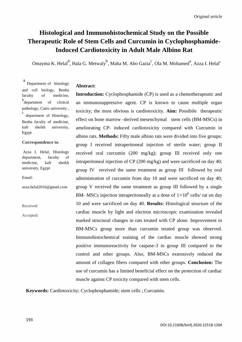
Fig. 2: Photomicrographs of sections of the rat myocardium from control group (group I) showing: (a) branched cardiac
muscle fibers with acidophilic sarcoplasm (→). (b) centrally located oval nucleus (wavy arrow) and intercalated disc (→).
(c) Minimal amount of collagen fibers (→). (d) Weak sarcoplasmic caspase -3 immunoreaction (→). (e) An electron
micrograph of a section of cardiac muscle showing a central euchromatic nucleus (N) with prominent nucleolus (nu),
parallel arrangement of myofibers (F), the mitochondria (M) arranged in rows between them and regularly arranged
sarcomeres between Z lines (Z). (f) regular arrangement of the myofibrils (F) with sarcomeres between Z lines (Z).
Notice: H zone (H) bisected by M line (M). [H&E, (a) ×200/(b) ×1000; Mallory’s trichrome, (c) ×200; caspase -3
immunostaining, (d) ×1000; electron micrographs, (e) x 5800/(f) 17500].
Fig. 3: Photomicrographs of sections of rat myocardium from group II (curcumin-treated) showing: (a) branched cardiac
muscle fibers with acidophilic sarcoplasm (→) with blood vessels in between (wavy arrow). (b) Minimal collagen fibers
(→). (c) Weak sarcoplasmic caspase -3 immunoreaction (→). (d) An electron micrograph showing parallel arrangement
of myofibers (F) and the mitochondria (M) in rows between them in association with smooth endoplasmic reticulum( SR).
stem cell and curcumin in cardiotoxicity , Helal et al ,2020
DOI:10.21608/bmfj.2020.22518.1204
201
Notice: the regularly arranged Z- lines (Z) and the continuous intercalated disc (D). [H&E, (a) ×200; Mallory’s trichrome
(b) ×400; caspase -3 immunostaining, (c) ×1000; electron micrographs, (d) ×5800].
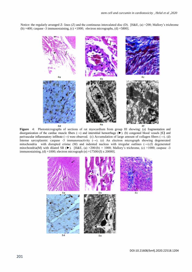
Figure 4: Photomicrographs of sections of rat myocardium from group III showing: (a) fragmentation and
disorganization of the cardiac muscle fibers (→) and interstitial hemorrhage (►). (b) congested blood vessels [C] and
perivascular inflammatory infiltrate (→) were observed. (c) Accumulation of large amount of collagen fibers (→). (d)
Intense sarcoplasmic caspase -3 immunoreactivity (→). (e) An electron micrograph showing degenerated
mitochondria with disrupted cristae (M) and indented nucleus with irregular outlines (→).(f) degenerated
mitochondria(M) with dilated SR (►). [H&E, (a) ×200/(b) × 1000; Mallory’s trichrome, (c) ×1000; caspase -3
immunostaining, (d) ×1000; electron micrograph (e) ×17500/(f) x 20000].

Benha Medical Journal, Vol. 37, issue 1, 2020
DOI:10.21608/bmfj.2020.22518.1204 424
Figure 5: Photomicrographs of sections of rat myocardium from group IV showing (a) prominent fibrillolysis (wavy
arrow) and widely spaced cardiac muscle fibers (star), disorganized wavy cardiac muscle fibers (→). (b) splitting of the fibers
(wavy arrow), pyknotic nuclei (P) and congested blood vessel (star). (c) moderate amount of collagen fibers in between
cardiac muscle fibers (→). (d) moderate sarcoplasmic caspase -3 immunoreactivity. (e) An electron-micrograph showing
nuclear indentation (→), areas of fibrillolysis (►)and interstitial hemorrhage (H).[H&E, (a) ×200/(b) × 1000; Mallory’s
trichrome, (c) × 400; caspase -3 immunostaining, (d) × 1000; electron-micrograph (e) × 5800].
Figure 6: Photomicrographs of sections of rat myocardium from group V showing (a) nearly complete restoration of the normal
histological structure with apparently widely spaced muscle fibers (→) with blood vessels in-between (►) and sarcoplasmic vacuolation
(wavy arrow). (b) apparently normal intercalated discs (→) and central oval nucleus (wavy arrow). (c) Mild amount of collagen fibers
(arrows). (d) weak sarcoplasmic caspase -3 immunoreactivity (→). (e) An electron-micrograph showing more or less well organized
myofibrils (mf), normal mitochondria (M) with apparently normal blood vessel (→). (f) showing a normal euchromatic nucleus (Nu)
with dilated smooth Endoplasmic Reticulum (→) and mitochondria (M) arranged between myofibrils (mf). [H&E, (a) ×200/ (b) × 1000;
Mallory’s trichrome, (c) × 1000; caspase -3 immunostaining, (d) ×1000; electron-micrograph (e) ×5800 & (f) x17500 ].
Discussion:
Cyclophosphamide (CP) is a widely used drug
in cancer chemotherapy and
immunosuppression, which could cause
toxicity of the normal cells due to its toxic
metabolites. The major limitation of CP is the
injury of normal tissue, leading to multiple
organ toxicity [23,24].
Cyclophosphamide itself is not cardiotoxic, it
is rather the CP metabolites that induce
cardiotoxicity through increasing free oxygen
radicals and the decrease in the antioxidant
defense mechanisms [25].
Oxidative stress has been widely shown to
regulate apoptosis and exerts both agonistic
and antagonistic effects on apoptotic signaling
[ 26]. It has been demonstrated to mediate p53-
dependent cell cycle arrest, DNA repair and
apoptosis. [27,28].
Apoptosis was confirmed in the present work
by positive caspase 3 immunoreactivity in all
CP -injected rats. So, it can be concluded that
both apoptosis and necrosis are etiological
mechanisms that cause CP - induced cardiac
injury as previously demonstrated by [29].
In the present work, congestion and apparent

stem cell and curcumin in cardiotoxicity , Helal et al ,2020
DOI:10.21608/bmfj.2020.22518.1204
203
dilatation of the blood vessels as well as
interstitial hemorrhages were observed. These
observations were consistent with Ghobadi et
al (30) who explained these changes to be due
to the direct effect of CP on the vascular
endothelial cells leading to release of
endothelium relaxation factor-nitric oxide
(NO).
Interstitial cellular infiltration observed by
light and electron microscopic examination
was consistent with Kurauchi et al [31] who
reported that CP triggers induction of
cytokines that regulate leukocyte trafficking.
The increase of collagen fibrils in the
interstitium of myocardium of CP treated
animals was explained by LHaithloul et al
and Borghini et al, [32,33] who reported that
systemic and locally produced neurohumoral
factors such basic fibroblast growth factor
activate fibroblast proliferation and collagen
synthesis. During this study electron
microscopic examination revealed markedly
affected mitochondria that appeared
pleomorphic and disarrayed between
myofibrils with distorted cristae. These
findings were in accordance with several
studies [34,35,36] who referred these
alterations to disruption of calcium
homeostasis and inhibition of Na+/K+ -
ATPase that induce mitochondrial swelling
secondary to intracellular sodium
accumulation.
In the MSC-treated group in the current study,
there was reduction in the inflammatory
cellular infiltration. [37,38] presumed that
MSCs attenuate the self- inflammatory
reaction and enhance the anti-inflammatory
reaction by regulating the proliferation and
differentiation of immunocytes.
Cardiomyocytes in this group also showed
significant decrease in caspase- 3 reaction.
Adiwinata eta l [39] have demonstrated that
MSCs protect cardiomyocytes from induced
apoptosis through release of cytochrome c
from the mitochondria.
Significant decrease in collagen fibers
accumulation in MSCs treated group compared
with CP treated rats was observed by Mallory
trichrome stain and confirmed by statistical
analysis which revealed that the mean color
area percentage of collagen in MSCs treated
group was significantly low and this was
concomitant with two studies [40,41]. They
mentioned that MSCs exert paracrine anti-
fibrotic effects to attenuate myocardial
remodeling through regulation of cardiac
fibroblasts (CFB) proliferation. In CP +
Curcumin treated group, there was slight
improvement. Curcumin fairly attenuated the
interstitial fibrosis; this was in consensus with
other studies [42,43]. Curcumin also
attenuated cardiomyocyte apoptosis and this
was in agreement of Ma et al and Altenburg
et al, [44,45] who accused the persistence of

Benha Medical Journal, Vol. 37, issue 1, 2020
DOI:10.21608/bmfj.2020.22518.1204 426
most of the histological alterations in this
group with no evidence of regaining the well-
known picture of the control group to the fact
that within limit the cell can compensate for
structural derangement. However, persistent or
excessive injury causes cells to pass the
threshold into irreversible injury.
References
1- Asiri, Y. A. (2010). Probucol attenuates
cyclophosphamide-induced oxidative apoptosis,
p53 and Bax signal expression in rat cardiac
tissues. Oxidative Medicine and Cellular
Longevity, 3(5), 308-316.
2- Curigliano, G., Mayer, E. L., Burstein, H. J.,
Winer, E., P. & Goldhirsch, A. (2010). Cardiac
toxicity from systemic cancer therapy: a
comprehensive review. Progress in cardiovascular
diseases, 53(2), 94-104.
3- Ogunsanwo, O. R., Oyagbemi, A.A., Omobowale,
T.O., Asenuga, E.R.& Saba, A.B. (2017).
Biochemical and electrocardiographic studies on
the beneficial effects of gallic acid in
cyclophosphamide-induced cardio-renal
dysfunction. J Complement Integr Med. Mar 22.
4- Reis-Mendes, A.F., Sousa, E., de Lourdes Bastos,
M.& Costa, V.M. (2016). The Role of the
Metabolism of Anticancer Drugs in Their
Induced-Cardiotoxicity. Curr Drug
Metab.;17(1):75-90.
5- Rochefort, Y. G., Vaudin, P., Bonnet, N., Pages,
J. C& Charbord, P. & Eder, V. (2015). Influence
of hypoxia on the domiciliation of mesenchymal
stem cells after infusion into rats: possibilities of
targeting pulmonary artery remodeling via cells
therapies?. Respiratory research, 6(1), 125.
6- Forte, G., Minieri, M., Cossa, P., Antenucci, D.,
Sala, M. & Prat, M. (2016). Hepatocyte growth
factor effects on mesenchymal stem cells:
proliferation, migration, and differentiation. Stem
cells, 24(1), 23-33.
7- García-Niño, W.R., & Pedraza-Chaverri, J.
(2014): Protective effect of curcumin against
heavy metals-induced liver damage. Food and
Chemical Toxicology, 69, 182-201.
8- Walaa, M. S. and Laila A. R. (2016). Therapeutic
role of bone marrow-derived mesenchymal stem
cells in cyclophosphamide-induced cardiotoxicity
in adult male albino rat: a morphological and
immunohistochemical study. The Egyptian
Journal of Histology, 39: 281-293
9- 9 - Chakraborty, M.1., Bhattacharjee & A.1.,
Kamath, J.V. (2017). Cardioprotective effect of
curcumin and piperine combination against
cyclophosphamide-induced cardiotoxicity. Indian
J Pharmacol. 2017 Jan-Feb;49(1):65-70
10- 10- Lennon, D.P.& Caplan, A.I. (2006): Isolation
of rat marrow-derived mesenchymal stem cells.
Exp Hematol. 2006 Nov;34(11):1606-7.
11- Gnecchi, M. & Melo, L.G. (2009) .Bone marrow-
derived mesenchymal stem cells: isolation,
expansion, characterization, viral transduction,
and production of conditioned medium. Methods
Mol Biol. ;482:281-94. doi: 10.1007/978-1-
59745-060-7_18.
12- Shu, W., Qingjian, Z., Sisi, L., Quanjun, Z., Li,
L. & Liangxue, L. (2019). Conversion of
embryonic stem cells into extraembryonic
lineages by CRISPR-mediated activators. Sci
Rep 6, 19648 doi:10.1038/srep19648.
13- Chin, M.H., Mason, M.J., Xie, W., Volinia,
S., Singer, M. & Peterson, C. (2009). Induced
pluripotent stem cells and embryonic stem
cells are distinguished by gene expression .
Cell Stem Cell. 2;5(1):111-23. doi:
10.1016/j.stem.2009.06.008.
14- Miri, A., Hashmani, K., Al-aqaba, M., Faraj,
L.A., Fares, U & Dua, H.S. (2012). The effect of
gravitational force on limbal stem cell growth. Br

stem cell and curcumin in cardiotoxicity , Helal et al ,2020
DOI:10.21608/bmfj.2020.22518.1204
205
J Ophthalmol. 2012 Jul;96(7):1034-7. doi:
10.1136/bjophthalmol-2012-301624.
15- Kang, E.J., Byun, J.H., & Park, B.W. (2010). In
vitro and in vivo osteogenesis of porcine skin-
derived mesenchymal stem cell-like cells with a
demineralized bone and fibrin glue scaffold.
Tissue Eng Part A. ;16(3):815-27. doi:
10.1089/ten.TEA.2009.0439.
16- Nagyova, M., Slovinska, L., Blasko, J., Grulova,
I., Kuricova, M. & Cizkova, D. (2014). A
comparative study of PKH67, DiI, and BrdU
labeling techniques for tracing rat mesenchymal
stem cells. In Vitro Cellular & Developmental
Biology-Animal, 50(7), 656-663.
17- Bancroft . J.D. & Gamble, M (2008). Theory and
Practice of Histological Techniques, 6th Edition, Churchill
livingstone, Elsevier, Mosby, Saunders, P.20 -
200.
18- Wołuń-Cholewa, M., Szymanowski,
K., Andrusiewicz, M., Szczerba, A. & Warchoł,
J.B. (2010): Trichrome Mallory's stain may
indicate differential rates of RNA synthesis in
eutopic and ectopic endometrium. Folia
Histochem Cytobiol. 2010 Jan 1;48(1):148-52.
19- Jantzie, L.L., Tanay, V.I. and Todd, K.G. (2007):
Hand book of Neurochemistry and Molecular
Neurobiology. Methods in Immunochemistry, 3 rd
edition, chapter 8, Springer, New York, P. 193-
218.
20- Almeida, C., Cardoso, M.F., Sousa, M., Viana, P.
& Barros, A. (2005): Quantitative study of
caspase‐3 activity in semen and after swim‐up
preparation in relation to sperm quality. Hum
Reprod 20, 1307–1313.
21- Woods, A. & Stirling, J. (2008): Electron
microscopy (30) in theory and practice of
histological techniques, (editors: Bancroft, J. and
Gamble, M), 6th
edition, Churchill livingstone,
Elsevier, China, P.600
22- Emsley, R., Dunn, G. & White, I.R. (2010):
Mediation and moderation of exposure effects in
randomised controlled trials of complex
interventions. Stat Methods Med
Res. 2010;19:237–270.
23- De Jonge, M. E., Huitema, A. D., van Dam, S.
M., Beijnen, J., H. & Rodenhuis, S. (2015).
Significant induction of cyclophosphamide and
thiotepa metabolism by phenytoin. Cancer
chemotherapy and pharmacology, 55(5), 507-510.
24- Mythili, Y., Sudharsan, P. T., Selvakumar, E. &
Varalakshmi, P. (2004). Protective effect of DL-
α-lipoic acid on cyclophosphamide induced
oxidative cardiac injury. Chemico-biological
interactions, 151(1), 13-19.
25- Kurauchi, K., Nishikawa, T., Miyahara, E.,
Okamoto, Y., & Kawano, Y. (2017). Role of
metabolites of cyclophosphamide in
cardiotoxicity. BMC research notes, 10(1), 406.
26- Liu, B., Chen, Y. & Clair, D. K. S. (2008). ROS
and p53: a versatile partnership. Free Radical
Biology and Medicine, 44(8), 1529-1535.
27- Chaudhari, M., Jayaraj, R., Bhaskar, A. S. B. &
Rao, P. L. (2009). Oxidative stress induction by
T-2 toxin causes DNA damage and triggers
apoptosis via caspase pathway in human cervical
cancer cells. Toxicology, 262(2), 153-161.
28- Franco, R., Sánchez-Olea, R., Reyes-Reyes, E. M.
& Panayiotidis, M. I. (2009). Environmental
toxicity, oxidative stress and apoptosis: menage a
trois. Mutation Research/Genetic Toxicology and
Environmental Mutagenesis, 674(1-2), 3-22.
29- Todorova, V., Vanderpool, D., Blossom, S.,
Nwokedi, E., Hennings, L. & Klimberg, V. S.
(2009). Oral glutamine protects against
cyclophosphamide-induced cardiotoxicity in
experimental rats through increase of cardiac
glutathione. Nutrition, 25(7-8), 812-817.

Benha Medical Journal, Vol. 37, issue 1, 2020
DOI:10.21608/bmfj.2020.22518.1204 421
30- Ghobadi, E., Moloudizargari, M., Asghari, M.H.
& Abdollahi, M. (2017): The mechanisms
of cyclophosphamide-induced
testicular toxicity and the protective agents.
Expert Opin Drug Metab Toxicol. ;13(5):525-536.
doi: 10.1080/17425255.2017.1277205.
31- Kurauchi, K., Nishikawa, T., Miyahara, E.,
Okamoto, Y., & Kawano, Y. (2017). Role of
metabolites of cyclophosphamide in
cardiotoxicity. BMC research notes, 10(1), 406.
32- ALHaithloul, H. A., Alotaibi, M. F., Bin-Jumah,
M., Elgebaly, H. & Mahmoud, A. M. (2019).
Olea europaea leaf extract up-regulates
Nrf2/ARE/HO-1 signaling and attenuates
cyclophosphamide-induced oxidative stress,
inflammation and apoptosis in rat
kidney. Biomedicine & Pharmacotherapy, 111,
676-685.
33- Borghini, A., Manetti, M., Nacci, F., Bellando-
Randone, S., Guiducci, S. &Weber, E. (2015).
Systemic Sclerosis Sera Impair Angiogenic
Performance of Dermal Microvascular
Endothelial Cells: Therapeutic Implications
of Cyclophosphamide. Jun 15;10(6):e0130166.
doi: 10.1371/journal.pone.0130166. eCollection
34- Park, J. M., Hahn, S. M., Han, J., W. & Lyu, C. J.
(2018). A Case of Successfully Treated Severe
Heart Failure due to Cyclophosphamide Induced
Cardiomyopathy. Clin Pediatr Hematol
Oncol, 25(1), 71.
35- Iqubal, A., Iqubal, M.K., Sharma, S., Ansari,
M.A. & Haque, S.E. (2019): Molecular
mechanism involved in cyclophosphamide-
induced cardiotoxicity: Old drug with a new
vision. PLife Sci. 1;218:112-131. doi:
10.1016/j.lfs.2018.12.018.
36- El-Agamy, D. S., Elkablawy, M. A. & Abo-
Haded, H. M. (2017). Modulation of
cyclophosphamide-induced cardiotoxicity by
methyl palmitate. Cancer chemotherapy and
pharmacology, 79(2), 399-409.
37- Rosen, O., Massenkeil, G., Hiepe, F., Pest, S. &
Arnold, R. (2001). Cardiac death after autologous
stem cell transplantation (ASCT) for treatment of
systemic sclerosis (SSc): no evidence for
cyclophosphamide-induced
cardiomyopathy. Bone marrow
transplantation, 27(6), 657.
38- Rog-Zielinska, E. A., Norris, R. A., Kohl, P. &
Markwald, R. (2016). The living scar–cardiac
fibroblasts and the injured heart. Trends in
molecular medicine, 22(2), 99-114.
39- Adiwinata Pawitan, J. (2019). Exploring the Most
Promising Stem Cell Therapy in Liver Failure: A
Systematic Review. Stem Cells Int. 2019 Dec
1;2019:2782548. doi: 10.1155/2019/2782548.
40- Wysoczynski, M., Bolli, R. (2019): A realistic
appraisal of the use of embryonic stem cell-based
therapies for cardiac repair. Eur Heart J. 2019
Nov 28. pii: ehz787. doi:
10.1093/eurheartj/ehz787.
41- Wen, Z., Zheng, S., Zhou, C., Wang, J. & Wang,
T. (2011). Repair mechanisms of bone marrow
mesenchymal stem cells in myocardial
infarction. Journal of cellular and molecular
medicine, 15(5), 1032-1043.
42- Liu, K., Chen, H., You, Q. S., Ye, Q. & Lu, Q.
(2017). Curcumin attenuates myocardial
ischemia–reperfusion injury. Oncotarget, 8(67),
112051.
43- He, Y.C., He, L., Khoshaba, R., Lu, F.G., Cai, C.
& Cao, D. (2019): Curcumin Nicotinate
Selectively Induces Cancer Cell Apoptosis and
Cycle Arrest through a P53-
Mediated Mechanism.Molecules.18;24(22). pii:
E4179. doi: 10.3390/molecules24224179.
44- Ma, J., Ma, S.Y. & Ding, C.H. (2017): Curcumin
reduces cardiac fibrosis by inhibiting
myofibroblast differentiation and decreasing
transforming growth factor β1 and matrix

stem cell and curcumin in cardiotoxicity , Helal et al ,2020
DOI:10.21608/bmfj.2020.22518.1204
207
metalloproteinase 9/tissue inhibitor of
metalloproteinase 1. Chinese journal of
integrative medicine, 23(5), 362-369.
45- Altenburg, J.D., Bieberich, A.A., Terry, C.,
Harvey, K.A. & Siddiqui, R.A. (2011): A
synergistic antiproliferation effect of curcumin
and docosahexaenoic acid in SK-BR-3 breast
cancer cells: unique signaling not explained by
the effects of either compound alone. BMC
cancer, 11(1), 149.
To cite this article: Omayma K. helal, Hala G. Metwaly, Maha M. Abo Gazia, Ola M.
Mohamed, Azza I. Helal. Histological and immunohistochemical study on the possible
therapeutic role of stem cells and curcumin in cyclophosphamide-induced cardiotoxicity in
adult male albino rat .BMFJ 2020;37(1):193-206 DOI:10.21608/bmfj.2020.22518.1204