histology lecture #2
TRANSCRIPT

Histology
Dr. Jamal Abu-Ghaida
Sura AL Faris
1 Of 9 | Hope group
Before you start
Before the beginning of the lecture, one student complained to the Dr. that when she came to study histology she got confused! The Dr. said it’s very normal, and told us that moreover for now (as beginners in Histology and Anatomy) we read and we ask ourselves, what did I read? And it’s nothing! Again very normal :) and this shouldn’t by no way frustrate you, and those who say I read it and got it all I know he has not read carefully!
You should have this way reading Anatomy and Histology, you should read the book first as if you are reading about someone relative to you somewhere in Australia :P like you are reading his CV, you read through only trying to know who he is, you can’t remember his birthdate or any details but you can tell that he might have studied in an Australian university from its name (Sidney University) use logic! And actually you didn’t memorize anything.
But once you do this (studying from the book) you should and you should and you SHOULD do the next step, which is studying the lecture, and if you attend then you already have this.

Histology
Dr. Jamal Abu-Ghaida
Sura AL Faris
2 Of 9 | Hope group
Matthias Jakob Schleiden (1804 - 1881), German scientist whose microscopic study of plant cells
helped establish the foundation of cell theory, 1870s.
Cytology
We will start talking about the cell, cytology is the study of the cell, it’s a science which has evolved so much through the years, at first it was morphological study of the cell.
The name cytology came from very simple recognition when they saw a plant cells “as you know it has very rigid membrane and regular borders, actually its walls” once they look at it, they compare the shape with the honey beehive.
In Latin cytos mean “حفرة العسل” , so scientist refer the name of the first plant cell they saw to cytos (because it look like honey beehive).
Definitions of the cell
You should consider two definitions of the cell:
1. Very old one by Schleiden in 19th century. The very old definition by Schleiden which is: “cell is a mass of protoplasm contain a nucleus, and it’s gifted with life’.
Protoplasm means “proto-primary, plasm- material” so it’s mass containing primary material with nucleus, this is the structure, then he said gifted not living “and his words always mean something he don’t use it such like that!” now if I’m gifted with something I don’t own it! It’s something I get and use.
From that time to now all the evidences still assure that
life cannot be defined, life cannot be created. All what we can is describe the existent of life, when we say someone is living we actually describe that he is breathing his heart is beating, but can you define

Histology
Dr. Jamal Abu-Ghaida
Sura AL Faris
3 Of 9 | Hope group
what life is? You don’t! At the same time you don’t give it.
From this arise that time the sentence in Latin: omnis cellae a-cellae; “omnis-every, cellae-cell, a-cellae: come from cell”. That means that you cannot create a cell. Now we know all of the compositions of the cell, but we cannot create a cell. It’s also same about nucleus, omnis nucleolus a-nucleolus, so it’s impossible to have a nucleus until another nucleus is divided. This is the old definition.
2. New definition is the cell is: “the smallest part of your organism…”
Actually that is not enough I need something more, it’s true that the cell is a morphological “morphology means structure” unit and functional unit, that was used in the near past, but science now developed.
For example, the neuron cell is by itself independent; can function by itself but our neuronal activity, our intelligence for example, cannot function or exist unless cells cooperate, if I deny a neuron by cutting its connections, or killing one adjacent neuron, the neuron cell will die, it depends on information which must reach it not the neuron itself.
We know now that cells should active as groups, as tissues, also cells need their extracellular matrix to function, another example is if the cell rely on a basement membrane in extracellular matrix, it secretes, divide doing everything normal, now if you just strip it’s basement membrane the cell goes crazy.

Histology
Dr. Jamal Abu-Ghaida
Sura AL Faris
4 Of 9 | Hope group
Nucleus is morphological unit. Also mitochondrion is an independent creature by itself! It has its own DNA, it can duplicate and produce energy.
So functional unit is true but it’s not complete.
Now there definition was the cell is “the smallest unit of your body which has the ability of living in vitro”
Vitro means outside the living body, in vivo means inside the living body. So the cell is the smallest unit of your body which has the ability of living outside living body. They said in vitro instead of outside your body. in vitro means in venture, which mean when I remove a cell, for example from your skin, and treat it with enzymes such as collagenase, cells will dissociate, will become indecisive, instead of being for skin it will be (kokteel) cell, it will swim as the blood, now if u captivate these cells in petri dish, give them some “nanna” “Mansaf for example!“ Which we call culture medium they live!
Studying of the cell
How do we study cell? Now the newest method in studying is what we call molecular biology.
Molecular biology is neutralization of the newest method in histology studying structure and function of the cell, we won’t be to separate structure and function, actually structure and function are directly related, there is no structure without a functional realties.
.( ا َ ذ َ َ ھ ت ْ ق َ ل َ ا خ َ ا م َ ن َّ ب َ ِ ر ار َّ َ الن اب َ ذ َ ا ع َ قِن َ كَ ف َ ان َ ح ْ ب ً سُ ال ِ اط َ ب ) :قال تعالى
Always a structure needs best function, it’s the best way to perform a function, because of that we always study structure with function, however this doesn’t applies only for the cell, it also applies subcellular (on molecules) and organelles, also it applies to tissues and organs.
There is a direct relationship between structure and function; we should always try to do this in anatomy, in fact, the cell is mandatory to do this, so cytology is actually structure
Histology
Dr. Jamal Abu-Ghaida
Sura AL Faris
5 Of 9 | Hope group
and function, even when we say cyto-physiology. Actually cytology by itself means study of structure and function.
Now molecular biology uses modern methods which are:
electron microscope immunocytochemistry incyto-hybridization
those three major methods are used to study the structure and the function. If we deal do this we are learning molecular biology. In fact, anatomy is molecular biology anatomy; dissecting in the past. we used to dissect muscles and skin, but now we dissect again but at molecular level.
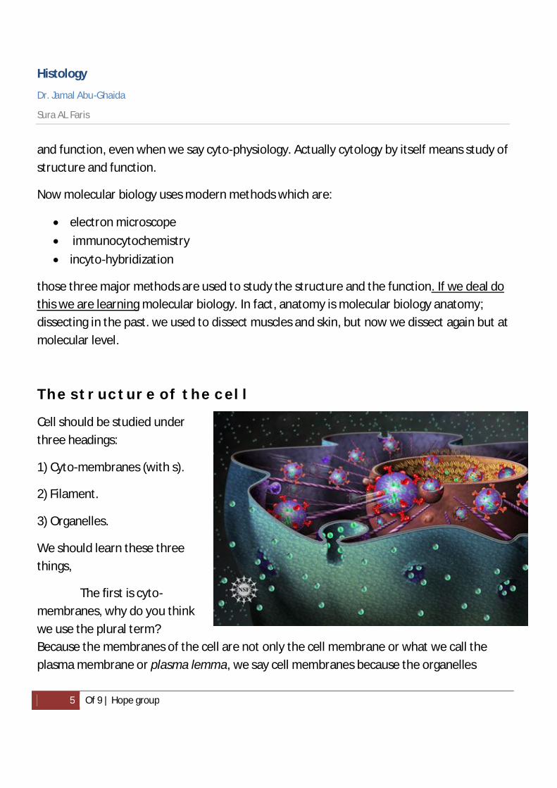
The structure of the cell
Cell should be studied under three headings:
1) Cyto-membranes (with s).
2) Filament.
3) Organelles.
We should learn these three things,
The first is cyto-membranes, why do you think we use the plural term? Because the membranes of the cell are not only the cell membrane or what we call the plasma membrane or plasma lemma, we say cell membranes because the organelles

Histology
Dr. Jamal Abu-Ghaida
Sura AL Faris
6 Of 9 | Hope group
within the cell full of membranes; membranes within the cell form dawn the rest of organelles, they form the Endoplasmic reticulum, they form the nuclear envelop, that means they are membranes, not a single one.
Organelles are metabolic active elements of the cell, and this differentiates it from what we call inclusions; inclusions within the cell are metabolically inactive structures, for example it contains pigments which are metabolically inactive.
Filaments are important to be studied as a one unit because the more we know about filaments within the cell the more we understand how important they are.
The filaments within the cell consisted in whole what we call the cytoskeleton; the skeleton of the cell which is very important structure in the cell for motion, transport, metabolic and biding activity.
Q: The cell membrane by itself is only 7.5 nanometers thickness. Can we see it using light microscope or not?
Note: Always when you face these kinds of questions “Is this structure seen using type of microscope?” you should always think of the resolution power of that optic system.
So if you remember our numbers of resolu on power was 0.1 mm 0.1 µm 0.1 nm for naked eye, light microscope, electron microscope respectively.
Now let’s go back to our question above, when we say that the thickness of plasma membrane is 7.5 nm, this is below the resolu on power of light microscopes so we shouldn’t be able to see the structure using it.
Histology
Dr. Jamal Abu-Ghaida
Sura AL Faris
7 Of 9 | Hope group
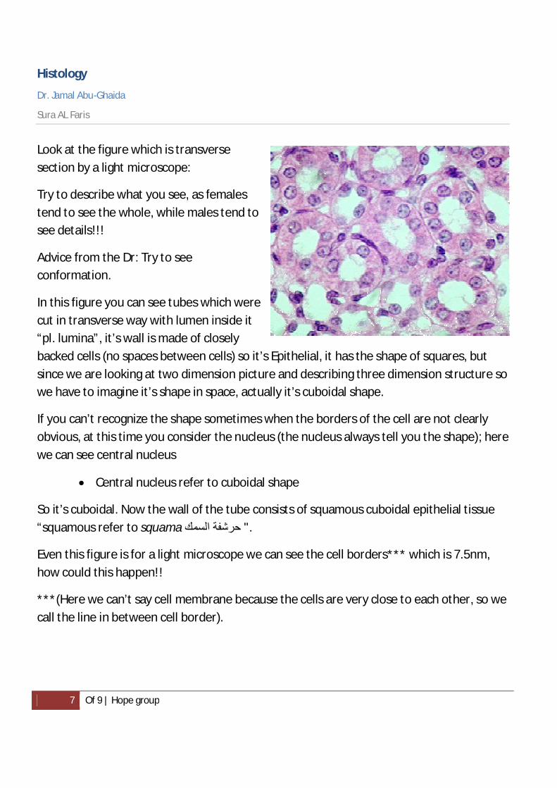
Look at the figure which is transverse section by a light microscope:
Try to describe what you see, as females tend to see the whole, while males tend to see details!!!
Advice from the Dr: Try to see conformation.
In this figure you can see tubes which were cut in transverse way with lumen inside it “pl. lumina”, it’s wall is made of closely backed cells (no spaces between cells) so it’s Epithelial, it has the shape of squares, but since we are looking at two dimension picture and describing three dimension structure so we have to imagine it’s shape in space, actually it’s cuboidal shape.
If you can’t recognize the shape sometimes when the borders of the cell are not clearly obvious, at this time you consider the nucleus (the nucleus always tell you the shape); here we can see central nucleus
Central nucleus refer to cuboidal shape
So it’s cuboidal. Now the wall of the tube consists of squamous cuboidal epithelial tissue “squamous refer to squama حرشفة السمك ".
Even this figure is for a light microscope we can see the cell borders*** which is 7.5nm, how could this happen!!
***(Here we can’t say cell membrane because the cells are very close to each other, so we call the line in between cell border).

Histology
Dr. Jamal Abu-Ghaida
Sura AL Faris
8 Of 9 | Hope group
Remember when one student asked me: Dose your methods in preparing tissues changing your tissue? My answer now is SURE.
This is what we call an equivalent picture not an error, because when you fix with formalin, proteins within the cell precipitate on the wall inside so it became thicker in other words light microscopically visible, in some cases when something adhered to the outer surface (we will learn about this), it also become visible using light microscope. Otherwise cell membranes is only visible using electron microscope
Cell membrane actually is very interesting! When I think about cell membrane it’s actually the same as my skin for my body, or the walls of my bones.
Cell membrane is related to protection, most of researches right now are about elements confirming the physical protective characteristic of this membrane, naturally the cell membrane provides protection but actually the most interesting function of it is communication. How does the cell communicate with its environment? Can you imagine that your skin become insensible to touch, temperature….or imagine that you have a house without windows, receivers, dishes.
Next lecture I’m going to discuss with you the molecular level of cell membrane, because unless we study it, we can’t understand how the cell knows what other cells do.
Imagine that you are a cell how could you know that your neighbor cell is contracting for example, cells actually know, they actually talk to each other. Another thing is how could the cell change its shape, its communication and its behavior during indigenize.

Histology
Dr. Jamal Abu-Ghaida
Sura AL Faris
9 Of 9 | Hope group
The sperm-cell is injected into the ovum with a fine needle
I’ll tell you a well-known example, that when they do ICSI (intracytoplasmic sperm injection or abbreviated) and inject the sperm in the egg with a micro needle, I wondered that time that why doesn’t the cell burst! Or why doesn’t it loose some of its cytoplasm!! The molecular compositions of the plasma membrane actually it’s so fascinated; this will be explained next lecture.
The end
Best wishes, Sura AL Faris :)