histology of the urinary system - medsyllabus.org filetortora & grabowski 9/e 2000 jws 26-6...
TRANSCRIPT

HISTOLOGY OF THE URINARY SYSTEM

Tortora & Grabowski 9/e 2000 JWS
26-2
The Urinary System
Kidneys, ureters, urinary bladder & urethra
Urine flows from each kidney, down its ureter to the bladder and to the outside via the urethra
Filter the blood and return most of water and solutes to the bloodstream

Tortora & Grabowski 9/e 2000 JWS
26-3
Overview of Kidney Functions Regulation of blood ionic composition
Na+, K+, Ca+2, Cl- and phosphate ions Regulation of blood pH, osmolarity & glucose Regulation of blood volume
conserving or eliminating water Regulation of blood pressure
secreting the enzyme renin adjusting renal resistance
Release of erythropoietin & calcitriol Excretion of wastes & foreign substances

Tortora & Grabowski 9/e 2000 JWS
26-4
External Anatomy of Kidney
Paired kidney-bean-shaped organ
4-5 in long, 2-3 in wide,1 in thick
Found just above the waist between the peritoneum & posterior wall of abdomen retroperitoneal along with
adrenal glands & ureters Protected by 11th & 12th
ribs with right kidney lower

External Anatomy of Kidney
Blood vessels & ureter enter hilus of kidney Renal capsule = transparent membrane maintains organ shape Adipose capsule that helps protect from trauma Renal fascia = dense, irregular connective tissue that holds
against back body wall

Tortora & Grabowski 9/e 2000 JWS
26-6
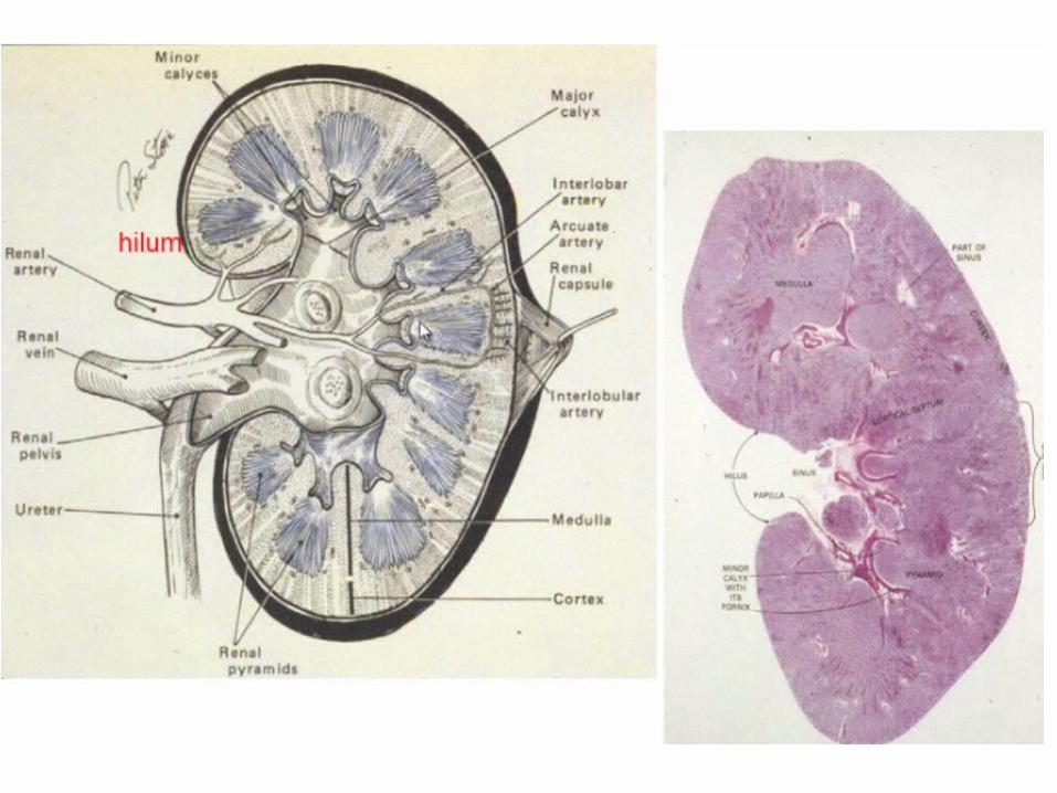
Internal Anatomy of the Kidneys Parenchyma of kidney
renal cortex = superficial layer of kidney renal medulla
inner portion consisting of 8-18 cone-shaped renal pyramids separated by renal columns
renal papilla point toward center of kidney
Drainage system fills renal sinus cavity cuplike structure (minor calyces) collect urine
from the papillary ducts of the papilla minor & major calyces empty into the renal pelvis
which empties into the ureter

Internal Anatomy of Kidney
What is the difference between renal hilus & renal sinus? Outline a major calyx & the border between cortex & medulla.

Tortora & Grabowski 9/e 2000 JWS
26-8
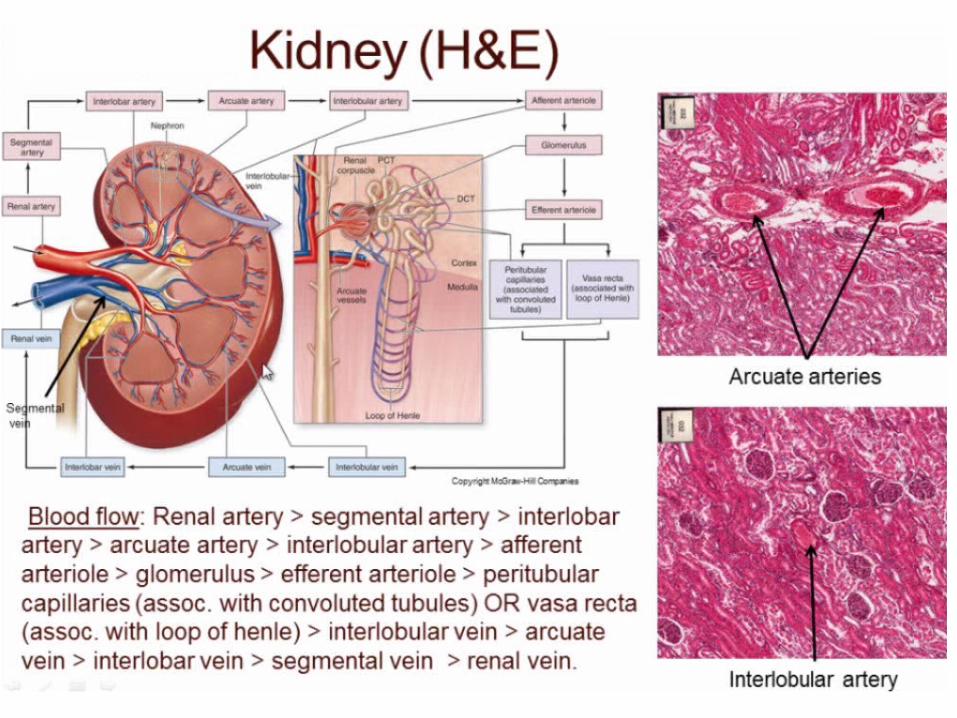
Blood & Nerve Supply of Kidney Abundantly supplied with blood vessels
receive 25% of resting cardiac output via renal arteries
Functions of different capillary beds glomerular capillaries where filtration of blood occurs
vasoconstriction & vasodilation of afferent & efferent arterioles produce large changes in renal filtration
peritubular capillaries that carry away reabsorbed substances from filtrate
vasa recta supplies nutrients to medulla without disrupting its osmolarity form
Sympathetic vasomotor nerves regulate blood flow & renal resistance by altering arterioles
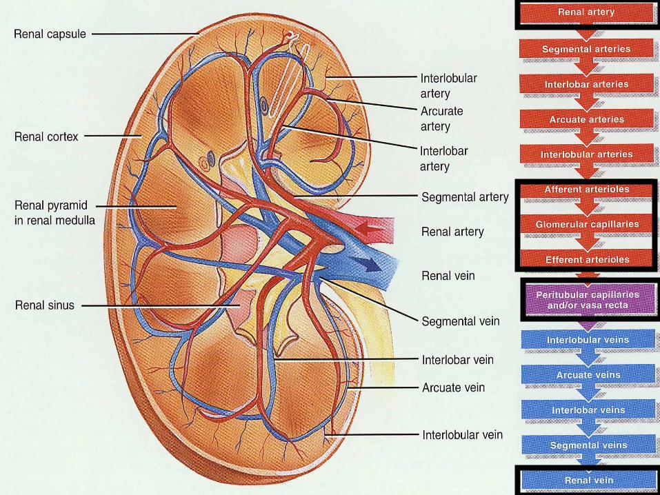

Blood Vessels around the Nephron
Glomerular capillaries are formed between the afferent & efferent arterioles
Efferent arterioles give rise to the peritubular capillaries and vasa recta

Blood Supply to the Nephron

Tortora & Grabowski 9/e 2000 JWS
26-12
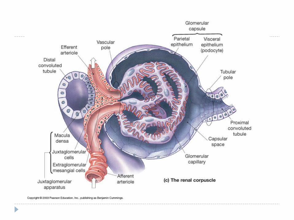
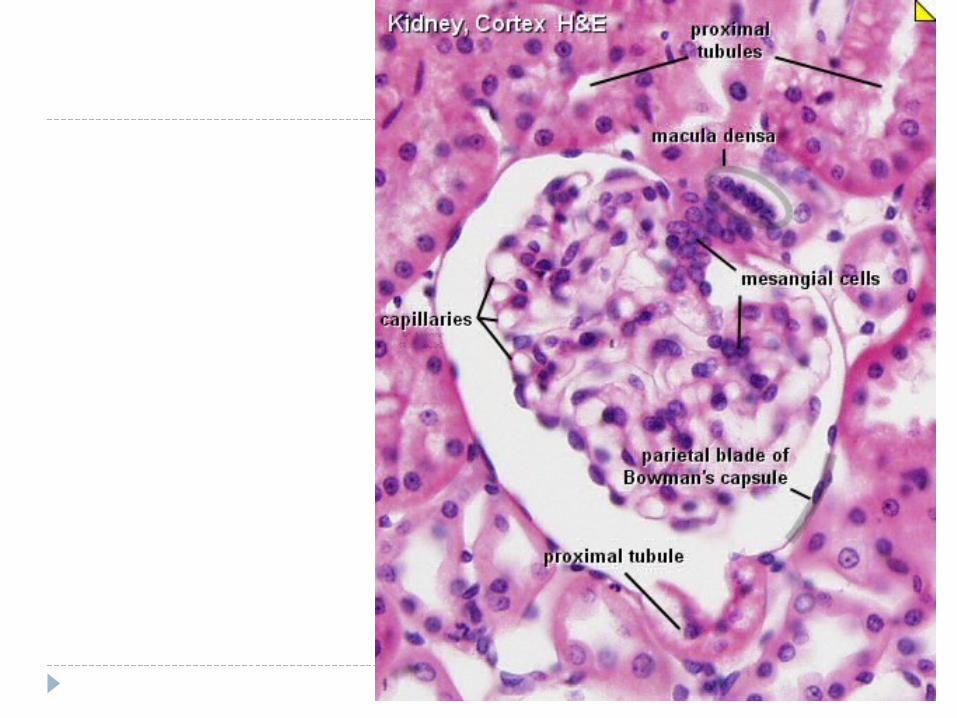
The Nephron Kidney has over 1 million nephrons composed of a
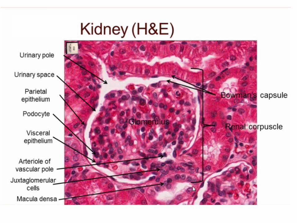
corpuscle and tubule Renal corpuscle = site of plasma filtration
glomerulus is capillaries where filtration occurs glomerular (Bowman’s) capsule is double-walled
epithelial cup that collects filtrate Renal tubule
proximal convoluted tubule loop of Henle dips down into medulla distal convoluted tubule
Collecting ducts and papillary ducts drain urine to the renal pelvis and ureter


Nephron
basic structural and functional unit of the kidney.
Its chief function is to regulate the concentration of water and soluble substances like sodium salts by filtering the blood, reabsorbing what is needed and excreting the rest as urine.

A nephron eliminates wastes from the
body, regulates blood volume and blood pressure, controls levels of electrolytes and metabolites, and regulates blood pH. Its functions are vital to life and are regulated by the endocrine system by hormones such as antidiuretic hormone, aldosterone, and parathyroid hormone.

microscopic units of a kidney, have 2 main parts,
1. renal corpuscle (Bowman's capsule with glomerulus)
2. renal tubule.


RENAL CORPUSCLE RENAL CORPUSCLE glomeruli surrounded by Bowman's capsules.
Bowman's capsule -- the cup-shaped top of a nephron. It is the sack-like Bowmans's capsule that surrounds the glomerulus.
Glomerulus -- a network of blood capillaries tucked into Bowman's capsule.


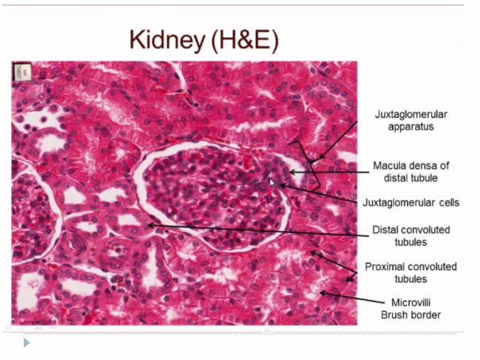
Histology of Renal Tubule & Collecting Duct
Proximal convoluted tubule simple cuboidal with brush border of
microvilli that increase surface area Descending limb of loop of Henle
simple squamous Ascending limb of loop of Henle
simple cuboidal to low columnar forms juxtaglomerular apparatus where
makes contact with afferent arteriole macula densa is special part of ascending limb
Distal convoluted & collecting ducts simple cuboidal composed of principal &
intercalated cells which have microvilli
26-25
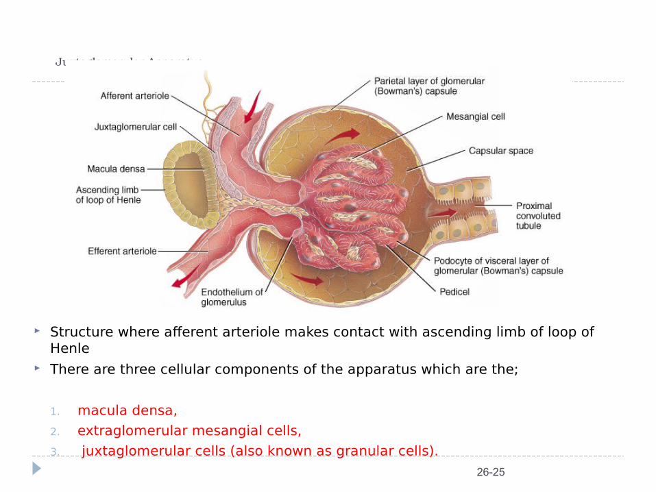
Juxtaglomerular Apparatus
Structure where afferent arteriole makes contact with ascending limb of loop of Henle
There are three cellular components of the apparatus which are the;
1. macula densa,
2. extraglomerular mesangial cells,
3. juxtaglomerular cells (also known as granular cells).

Tortora & Grabowski 9/e 2000 JWS
26-26
Number of Nephrons Remains constant from birth
any increase in size of kidney is size increase of individual nephrons
If injured, no replacement occurs Dysfunction is not evident until function
declines by 25% of normal (other nephrons handle the extra work)
Removal of one kidney causes enlargement of the remaining until it can filter at 80% of normal rate of 2 kidneys

Tortora & Grabowski 9/e 2000 JWS
26-27
Overview of Renal Physiology Nephrons and collecting ducts perform 3 basic
processes glomerular filtration
a portion of the blood plasma is filtered into the kidney tubular reabsorption
water & useful substances are reabsorbed into the blood tubular secretion
wastes are removed from the blood & secreted into urine
Rate of excretion of any substance is its rate of filtration, plus its rate of secretion, minus its rate of reabsorption

Overview of Renal Physiology
Glomerular filtration of plasma Tubular reabsorption Tubular secretion

Glomerular Filtration Blood pressure produces glomerular filtrate Filtration fraction is 20% of plasma 48 Gallons/day
filtrate reabsorbedto 1-2 qt. urine
Filtering capacityenhanced by: thinness of membrane & large surface area of
glomerular capillaries glomerular capillary BP is high due to small size of
efferent arteriole

Filtration Membrane
#1 Stops all cells and platelets #2 Stops large plasma proteins #3 Stops medium-sized proteins, not small ones
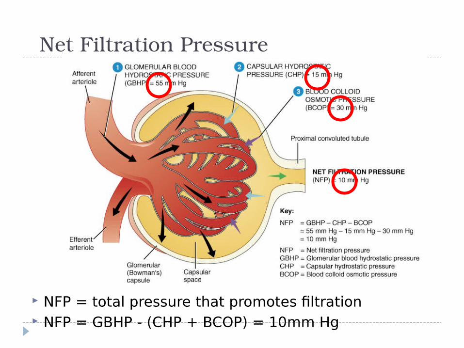
Net Filtration Pressure
NFP = total pressure that promotes filtration NFP = GBHP - (CHP + BCOP) = 10mm Hg
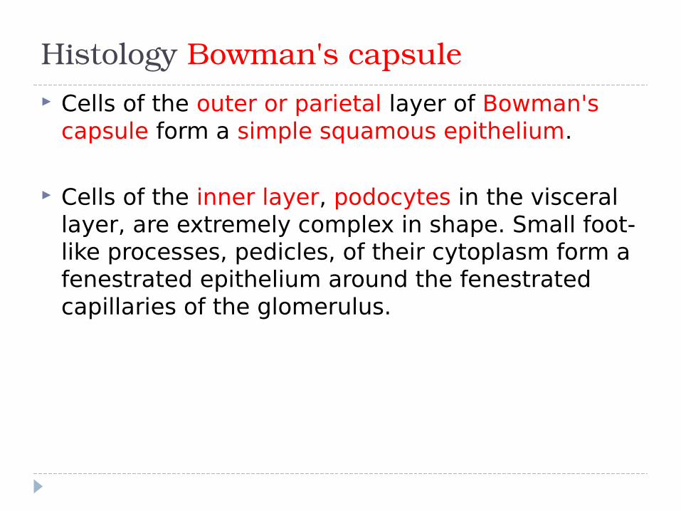
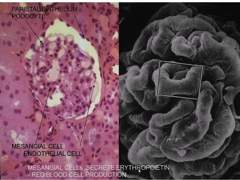
Histology Bowman's capsule Cells of the outer or parietal layer of Bowman's
capsule form a simple squamous epithelium.
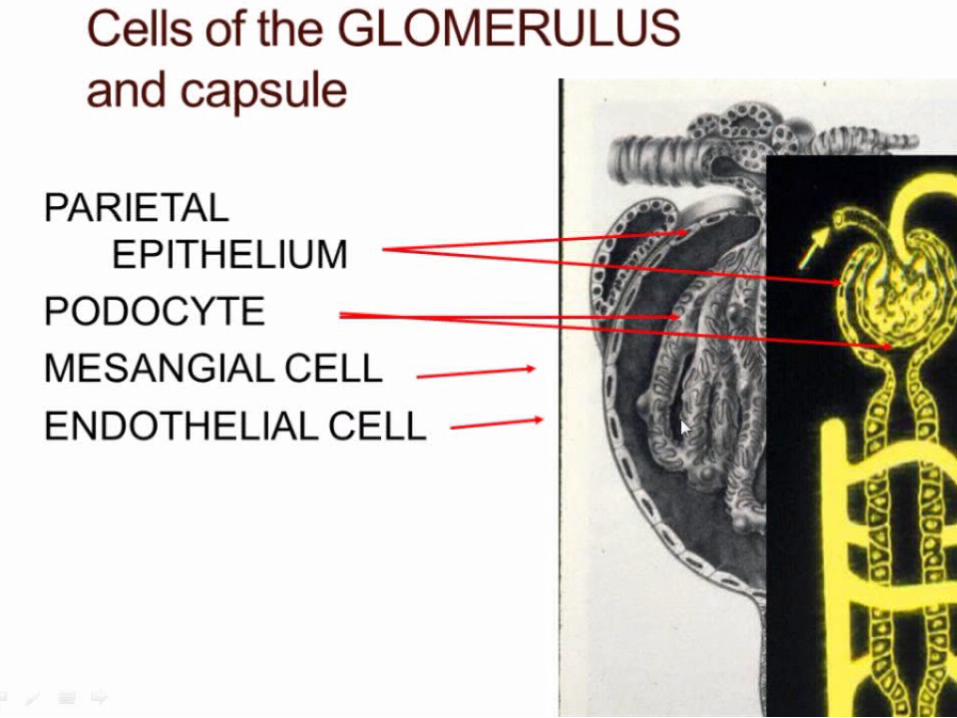
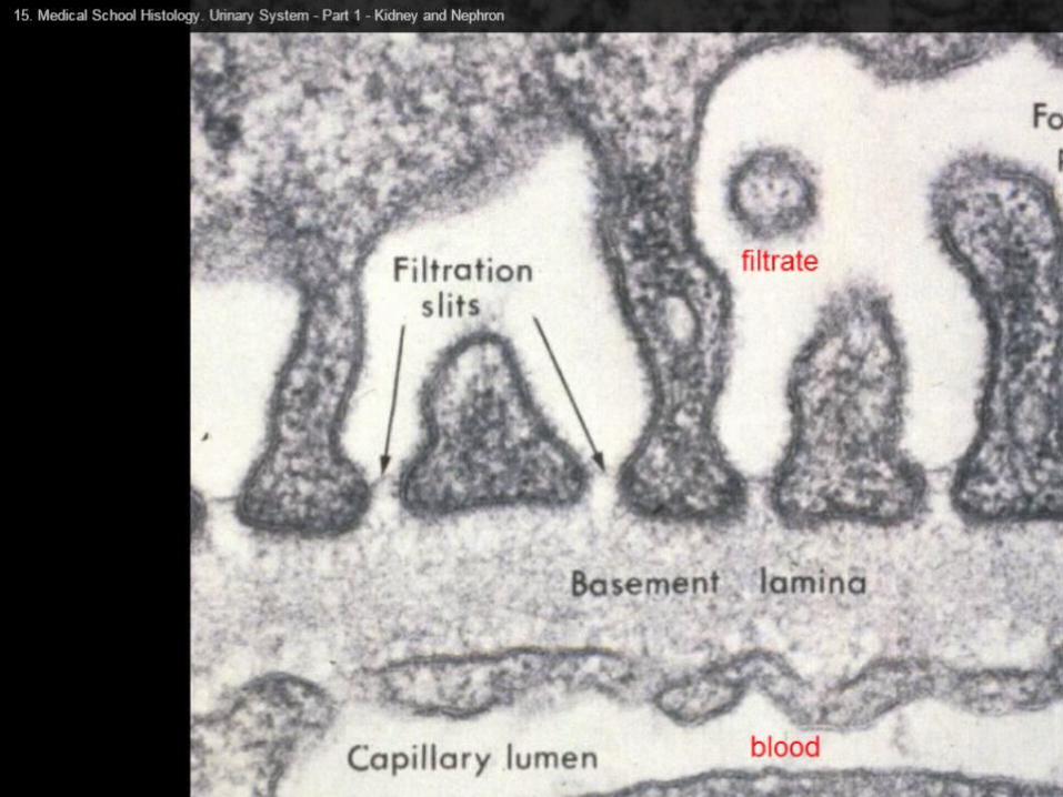
Cells of the inner layer, podocytes in the visceral layer, are extremely complex in shape. Small foot-like processes, pedicles, of their cytoplasm form a fenestrated epithelium around the fenestrated capillaries of the glomerulus.

The openings between the pedicles are called filtration slits. They are spanned by a thin membrane, the filtration slit membrane. Between the podocytes and the endothelial cells of the capillaries a comparatively thick basal lamina, which can be subdivided into an outer lamina externa, a middle lamina densa and an inner lamina interna.
The basal lamina and the slit membranes form the glomerular filtration barrier, which prevents some large molecules from entering the capsular space between the outer and inner epithelial layers of Bowman's capsule.
Mesangial cells in the glomerulus form the connective tissue that gives structural support to podocytes and vessels.


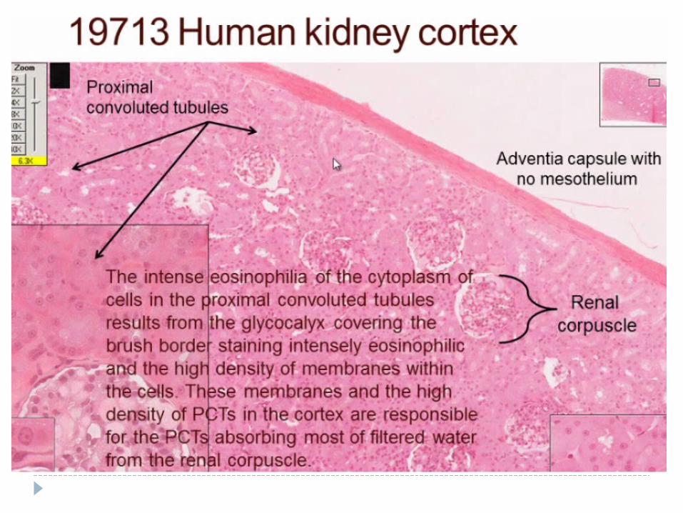


Histo: proximal tubule walls - low columnar epithelium. The eosinophilic cells of the epithelium have a
wide brush border and are active in endocytosis.

Histo of Loop of Henle It is 'U' shaped and has descending and
ascending segments. Thin descending segment has flattened
epithelium( squamous). It is permeable to water but not solutes.

Histo: Distal convoluted tubule
straight part of the DCT is formed by the low cuboidal cells without a brush border. The diameter of the tubule gradually expands to about 35 microns.
convoluted part and comes in contact with the Glomerulus forming the Macula Densa.


Nephron structure characteristic
Renal corpusle Capillary ball covered by podocyte & surrounded by simple squamous epithelial capsule, capsular space.
Proximal convulated Lined with simple cubodial epithelium & prominent brush border
Loop of Henle Tubule that form a loop, there are thick & thin ascending & desecnding portion; the most distal part of the loop often extends into the medulla.
Thick limb are lined with simple squamous/ cuboidal epithelium
Thin limb are lined with simple squamous epithelium
Distal convulated Lined with simple cuboidal with only sparse brush border; cytoplasm of cells tend to be paler than that of proximal convulated
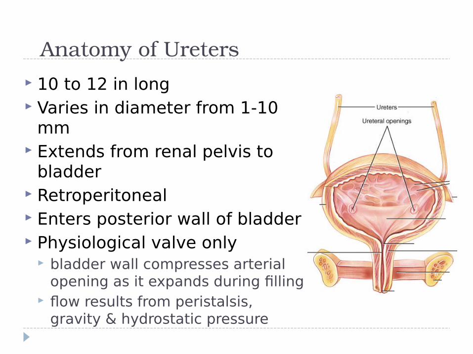
Anatomy of Ureters 10 to 12 in long Varies in diameter from 1-10
mm Extends from renal pelvis to
bladder Retroperitoneal Enters posterior wall of bladder Physiological valve only
bladder wall compresses arterial opening as it expands during filling
flow results from peristalsis, gravity & hydrostatic pressure

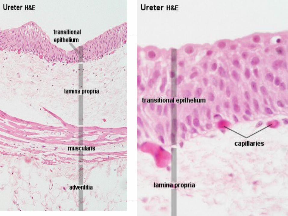
Ureter The wall of the ureter is made 3 layers. From
inside outwards they are :1. Mucosa- which is made up of epithelium &
lamina propria2. Muscular coat – made of smooth
muscles3. Fibrosa – made of fibrous connective
tissue Mucosa- is thrown into folds and thus gives the
appearance of star shaped lumen.
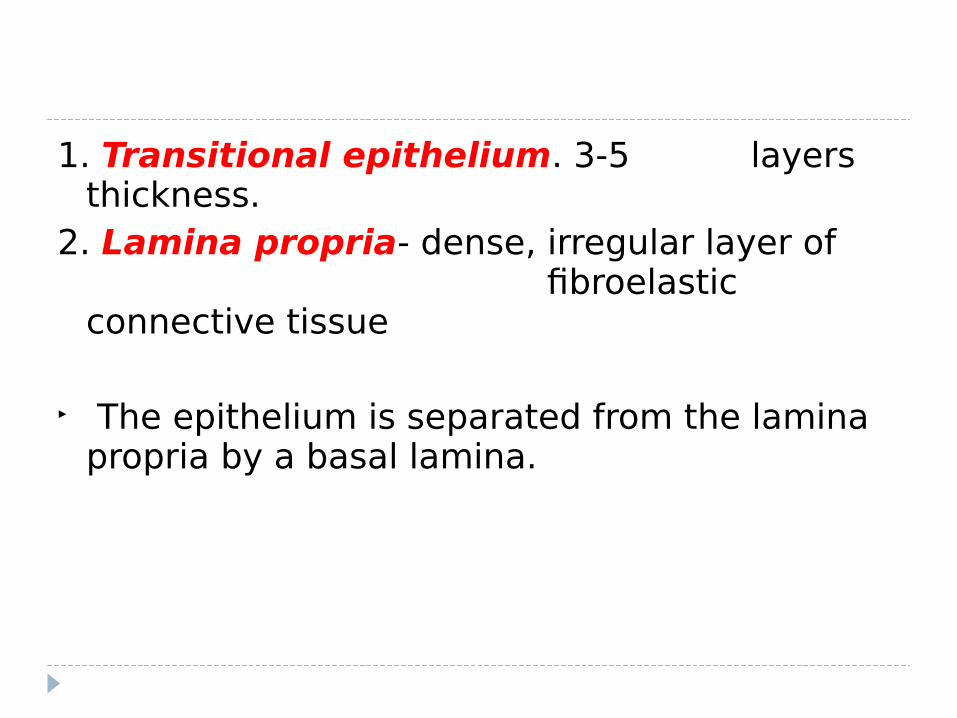
1. Transitional epithelium. 3-5 layers thickness.
2. Lamina propria- dense, irregular layer of fibroelastic connective tissue
The epithelium is separated from the lamina propria by a basal lamina.

Muscular layer – Upper 2/3 of the ureter is made of two layers of smooth muscle cells. Inner longitudinal and outer circular layer( in
contrast to the wall of GIT which has inner circular & outer longitudinal !).
Lower 1/3 of the ureter has a third outer layer of longitudinal muscles( inner longitudinal, middle circular, outer loingitudinal ).
Fibrous coat – is made up of fibrous connective tissue


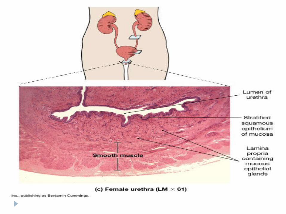
The urethra:
Female: relatively short, exits just anterior to the vagina
Male: longer, divided into three sections the prostatic, membranous and spongy urethra. The prostatic urethra is enclosed in the prostate gland. The membranous urethra is a short section that penetrates the urogenital diaphragm. The spongy urethra or penile urethra extends from the membranous urethra to the external urethral orifice (meatus).

Histology: In both male and females the urethra starts out as transitional cell but quickly becomes stratified squamous in the female.
The male urethra is more variable but ends up stratified squamous as well.

Histology of the bladder
mucosa of transitional epithelium, Submucosa, and thick muscular layer know as the detrusor
muscle


Clinical correlates
Urinary incontinence Childbirth and other events can injure the scaffolding
that helps support the bladder in women. Pelvic floor muscles, the vagina, and ligaments support your bladder.
Overactive bladderSpecifically, the symptoms of overactive bladder includeurinary frequency—bothersome urination eight or more times a day or two or more times at night urinary urgency—the sudden, strong need to urinate immediately urge incontinence—leakage or gushing of urine that follows a sudden, strong urge nocturia—awaking at night to urinate