histologyofthelarvalneodiprionabietis (hymenoptera
TRANSCRIPT

Hindawi Publishing CorporationPsycheVolume 2011, Article ID 910286, 10 pagesdoi:10.1155/2011/910286
Research Article
Histology of the Larval Neodiprion abietis(Hymenoptera: Diprionidae) Digestive Tract
Christopher J. Lucarotti,1, 2 Beatrixe H. Whittome-Waygood,3 and David B. Levin3, 4
1 Natural Resources Canada, Canadian Forest Service Atlantic Forestry Centre, 1350 Regent Street, P.O. Box 4000,Fredericton, NB, Canada E3C 2G6
2 Faculty of Forestry and Environmental Management, University of New Brunswick, Fredericton, NB, Canada E3B 6C23 Department of Biology, University of Victoria, Victoria, BC, Canada V8W 2Y24 Department of Biosystems Engineering, University of Manitoba, E2-376 EITC, Winnipeg, MB, Canada R3T 5V6
Correspondence should be addressed to Christopher J. Lucarotti, [email protected]
Received 18 April 2011; Revised 17 August 2011; Accepted 6 September 2011
Academic Editor: Robert Matthews
Copyright © 2011 Christopher J. Lucarotti et al. This is an open access article distributed under the Creative Commons AttributionLicense, which permits unrestricted use, distribution, and reproduction in any medium, provided the original work is properlycited.
The alimentary canal of Neodiprion abietis larvae is a straight tube divided into foregut, midgut, and hindgut. Posterior to themouth, the foregut is further divided into the pharynx, esophagus (crop), and proventriculus, all of which are lined with cuticle.A pair of muscular, chitin-lined pouches branch off the anterior foregut and lie lateral to the alimentary canal. Gastric caeca arelocated at the anterior end of the midgut, where the peritrophic membrane is formed and was observed throughout the midgut. Asingle layer of midgut columnar epithelial cells abuts on the basal lamina at one end with microvilli extending into the gut lumenat the other. Nidi of regenerative cells were observed between columnar epithelial cells at the basal lamina. Malpighian tubulesare attached to the posterior end of the midgut. The hindgut consists of the pylorus, a muscular ileum connecting to a bulbousrectum, which then opens to the anus.
1. Introduction
Insect gut morphology and function are dependent onseveral factors including insect taxon, developmental stage,feeding behavior, and food source [1, 2], but all insect gutsfollow the same basic plan. The fore- and hindguts originatefrom the embryonic ectoderm and are lined with cuticle[2–4]. The middle component, or midgut, has no cuticularcovering and is generally thought to originate from theembryonic endoderm [2, 5, 6]. The foregut typically func-tions for short-term food storage and transport to the midgut[3], where food is digested by enzymes and nutrients areabsorbed across a columnar epithelium. The hindgut isdivided into the pylorus, ileum, and rectum, where waterand salts may be absorbed, and the anus through which fecespass from the body [2, 4]. Malpighian tubules attach at thejunction of the midgut and hindgut and may be on one sideor the other of this junction depending on the insect [7, 8]. In1895, Bordas [9] described the gut morphologies of selected
examples from every family in the Hymenoptera. Sixty yearslater, Maxwell [10] made extensive comparisons of the inter-nal larval anatomies of 132 different species from 11 familiesof sawflies, collected worldwide, in an effort to resolve certainissues related to the taxonomy of sawflies. Maxwell [10]determined that the two major internal anatomical featuresfor establishing evolutionary relationships amongst andbetween sawfly taxa were the salivary glands and Malpighiantubules.
The balsam fir sawfly, (Neodiprion abietis) (Hymenop-tera: Diprionidae), is indigenous and widespread in NorthAmerica, where the larvae feed predominantly on balsamfir (Abies balsamea), white spruce (Picea glauca), and blackspruce (Picea mariana) [11]. Neodiprion abietis is likely aspecies complex where temporal differences in life historiesand host-plant selection for oviposition and feeding mayprovide an isolating mechanism for strains that can other-wise freely interbreed [12]. On the island of Newfoundland(Province of Newfoundland and Labrador (NL), Canada),
2 Psyche
outbreak populations of balsam fir sawflies typically occurin 5- to 15-year cycles and last 4 to 5 years [13]. Here, balsamfir sawfly larvae emerge in late spring and early summerafter overwintering as eggs that had been oviposited the yearbefore in the then current-year needles of balsam fir trees.Male larvae pupate after the fifth larval stadium, whereasfemale larvae may go through an addition instar beforepupating [14]. The adults emerge in late summer, and matedfemales will lay female eggs and unmated females, male eggs(arrhenotoky). Outbreak populations of balsam fir sawfliesare brought down by epizootics of a naturally occurringnucleopolyhedrovirus (NeabNPV: Gammabaculovirus: Bac-uloviridae [15]) [16]. Sawfly NPVs only infect the midgut[17], so prior to examining the pathology of NeabNPV inlarval balsam fir sawflies and because of the general paucityof reports on sawfly gut anatomy, we have first undertakenan examination of the anatomy and histology of the healthyalimentary canal of the balsam fir sawfly larva.
2. Materials and Methods
2.1. Larval Collection. Balsam fir branches, containing N.abietis larvae, were collected from the leading edge of thebalsam fir sawfly outbreak near Old Man’s Pond, NL, Canada(49◦7′23.3 ′′N: 57◦51′45.6′′ W) between 18–21 July 2003.Larvae were maintained on balsam fir foliage in 20-kg brownpaper bags at 4◦C for a maximum of 48 h. Larvae wereremoved from the foliage, and head capsule measurementswere taken using a dissecting microscope equipped with acalibrated micrometer in the objective lens. Larvae with headcapsule widths between 0.96 to 1.5 mm, which correspond tothird- to fifth-instar larvae [14], were transferred to sterile100 mm × 15 mm plastic Petri dishes for a 12- to15-hstarvation period. Larvae were then allowed to imbibe anaqueous solution of 10% pasteurized honey and were placedon clean (5-min soak in 0.25% aqueous NaOCl followed bythree 15-min rinses in tap water), fresh balsam fir sprigs, for72 h at ambient room temperature (approximately 20◦C) tomonitor for signs of NeabNPV infection.
2.2. Histological Preparations
2.2.1. Paraplast Sections. Larvae were submerged whole inBouin’s fixative (Electron Microscope Sciences, Hatfield, Pa,USA) for 24 to 48 h. Larvae were rinsed and dehydratedin a graded ethanol series to butanol and embedded inParaplast+ (Sherwood Medical, St. Louis, Mo, USA). Embed-ded material was sectioned using a steel histological knifeon an American Optical (Buffalo, NY) 820 Spencer rotarymicrotome set to cut sections 10 μm thick. Serial sectionson clean glass microscope slides were stained using a mod-ified azan staining technique [18], dehydrated in ethanolto Hemo-D (Fisher Scientific, Fair Lawn, NJ, USA) andmounted in Permount (Fisher Scientific, Fair Lawn, NJ,USA).
2.2.2. Epon-Araldite Sections. Larvae harvested for whole-mount preparations and epoxy embedding were first injected
with fixative (2.5% gluteraldehyde—0.05 M sodium cacodyl-ate—0.1 M sucrose pH 7.4) using a 1-cc, 27G1/2 syringeand needle (Becton Dickinson, Franklin Lakes, NJ, USA).The heads and tails were removed at the head capsule andeighth proleg, respectively, and then, the gut was pulledfrom the hemocoel into fresh fixative, using fine forceps.Guts excised for epoxy embedding were then embedded in2% low-gelling-temperature agarose (SeaPlaque: FMC Bio-Products, Rockland, Me, USA) to preserve the integrity ofthe gut contents [19]. Guts were then cut roughly into fore-,mid-, and hindgut sections, re-embedded in agarose, andtransferred to 1.5-mL microcentrifuge tubes containing freshfixative overnight at 4◦C. Gut samples were rinsed at 20◦Cfor 15 min each in 0.05 M sodium cacodylate containing0.1 M sucrose, 0.05 M sucrose and no sucrose followed bypostfixation in 1% OsO4 in 0.05 M sodium cacodylate buffer,pH 7.4 for 1 h at 20◦C. Samples received two 15-min rinsesin the same buffer followed by two 10-min washes in distilledwater prior to en bloc staining in 4% aqueous uranyl acetateovernight and in the dark at 4◦C [19]. Two additional 10-min water washes were followed by dehydration in a gradedacetone series (30–100%) and embedding in Epon-Araldite[20] (Epon-5 g, Araldite-5 g, DDSA-12 g, DMP-30–500 μL)(Electron Microscope Sciences, Hatfield, Pa, USA). Sectionswere cut using an RMC MT-7 ultramicrotome at 1 μm forlight microscopy and 80–90 nm for electron microscopyusing Diatome (Biel, Switzerland) Histo and diamondknives, respectively. Thin sections were placed on clean glassslides, stained with toluidine blue-basic fuchsin (Canemco-Marivac, St. Laurent, QC, Canada) and were mounted inPermount. Ultrathin sections on 100-mesh copper gridswere stained with 2% uranyl acetate and 0.01% lead citrate.Digital light microscope images were captured using a NikonEclipse 80i/DS camera (Nikon Instruments, Melville, NY,USA). Electron microscope images were taken on an HitachiH7000 transmission electron microscope (Mississauga, ON,Canada) at 75 kV.
3. Results
3.1. The Alimentary Canal. The N. abietis larval alimentarycanal is straight and runs the full length of the body ofthe larva, between the anterior mouth and posterior anus,and is divided into three distinct regions (Figures 1 and 2):foregut (Figures 3–9), midgut (Figures 9–13), and hindgut(Figures 13–19). The foregut forms approximately 22% ofthe total length of the gut. A pair of muscular diverticularpouches branch off the anterior foregut (Figures 1, 3, and4) and connect to the buccal cavity (Figure 5). Like all com-ponents of the foregut, these pouches are lined with cuticle.Posterior to the buccal cavity is the muscular pharynx withposteriorly directed cuticular spines (Figures 6–8) followedby the esophagus that enlarges posteriorly to form the crop(Figures 6 and 9). The proventriculus (Figures 9 and 10) ischaracterized by a thick cuticle and is the last part of theforegut before the midgut. Gastric caeca encircle the anteriorend of the midgut, and it is in this region that peritroph-ic membrane is formed by cells of the cardia and anterior

Psyche 3
Foregut
DP
G G
GG
Midgut Hindgut
Salivary gland
Malpighian tubules
Figure 1: Diagrammatic representation of larval Neodiprion abietissalivary glands, gland cells (G), diverticular pouches (DP), and ali-mentary canal (after Maxwell [10]).
FG MG HG
Figure 2: Photomontage of a whole mount of the foregut (FG),midgut (MG), and hindgut (HG) of a balsam fir sawfly larva. Thearrow on the left indicates the junction of the foregut and midgutand the arrow on the right indicates the junction of the midgut andhindgut, where the Malpighian tubules (to the right of the arrow)are inserted. Scale bar = 2 mm.
midgut (Figures 9 and 10). The midgut is about 63% of thelength of the alimentary canal, the majority of which consistsof columnar epithelial cells butting onto the basal laminawhich is then surrounded by circular and longitudinalmuscles (Figures 11 and 12). Nidi of regenerative cells liebetween columnar epithelial cells and adjacent to the basallamina (Figures 11). At the posterior end of the midgut, justin front of the pylorus of the hindgut, the epithelium formsfolds (Figures 13 and 14), and the cells in this region appearto be contributing material to the peritrophic membrane(Figure 15). Malpighian tubules were observed to insert intothe posterior end of the midgut just anterior to the pylorus(Figure 14). The hindgut makes up the remaining 15% of thealimentary canal, consisting of the pylorus, ileum, rectum,and anus (Figures 13–19), all of which are lined with cuticle.The pylorus is not as wide as the midgut and constrictsfurther at the ileum (Figures 2 and 13), which forms a mus-cular tube characterized by a thick cuticle (Figures 16 and17). The epithelium undulates and consists of cuboid cells(Figure 17). Posterior to the ileum is the bulbous rectum,where waste plant material could be seen encased in a sheathformed by the peritrophic membrane (Figure 18). The outersurface of the cuticle lining of the anus had posteriorlydirected spines and innervated setae (Figure 19).
3.2. The Salivary Glands. A pair of salivary glands flank thealimentary canal and open into the buccal cavity (Figure 1).The glands consisted of pair of large ducts, each with numer-ous gland cells on their surfaces (Figures 20–25). Granuleswere observed within and outside the cytoplasm of the glandcells (Figures 22 and 23). The salivary ducts consisted ofa single layer of epithelial cells with microvilli facing thecentral lumen of the ducts, where secretions were observed(Figures 20, 24, and 25).
H
T
DP
DP
Figure 3: Longitudinal section through the head (H) and thorax(T) of a N. abietis larva showing the paired diverticular pouches(DP) and the tubule (arrows) that connects one of the pouches tothe buccal cavity. (Note that serial sections showed the continuityof the tubule between the diverticular pouch and the buccal cavity.)Scale bar = 0.5 mm.
Tu
Figure 4: Detail of the musculature (arrows) and cuticle lining(arrowheads) of a diverticular pouch (different section but samepouch as the one on right in Figure 3) and tubule (Tu) with contentsof a second pouch (on left in Figure 3). Scale bar = 0.1 mm.
Figures 3–6, 9, 10, 13–16, and 18–21 are light micro-graphs of paraplast sections. Figures 7, 8, 11, 17, and 22–25are light micrographs of epoxy sections, and Figure 12 is anelectron micrograph of an epoxy section.
4. Discussion
Sawflies are phytophagous and the Diprionidae are defo-liators of conifer trees (Pinaceae) with digestive systemsadapted to that purpose. The diverticular pouches, whichbranch off the diprionid larval foregut, are used to store host-plant-derived terpenoids that are regurgitated in defensivebehaviors against predators [21–27]. Like N. abietis, thediverticular pouches of N. sertifer larvae have been shown tobe muscular sacs lined with an impervious layer of cuti-cle [21]. The chemistry of the contents of the pouchesof conifer-feeding diprionid sawflies [21–24] and pergidsawflies feeding on eucalyptus [28] reflect the chemistry ofthe food source. Food source and larval stadium can also

4 Psyche
BC
Tu
Figure 5: Detail of tubule (Tu) extending into the buccal cavity(BC) in the head of a N. abietis larva. (Same larva, different sectionas Figure 3.) Scale bar = 0.1 mm.
Ph
M
Cr
Figure 6: Muscular (M) pharynx (Ph) and anterior crop (Cr) ofa N. abietis larva. Cuticular spines of the pharynx (arrows) aredirected posteriorly. Scale bar = 0.1 mm.
affect the volume of fluid regurgitated. Redheaded pinesawfly (N. lecontei) larvae fed Pinus banksiana regurgitated0.26 ± 0.02 μL compared with 0.07 ± 0.04 μL when fed P.resinosa [24]; second-instar red pine sawflies (N. nanulusnanulus) released 58.9 ± 2.3% of the regurgitated volumeon the first expulsion of fluid and lower amounts with eachof the next two expulsions, whereas fifth instars released40.5 ± 2.0% on the first regurgitation and lower but similaramounts on the next two regurgitations [24]. Diprion pinilarvae fed a high-resin diet produced higher amounts of fluidand had higher pupal weights than larvae fed low-resin diets,indicating that the cost and maintenance of this chemicaldefense was low [27]. However, there may be a balancebetween negative effects of high-resin acid contents to earlyinstars (e.g., longer development times) and advantages ofpositive effects (chemical defense) to late instars in N. sertiferlarvae [23]. Balsam fir sawfly larvae have been shown toperform best (better survival and cocoon weights) when theycould feed on all age classes of foliage [29].
The rest of the foregut is involved in the intake of food, itstrituration, and movement back towards the midgut [2, 3].The musculature and posteriorly directed spines on thecuticle of the pharynx of N. abietis larvae would aid in thesefunctions. The esophagus is enlarged to serve as a temporary
M
Ep
B
Figure 7: Posteriorly directed cuticular spines (arrows) subtendedby a single cell layer of epithelium (Ep) and muscle (M). Bacteria(B) can be seen in the lumen of the pharynx. Scale bar = 30 μm.
C
Ep
M
Figure 8: Detail of the epithelium (Ep), muscles (M), and cuticle(C) of the pharynx of a N. abietis larva. Scale bar = 30 μm.
GC
Cr
C
Pr
Ep
PM
Figure 9: Posterior end of the foregut (left) showing the crop (Cr)and proventriculus (Pr) with its thick cuticle (C) and subtendingepithelium (Ep) and the anterior end of the midgut (right) witha gastric caecum (GC) and peritrophic membrane (PM) beingproduced by cells of the cardia (arrows). Scale bar = 0.2 mm.

Psyche 5
GC
PrPM
Figure 10: Junction of the foregut (left) and midgut (right) showingthe proventriculus (Pr), a gastric caecum (GC), and the origin(arrow) of the peritrophic membrane (PM). The anterior cells ofthe midgut also appear to contribute secreted material (arrowhead)to the peritrophic membrane. Scale bar = 0.1 mm.
CM
LM
N
Mv
Figure 11: Nucleated (N) columnar epithelial cells of the midgutwith microvilli (Mv) extending into the lumen (endoperitrophicspace) of the gut. A regenerative cell (arrow) lies between twocolumnar epithelial cells beneath layers of circular (CM) and lon-gitudinal (LM) muscle. Scale bar = 30 μm.
storage organ, and the proventriculus, with its thick cuticlecovering, is involved in the maceration of food [2, 3]. Thecardia are specialized cells of the proventriculus that secretethe peritrophic membrane [30]. Peritrophic membranes laiddown by cardia cells only are considered type II peritrophicmembranes and are found, for example, in larval Dipteraand a few adult Lepidoptera [31]. Type I peritrophic mem-branes are produced along the entire length or at eitherend of the midgut [1]. Type I peritrophic membranes arefound in Coleoptera (beetles), Dictyoptera (cockroaches),Hymenoptera (bees, ants, wasps), Lepidoptera (moths andbutterflies), and adult hematophagous Diptera (e.g., femalemosquitoes) [31]. In larval N. abietis, material used in theformation of the peritrophic membrane was observed beingsecreted by the cells of the cardia (proventriculus) and ante-rior and posterior midgut epithelium. The evolution of thecardia may be an adaptation allowing insects to produce large
BLCM
Figure 12: The base of a midgut columnar epithelial abuts againstthe basal lamina (BL) and circular muscle (CM). Fine, finger-likeprojections of the basal lamina cause invaginations of the epithelialcell plasma membrane (arrowheads). Mitochondria in the epithelialcell are indicted by the arrows. Scale bar = 1 μm.
MG PyIm
RSG
MT
Figure 13: Longitudinal section (dorsal side up) of a N. abietislarva showing folds (arrows) in the epithelium at posterior end ofthe midgut (MG) and the hindgut showing the pylorus (Py), ileum(Im), and rectum (R). Malpighian tubules (MT) and salivary gland(SG) are visible in the hemocoel. Scale bar = 0.5 mm.
quantities of peritrophic membranes at a localized region ofthe gut [32]. In larval sawflies, the importance of abundantproduction of peritrophic membrane in the anterior regionof the midgut would be for the protection of the midgutepithelium from abrasion by the jagged edges of the partiallymasticated food [31] and potentially harmful microbialpathogens while allowing for the passage of molecules(enzymes and products of digestion) between the endo- andectoperitrophic spaces [30]. In Hymenoptera, initial stages ofdigestion occur in the endoperitrophic space, intermediatestages in the ectoperitrophic space, and final stages at thesurface of the midgut epithelium [31]. Only the enzymes in-volved in the initial stages of digestion are free to move acrossthe peritrophic membrane between the endo- and ectoper-itrophic spaces [31]. A counterflow of water from the poste-rior midgut to the caeca is thought to recirculate these en-zymes, in the ectoperitrophic space, to the anterior midgut,where they can re-enter the endoperitrophic space [31].

6 Psyche
MG
Py
MT
Figure 14: Insertion point (arrow) of a Malpighian tubule (MT) atthe posterior end of the midgut (MG) just anterior of the pylorus(Py). Note the layer of cuticle (arrowheads) lining the pylorus,which is typical of the hindgut. Scale bar = 0.1 mm.
PM
Py
Figure 15: Cells contributing material (arrows) to the peritrophicmembranes (PM) at the posterior end of the midgut just anterior ofthe pylorus (Py). Scale bar = 0.1 mm.
Sawfly larvae have distinct gastric caeca [10]; however,there is an evolutionary trend in the Hymenoptera, towardsthe Apocrita, where gastric caeca are lost and their functionreplaced by cells in the anterior midgut [31]. At the otherend of the N. abietis larval midgut, Malpighian tubules emptyinto the ectoperitrophic space just anterior to the pylorusof the hindgut. Maxwell [10] reports that there are approxi-mately 28 Malpighian tubules in N. swainei larvae and as shestates that, “The general anatomy of all species of Neodiprionis monotonously similar; differences found on a minor levelare described for swainei, abietis, and lecontei”, and one mayassume a similar number are present in N. abietis. In her
FBC
A
HG
Figure 16: Longitudinal section (dorsal side up) of the posteriorend of a N. abietis larva showing the muscular and chitin-linedhindgut (HG), which opens to the exterior through the anus (A).Anal setae are indicated by the arrows. Also visible are fat body (FB)and the exterior cuticle (C) of the larva. Scale bar = 0.2 mm.
Ep
CM
MT
B
C
Figure 17: Detail of the ileum showing bacteria (B) in the gutlumen, the thick cuticle (C), single layer of epithelial cells (Ep), andlayers of circular muscles (CM). A Malpighian tubule (MT) is visiblein the hemocoel. Scale bar = 30 μm.
thesis, Maxwell [33] provides a line drawing of the larvaldigestive tract of N. abietis drawn from histological sections(her plate II, E) that shows the structure of the digestivetract to be similar to our observations. In Maxwell’s drawing,however, the folds in the epithelium appear to be in themiddle region of the midgut, whereas we observed them atthe anterior and posterior ends of the midgut (Figures 2,9, 13, and 14). The reason for this difference is not clear,but it could be that Maxwell sectioned different larval stadiafrom those we sectioned, or the differences could be due tofixation artifact or it could be that the material examinedby Maxwell was infected with NeabNPV. NeabNPV is highlycontagious in populations of balsam fir sawfly larvae andNeabNPV can be acquired by larvae shortly after emergencefrom the egg and quickly transmitted to other larvae [34]. Inthe material we examined, columnar epithelial cells and nidiof regenerative cells were the principle cell types observedin the middle region of midgut of larval N. abietis. Thesimplicity of the larval gut of N. abietis is perhaps reflected
Psyche 7
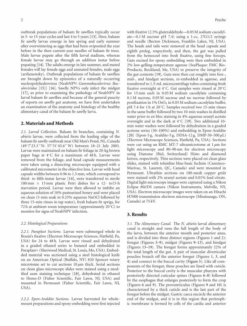
Figure 18: Fecal pellet surrounded by a sheath (arrow) formedfrom the peritrophic membrane in the bulbous rectum of a N.abietis larva. Scale bar = 0.2 mm.
C
S
Fb
Ep
Figure 19: Longitudinal section from the anus of a N. abietis larvashowing an innervated (arrowhead) seta (S) extending from thecuticle (C), which is underlain by a single layer of epithelial cells(Ep) and fat body (FB). Posteriorly directed cuticular spines areindicated by the arrows. Scale bar = 60 μm.
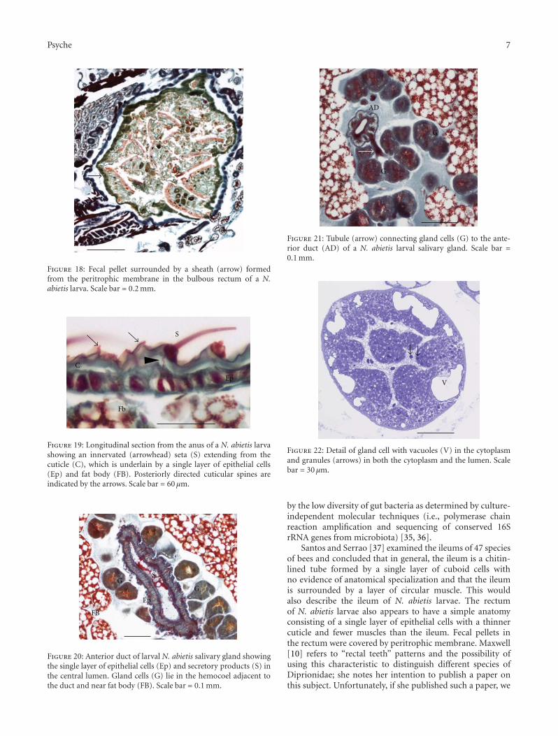
GS
G
Ep
FB
Figure 20: Anterior duct of larval N. abietis salivary gland showingthe single layer of epithelial cells (Ep) and secretory products (S) inthe central lumen. Gland cells (G) lie in the hemocoel adjacent tothe duct and near fat body (FB). Scale bar = 0.1 mm.
AD
G
G
Figure 21: Tubule (arrow) connecting gland cells (G) to the ante-rior duct (AD) of a N. abietis larval salivary gland. Scale bar =0.1 mm.
V
Figure 22: Detail of gland cell with vacuoles (V) in the cytoplasmand granules (arrows) in both the cytoplasm and the lumen. Scalebar = 30 μm.
by the low diversity of gut bacteria as determined by culture-independent molecular techniques (i.e., polymerase chainreaction amplification and sequencing of conserved 16SrRNA genes from microbiota) [35, 36].
Santos and Serrao [37] examined the ileums of 47 speciesof bees and concluded that in general, the ileum is a chitin-lined tube formed by a single layer of cuboid cells withno evidence of anatomical specialization and that the ileumis surrounded by a layer of circular muscle. This wouldalso describe the ileum of N. abietis larvae. The rectumof N. abietis larvae also appears to have a simple anatomyconsisting of a single layer of epithelial cells with a thinnercuticle and fewer muscles than the ileum. Fecal pellets inthe rectum were covered by peritrophic membrane. Maxwell[10] refers to “rectal teeth” patterns and the possibility ofusing this characteristic to distinguish different species ofDiprionidae; she notes her intention to publish a paper onthis subject. Unfortunately, if she published such a paper, we

8 Psyche
Figure 23: Further detail of granules (arrows) that originate fromthe gland cell shown in Figure 22. Scale bar = 30 μm.
Ep
FB
L
Figure 24: Anterior duct of a N. abietis larva salivary gland showingthe single layer of epithelial cells (Ep) with a border of microvilli(arrow) lining the lumen (L) of the duct. Fat body (FB) cells arepressed against the duct. Scale bar = 0.1 mm.
have been unable to find it. In general, Maxwell [10] reportstwo rows of rectal teeth in the Diprionidae (e.g., N. swainei,Diprion (Gilpinia) hercyniae). We observed numerous, saw-like teeth and setae lining the cuticle of the anal canal of N.abietis, but it is unclear whether Maxwell was referring toeither of these structures.
Saliva in insects serves as a lubricant for food entering thedigestive tract; it may contain enzymes and may [38] or maynot [31] be involved in digestion. Salivary glands of sawfliesand higher Hymenoptera are labial glands. Unlike thesalivary glands of sawflies, where the gland cells are clearlyevident on the salivary glands [10, 39], the gland cells ofhigher Hymenoptera have been incorporated into the liningof the salivary ducts [40]. In addition to the productionof saliva, the salivary glands of sawflies may also functionas silk glands for cocoon production in some groups,such as Xyelidae, Cephidae, and Tenthredinoidae (exceptBlasticotomidae) but not others, for example, Pamphiliidae,Siricidae, and Xiphydriidae (see [41]). In the honey bee (Apismellifera), silk production begins and ends in the fifth larvalstadium prior to the prepupal period [42, 43]. Presumably, asimilar process would occur in the larval stadium just priorto pupation in N. abietis and other diprionid sawflies.
Maxwell [10, 33] undertook her study of the internalanatomy of 132 species of sawfly larvae to determine thevalue of internal characteristics as indicators of taxonomicand phylogenetic relationships with the view that sawflies
Mv
N
Ep
L
Figure 25: Detail of a N. abietis larva salivary gland anterior ductshowing the nucleus (N) and microvilli (Mv) of epithelial cells (Ep)and secretory products (arrows) in the lumen (L) of the duct. Scalebar = 30 μm.
were part of a monophyletic group, the Symphyta. Morerecent studies, however, indicate that the sawflies are notmonophyletic [44, 45]. Instead, the different superfamilies ofsawflies form branches off of the main evolutionary line froma common hymenopteran ancestor to the Euhymenoptera(Orussoidea and Apocrita) [45]. The differences in internalanatomies observed by Maxwell [10, 33] likely representthe long-standing and separate evolutionary histories ofthe different groups of sawflies examined. The particularreleveance of an examination of the larval gut histology ofa diprionid sawfly such as N. abietis is that the Diprionidae isthe only family of sawflies, where gammabaculoviruses havebeen identified, isolated, and verified [46, 47]. Thus, thiscurrent paper provides information on the healthy digestivetract of a diprionid sawfly larva against which studies on thepathology of gammabaculoviruses in diprionid sawflies canbe compared.
Acknowledgments
This work was supported by grants-in-aid of research toC. J. Lucarotti and D. B. Levin from the Natural Sciencesand Engineering Research Council (NSERC) through anIndustrial Research Partnership with Forest Protection Lim-ited (Lincoln, New Brunswick) and the NSERC BioCon-trol Network, and through grants from Natural ResourcesCanada and the Canadian Forest Service to C. J. Lucarotti.The authors gratefully acknowledge the assistance of AlisonConnors in sectioning paraffin-embedded material and RobJohns and Renee Lapointe for critical reviews of the paper.
References
[1] V. B. Wigglesworth, “Digestion and nutrition,” in The Princi-ples of Insect Physiology, V. B. Wigglesworth, Ed., pp. 476–536,Chapman and Hall, London, UK, 7th edition, 1972.
[2] R. F. Chapman, “Structure of the digestive system,” in Com-prehensive Insect Physiology, Biochemistry, and Pharmacology,G. A. Kerkut and L. L. Gilbert, Eds., pp. 165–211, PergamonPress, Oxford, UK, 1985.

Psyche 9
[3] M. J. Lehane, “The foregut,” in Microscopic Anatomy of Inver-tebrates, F. W. Harrison and M. Locke, Eds., vol. 11 of Insecta,pp. 713–724, Wiley-Liss, New York, NY, USA, 1998.
[4] J. Noble-Nesbitt, “Hindgut with rectum,” in Microscopic Anat-omy of Invertebrates, F. W. Harrison and M. Locke, Eds., vol. 11of Insecta, pp. 759–808, Wiley-Liss, New York, NY, USA, 1998.
[5] M. J. Lehane, “The midgut,” in Microscopic Anatomy of Inver-tebrates, F. W. Harrison and M. Locke, Eds., vol. 11 of Insecta,pp. 725–746, Wiley-Liss, New York, NY, USA, 1998.
[6] H. Mori, “Origin, development, functions and phylogeny ofthe embryonic midgut epithelium of insects,” EntomologiaGeneralis, vol. 8, pp. 135–154, 1983.
[7] V. B. Wigglesworth, The Principles of Insect Physiology, Me-thuen and Company, London, UK, 6th edition, 1965.
[8] T. J. Bradley, “The excretory system: structure and physiology,”in Comprehensive Insect Physiology, Biochemistry, and Phar-macology, G. A. Kerkut and L. L. Gilbert, Eds., pp. 421–465,Pergamon Press, Oxford, UK, 1985.
[9] M. L. Bordas, “Appareil glandulaire des Hymenopteres (glan-des salivaires, tube digestif, tubes de Malpighi et glandesvenineuses),” Annales des Sciences Naturelles Zoologie et Biolo-gie Animale, vol. 19, pp. 1–362, 1895.
[10] D. E. Maxwell, “The comparative internal larval anatomy ofsawflies (Hymentoptera: Symphyta),” The Canadian Entomol-ogist, vol. 87, pp. 1–132, 1955.
[11] D. R. Wallace and J. C. Cunningham, “Diprionid sawflies,” inForest Insect Pests in Canada, J. A. Armstrong and W. G. H.Ives, Eds., pp. 193–232, Natural Resources Canada, Ottawa,ON, Canada, 1995.
[12] G. Knerer and C. E. Atwood, “Evolutionary trends in thesubsocial sawflies belonging to the Neodiprion abietis complex(Hymenoptera: Tenthredinoidea),” The American Zoologist,vol. 12, pp. 407–418, 1972.
[13] G. Moreau, “Past and present outbreaks of the balsam firsawfly in western Newfoundland: an analytical review,” ForestEcology and Management, vol. 221, no. 1–3, pp. 215–219, 2006.
[14] W. J. Carroll, Some aspects of the Neodiprion abietis (Harr.)complex in Newfoundland, Ph.D. thesis, State University ofForestry, Syracuse University, Syracuse, NY, USA, 1962.
[15] J. A. Jehle, G. W. Blissard, B. C. Bonning et al., “On the clas-sification and nomenclature of baculoviruses: a proposal forrevision,” Archives of Virology, vol. 151, no. 7, pp. 1257–1266,2006.
[16] G. Moreau, C. J. Lucarotti, E. G. Kettela et al., “Aerial appli-cation of nucleopolyhedrovirus induces decline in increasingand peaking populations of Neodiprion abietis,” BiologicalControl, vol. 33, no. 1, pp. 65–73, 2005.
[17] B. A. Federici, “Baculovirus pathogenesis,” in The Baculovirus-es, L. K. Miller, Ed., pp. 33–56, Plenum Press, New York, NY,USA, 1997.
[18] J. J. Hamm, “A modified azan staining technique for inclusionbody viruses,” Journal of Invertebrate Pathology, vol. 8, no. 1,pp. 125–126, 1966.
[19] C. J. Lucarotti, “Cytology of Leidyana canadensis (Apicom-plexa: Eugregarinida) in Lambdina fiscellaria fiscellaria larvae(Lepidoptera: Geometridae),” Journal of Invertebrate Pathol-ogy, vol. 75, no. 2, pp. 117–125, 2000.
[20] H. H. Mollenhauer, “Plastic embedding mixtures for use inelectron microscopy,” Stain Technology, vol. 39, pp. 111–114,1964.
[21] T. Eisner, J. S. Johnessee, J. Carrel, L. B. Hendry, and J. Mein-wald, “Defensive use by an insect of a plant resin,” Science, vol.184, pp. 996–999, 1973.
[22] T. Ikeda, F. Matsumura, and D. M. Benjamin, “Chemical basisfor feeding adaptation of pine sawflies Neodiprion rugifronsand Neodiprion swainei,” Science, vol. 197, no. 4302, pp. 497–499, 1977.
[23] S. Larsson, C. Bjorkman, and R. Gref, “Responses of Neodipri-on sertifer (Hym., Diprionidae) larvae to variation in needleresin acid concentration in Scots pine,” Oecologia, vol. 70, no.1, pp. 77–84, 1986.
[24] S. G. Codella Jr. and K. R. Raffa, “Host plant influence onchemical defense in conifer sawflies (Hymenoptera: Diprion-idae),” Oecologia, vol. 104, no. 1, pp. 1–11, 1995.
[25] J. T. Costa and R. W. Louque, “Group foraging and trailfollowing behavior of the red-headed pine sawfly Neodiprionlecontei (Fitch) (Hymenoptera: Symphyta: Diprionidae),”Annals of the Entomological Society of America, vol. 94, no. 3,pp. 480–489, 2001.
[26] L. Carita, M. Johanna, P. Jussi, and V. Martti, “Effects of groupsize and pine defence chemicals on Diprionid sawfly survivalagainst ant predation,” Oecologia, vol. 150, no. 3, pp. 519–526,2006.
[27] C. Lindstedt, H. Huttunen, M. Kakko, and J. Mappes, “Diseng-tangling the evolution of weak warning signals: high detectionrisk and low production costs of chemical defences in gregari-ous pine sawfly larvae,” Evolutionary Ecology, pp. 1–18, 2011.
[28] P. A. Morrow, T. E. Bellas, and T. Eisner, “Eucalyptus oils inthe defensive oral discharge of Australian sawfly larvae (Hy-menoptera: Pergidae),” Oecologia, vol. 24, no. 3, pp. 193–206,1976.
[29] G. Moreau, D. T. Quiring, E. S. Eveleigh, and E. Bauce, “Ad-vantages of a mixed diet: feeding on several foliar age classesincreases the performance of a specialist insect herbivore,”Oecologia, vol. 135, no. 3, pp. 391–399, 2003.
[30] K. C. Binnington, M. J. Lehane, and C. D. Beaton, “The per-itrophic membrane,” in Microscopic Anatomy of Invertebrates,F. W. Harrison and M. Locke, Eds., vol. 11 of Insecta, pp. 759–808, Wiley-Liss, New York, NY, USA, 1998.
[31] W. R. Terra and C. Ferreira, “Biochemistry of digestion,” inComprehensive Molecular Insect Science, L. I. Gilbert, K. Iatrou,and S. S. Gill, Eds., vol. 4 of Biochemistry and Molecular Bio-logy, pp. 171–224, Elsvevier Pergamon, Oxford, UK, 2005.
[32] C. Eisemann, G. Wijffels, and R. L. Tellam, “Secretion of thetype 2 peritrophic matrix protein, peritrophin-15, from thecardia,” Archives of Insect Biochemistry and Physiology, vol. 47,no. 2, pp. 76–85, 2001.
[33] D. E. Maxwell, Cytology and correlated morphology of the genusNeodiprion Rohwer (Hymenoptera: Symphyta), Ph.D. thesis,McGill University, Montreal, Canada, 172 p., XCIV plates,1955.
[34] C. S. Campbell, D. T. Quiring, E. G. Kettela, and C. J. Lucarotti,“Application of balsam fir sawfly nucleopolyhedrovirus againstits natural host Neodiprion abietis (Hymenoptera: Dipri-onidae),” in Proceedings of the IUFRO Workshop on ForestInsect Population Dynamics and Host Influences, pp. 86–89,Kanazawa, Japan, September 2003.
[35] B. Whittome, R. I. Graham, and D. B. Levin, “Preliminaryexamination of gut bacterium from Neodiprion abietis (Hy-menoptera: Diprionidae) larvae,” Journal of the EntomologicalSociety of Ontario, vol. 138, pp. 49–63, 2007.
[36] R. I. Graham, V. Zahner, and C. J. Lucarotti, “An intracel-lular symbiont and other microbiota associated with field-collected populations of sawflies (Hymenoptera: Symphyta),”The Canadian Journal of Microbiology, vol. 54, no. 9, pp. 758–768, 2008.

10 Psyche
[37] C. G. Santos and J. E. Serrao, “Histology of the ileum in bees(Hymentoptera, Apoidea),” The Brazilian Journal of Morpho-logical Science, vol. 23, pp. 405–413, 2006.
[38] R. F. Chapman, “Coordination of digestion,” in ComprehensiveInsect Physiology, Biochemistry, and Pharmacology, G. A.Kerkut and L. L. Gilbert, Eds., pp. 213–239, Pergamon Press,Oxford, UK, 1985.
[39] W. Kenchington, “Variations in silk gland morphology amongsawfly larvae (Hymenoptera: Symphyta),” Journal of Entomol-ogy, vol. 46, pp. 111–116, 1972.
[40] F. J. Zara and F. H. Caetano, “Ultramorphology and histologyof the larval salivary gland of Pachycondyla villosa (Fabricius)(Hymenoptera: Formicidae, Ponerinae),” Neotropical Ento-mology, vol. 32, no. 1, pp. 59–68, 2003.
[41] R. Wharton, L. Vilhelmsen, and G. A. P. Gibson, “Characteriz-ing basal apocritans (Hymenoptera: Apocrita),” in Proceedingsof the Russian Entomological Society, vol. 75, pp. 17–23, St.Petersburg, Russia, 2004.
[42] E. C. M. Silva-Zacarin, R. L. M. Silva De Moraes, and S.R. Taboga, “Silk formation mechanisms in the larval salivaryglands of Apis mellifera (Hymenoptera: Apidae),” Journal ofBiosciences, vol. 28, no. 6, pp. 753–764, 2003.
[43] E. C. M. Silva-Zacarin, G. A. Tomaino, M. R. Brocheto-Braga,S. R. Taboga, and R. L. M. Silva De Moraes, “Programmedcell death in the larval salivary glands of Apis mellifera(Hymenoptera, Apidae),” Journal of Biosciences, vol. 32, no. 2,pp. 309–328, 2007.
[44] S. Schulmeister, “Simultaneous analysis of basal Hymenoptera(Insecta): Introducing robust-choice sensitivity analysis,” Bio-logical Journal of the Linnean Society, vol. 79, no. 2, pp. 245–275, 2003.
[45] S. M. Farris and S. Schulmeister, “Parasitoidism, not sociality,is associated with the evolution of elaborate mushroom bodiesin the brains of hymenopteran insects,” Proceedings of the RoyalSociety B Biological Sciences, vol. 278, pp. 940–951, 2011.
[46] S. P. Duffy, A. M. Young, B. Morin, C. J. Lucarotti, B. F. Koop,and D. B. Levin, “Sequence analysis and organization of theNeodiprion abietis nucleopolyhedrovirus genome,” Journal ofVirology, vol. 80, no. 14, pp. 6952–6963, 2006.
[47] H. A. M. Lauzon, A. Garcia-Maruniak, P. M. de et al., “Ge-nomic comparison of Neodiprion sertifer and Neodiprionlecontei nucleopolyhedroviruses and identification of potentialhymenopteran baculovirus specific ORFs,” Journal of GeneralVirology, vol. 87, no. 6, pp. 1477–1489, 2006.

Submit your manuscripts athttp://www.hindawi.com
Hindawi Publishing Corporationhttp://www.hindawi.com Volume 2014
Anatomy Research International
PeptidesInternational Journal of
Hindawi Publishing Corporationhttp://www.hindawi.com Volume 2014
Hindawi Publishing Corporation http://www.hindawi.com
International Journal of
Volume 2014
Zoology
Hindawi Publishing Corporationhttp://www.hindawi.com Volume 2014
Molecular Biology International
GenomicsInternational Journal of
Hindawi Publishing Corporationhttp://www.hindawi.com Volume 2014
The Scientific World JournalHindawi Publishing Corporation http://www.hindawi.com Volume 2014
Hindawi Publishing Corporationhttp://www.hindawi.com Volume 2014
BioinformaticsAdvances in
Marine BiologyJournal of
Hindawi Publishing Corporationhttp://www.hindawi.com Volume 2014
Hindawi Publishing Corporationhttp://www.hindawi.com Volume 2014
Signal TransductionJournal of
Hindawi Publishing Corporationhttp://www.hindawi.com Volume 2014
BioMed Research International
Evolutionary BiologyInternational Journal of
Hindawi Publishing Corporationhttp://www.hindawi.com Volume 2014
Hindawi Publishing Corporationhttp://www.hindawi.com Volume 2014
Biochemistry Research International
ArchaeaHindawi Publishing Corporationhttp://www.hindawi.com Volume 2014
Hindawi Publishing Corporationhttp://www.hindawi.com Volume 2014
Genetics Research International
Hindawi Publishing Corporationhttp://www.hindawi.com Volume 2014
Advances in
Virolog y
Hindawi Publishing Corporationhttp://www.hindawi.com
Nucleic AcidsJournal of
Volume 2014
Stem CellsInternational
Hindawi Publishing Corporationhttp://www.hindawi.com Volume 2014
Hindawi Publishing Corporationhttp://www.hindawi.com Volume 2014
Enzyme Research
Hindawi Publishing Corporationhttp://www.hindawi.com Volume 2014
International Journal of
Microbiology