histopathological analysis of schistosomal appendicitis in
TRANSCRIPT
Indian Journal of Pathology and Oncology 2021;8(2):267–270
Content available at: https://www.ipinnovative.com/open-access-journals
Indian Journal of Pathology and Oncology
Journal homepage: www.ijpo.co.in
Original Research Article
Histopathological analysis of schistosomal appendicitis in Kano North-WesternNigeria
Haruna Muhammad Sanusi1, Usman Bello
2,*1Dept. of Pathology, Aminu Kano Teaching Hospital, Kano, Nigeria2Dept. of Morbid Anatomy and Forensic Medicine, College of Health Sciences, Usmanu Danfodio University, Sokoto, Nigeria
A R T I C L E I N F O
Article history:Received 07-01-2021Accepted 15-02-2021Available online 19-05-2021
Keywords:AppendixSchistosoma ovaPathology
A B S T R A C T
Background: Schistosomiasis is a chronic granulomatous inflammation affecting many organs and systemsof the body including the gastrointestinal tract with frequent involvement of the appendix. It presents withclinical diagnostic challenge without biopsy and histopathologic evaluation. The study aims to documentsthe histopathologic pattern of the appendiceal schistosomiasis in our environment.Material and Methods: A 15-year (2004 to 2018) review of all appendix specimens received in thedepartment of Histopahology, Aminu Kano University Teaching Hospital. Information on age, sex andduration of disease was retrieved from request cards and patient case notes. Cases with histological evidenceof Schistosomiasis were selected for the study. Data presented as frequency distribution tables, charts andphotomicrographs.Results: Thirty six (36) cases of histopathologically confirmed Schistosomal appendicitis constituting3.2% of all appendectomy specimen with male to female ratio of 9:1, and peak incidence in the third decadeof life. Majority (83.3%) of cases show granuloma formation with either calcified or viable schistosomaova or both. Only 6 cases show Minimal inflammatory response and calcified schistosoma ova. The mostcommon presentation was recurrent and acute appendicitis.Conclusion: Appendiceal schistosomiasis constitutes 3.2% of appendiceal lesions in our settings, affectingpredominantly young male. High index of suspicions is therefore advocated in all cases of acute recurrentabdominal pains to avoid unnecessary surgical trauma.
© This is an open access article distributed under the terms of the Creative Commons AttributionLicense (https://creativecommons.org/licenses/by/4.0/) which permits unrestricted use, distribution, andreproduction in any medium, provided the original author and source are credited.
1. Introduction
Schistosomiasis is waterborne infestation in humans thatis widely distributed and endemic in distinct geographicalareas, mostly developing countries.1,2 Causative organismis a trematode of the genus Schistosoma. Major speciesimplicated in human diseases include Schistosoma mansoni,japonicum, mecongi, intercalatum and haematobium, allof which utilizes various species of fresh water snailas intermediate host.3 It affects more than 200 millionpeople, majority of which are in sub sahara Africa. Theincidence of schistosomiasis in Africa and other endemicareas is 0.02-6%, while in Nigeria; the reported incidence
* Corresponding author.E-mail address: [email protected] (U. Bello).
is between 0.9-4.2%.4 Schistosomiasis is considered onethe neglected tropical diseases (NTD) by World HealthOrganization.4 It is primarily a disease of genitourinary andhepato-biliary systems; however the disease can affect otherorgans/systems in the body.5
Globally, Schistosomiasis of vermiform appendix isa very rare in developed countries and mostly reportedin patients that had recent history of travelling toschistosomiasis endemic regions.6–8 In endemic areasappendicular Schistosomiasis is uncommon.9,10 The aim ofthis review is therefore to document incidence, clinical andhistopathological features of Schistosomal appendicitis inKano, North-Western Nigeria.
https://doi.org/10.18231/j.ijpo.2021.0512394-6784/© 2021 Innovative Publication, All rights reserved. 267
268 Sanusi and Bello / Indian Journal of Pathology and Oncology 2021;8(2):267–270
2. Materials and Methods
This is a 15-year retrospective review of all appendixspecimens received from January 2004 to December 2018in histopathology department of Aminu Kano TeachingHospital. All specimens were fixed in 10% formal salineembedded in paraffin wax. Microtome sections were made4um thick and stained with Haematoxylin and Eosin.New histological slides sections were made from tissueblocks where necessary. All cases were reviewed andcases with diagnosis of schistosomiasis were selected.Clinical information including age, gender and modeof presentations were obtained from patient laboratoryrequest cards and case notes. Cases with incompletebiodata and or clinical information and/or missing tissueblocks were excluded. Collected data was analyzed andpresented as frequency distribution tables and charts, andphotomicrographs.
3. Results
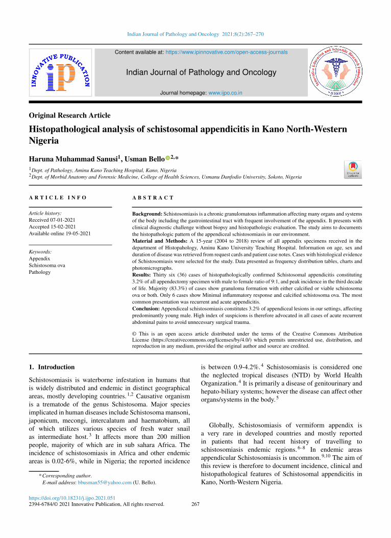
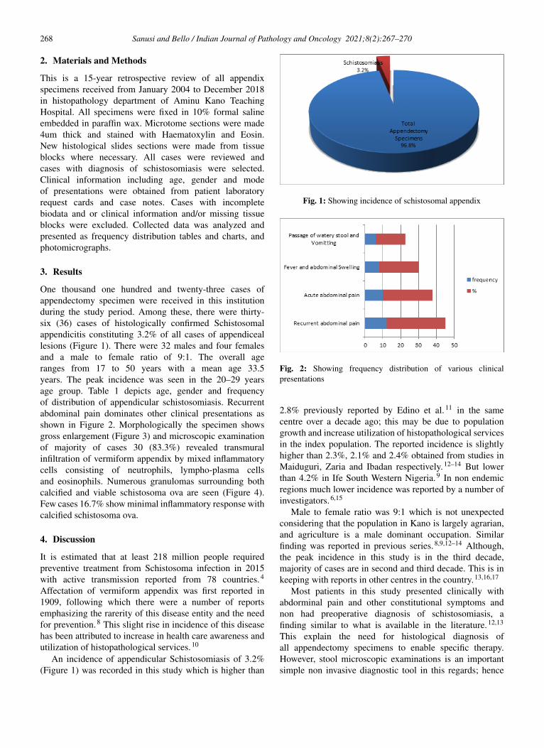
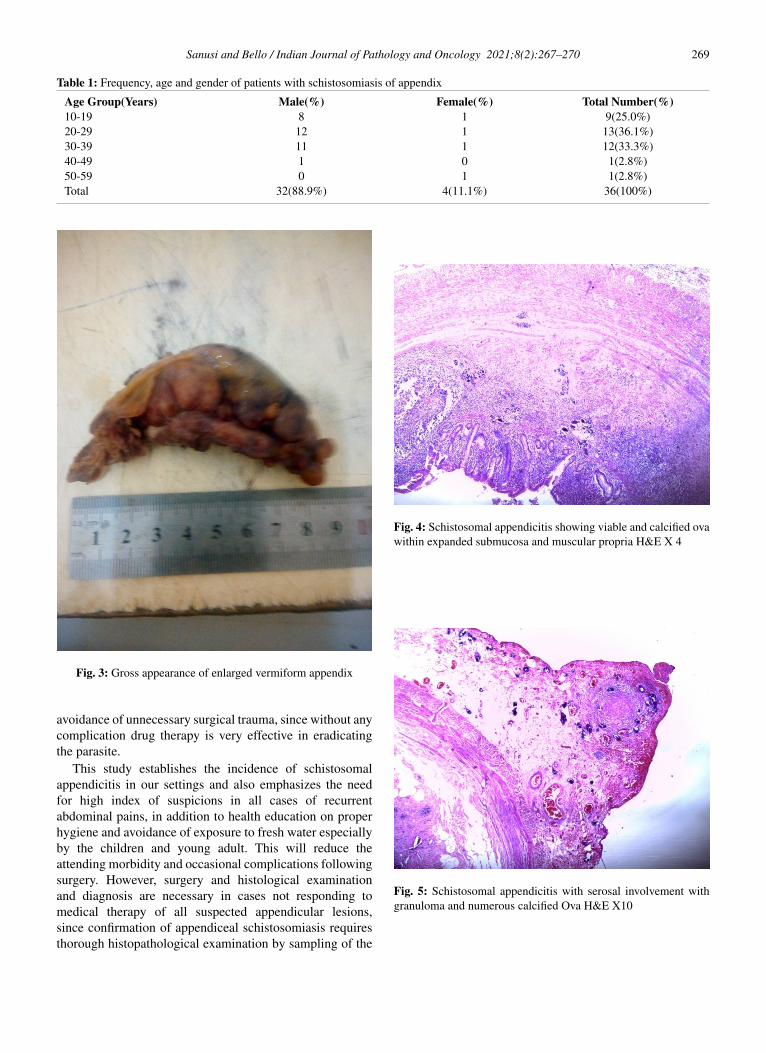
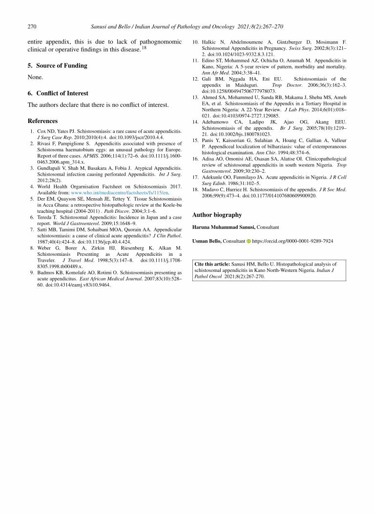
One thousand one hundred and twenty-three cases ofappendectomy specimen were received in this institutionduring the study period. Among these, there were thirty-six (36) cases of histologically confirmed Schistosomalappendicitis constituting 3.2% of all cases of appendiceallesions (Figure 1). There were 32 males and four femalesand a male to female ratio of 9:1. The overall ageranges from 17 to 50 years with a mean age 33.5years. The peak incidence was seen in the 20–29 yearsage group. Table 1 depicts age, gender and frequencyof distribution of appendicular schistosomiasis. Recurrentabdominal pain dominates other clinical presentations asshown in Figure 2. Morphologically the specimen showsgross enlargement (Figure 3) and microscopic examinationof majority of cases 30 (83.3%) revealed transmuralinfiltration of vermiform appendix by mixed inflammatorycells consisting of neutrophils, lympho-plasma cellsand eosinophils. Numerous granulomas surrounding bothcalcified and viable schistosoma ova are seen (Figure 4).Few cases 16.7% show minimal inflammatory response withcalcified schistosoma ova.
4. Discussion
It is estimated that at least 218 million people requiredpreventive treatment from Schistosoma infection in 2015with active transmission reported from 78 countries.4
Affectation of vermiform appendix was first reported in1909, following which there were a number of reportsemphasizing the rarerity of this disease entity and the needfor prevention.8 This slight rise in incidence of this diseasehas been attributed to increase in health care awareness andutilization of histopathological services.10
An incidence of appendicular Schistosomiasis of 3.2%(Figure 1) was recorded in this study which is higher than
Fig. 1: Showing incidence of schistosomal appendix
Fig. 2: Showing frequency distribution of various clinicalpresentations
2.8% previously reported by Edino et al.11 in the samecentre over a decade ago; this may be due to populationgrowth and increase utilization of histopathological servicesin the index population. The reported incidence is slightlyhigher than 2.3%, 2.1% and 2.4% obtained from studies inMaiduguri, Zaria and Ibadan respectively.12–14 But lowerthan 4.2% in Ife South Western Nigeria.9 In non endemicregions much lower incidence was reported by a number ofinvestigators.6,15
Male to female ratio was 9:1 which is not unexpectedconsidering that the population in Kano is largely agrarian,and agriculture is a male dominant occupation. Similarfinding was reported in previous series.8,9,12–14 Although,the peak incidence in this study is in the third decade,majority of cases are in second and third decade. This is inkeeping with reports in other centres in the country.13,16,17
Most patients in this study presented clinically withabdorminal pain and other constitutional symptoms andnon had preoperative diagnosis of schistosomiasis, afinding similar to what is available in the literature.12,13
This explain the need for histological diagnosis ofall appendectomy specimens to enable specific therapy.However, stool microscopic examinations is an importantsimple non invasive diagnostic tool in this regards; hence
Sanusi and Bello / Indian Journal of Pathology and Oncology 2021;8(2):267–270 269
Table 1: Frequency, age and gender of patients with schistosomiasis of appendix
Age Group(Years) Male(%) Female(%) Total Number(%)10-19 8 1 9(25.0%)20-29 12 1 13(36.1%)30-39 11 1 12(33.3%)40-49 1 0 1(2.8%)50-59 0 1 1(2.8%)Total 32(88.9%) 4(11.1%) 36(100%)
Fig. 3: Gross appearance of enlarged vermiform appendix
avoidance of unnecessary surgical trauma, since without anycomplication drug therapy is very effective in eradicatingthe parasite.
This study establishes the incidence of schistosomalappendicitis in our settings and also emphasizes the needfor high index of suspicions in all cases of recurrentabdominal pains, in addition to health education on properhygiene and avoidance of exposure to fresh water especiallyby the children and young adult. This will reduce theattending morbidity and occasional complications followingsurgery. However, surgery and histological examinationand diagnosis are necessary in cases not responding tomedical therapy of all suspected appendicular lesions,since confirmation of appendiceal schistosomiasis requiresthorough histopathological examination by sampling of the
Fig. 4: Schistosomal appendicitis showing viable and calcified ovawithin expanded submucosa and muscular propria H&E X 4
Fig. 5: Schistosomal appendicitis with serosal involvement withgranuloma and numerous calcified Ova H&E X10
270 Sanusi and Bello / Indian Journal of Pathology and Oncology 2021;8(2):267–270
entire appendix, this is due to lack of pathognomomicclinical or operative findings in this disease.18
5. Source of Funding
None.
6. Conflict of Interest
The authors declare that there is no conflict of interest.
References1. Cox ND, Yates PJ. Schistosomiasis: a rare cause of acute appendicitis.
J Surg Case Rep. 2010;2010(4):4. doi:10.1093/jscr/2010.4.4.2. Rivasi F, Pampiglione S. Appendicitis associated with presence of
Schistosoma haematobium eggs: an unusual pathology for Europe.Report of three cases. APMIS. 2006;114(1):72–6. doi:10.1111/j.1600-0463.2006.apm_314.x.
3. Gundlapali V, Shah M, Basakara A, Fobia J. Atypical Appendicitis.Schistosomal infection causing perforated Appendicitis. Int J Surg.2012;28(2).
4. World Health Orgarnisation Factsheet on Schistosomiasis 2017.Available from: www.who.int/mediacentre/factsheets/fs/115/en.
5. Der EM, Quayson SE, Mensah JE, Tettey Y. Tissue Schistosomiasisin Acca Ghana: a retrospective histopathologic review at the Koele-buteaching hospital (2004-2011) . Path Discov. 2004;3:1–6.
6. Tereda T. Schistosomal Appendicitis: Incidence in Japan and a casereport. World J Gastroenterol. 2009;15:1648–9.
7. Satti MB, Tamimi DM, Sohaibani MOA, Quorain AA. Appendicularschistosomiasis: a cause of clinical acute appendicitis? J Clin Pathol.1987;40(4):424–8. doi:10.1136/jcp.40.4.424.
8. Weber G, Borer A, Zirkin HJ, Riesenberg K, Alkan M.Schistosomiasis Presenting as Acute Appendicitis in aTraveler. J Travel Med. 1998;5(3):147–8. doi:10.1111/j.1708-8305.1998.tb00489.x.
9. Badmos KB, Komolafe AO, Rotimi O. Schistosomiasis presenting asacute appendicitus. East African Medical Journal. 2007;83(10):528–60. doi:10.4314/eamj.v83i10.9464.
10. Halkic N, Abdelmoumene A, Gintzburger D, Mosimann F.Schistosomal Appendicitis in Pregnancy. Swiss Surg. 2002;8(3):121–2. doi:10.1024/1023-9332.8.3.121.
11. Edino ST, Mohammed AZ, Ochicha O, Anumah M. Appendicitis inKano, Nigeria: A 5-year review of pattern, morbidity and mortality.Ann Afr Med. 2004;3:38–41.
12. Gali BM, Nggada HA, Eni EU. Schistosomiasis of theappendix in Maiduguri. Trop Doctor. 2006;36(3):162–3.doi:10.1258/004947506777978073.
13. Ahmed SA, Mohammed U, Sanda RB, Makama J, Shehu MS, AmehEA, et al. Schistosomiasis of the Appendix in a Tertiary Hospital inNorthern Nigeria: A 22-Year Review. J Lab Phys. 2014;6(01):018–021. doi:10.4103/0974-2727.129085.
14. Adebamowo CA, Ladipo JK, Ajao OG, Akang EEU.Schistosomiasis of the appendix. Br J Surg. 2005;78(10):1219–21. doi:10.1002/bjs.1800781023.
15. Panis Y, Kaisserian G, Sulahian A, Hoang C, Gallian A, ValleurP. Appendiceal localization of bilharziasis: value of extemporaneoushistological examination. Ann Chir. 1994;48:374–6.
16. Adisa AO, Omonisi AE, Osasan SA, Alatise OI. Clinicopathologicalreview of schistosomal appendicitis in south western Nigeria. TropGastroenterol. 2009;30:230–2.
17. Adekunle OO, Funmilayo JA. Acute appendicitis in Nigeria. J R CollSurg Edinb. 1986;31:102–5.
18. Madavo C, Hurriez H. Schistosomiasis of the appendix. J R Soc Med.2006;99(9):473–4. doi:10.1177/014107680609900920.
Author biography
Haruna Muhammad Sanusi, Consultant
Usman Bello, Consultant
https://orcid.org/0000-0001-9289-7924
Cite this article: Sanusi HM, Bello U. Histopathological analysis ofschistosomal appendicitis in Kano North-Western Nigeria. Indian JPathol Oncol 2021;8(2):267-270.