hiv-associated conditions occurring in multiple head ... · pdf filehiv-associated conditions...
TRANSCRIPT

HIV-ASSOCIATED CONDITIONS OCCURRING IN MULTIPLE HEAD & NECK ANATOMIC SITES
Sharon Ramos, MSIV
Faculty Advisor: Harold Pine, MD
The University of Texas Medical Branch
Department of Otolaryngology
Grand Rounds Presentation
October 27, 2010
OUTLINE
HIV/AIDS
Kaposi’s Sarcoma (KS)
Non-Hodgkin’s Lymphoma
Lymphoid Hyperplasia
Herpes Zoster
Case Report
Conclusion

HIV/AIDS
Structure of HIV
2 copies of positive
single-stranded RNA
Reverse transcriptase
Enclosed by capsid
(p24)
Viral envelope
(phospholipid bilayer)
HIV/AIDS
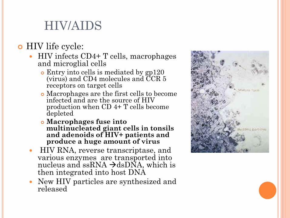
HIV life cycle: HIV infects CD4+ T cells, macrophages
and microglial cells Entry into cells is mediated by gp120
(virus) and CD4 molecules and CCR 5 receptors on target cells
Macrophages are the first cells to become infected and are the source of HIV production when CD 4+ T cells become depleted
Macrophages fuse into multinucleated giant cells in tonsils and adenoids of HIV+ patients and produce a huge amount of virus
HIV RNA, reverse transcriptase, and various enzymes are transported into nucleus and ssRNA dsDNA, which is then integrated into host DNA
New HIV particles are synthesized and released
HIV/AIDS
Incidence as of 2008:
33.4 million people are living with HIV/AIDS
15.7 million are women
2.1 million are children
2.7 million were newly infected with HIV
2.0 million AIDS related deaths
Median survival without treatment is about 10yrs
HAART has reduced death rate from disease by 80%,
and raised life expectancy for a newly diagnosed HIV
infected person 20-50yrs
AIDS:
CD4 < 200/ul
Presence of opportunistic pathogens

KAPOSI’S SARCOMA
Most common malignancy associated with advanced HIV/AIDS
It is an AIDS defining illness (CD4+ <200/ul)
Occurs in:
43% of homosexual or bisexual men
4% of IVD
KS is caused by HHV-8
AIDS related KS is very aggressive and affects:
Oral and pharyngeal mucosa
Neck masses appear secondary to lymph node infiltration
Cutaneous lesions of face, trunk, and lower extremities

KAPOSI’S SARCOMA
Typical KS lesions are pink-purplish and slightly raised or nodular and non-tender Slow progression, many lesions regress
with HAART
Secondary Infection is a major complication
Diagnosis is made by history and characteristic appearance of lesion, biopsy allows confirmation
Treatment: Low-dose radiation therapy,
chemotherapy, and immunotherapy
Low-dose radiation therapy is effective for dermatologic lesions, mucosal lesions are more resistant to therapy Painful mucositis occurs at much lower
radiation doses in HIV/AIDS patients

ORAL AND OROPHARYNGEAL KAPOSI’S
SARCOMA
KS is the most common oral malignancy in HIV disease 95% occur on the palate
and tongue
gingival surfaces and the oropharynx
In one study, 44% of patients with dermatologic KS also had submucosal lesions of their aerodigestive tract
Lesions are initially flat and asymptomatic but they often become exophytic and ulcerated
ORAL AND OROPHARYNGEAL KAPOSI’S
SARCOMA
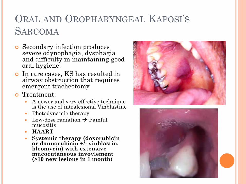
Secondary infection produces severe odynophagia, dysphagia and difficulty in maintaining good oral hygiene.
In rare cases, KS has resulted in airway obstruction that requires emergent tracheotomy
Treatment: A newer and very effective technique
is the use of intralesional Vinblastine
Photodynamic therapy
Low-dose radiation Painful mucositis
HAART
Systemic therapy (doxorubicin or daunorubicin +/- vinblastin, bleomycin) with extensive mucocutaneous invovlement (>10 new lesions in 1 month)
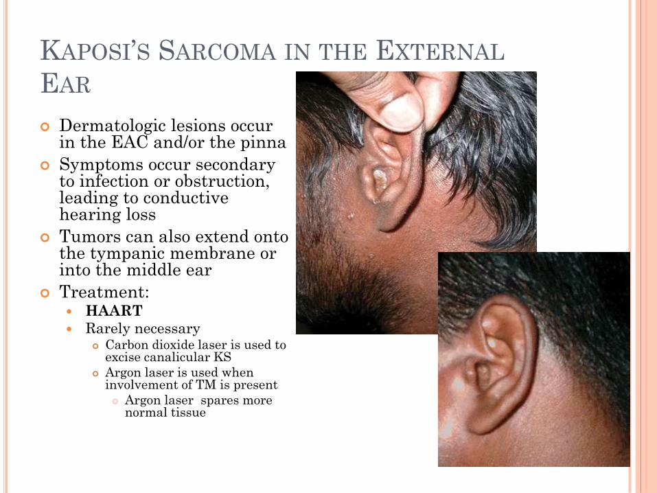
KAPOSI’S SARCOMA IN THE EXTERNAL
EAR
Dermatologic lesions occur in the EAC and/or the pinna
Symptoms occur secondary to infection or obstruction, leading to conductive hearing loss
Tumors can also extend onto the tympanic membrane or into the middle ear
Treatment: HAART
Rarely necessary Carbon dioxide laser is used to
excise canalicular KS
Argon laser is used when involvement of TM is present
Argon laser spares more normal tissue

OTHER LOCATIONS FOR KAPOSI’S
SARCOMA
Sinonasal KS
Is very rare and usually associated with concurrent
cutaneous lesions of nose and face
Symptoms include:
Nasal obstruction
Intermittent epistaxis
Rhinorrhea
Cervical Lymphadenopathy
KS can initially present as an enlarging cervical
mass or parotid gland swelling
FNAB of lymphnodes can diagnose KS but its
differentiation from bacillary epitheliod angiomatosis
(cat-scratch disease) may require an open biopsy

NON-HODGKIN’S LYMPHOMA
NHL is the second most common malignancy associated with HIV 100-fold increase risk if HIV+
NHL is much more aggressive and high grade (Large B-cell) in HIV patients Extra-nodal sites are involved in 89% of
patients 10% of patients have involvement of the Head
and Neck
42% have CNS extension
Patients present with a non-tender, rapidly enlarging neck mass, fever, night sweats and significant weight loss
Diagnosis: multiple FNABs because NHL can present
in the background of benign follicular hyperplasia (HIV lymphadenopathy)
Open biopsy

NON-HODGKIN’S LYMPHOMA
Criteria for open excisional biopsy
Constitutional Symptoms
Localized lymphadenopathy
Disproportionately large node in a patient with
persistent generalized lymphadenopathy
Cytopenia or elevated erythrocyte sedimentation rate
or both in a patient with otherwise negative
evaluation
NON-HODGKIN’S LYMPHOMA
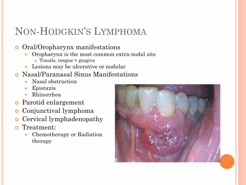
Oral/Oropharynx manifestations Oropharynx is the most common extra-nodal site
Tonsils, tongue > gingiva
Lesions may be ulcerative or nodular
Nasal/Paranasal Sinus Manifestations Nasal obstruction
Epistaxis
Rhinorrhea
Parotid enlargement
Conjunctival lymphoma
Cervical lymphadenopathy
Treatment: Chemotherapy or Radiation
therapy
LYMPHOID HYPERPLASIA
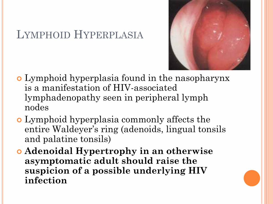
Lymphoid hyperplasia found in the nasopharynx is a manifestation of HIV-associated lymphadenopathy seen in peripheral lymph nodes
Lymphoid hyperplasia commonly affects the entire Waldeyer’s ring (adenoids, lingual tonsils and palatine tonsils)
Adenoidal Hypertrophy in an otherwise asymptomatic adult should raise the suspicion of a possible underlying HIV infection
LYMPHOID HYPERPLASIA
Nasopharyngeal lymphoid hyperplasia results in
nasal obstruction, serous otitis media, recurrent acute
otitis media and oropharyngeal airway compromise
(very rare).
Diagnosis is based on history and physical exam
including indirect laryngoscopy.
MRI or CT scan of the nasopharynx is recommended to rule
out an erosive skull base process or asymmetric adenoid
hypertrophy which can suggest Lymphoma/KS.
FNAB is helpful to establish a diagnosis but does not rule
out lymphoma
Treatment:
Systemic antibiotic and topical steroid sprays
Adenoidectomy

HERPES ZOSTER-RAMSAY HUNT SYNDROME
Etiology: Reactivation of VZV in Geniculate
ganglion
Initial encephalomeningomyelitis that secondarily spreads from the CSF to the labyrinth
Inflammatory neuritis of facial, cochlear and vestibular nerves Severe facial nerve
paralysis+SNHL+vertigo
Painful vesicular lesions in the concha or EAC Often misdiagnosed as otitis externa
Dysgeusia (chorda tympani)
Hyperacusis (stapedius)
More common in HIV than non-HIV patients In a prospective study of 48 high-risk
patients with VZV revealed that 73% were seropositive for HIV and 17% developed AIDS during 10-24 month follow-up
HERPES ZOSTER-RAMSAY HUNT SYNDROME
Diagnosis is based on history and physical, virus
isolation from vesicles, acute and convalescent
serum titers (4-fold increase)
Treatment:
Acyclovir
High dose corticosteroids (concurrent opportunistic
infections contraindicate use of systemic steroids)
Eye protection
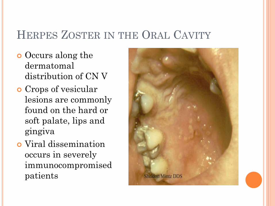
HERPES ZOSTER IN THE ORAL CAVITY
Occurs along the
dermatomal
distribution of CN V
Crops of vesicular
lesions are commonly
found on the hard or
soft palate, lips and
gingiva
Viral dissemination
occurs in severely
immunocompromised
patients
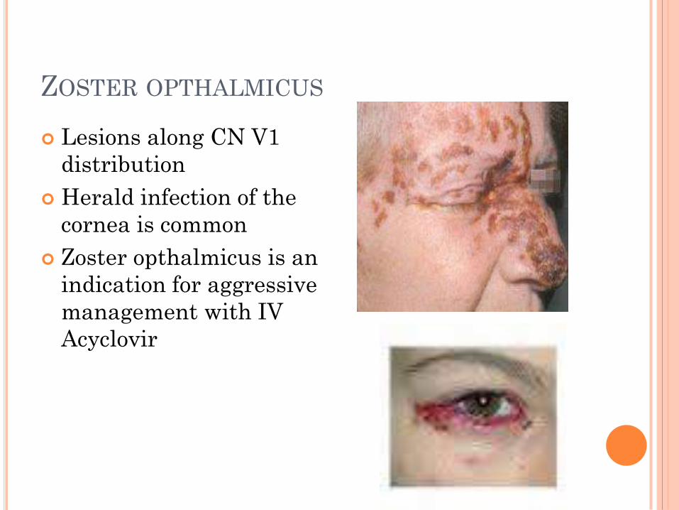
ZOSTER OPTHALMICUS
Lesions along CN V1
distribution
Herald infection of the
cornea is common
Zoster opthalmicus is an
indication for aggressive
management with IV
Acyclovir
CASE REPORT HPI: A 33-year old, 13 weeks pregnant female presents with shortness of
breath and noisy breathing. She also has odynophagia, bilateral neck swelling, sore throat and night sweats for the past 3 days. She has a discharging ear and a chronic non-productive cough for 3-6 weeks. She was started on Distaclor (Cefaclor) and discontinued when she was confirmed to be pregnant. She is a non smoker and social drinker. No history of IVD or other risk factors of HIV infection.
PE: She is febrile, has inspiratory stridor, tachycardia and tachypnea
Oral cavity/Oropharynx: Extensive candiasis
Neck: Bilateral Lymphadenopathy
Fiberoptic laryngoscopy: Very large, edematous and inflamed epiglottis with extensive white patches. Vocal cords could not be visualized only the posterior portion of arytenoids were seen
Lateral x-ray of the neck: enlarged epiglottis and a normal trachea
U/S: nonviable pregnancy
Labs: WBC 6.1x10³/ul, Hb 13.2g/dl, PLT 215x103/ul,
CD4+ 44/mm3
Assesment: Patient tested positive for HIV and is diagnosed with severe fungal /bacterial epiglottitis.
Plan: Patient is started on IV fluconazole, cefuroxime, metronidazole, nystatin mouth washes, HAART, Bactrim, azithromycin and dapsone.

CASE REPORT
Patient is examined 2 weeks later
Flexible fiberoptic laryngoscopy :
Epiglottis still appeared inflamed and grossly
swollen
CT scan of neck and upper thorax:
Showed a 4 cm swelling of the epiglottis
extending down into the aryepiglottic folds
and into the vestibule. Bilateral cervical
lymphadenopathy was also noted.
In view of her slow recovery, a biopsy of her
epiglottis was obtained.
Histopathology: Necrotic inflammatory tissue
with ulceration and dense proliferation of
anastomosing vascular channels. Patient was
diagnosed with Kaposi’s Sarcoma.
Patient continued on HAART and 8 weeks
after her initial presentation her larynx
appeared normal, her viral load was
undetectable and CD4 count increased.
CONCLUSION
About 80% of patients with HIV infections
present with otolaryngological symptoms.
Often, the otolaryngologist is the primary
physician who diagnosis the HIV infection
Oropharyngeal findings are the most common
and seen in 59% of patients, followed by cervical
lymphadenopathy in 42% of patients.
Combination of findings are common in some
patients particularly cervical lymphadenopathy
coexisting with oral and nasal manifestations
this should be a clue that HIV ought to be ruled
out.

DISCUSSION: HAROLD PINE, MD, FAAP
Sharon, that was a nice review of ENT manifestations of HIV. It certainly highlights the fact that many of these patients are going to get a problem in the head and neck and these can range from commonplace issues to the exotic. The causes of most otolaryngologic manifestations of HIV disease fall into the following three categories: infections, neoplasms, and primary neurologic damage caused by HIV.
As otolaryngologists we certainly see our share of patients with HIV and even full blown AIDS. There are certain things like seeing adenoids in adults that should raise your suspicions about underlying HIV disease. Large parotid cysts or unexplained persistent adenopathy should also make you think possible HIV.
A word of caution: If you order tests, be prepared to follow up the results. Especially with sensitive things like HIV testing, make sure you are willing to be responsible for following up the results.
Instead of being a death sentence, having HIV is now quite manageable if found early and treated aggressively. An astute otolaryngologist can pick up on things during a routine head and neck exam that can lead to an early diagnosis and treatment for the patient.
Harold Pine, MD, FAAP

REFERENCES
Delbrouck C, Kapouridis S, Chantrain G. An unusual localisation of Kaposi's
sarcoma: the external auditory canal. Acta Otorhinolaryngol Belg. 1998;52(1):29-36.
Sinonasal Malignancies website:
http://www.hopkins-aids.edu/diagnosis/malignancies/sinonasal_malignancies.
Truitt T, Tami T. Otolaryngologic manifestations of HIV infection. Medical Clinics of
North America. 1999; 83(1)
Pavan-Langston D. Clinical manifestations and therapy of herpes zoster
ophthalmicus. Comp Ophthalm Update. 2002;3:217.
Prasad H, Bhojwani K, Shenoy V, Prasad S. HIV manifestations in Otolaryngology.
American Journal of Otolaryngology. 2006;27: 179-185
Dhanasekar G, Robertson A, Nicholson K. Atypical presentation of HIV in a
pregnant patient. The Ulster Medical Journal. 2006; 75(2): 160-161
Pasha R, Golub J. Otolaryngology: Head and neck surgery: clinical reference guide.
pages 73-74 San Diego: Plural pub., 2009.
Bailey B., Johnson J, Newlands S. Head & Neck Surgery – Otolaryngology. pages
263-273. Lippincott Williams & Wilkins, 2006
Lee K, Thomas T, Otolaryngologic Manifestations of HIV. HIV InSite Knowledge
Base Chapter http://hivinsite.ucsf.edu
"CDC HIV/AIDS." Centers for Disease Control and Prevention. Web. 27 Oct. 2010.
<http://www.cdc.gov/hiv/>.