hochman-mendez, c., cantini, m., moratal, d., salmeron ...eprints.gla.ac.uk/98139/1/98139.pdf ·...
TRANSCRIPT
n
Hochman-Mendez, C., Cantini, M., Moratal, D., Salmeron-Sanchez, M., and Coelho-Sampaio, T. (2014) A fractal nature for polymerized laminin. PLoS ONE, 9 (10). e109388. ISSN 1932-6203 Copyright © 2014 The Authors http://eprints.gla.ac.uk/98139/ Deposited on: 09 October 2014
Enlighten – Research publications by members of the University of Glasgow
http://eprints.gla.ac.uk

A Fractal Nature for Polymerized LamininCamila Hochman-Mendez1,2, Marco Cantini3, David Moratal4, Manuel Salmeron-Sanchez3,
Tatiana Coelho-Sampaio2*
1 Institute of Biomedical Sciences, Federal University of Rio de Janeiro, Rio de Janeiro, Brazil, 2 Institute of Biophysics Carlos Chagas Filho, Federal University of Rio de
Janeiro, Rio de Janeiro, Brazil, 3 Division of Biomedical Engineering, School of Engineering, University of Glasgow, Glasgow, United Kingdom, 4 Center for Biomaterials and
Tissue Engineering, Universitat Politecnica de Valencia, Valencia, Spain
Abstract
Polylaminin (polyLM) is a non-covalent acid-induced nano- and micro-structured polymer of the protein laminin displayingdistinguished biological properties. Polylaminin stimulates neuritogenesis beyond the levels achieved by ordinary lamininand has been shown to promote axonal regeneration in animal models of spinal cord injury. Here we used confocalfluorescence microscopy (CFM), scanning electron microscopy (SEM) and atomic force microscopy (AFM) to characterize itsthree-dimensional structure. Renderization of confocal optical slices of immunostained polyLM revealed the aspect of aloose flocculated meshwork, which was homogeneously stained by the antibody. On the other hand, an ordinary matrixobtained upon adsorption of laminin in neutral pH (LM) was constituted of bulky protein aggregates whose interior was notaccessible to the same anti-laminin antibody. SEM and AFM analyses revealed that the seed unit of polyLM was a flatpolygon formed in solution whereas the seed structure of LM was highly heterogeneous, intercalating rod-like, sphericaland thin spread lamellar deposits. As polyLM was visualized at progressively increasing magnifications, we observed thatthe morphology of the polymer was alike independently of the magnification used for the observation. A search for theHausdorff dimension in images of the two matrices showed that polyLM, but not LM, presented fractal dimensions of 1.55,1.62 and 1.70 after 1, 8 and 12 hours of adsorption, respectively. Data in the present work suggest that the intrinsic fractalnature of polymerized laminin can be the structural basis for the fractal-like organization of basement membranes in theneurogenic niches of the central nervous system.
Citation: Hochman-Mendez C, Cantini M, Moratal D, Salmeron-Sanchez M, Coelho-Sampaio T (2014) A Fractal Nature for Polymerized Laminin. PLoS ONE 9(10):e109388. doi:10.1371/journal.pone.0109388
Editor: Heidar-Ali Tajmir-Riahi, University of Quebec at Trois-Rivieres, Canada
Received June 9, 2014; Accepted September 2, 2014; Published October 8, 2014
Copyright: � 2014 Hochman-Mendez et al. This is an open-access article distributed under the terms of the Creative Commons Attribution License, whichpermits unrestricted use, distribution, and reproduction in any medium, provided the original author and source are credited.
Data Availability: The authors confirm that all data underlying the findings are fully available without restriction. All relevant data are within the paper and itsSupporting Information files.
Funding: This work was supported by a grant from the Brazilian National Research Council (CNPq; 476772/2008-7) to TCS. MSS acknowledges support from theEuropean Research Council through ERC - 306990. The funders had no role in study design, data collection and analysis, decision to publish, or preparation of themanuscript.
Competing Interests: The authors have declared that no competing interests exist.
* Email: [email protected]
Introduction
Laminin is the major molecular component of the basement
membrane, a specialized type of extracellular matrix characterized
by a flat sheet-like geometry. Laminin regulates a variety of
biological phenomena, including the provision of boundaries
between neighboring tissues, the establishment of molecular filters
and the modulation of cell behavior [1-3]. The quaternary
structure of laminin is given by three different polypeptide chains,
which associate to form a cross-shaped heterotrimer, exhibiting
one long and three shorter arms. In the three-dimensional space
laminin has the shape of a three-leafed clover, in which the leaves
correspond to the three short arms while the stem corresponds to
the long arm. This 3-D structure is particularly adequate to favor
the assembly of a sheet-like polymer, where the three short arms
simultaneously interact with each other within a single spatial
plane, while the long arm is left available to interact with the
surface of contiguous cells [4].
As a consequence of its structural properties, laminin can
spontaneously self-polymerize in a test tube, requiring either a
minimal protein concentration [5] or a decrease in the solution pH
[6,7]. Polymers formed upon pH acidification, designated as
polylaminin (polyLM1), present specific signaling properties and
have been shown to stimulate the outgrowth of neurites with at
least twice the efficiency of ordinary laminin (LM), namely a
matrix obtained by adsorbing the protein diluted in neutral pH
onto a glass coverslip [8]. PolyLM was also shown to reverse the
loss of migratory and neuritogenic potentials of cortical neurons
and to promote the survival and the proliferation of axotomized
retinal ganglion cells, both isolated from newborn rodents [9].
Finally, it was demonstrated that polyLM, but not the laminin
protein diluted in neutral buffer, promoted axonal regeneration
and functional recovery after spinal cord injury in rats [10].
The morphology of polyLM has previously been studied both at
the micro and at the nanometer scales. Using negative staining
followed by transmission electron microscopy it was possible to
characterize it as a regular polygonal network displaying the same
features of the natural laminin networks assembled by living cells
[11,12]. In such polymers the unit polygon was a hexagon of
approximately 30 nm of side, which well corresponded to the size
of each short arm in the laminin molecule. Curiously, a polygonal
array of comparable features was observed at a three orders of
magnitude larger scale when immunolabeled polyLM was
analyzed by fluorescence microscopy [7,8,13].
PLOS ONE | www.plosone.org 1 October 2014 | Volume 9 | Issue 10 | e109388

The fact that polyLM observed at different magnifications
showed similar structures suggested that the polymer could be a
fractal structure. Nevertheless, the lack of images obtained at
intermediary magnifications, as well as the insufficient resolution
of previous epifluorescence photomicrographs, prevented the
search for a fractal dimension of polyLM. In the present work
we used confocal fluorescence microscopy (CFM), scanning
electron microscopy (SEM) and atomic force microscopy (AFM)
to obtain a detailed structural characterization of polyLM that
would permit assessment of its putative fractal nature. We showed
that polyLM presented a fractal dimension, which increased its
complexity upon accumulation of the polymer on a flat substrate.
These findings may have important implications as they can
provide an intrinsic molecular basis for the fractal-like organiza-
tion of the basement membranes present at the neurogenic niches
in the adult central nervous system.
Experimental Procedures
Preparation of laminin matricesPolyLM was produced by diluting EHS laminin (laminin 111;
Invitrogen) to 50 mg/mL with 20 mM sodium acetate (pH 4),
containing 1 mM CaCl2. The laminin protein has previously been
shown to polymerize in solution within a few minutes after dilution
in acidic buffer, independently of its concentration [6]. The
polymers produced in solution (polyLM) were adsorbed to glass
coverslips to produce the matrices used here for microscopic
analyses. LM was produced by diluting EHS laminin to 50 mg/mL
with 20 mM Tris-HCl (pH 7) containing 1 mM CaCl2. This
concentration was below the critical protein concentration
necessary to trigger laminin polymerization in solution at neutral
pH [5], so that the LM matrix was formed as the protein decanted
and raised its concentration at the glass surface. In order to avoid
unwanted polymerization in the highly concentrated stock solution
of EHS laminin (0.5–1.5 mg/mL), working aliquots (2–10 mL)
were stored frozen and individually thawed in ice immediately
before dilution. Unless otherwise indicated, incubations with glass
coverslips were carried out at 37uC for 12 hours, which is known
to be sufficient to warrant that at least 60% of the protein would
decant and adsorb regardless of the solution pH [8].
Immunostaining and confocal microscopyLaminin matrices adsorbed on glass coverslips were fixed with
paraformadehyde 4% for 20 min and prepared for indirect
immunofluorescence analysis. Coverslips were washed 3 times
for 5 min in PBS and incubated with bovine serum albumin 5% in
PBS (PBS-BSA 5%) for 30 min. The primary antibody was a
polyclonal rabbit anti-laminin antibody (1:30, Sigma-Aldrich,
no. L9393). After overnight incubation at 4uC, coverslips were
washed 3 times for 5 min in PBS and incubated with an Alexa
Fluor 488 anti-rabbit secondary antibody (1:300; Life Technology,
no. A-11001) for 1 hour at room temperature. They received
three 5-min washes with PBS and one with distilled water before
being mounted in n-propyl gallate in 80% glycerol (Sigma-
Aldrich). Confocal images were obtained in a Leica TCS-SP5
confocal laser scanning microscope using a HCX PL APO lambda
blue 63X objective for oil immersion (1.4 of numerical aperture).
Images correspond to renderized stacks of 74 optical slices
obtained with a zoom of 3.2 at each 21.4 mm (total width of
1530.8 mm).
Scanning electron microscopyLaminin matrices attached to coverslips were fixed in
Karnowsky reagent (4% PA and 0.5% glutaraldehyde in 0.1 M
cacodylate buffer, pH 7.2) for 2 hours, washed three times with
sodium cacodylate buffer 0.1 M, pH 7.2, dehydrated through
increasing concentrations of ethanol and dried in E300 (Polaron,
Quorum Technologies Ltd, Laughton, United Kingdom) critical
point. The samples were then coated with a thin layer of gold
sputter (Leica EM MED020) and viewed under a scanning
electron microscope Jeol JSM6300.
Transmission electron microscopyTransmission electron microscopy after negative staining was
carried out as previously described [11]. Briefly, 5 ml of laminin in
acidic buffer (polyLM) was deposited on a Formvar-coated copper
grid and a 5 ml drop of 2% uranyl acetate was added over it.
Samples were visualized in a Zeiss EM 900 transmission electron
microscope operated at 80 kV.
Atomic force microscopyLaminin matrices were fixed in 4% paraformaldehyde for 20
minutes and dried in critical point dryer E300 (Polaron, Quorum
Technologies Ltd, Laughton, United Kingdom). AFM analyses
were performed using a Multimode AFM equipped with a
NanoScope IIIa controller (Bruker) operating in tapping mode
in air; the Nanoscope 5.30r2 software version was used for image
processing and analysis. Si-cantilevers MPP-21120 from Bruker
were used, with force constant of 3 N.m21 and resonance
frequency of 75 kHz. The phase signal was set to zero at a
frequency 5–10% lower than the resonance one. Drive amplitude
was 200 mV and the amplitude set-point (Asp) was 1.4 V. The
ratio between the amplitude set-point and the free amplitude (Asp/
A0) was kept equal to 0.7.
Quantification of laminin adsorptionThe amount of adsorbed laminin was measured using a Micro
BCA Protein Assay Kit (23235#, Thermo Scientific Pierce).
Following the standard protocol, the working reagent (WR) was
prepared from 25 parts of MA (sodium carbonate, sodium
bicarbonate and sodium tartrate in 0.2 N NaOH), 24 parts of
MB (4% BCA in water) and 1 part of MC (4% cupric sulfate,
pentahydrate in water). As standards, nine bovine serum albumin
(BSA) solutions with concentrations ranging from 0.0 to 200 mg/
mL were prepared by dissolving BSA in the buffers used for
laminin adsorption (acidic buffer and neutral buffer). The amount
of adsorbed protein was calculated by measuring the amount of
protein remaining in the supernatant at each time point. The
samples and the standards were incubated with WR 1:1 at 37uCfor two hours before cooling to room temperature. Then, the
absorbance at 562 nm was measured with the plate reader Victor3
(PerkinElmer, Waltham, Massachusetts). All the absorbance values
were corrected by the average 562 nm absorbance reading of the
blank standard replicates. Each measurement was performed in
duplicate.
Calculus of the fractal or the Hausdorff dimensionAll image processing and analysis was done using an in-house
software developed under MATLAB R2006a (The MathWorks,
Inc., Natick, MA). The fractal dimension was determined using a
box-counting dimension estimate of the Hausdorff dimension,
which is a descriptor of the complexity of geometry of a given set
[14]. As the definition of the Hausdorff dimension does not offer
any guideline to an estimate calculation, the box-counting
dimension has been used. The box-counting dimension is an
estimate of the Hausdorff dimension based on covering the
investigated set with a fixed grid of size r [15,16]. This
A Fractal Nature for Polymerized Laminin
PLOS ONE | www.plosone.org 2 October 2014 | Volume 9 | Issue 10 | e109388

box-counting dimension can be calculated using the equation (1):
DH ~{D log k rð Þ½ �
D log rð1Þ
where k(r) is a number of grid boxes that contain any part of the
investigated set, and where the process is repeated with different
values of the grid size. DH is known as the Hausdorff dimension,
the Minkowski-Bouligand dimension, the Kolmogorov capacity or
dimension, or simply the box-counting dimension.
Before applying the box-counting algorithm, the grayscale
image histogram was equalized and the resulting image was
binarized using the Otsu’s method [17]. Finally, the box-counting
estimate was calculated on this image. All this process was
performed on ten different image crops of the same size (100
pixels6100 pixels) of the original images, and all these image crops
were rescaled ten times to obtain an image big enough to apply the
box-counting algorithm.
Results
Three-dimensional structures of polyLM and LM assessedby confocal fluorescence and scanning electronmicroscopy
Matrices of adsorbed laminin obtained at acidic (polyLM) or
neutral pH (LM) were initially analyzed by reconstructing a series
of 74 confocal optical slices renderized to reveal their 3-D
structure. PolyLM corresponded to a sponge-like network of an
apparently homogeneous density (Fig. 1A). Labeling was equally
distributed, indicating that the antibody could evenly access
protein epitopes within the polymer. On the other hand, LM
presented a branched morphology, resembling that of a marine
coral, which protruded from the glass surface (Fig. 1B). The
antibody did not penetrate the spherical protein clumps and only
their contours were brightly stained. Noteworthy was the
observation that in LM a significant amount of protein adsorbed
directly to the glass coverslip. This likely corresponds to the
protein not incorporated to the aggregates. As a comparison, the
background of the image depicting polyLM was dark, indicating
that virtually all laminin protein engaged into that polymer. The 3-
D structures of the two laminin matrices can be better appreciated
in the animation movies presented as Movies S1 and S2.
The 3-D structures of polyLM and LM were additionally
investigated by using SEM. While polyLM was again seen as a
homogeneous mesh, LM showed at least two structural compo-
nents (Fig. 1C and D). Besides the spherical aggregates already
identified by CFM, it was possible to devise the presence of rod-
like structures. In addition, the tips of these rods possessed lamellar
terminations, suggesting the occurrence a third structural compo-
nent of the LM matrix (arrows in Fig. 1D).
Kinetics of formation of laminin matricesThe amount of adsorbed laminin was calculated by measuring
the concentration of protein remaining in solution at 20, 40 and
60 min and after 4, 8 and 12 hours of adsorption (Fig. 2A).
Laminin decanted more quickly at neutral pH to form the LM
matrix. More than 60% of the protein was absorbed after 20 min
of incubation and such amount slightly increased up to 80% at
12 hours. On the other hand, at acidic pH the kinetic of
adsorption was more linear, whereas 45% of the protein was
adsorbed at 20 min and such proportion increased to 90% at
12 hours. The morphologies of the two matrices were analyzed at
1, 8 and 12 hours by SEM (Fig. 2B–G). Already at 1 hour polyLM
decanted exhibiting a structured 2-D morphology consistent with
the formation of the meshwork observed at 12 hours (Fig. 2B, D).
Characterization of the seed units of polyLM and LMWhen the seed unit of polyLM was observed at higher
magnification one could see that it was itself composed of polygons
in a planar organization. Such seed unit was consistently present in
matrices obtained within one hour of incubation (Fig. 3A–C). The
extent of their longest axes ranged between 14 and 28 mm
(Fig. 3D). After 8 and 12 hours of incubation we could already
observe the presence of a mesh-like network whose morphology
was compatible with the overlay of the seed units observed at one
hour; these units however could no longer be distinguished within
the meshwork (Fig. 3E, F). In LM we could observe lamellar-like
deposits adsorbed directly to the coverslips (pseudo colored green
in Fig. 3). These lamellar deposits were either located at the end of
rod-like structures (yellow) or they appeared as individual patches
(arrowheads in Fig. 3I). The seed unit was pseudo colored as to
reveal the three types of deposits seen in LM, namely spheres
(pink), rods (yellow) and lamellar deposits (green) (Fig. 3G–I).
Three-dimensional structures of polyLM and LM assessedby atomic force microscopy
We next examined the 3-D features of polyLM and LM using
AFM. When areas of 50650 mm were scanned, the overall
appearances of polyLM and LM were comparable to those
visualized by CFM and SEM (Fig. 1). PolyLM displayed the
morphology of a multilayered meshwork containing homogeneous
struts (Fig. 4A), while LM exhibited rods and lamellar deposits
(Fig. 4B). The spherical aggregates previously seen under confocal
fluorescence (Fig 1B) and SEM (Fig 1D and 3G–I) were not
Figure 1. Three-dimensional structure of laminin polymersunder confocal fluorescence microscopy and scanning electronmicroscopy. Laminin was incubated on glass coverslip for 12 hours inacidic (polyLM) or in neutral buffer (LM). (A, B) Indirect immunofluo-rescence was performed using a polyclonal antibody against laminin.The images depict z-stacks obtained by the superposition of 74confocal slices renderized using the software 7.2.3 (Bit-plane; free trial).(C, D). Scanning electron micrographies (SEM) of the polymers shown ata similar magnification. Arrows in D point to lamellar deposits oflaminin. The scale bars apply to panels A–D and represent 10 mm.doi:10.1371/journal.pone.0109388.g001
A Fractal Nature for Polymerized Laminin
PLOS ONE | www.plosone.org 3 October 2014 | Volume 9 | Issue 10 | e109388

observed under AFM due to their large size, which was beyond the
Z scan range of the AFM.
To further characterize the homogeneity of the struts in
polyLM, the matrix was scanned at higher magnifications
(Fig. 5A, B). In fields of 0.560.5 mm, we measured the heights
of 10 individual struts of the mesh, chosen as those in direct
contact with the glass support, i.e., those in the bottom layer of the
mesh. We found values ranging between 50 and 73 nm, with a
mean height of 60.2561.764 nm (Fig. 5C, D). In the heteroge-
neous LM matrix, we measured the heights of rods and lamellar
deposits, which displayed average heights of 12136134.6 nm
(Fig. 5E–H) and 125.1613.51 nm (Fig.5I–L), respectively.
Evidence of a fractal nature for polyLMWhen polyLM was observed at a higher magnification with
AFM it was possible to observe the occurrence of figures that
matched the hexagon-like shape of polymerized laminin at the
molecular level (Fig. 6A–C). Such figures were still one order of
magnitude larger than the basic hexagons formed by the
association of the short arms of individual laminin molecules
(Fig. 6G, H) [4,12]. In that structure, each side of the hexagon
possesses ,30 nm, resulting from the interaction of the laminin
short arms (35–50 nm long). The sides of the putative hexagons
observed here were larger and their length was in the range of a
few hundreds of nanometers. Nevertheless, these polygons were
made out of small globules with a diameter and a spacing around
30–40 nm, compatible with the characteristic length of laminin
polymerized through interaction between short arms (Fig. 6D–F).
These structures could not be further resolved via AFM.
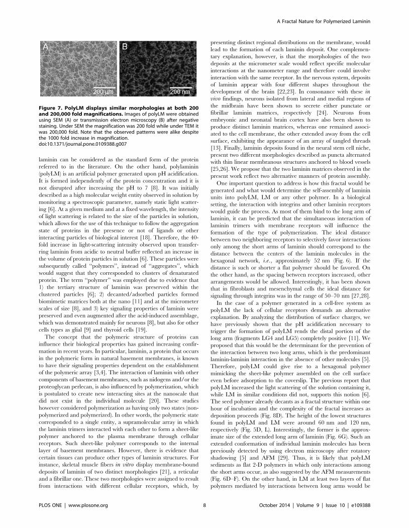
The results obtained up this point suggested that the hexagonal
network formed by the association of individual laminin molecules
could reproduce itself at higher levels of organization. In order to
investigate this hypothesis we compared images of polyLM
obtained with SEM and with transmission electron microscopy
after negative staining (Fig. 7). Surprisingly, the morphologies of
polyLM were very similar under SEM (Fig. 7A) and transmission
electron microscopy (Fig. 7B) regardless of a difference in
magnification of 1,000 fold. This observation suggests that polyLM
presents a fractal nature.
Determination of the fractal dimension of polyLMBased on evidence that polyLM possessed a fractal structure, we
analyzed images of polyLM obtained at increasing incubation
times in search for its fractal dimension. Fractal or Hausdorff
dimensions of increasing complexities were found for polyLM as
the adsorption time increased from 1 to 12 h (Fig. 8). The
calculated values were 1.55, 1.62 and 1.70 after 1, 8 and 12 hours
of adsorption, respectively. By contrast, the LM matrix did not
present a fractal structure from which a fractal dimension could be
obtained.
Figure 2. Kinetics of adsorption of polyLM and LM. (A) Laminin was incubated in acidic (polyLM) or neutral buffer (LM) and a kinetic ofadsorption was carried out by collecting aliquots of the supernatant at 10 minutes, 30 minutes, 1 hour, 4 hours, 8 hours and 12 hours forquantification of the protein content remaining in solution. Open symbols represent polyLM and closed symbols represent the LM. (B–G) SEM imagesshow the polymers obtained in acidic (B–D) or neutral (E–G) buffers at the indicated times. The arrows (B) point to structured polymers observed at1 hour of incubation.doi:10.1371/journal.pone.0109388.g002
A Fractal Nature for Polymerized Laminin
PLOS ONE | www.plosone.org 4 October 2014 | Volume 9 | Issue 10 | e109388

Discussion
In the present work we described that two matrices of laminin,
polyLM and LM, obtained in different conditions presented highly
different structures when observed at a wide range of magnifica-
tions. In a previous study [11], we had already shown that one of
these polymers, polyLM, displayed the same nanostructure
reported for laminin arrays secreted by cells [12]. Moreover, we
had described that polyLM and LM presented different morphol-
ogies in the range of tens of micrometers [8]. Nevertheless
intermediary magnifications between these two ranges of sizes had
never been assessed before. Using confocal, electronic and atomic
force microscopy we filled in this gap and found that surprisingly
polyLM presented similar structures independently of the magni-
fication used to observe it. Since this property is a feature of
fractals, we searched for a possible fractal dimension and
confirmed that polyLM indeed corresponded to a fractal structure.
Conversely, the second polymer, LM, was more heterogeneous
and did not present a fractal nature.
Before addressing the biological significance of the present
findings it is important to recapitulate the features of the polymers
studied here. The term ordinary laminin (LM) is used to refer to
Figure 3. Characterization of polymer units in polyLM and LM. Laminin polymers were analyzed at high magnification in order tocharacterize the morphologies of the seed units of each polymer. (A–C) At 1 hour polyLM forms star-like 2D structures as exemplified in the threepanels. (D) The sizes of the longer axes in these structures were quantified and shown to average at 20.8465.449 mm. (E, F) High magnificationimages of polyLM at 8 and 12 hours show a meshwork pattern compatible with the deposition of the star-like structures. (G–I) LM observed at highmagnification reveals three types of seed structures: rods (pseudocolored yellow), spheres (pseudocolored pink) and lamellas (pesudocolored green).The scale bar in I applies to all panels and represents 10 mm.doi:10.1371/journal.pone.0109388.g003
Figure 4. Overall morphology of polyLM and LM under AFM. Atomic force microscopy images of polyLM (A) and LM (B) are shown in heightmode after critical-point drying of the samples. Both matrices were obtained by incubating laminin with glass coverslips in the appropriate buffers for12 hours. The scanned area was 2500 mm2.doi:10.1371/journal.pone.0109388.g004
A Fractal Nature for Polymerized Laminin
PLOS ONE | www.plosone.org 5 October 2014 | Volume 9 | Issue 10 | e109388

Figure 5. AFM analysis of polyLM and LM at increasing magnifications. PolyLM (A, B) and LM (E, F, I, J) obtained as described in Figure 4were scanned in areas of 225 mm2 (A, E, I) or 0.25 mm2 (B, F, J) and shown in height mode. In order to determine the thickness of the structural unitsforming each polymer, the heights of 10 struts were calculated in the fields depicted in B (struts of the polyLM mesh), F (rods in LM) and J (lamellas inLM). Considering that both matrices were multilayered, each structure selected for measurement followed the criteria of being the closest possible tothe support (glass coverslip). Panels C, G and K depict examples of three measurements and panels D, H and L show the distribution of the valuesobtained for each 10 structures. The white square in I represents an area at the edge of the lamellar structure used for the height measurement. PanelM shows the distribution of heights obtained at each condition all together for comparison.doi:10.1371/journal.pone.0109388.g005
A Fractal Nature for Polymerized Laminin
PLOS ONE | www.plosone.org 6 October 2014 | Volume 9 | Issue 10 | e109388

clusters of laminin adsorbed onto a glass surface at a concentration
below the critical concentration of 60–100 mg/ml, previously
shown to induce solution polymerization at pH 7 [5]. In this
condition the protein does not self-assemble in solution but it tends
to form clusters as its concentration increases at the glass surface
upon decantation/adsorption. Since laminin is used as a coating
substrate for cell attachment at concentrations below the critical
concentration (typically between 1 and 20 mg/ml), ordinary
Figure 6. Atomic force microscopy reveals the occurrence of hexagonal-like figures in polyLM. AFM was performed on polyLM matricesobtained as described in Figure 4 and areas of 1 (A, B) or 0.25 mm2 (C) were scanned in height mode. Hexagons-like figures similar to those occurringin natural laminin polymers [12] were identified. These hexagons were visible at different magnifications (A–C) and presented variable side lengths(sketched with white dashed lines), but they were never as short as 30 nm as they should be to correspond to the short arm of a laminin molecule.The smallest distinguishable structures contained within the sides of the hexagons were little globules (D) whose size and spacing was measured inimages of 0.02 mm2 (D). Panel E shows the distribution of spacing values, which are compatible with the characteristic length (,30 nm) of lamininpolymerized via the short arms. Panel F depicts a three-dimensional reconstruction of the same area shown in panel D, with superposition ofcompatible locations of laminin molecules. Schemes of one individual laminin molecule (long arm dashed and short arms colored blue, green andorange), with indication of its characteristic dimensions (G) and of the hexagonal polymer generated by the interaction between individual lamininmolecules (H) are also shown.doi:10.1371/journal.pone.0109388.g006
A Fractal Nature for Polymerized Laminin
PLOS ONE | www.plosone.org 7 October 2014 | Volume 9 | Issue 10 | e109388
laminin can be considered as the standard form of the protein
referred to in the literature. On the other hand, polylaminin
(polyLM) is an artificial polymer generated upon pH acidification.
It is formed independently of the protein concentration and it is
not disrupted after increasing the pH to 7 [8]. It was initially
described as a high molecular weight entity observed in solution by
monitoring a spectroscopic parameter, namely static light scatter-
ing [6]. At a given medium and at a fixed wavelength, the intensity
of light scattering is related to the size of the particles in solution,
which allows for the use of this technique to follow the aggregation
state of proteins in the presence or not of ligands or other
interacting particles of biological interest [18]. Therefore, the 40-
fold increase in light-scattering intensity observed upon transfer-
ring laminin from acidic to neutral buffer reflected an increase in
the volume of protein particles in solution [6]. These particles were
subsequently called ‘‘polymers’’, instead of ‘‘aggregates’’, which
would suggest that they corresponded to clusters of denaturated
protein. The term ‘‘polymer’’ was employed due to evidence that
1) the tertiary structure of laminin was preserved within the
clustered particles [6]; 2) decanted/adsorbed particles formed
biomimetic matrices both at the nano [11] and at the micrometer
scales of size [8], and 3) key signaling properties of laminin were
preserved and even augmented after the acid-induced assemblage,
which was demonstrated mainly for neurons [8], but also for other
cells types as glial [9] and thyroid cells [19].
The concept that the polymeric structure of proteins can
influence their biological properties has gained increasing confir-
mation in recent years. In particular, laminin, a protein that occurs
in the polymeric form in natural basement membranes, is known
to have their signaling properties dependent on the establishment
of the polymeric array [3,4]. The interaction of laminin with other
components of basement membranes, such as nidogens and/or the
proteoglycan perlecan, is also influenced by polymerization, which
is postulated to create new interacting sites at the nanoscale that
did not exist in the individual molecule [20]. These studies
however considered polymerization as having only two states (non-
polymerized and polymerized). In other words, the polymeric state
corresponded to a single entity, a supramolecular array in which
the laminin trimers interacted with each other to form a sheet-like
polymer anchored to the plasma membrane through cellular
receptors. Such sheet-like polymer corresponds to the internal
layer of basement membranes. However, there is evidence that
certain tissues can produce other types of laminin structures. For
instance, skeletal muscle fibers in vitro display membrane-bound
deposits of laminin of two distinct morphologies [21], a reticular
and a fibrillar one. These two morphologies were assigned to result
from interactions with different cellular receptors, which, by
presenting distinct regional distributions on the membrane, would
lead to the formation of each laminin deposit. One complemen-
tary explanation, however, is that the morphologies of the two
deposits at the micrometer scale would reflect specific molecular
interactions at the nanometer range and therefore could involve
interaction with the same receptor. In the nervous system, deposits
of laminin appear with four different shapes throughout the
development of the brain [22,23]. In consonance with these invivo findings, neurons isolated from lateral and medial regions of
the midbrain have been shown to secrete either punctate or
fibrillar laminin matrices, respectively [24]. Neurons from
embryonic and neonatal brain cortex have also been shown to
produce distinct laminin matrices, whereas one remained associ-
ated to the cell membrane, the other extended away from the cell
surface, exhibiting the appearance of an array of tangled threads
[13]. Finally, laminin deposits found in the neural stem cell niche,
present two different morphologies described as puncta alternated
with thin linear membranous structures anchored to blood vessels
[25,26]. We propose that the two laminin matrices observed in the
present work reflect two alternative manners of protein assembly.
One important question to address is how this fractal would be
generated and what would determine the self-assembly of laminin
units into polyLM, LM or any other polymer. In a biological
setting, the interaction with integrins and other laminin receptors
would guide the process. As most of them bind to the long arm of
laminin, it can be predicted that the simultaneous interaction of
laminin trimers with membrane receptors will influence the
formation of the type of polymerization. The ideal distance
between two neighboring receptors to selectively favor interactions
only among the short arms of laminin should correspond to the
distance between the centers of the laminin molecules in the
hexagonal network, i.e., approximately 52 nm (Fig. 6). If the
distance is such or shorter a flat polymer should be favored. On
the other hand, as the spacing between receptors increased, other
arrangements would be allowed. Interestingly, it has been shown
that in fibroblasts and mesenchymal cells the ideal distance for
signaling through integrins was in the range of 50–70 nm [27,28].
In the case of a polymer generated in a cell-free system as
polyLM the lack of cellular receptors demands an alternative
explanation. By analyzing the distribution of surface charges, we
have previously shown that the pH acidification necessary to
trigger the formation of polyLM rends the distal portion of the
long arm (fragments LG4 and LG5) completely positive [11]. We
proposed that this would be the determinant for the prevention of
the interaction between two long arms, which is the predominant
laminin-laminin interaction in the absence of other molecules [5].
Therefore, polyLM could give rise to a hexagonal polymer
mimicking the sheet-like polymer assembled on the cell surface
even before adsorption to the coverslip. The previous report that
polyLM increased the light scattering of the solution containing it,
while LM in similar conditions did not, supports this notion [6].
The seed polymer already decants as a fractal structure within one
hour of incubation and the complexity of the fractal increases as
deposition proceeds (Fig. 8D). The height of the lowest structures
found in polyLM and LM were around 60 nm and 120 nm,
respectively (Fig. 5D, L). Interestingly, the former is the approx-
imate size of the extended long arm of laminin (Fig. 6G). Such an
extended conformation of individual laminin molecules has been
previously detected by using electron microscopy after rotatory
shadowing [5] and AFM [29]. Thus, it is likely that polyLM
sediments as flat 2-D polymers in which only interactions among
the short arms occur, as also suggested by the AFM measurements
(Fig. 6D–F). On the other hand, in LM at least two layers of flat
polymers mediated by interactions between long arms would be
Figure 7. PolyLM displays similar morphologies at both 200and 200,000 fold magnifications. Images of polyLM were obtainedusing SEM (A) or transmission electron microscopy (B) after negativestaining. Under SEM the magnification was 200 fold while under TEM itwas 200,000 fold. Note that the observed patterns were alike despitethe 1000 fold increase in magnification.doi:10.1371/journal.pone.0109388.g007
A Fractal Nature for Polymerized Laminin
PLOS ONE | www.plosone.org 8 October 2014 | Volume 9 | Issue 10 | e109388

necessary to account for the 120 nm height observed in the lowest
deposits.
In the present work we demonstrate that the protein laminin
can give rise to a fractal structure. The observation that the
supramolecular organization of a pure protein (polyLM) is fractal
implies that the information contained ultimately within its
primary sequence is sufficient to determine the morphology of
larger structures that will spatially organize tissue compartments.
This is particularly interesting because it correlates with the
‘‘fractal-like’’ organization of the niche for stem cells in the
subventricular zone of the adult brain. In this case, a laminin-rich
basement membrane, named ‘‘fractone’’, has been proposed to
orient the binding of proteoglycans, which, in turn, organize the
distribution of the growth factors controlling the maintenance of
the stem cell niche [26,30], as shown to be the case for bFGF [31].
Although fractones have been described only in the central
nervous system, it is well possible that a similar fractal-like
extracellular matrix is present in other stem cell niches. Adult stem
Figure 8. Calculation of the fractal dimension (Hausdorff estimate). (A, B) Image processing in order to prepare the image for the box-counting algorithm for LM (A) and polyLM (B). 1, original image; 2, original image in which the histogram has been equalized; 3, binarized imageusing Otsu’s method). (C) From images A.3 and B.3 the Hausdorff dimension estimates can be calculated superimposing a grid of variable size (C.1-C.4, examples of the same image on which a grid of variable size has been superimposed). (D) Repeating the previous process for different values ofgrid size and computing the number of grid boxes that contain any part of the investigated set, the Hausdorff dimension or simply the box-countingdimension can be calculated. (E) Fractal dimension calculated for polyLM structures as a function of time.doi:10.1371/journal.pone.0109388.g008
A Fractal Nature for Polymerized Laminin
PLOS ONE | www.plosone.org 9 October 2014 | Volume 9 | Issue 10 | e109388

cell niches manifest under certain restricted microenvironments
that host tissue-specific stem cells and regulate their physiology.
They exhibit complex cytoarchitectures composed of stem cells,
progenitor cells, supportive cells and a laminin-rich basement
membrane. The basement membrane regulates cell division and
differentiation within the niche due to several of its properties [32].
First, its molecular components interact with integrins to regulate
the cytoskeletal assembly. It also harbors and controls the
availability of growth factors and cytokines. A basement
membrane provides an orienting surface for asymmetric cell
division. Finally, it guides the traffic of progenitor cells within the
niche. It is known that the assembly of basement membranes is
initiated and dictated by laminin secretion and polymerization at
the cell surface [33]. In addition, laminin is able to interact with
integrins, to bind heparan sulfate proteoglycans, which, in turn,
present soluble factors. It is thus very likely that laminin is the key
component in basement membranes to play a regulatory role
controlling the physiology of the stem cell niche. The importance
of laminin for the maintenance of the stem cell niches has been
demonstrated in several cases such as in pancreatic islets [34,35],
the germline niche [36], skeletal muscle [37] and the embryonic
neocortical stem cell niche [38].
It is tempting to speculate that the fractal-like organization of
stem cell niches can not only control the distribution of growth
factors but also provide a physical constrain for progenitor cells
during differentiation. The representation of a differentiation
pathway as a tree, in which the stem corresponds to the stem cell
and the branches, to progenitors is per se a fractal. In this scenario,
the propagation of laminin polymerization throughout lager size
scales could be a key step for the organization of multicellular
organisms, which is in line with the observations that laminin-
containing basement membranes are the first assembled extracel-
lular matrix appearing during mammalian development and that
laminin is present in virtually all metazoans [39].
Supporting Information
Movie S1 Animated view of the three-dimensionalstructure of polyLM. The animation was generated from a
series of confocal optical slices (the same shown in Figure 1A),
using the software Imaris, version 7.2.3.
(AVI)
Movie S2 Animated view of the three-dimensionalstructure of LM. The animation was generated from a series
of confocal optical slices (the same shown in Figure 1B), using the
software Imaris, version 7.2.3.
(AVI)
Acknowledgments
We thank Laina Cunha for the excellent technical assistance.
Author Contributions
Conceived and designed the experiments: CHM MSS TCS. Performed the
experiments: CHM MC DM. Analyzed the data: CHM MC TCS MSS
DM. Contributed reagents/materials/analysis tools: TCS MSS DM.
Wrote the paper: TCS MSS DM.
References
1. Durbeej M (2010) Laminins. Cell Tissue Res 339: 259–268.
2. Miner JH, Yurchenco PD (2004) Laminin functions in tissue morphogenesis.
Annu Rev Cell Dev Biol 20: 255–284.
3. Yurchenco PD (2011) Basement membranes: cell scaffoldings and signaling
platforms. Cold Spring Harb Perspect Biol 3: a004911
4. Hohenester E, Yurchenco PD (2013) Laminins in basement membraneassembly. Cell Adhes Migr 7: 56–63.
5. Yurchenco PD, Tsilibary EC, Charonis AS, Furthmayr H (1985) Laminin
polymerization in vitro. Evidence for a two-step assembly with domain
specificity. J Biol Chem 260: 7636–7644.
6. Freire E, Coelho-Sampaio T (2000) Self-assembly of laminin induced by acidicpH. J Biol Chem 275: 817–822.
7. Freire E, Barroso MM, Klier RN, Coelho-Sampaio T (2012) Biocompatibility
and structural stability of a laminin biopolymer. Macromol Biosci 12: 67–74.
8. Freire E, Gomes FC, Linden R, Neto VM, Coelho-Sampaio T (2002) Structure
of laminin substrate modulates cellular signaling for neuritogenesis. J Cell Sci115: 4867–4876.
9. Hochman-Mendez C, de Menezes JR, Sholl-Franco A, Coelho-Sampaio T
(2014) Polylaminin recognition by retinal cells. J Neurosci Res 92: 24–34.
10. Menezes K, de Menezes JR, Nascimento MA, Santos RS, Coelho-Sampaio T
(2010) Polylaminin, a polymeric form of laminin, promotes regeneration afterspinal cord injury. FASEB J 24: 4513–4522.
11. Barroso MM, Freire E, Limaverde GS, Rocha GM, Batista EJ, et al. (2008)
Artificial laminin polymers assembled in acidic pH mimic basement membrane
organization. J Biol Chem 283: 11714–11720.
12. Yurchenco PD, Cheng YS, Colognato H (1992) Laminin forms an independentnetwork in basement membranes. J Cell Biol 117: 1119–1133.
13. Freire E, Gomes FC, Jotha-Mattos T, Neto VM, Silva Filho FC, et al. (2004)
Sialic acid residues on astrocytes regulate neuritogenesis by controlling theassembly of laminin matrices. J Cell Sci 117: 4067–4076.
14. Hausdorff F (1919) Dimension und auberes Mab. Mathematische Annalen 79:157–159.
15. Soille P, Rivest JF (1996) On the validity of fractal dimension measurements in
image analysis. J Vis Commun Image Repres 7: 217–229.
16. Theiler J (1990) Estimating fractal dimension. J Opt Soc Am A 7: 1055–1073.
17. Otsu N (1979) A threshold selection method from gray-level histograms. IEEE
Trans Syst Man Cybern 9: 62–66.
18. Hediyeh I, Rajabi O, Salari R, Chamani J (2012) Probing the interaction ofhuman serum albimuni with ciprofloxacin in the presence of silver nanoparticles
of three sizes: multispectroscopic and f potential investigation. J Phis Chem B
116: 1951–1964.
19. Palmero CY, Miranda-Alves L, Sant’Ana Barroso MM, Souza EC, Machado
DE, et al. (2013) The follicular thyroid cell line PCCL3 responds differently to
laminin and to polylaminin, a polymer of laminin assembled in acidic pH. Mol
Cell Endocrinol 376: 12–22.
20. Behrens DT, Villone D, Koch M, Brunner G, Sorokin L, et al. (2012) The
epidermal basement membrane is a composite of separate laminin- or collagen
IV-containing networks connected by aggregated perlecan, but not by nidogens.
J Biol Chem 287: 18700–18709.
21. Colognato H, Winkelmann DA, Yurchenco PD (1999) Laminin polymerization
induces a receptor-cytoskeleton network. J Cell Biol 145: 619–631.
22. Liesi P, Silver J (1988) Is astrocyte laminin involved in axon guidance in the
mammalian CNS? Dev Biol 130: 774–785.
23. Zhou FC (1990) Four patterns of laminin-immunoreactive structure in
developing rat brain. Brain Res Dev Brain Res 55: 191–201.
24. Garcia-Abreu J, Cavalcante LA, Moura Neto V (1995) Differential patterns of
laminin expression in lateral and medial midbrain glia. Neuroreport 6: 761–764.
25. Kazanis I, Ffrench-Constant C (2011) Extracellular matrix and the neural stem
cell niche. Dev Neurobiol 71: 1006–1017.
26. Mercier F, Schnack J, Chaumet MSG (2011) Chapter 4 Fractones: home and
conductors of the neural stem cell niche. In: Seki, T., Sawamoto, K., Parent, J.
M., Alvarez-Buylla, A., (Eds.) Neurogenesis in the adult brain I: neurobiology.
Springer. pp 109–133.
27. Cavalcanti-Adam EA, Micoulet A, Blummel J, Auernheimer J, Kessler H, et al.
(2006) Lateral spacing of integrin ligands influences cell spreading and focal
adhesion assembly. Eur J Cell Biol 85: 219–224.
28. Frith JE, Mills RJ, Cooper-White JJ (2012) Lateral spacing of adhesion peptides
influences human mesenchymal stem cell behavior. J Cell Sci 125: 317–27.
29. Rodrıguez Hernandez JC, Salmeron-Sanchez M, Soria JM, Gomez Ribelles JL,
Monleon Pradas M (2007) Substrate chemistry-dependent conformations of
single laminin molecules on polymer surfaces are revealed by the phase signal of
atomic force microscopy. Biophys J 93: 202–207.
30. Chyba M, Mercier F, Rader J, Douet V, Arikawa-Hirasawa E, et al. (2011)
Dynamic mathematical modeling of cell-fractone interactions. Journal of Math
for Industry 3: 79–88.
31. Douet V, Kerever A, Arikawa-Hirasawa E, Mercier F (2013) Fractone-heparan
sulphates mediate FGF-2 stimulation of cell proliferation in the adult
subventricular zone. Cell Prolif 46: 137–145.
32. Nikolova G, Strilic B, Lammert E (2007) The vascular niche and its basement
membrane. Trends Cell Biol 17: 19–25.
A Fractal Nature for Polymerized Laminin
PLOS ONE | www.plosone.org 10 October 2014 | Volume 9 | Issue 10 | e109388

33. Yurchenco PD, Amenta PS, Patton BL (2004) Basement membrane assembly,
stability and activities observed through a developmental lens. Matrix Biol 22:521–538.
34. Nikolova G, Jabs N, Konstantinova I, Domogatskaya A, Tryggvason K, et al.
(2006) The vascular basement membrane: a niche for insulin gene expressionand beta cell proliferation. Dev Cell 10: 397–405.
35. Qu H, Liu X, Ni Y, Jiang Y, Feng X, et al. (2014) Laminin 411 acts as a potentinducer of umbilical cord mesenchymal stem cell differentiation into insulin-
producing cells. J Transl Med 12: 135.
36. Kanatsu-Shinohara M, Shinohara T (2013) Spermatogonial stem cell self-
renewal and development. Annu Rev Cell Dev Biol 29: 163–187.37. Lander AD, Kimble J, Clevers H, Fuchs E, Montarras D, et al. (2012) What does
the concept of the stem cell niche really mean today? BMC Biol 10: 19.
38. Loulier K, Lathia JD, Marthiens V, Relucio J, Mughal MR, et al. (2009) Beta1integrin maintains integrity of the embryonic neocortical stem cell niche. PLoS
Biol 7: e1000176.39. Miner J, Yurchenco PD (2004) Laminin functions in tissue morphogenesis. Annu
Rev Cell Dev Biol 20: 255–284.
A Fractal Nature for Polymerized Laminin
PLOS ONE | www.plosone.org 11 October 2014 | Volume 9 | Issue 10 | e109388