host cell invasion by apicomplexan parasites: the junction
TRANSCRIPT
HAL Id: inserm-01075112https://www.hal.inserm.fr/inserm-01075112
Submitted on 16 Oct 2014
HAL is a multi-disciplinary open accessarchive for the deposit and dissemination of sci-entific research documents, whether they are pub-lished or not. The documents may come fromteaching and research institutions in France orabroad, or from public or private research centers.
L’archive ouverte pluridisciplinaire HAL, estdestinée au dépôt et à la diffusion de documentsscientifiques de niveau recherche, publiés ou non,émanant des établissements d’enseignement et derecherche français ou étrangers, des laboratoirespublics ou privés.
Host Cell Invasion by Apicomplexan Parasites: TheJunction Conundrum
Daniel Bargieri, Vanessa Lagal, Nicole Andenmatten, Isabelle Tardieux,Markus Meissner, Robert Ménard
To cite this version:Daniel Bargieri, Vanessa Lagal, Nicole Andenmatten, Isabelle Tardieux, Markus Meissner, et al.. HostCell Invasion by Apicomplexan Parasites: The Junction Conundrum. PLoS Pathogens, Public Libraryof Science, 2014, pp.e1004273. �10.1371/journal.ppat.1004273�. �inserm-01075112�

Review
Host Cell Invasion by Apicomplexan Parasites: TheJunction ConundrumDaniel Bargieri1, Vanessa Lagal2, Nicole Andenmatten3, Isabelle Tardieux2, Markus Meissner3,
Robert Menard1*
1 Institut Pasteur, Malaria Biology and Genetics Unit, Department of Parasitology and Mycology, Paris, France, 2 Institut Cochin, Laboratory Barriers and Pathogens,
INSERM U-1016, CNRS UMR-8104, University of Paris Descartes, Paris, France, 3 Institute of Infection, Immunity and Inflammation, Wellcome Centre for Molecular
Parasitology, Glasgow Biomedical Research Centre, University of Glasgow, Glasgow, United Kingdom
Introduction
Apicomplexans form a large phylum of parasitic protists, some
of which cause severe diseases in humans. Most notorious is
Plasmodium, the agent of malaria, which kills around a million
people each year, mostly young children in Africa. Most successful
is Toxoplasma, which parasitizes nearly a third of the human
population, making those people at risk of life-threatening
complications, primarily encephalitis or pneumonia, in case of
immunosuppression. Other apicomplexans of human importance
include Cryptosporidium, Isospora, and Sarcocystis, which are
opportunistic pathogens that cause severe diarrhea often associ-
ated with AIDS. Several apicomplexan parasites cause heavy
losses in livestock, particularly Theileria and Babesia in cattle and
Eimeria in poultry.
Most apicomplexans are obligate intracellular parasites. Their
extracellular stages, called zoites, display several conserved
features: they are elongated and polarized cells, their shape is
maintained by a set of microtubules running longitudinally, and
their anterior pole contains secretory vesicles, called micronemes
and rhoptries, which secrete their content at the anterior tip of the
parasite. Most zoites also share two unique properties among
eukaryotic cells. They move on substrate by a gliding type of
motility, i.e., without overt deformation of the cell shape, at speeds
of several microns per second. They also typically invade host cells
by forming a ring-like junction with the host cell membrane. Zoites
slide through the junction into an invagination of the host cell
surface that becomes the parasitophorous vacuole (PV) after
pinching off from the host cell plasma membrane, in a process that
takes less than a minute. Once inside the PV niche, the zoite can
multiply into multiple new zoites that eventually egress the infected
cell to infect new host cells.
Much work has been performed since the late 1970s to
understand the cellular and molecular bases of host cell invasion
by apicomplexans, using various zoites as models. The overall
invasion process encompasses several steps, including loose
followed by intimate attachment, reorientation relative to the host
cell surface, and organelle discharge with junction formation. The
ultimate step, sliding through the junction inside the PV and called
here internalization, is commonly viewed as powered by the zoite
submembrane actin-myosin motor. The junction is thought to act
as a traction point for the motor, to bridge the cortical
cytoskeletons in the two cells, and to be made of parasite proteins
conserved in the apicomplexan phylum. In this review, we
confront these established notions with genetic data recently
obtained in Plasmodium and Toxoplasma parasites.
The Junction: From ‘‘Moving’’ to StationaryThe first observation of a junction between an apicomplexan
zoite and its host cell was made using Plasmodium merozoites and
their target cells, erythrocytes [1]. Electron microscopy showed
that the merozoite, after initial random binding, reorients so that
its apical tip faces the erythrocyte surface, and then induces a
circumferential zone of close apposition of the zoite and
erythrocyte membranes over ,250 nm and the thickening of
the inner leaflet of the erythrocyte membrane [1]. This junctional
area was described as ‘‘actively moving down the body of the
merozoite,’’ since the poorly motile merozoite was not thought to
be capable of actively moving inside the cell, and was thus termed
‘‘moving junction’’ [1].
Studies in the 1980s focused on the highly motile Eimeriasporozoites. They showed that several activities at the zoite surface
were dependent on parasite actin, including the posterior
translocation (capping) of various surface ligands and beads [2].
Videomicroscopic studies revealed that host cell invasion by
Eimeria sporozoites was continuous with extracellular gliding [3].
This led to the proposal that the zoite actin-based system would
power both gliding motility and host cell invasion by capping
substrate-binding ligands or the junction, respectively, which
implied that the zoite actively moved inside the host cell [3,4].
After myosins were identified in Toxoplasma [5] and in
Plasmodium [6], it was assumed that an actin-myosin motor
powered the zoite motile processes.
The role of the host cell during zoite invasion has been studied
mainly with Toxoplasma tachyzoites, which can be made to invade
host cells at high frequency and synchronicity. The host cell was
initially described as displaying no detectable actin reorganization
and playing no active role during tachyzoite invasion [7,8]. More
Citation: Bargieri D, Lagal V, Andenmatten N, Tardieux I, Meissner M, et al.(2014) Host Cell Invasion by Apicomplexan Parasites: The Junction Conundrum. PLoS
Pathog 10(9): e1004273. doi:10.1371/journal.ppat.1004273
Editor: Chetan E. Chitnis, International Centre for Genetic Engineering andBiotechnology, India
Published September 18, 2014
Copyright: � 2014 Bargieri et al. This is an open-access article distributedunder the terms of the Creative Commons Attribution License, which permitsunrestricted use, distribution, and reproduction in any medium, provided theoriginal author and source are credited.
Funding: This work was supported by funds from the Pasteur Institute, theFrench National Research Agency, the EviMalar, the Wellcome Trust, and theFondation pour la Recherche Medicale (FRM team support, Barriers andPathogens). DB was a recipient of a Manlio Cantarini Fellowship, NA is supportedby an EviMalaR PhD fellowship (European FP7/2007-2013, grant number 242095),VL is supported by an EC Marie Curie integration grant (PIRG05-GA-2009-249158),and MM is funded by a Wellcome Trust Senior Fellowship (087582/Z/08/Z). TheWellcome Trust Centre for Molecular Parasitology is supported by core fundingfrom the Wellcome Trust (085349). The funders had no role in study design, datacollection and analysis, decision to publish, or preparation of the manuscript.
Competing Interests: The authors have declared that no competing interestsexist.
* Email: [email protected]
PLOS Pathogens | www.plospathogens.org 1 September 2014 | Volume 10 | Issue 9 | e1004273

recent work found that Toxoplasma tachyzoites induced, specifi-
cally at the junction, host actin polymerization and recruitment of
the Arp2/3 complex, an actin-nucleating factor, which is
important for tachyzoite entry [9]. Videomicroscopic studies
showed a stationary ring of host F-actin at the parasite
constriction, in agreement with the junction acting as an anchor
for zoite traction inside the cell. In addition to de novo actin
polymerization at the junction, tachyzoite invasion also requires
disorganization of the host cortical actin meshwork. This activity is
in part dependent on Toxofilin [10], a Toxoplasma protein that
sequesters actin monomers in vitro [11] and promotes actin
turnover at the leading edge of the cell [10]. Localized actin
disassembly might thus release G-actin necessary to feed actin
reassembly at the junction, regulated by recruited Arp2/3
complex, to anchor the junction to the host cortical cytoskeleton.
The Parasite MotorThe motor is located in the space (,20 nm) between the zoite
plasma membrane and the inner membrane complex (IMC), a
continuous layer of flattened vesicles apposed onto the microtu-
bule structure typical of alveolates (Figure 1A) [12]. The
development in the 1990s of gene targeting techniques in
Toxoplasma and Plasmodium has allowed the identification of
some of the main players of the apicomplexan motor. The first
proposed motor-substrate link was the thrombospondin-related
anonymous protein (TRAP), a transmembrane protein of Plas-modium sporozoites conserved in the phylum that was found to be
essential for sporozoite gliding [13]. The first (and, to date, only)
myosin shown to operate during apicomplexan gliding is MyoA, a
single-headed unconventional myosin of the apicomplexan-specific
XIV class, which was shown to be crucial for gliding of
Toxoplasma tachyzoites [14]. The orientation of the motor was
determined by two main findings: the association of the
cytoplasmic tails of Plasmodium TRAP and its MIC2 ortholog
in Toxoplasma with actin [15] and the localization of the MyoA
light chain (also called myosin A tail domain interacting protein,
MTIP) in the IMC [16].
Actin polymerization in apicomplexans has been studied in
some detail. In these parasites, which contain a limited repertoire
of actin-binding factors [17], actin forms inherently short and
unstable filaments [18], and normal zoite gliding requires rapid
actin dynamics [19]. In the absence of Arp2/3 complex in these
parasites, F-actin nucleation is promoted by formins [20,21],
which were detected as an apical ring at the junction, like actin
itself [22], in the invading Plasmodium merozoite [20]. Actin
depolymerization is controlled by actin-depolymerizing factor
(ADF) [23] while profilin sequesters monomeric actin [24,25].
How actin filaments are connected to the cytoplasmic tails of the
TRAP/MIC2 proteins remains unclear. The link was originally
thought to occur via aldolase [15], a glycolytic enzyme that binds
the cytoplasmic tails of the TRAP/MIC2 proteins and is known
to bundle actin filaments in mammalian cells. However, genetic
data have now clearly shown that aldolase does not play such a
mechanical, bridging role during gliding motility and invasion
[26,27] but is important for providing energy via its glycolytic
activity [28]. Finally, structural components of the motor
complex have also been identified, primarily in Toxoplasma,
called gliding-associated protein 45 (GAP45) [29,30], GAP50
[29], and GAP40 [31]. Although the individual contributions of
the GAP proteins during gliding motility and invasion remain
uncertain, they are thought to maintain the cohesion and
integrity of the pellicle during zoite gliding and invasion,
especially via GAP45 that spans the entire space between the
plasma membrane and the IMC and anchors the motor complex
at the IMC (Figure 1A) [31,32].
The motor is still typically viewed as linear, i.e., as ‘‘linear
arrangements of transmembrane proteins transducing the force
generated by the actin-myosin motor and posteriorly capped,’’ as
originally proposed by King [2]. However, recent studies using
biophysical approaches to measure the force Plasmodium sporo-
zoites exert on the surface during gliding indicate that zoite
movement is not continuous, as predicted by the linear motor, but
follows a stick-and-slip pattern [33]. This involves, in addition to
backward capping of adhesion proteins, the formation/disengage-
ment of adhesion sites at the front and rear ends of the zoite.
Interestingly, TRAP appears to have a key role in the release of
adhesion sites, not in retrograde capping (in stick but not in slip),
while actin is important for both processes. Moreover, sporozoites
lacking the motor-binding TRAP-like protein (TLP), which glide
less efficiently by more frequently detaching from the substrate,
were complemented by the addition of actin stabilizing drugs [34].
These data illustrate the complex bases of apicomplexan gliding,
which may be more akin to crawling of mammalian cells than
previously anticipated.
What Is in the Junction?Zoite-specific proteins? Numerous proteins in Plasmodium
merozoites have been described as being released from apical
organelles, important for invasion and found at the junction. This
is the case of several members of the micronemal erythrocyte
binding like (EBL) family of proteins, such as Plasmodiumfalciparum erythrocyte-binding antigen 175 (EBA175) [35] and
P. knowlesi Duffy binding protein (DBP) [36]. This is also the case
of several rhoptry proteins, including the P. falciparum reticulo-
cyte-binding like (RBL) homologue PfRh1 [37], PfRh2a [38], and
PfRh5 [39], and of the rhoptry-associated leucine zipper-like
protein 1 (RALP1) [40]. However, function is difficult to assess in
the Plasmodium merozoite, typically relying on antibody inhibition
and negative transfection experiments. In Toxoplasma, one
member of the micronemal MIC family of proteins, MIC8, was
shown to be specifically crucial for junction formation [41].
Importantly, none of these proteins is conserved across the phylum
and most are stage-specific. If these proteins are indeed part of the
junction, the latter might then be at least in part zoite-specific. If
instead the junction is composed of a molecular core conserved
across apicomplexans, then these stage-specific proteins may
constitute adaptations to particular zoite-host cell combinations.
Motor-binding proteins? Since the junction is viewed as a
traction point for the motor, other candidates for junction
components were the transmembrane proteins involved in gliding
motility, i.e., the TRAP family of proteins including TRAP, TLP,
and TRAP-related protein (TREP) in the Plasmodium sporozoite
and MIC2 in the Toxoplasma tachyzoite [42]. However, there is
no evidence that any of these proteins participates specifically at
the junction during host cell invasion. In the Toxoplasmatachyzoite, inactivation of MIC2 impairs but does not preclude
motility or invasion [43] and MIC2 is not specifically enriched at
the junction during tachyzoite internalization. The hypothesis that
TRAP/MIC2 might play a role as junction components was also
favored by the presence in their extracellular domains of one or
more A domains of von Willebrand factor, which are homologous
to integrin I domains and thus potential ligands of host cell surface
receptors [44]. However, the A domains of the TRAP family
member CS and TRAP-related protein (CTRP), expressed by the
Plasmodium motile ookinete stage that does not invade host cells,
were shown to be important for gliding motility [45]. Together,
PLOS Pathogens | www.plospathogens.org 2 September 2014 | Volume 10 | Issue 9 | e1004273

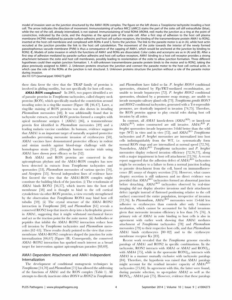
Figure 1. Molecular models of apicomplexan gliding and invasion. A. The parasite motor (glideosome) is located in the space between theparasite plasma membrane (PPM) and the inner membrane complex (IMC) apposed to the microtubules. Gliding motility is mediated by the bindingof the ectodomain of transmembrane TRAP-family proteins to a solid substrate, while the cytoplasmic tail of the protein is linked to the parasitemotor. The integrity of the glideosome is maintained by the gliding-associated protein 45 (GAP45), which is anchored to the PPM at one end and tothe IMC, via GAPs 40 and 50, at the other end. The link between the GAPs, and ultimately the IMC, to actin is provided by Myosin A (MyoA) and theMyoA Light Chain 1 (MLC1). The movement of the cell is the consequence of the capping, by myosin-actin activity, of the TRAP-family protein. B. The
PLOS Pathogens | www.plospathogens.org 3 September 2014 | Volume 10 | Issue 9 | e1004273
these data favor the view that the TRAP family of proteins is
involved in gliding motility, but not specifically for host cell entry.
AMA1-RON complexes? In 2005, two papers identified a set
of parasite proteins in Toxoplasma tachyzoites, called rhoptry neck
proteins (RON), which specifically marked the constriction around
invading zoites in a ring-like manner (Figure 1B) [46,47]. Later, a
ring-like staining of RON proteins was also shown in invading
Plasmodium merozoites [48]. It was additionally found that in
tachyzoite extracts, several RON proteins formed a complex with
apical membrane antigen 1 (AMA1) [46], a transmembrane
protein first identified in Plasmodium merozoites [49] and a
leading malaria vaccine candidate. In humans, evidence suggests
that AMA1 is an important target of naturally acquired protective
antibodies preventing merozoite invasion of erythrocytes [50].
AMA1 vaccines have demonstrated protective efficacy in rodent
and simian models against blood-stage challenge with the
homologous strain [51], although human vaccine trials using
AMA1 have shown poor efficacy so far [52].
Both AMA1 and RON proteins are conserved in the
apicomplexan phylum and the AMA1-RON complex has now
been detected in extracts from Toxoplasma tachyzoites and
sporozoites [53], Plasmodium merozoites of various species [54],
and Neospora [55]. Several independent lines of evidence have
first favored the view that the AMA1-RON complex might
constitute the building block of the junction. (i) The ectodomain of
AMA1 binds RON2 [56,57], which inserts into the host cell
membrane [58] and is thought to bind to the cell cortical
cytoskeleton via other RON proteins, a view recently strengthened
by the observation that Toxoplasma RON4 may bind host cell
tubulin [59]. (ii) The crystal structure of the AMA1–RON2
interaction in Toxoplasma [60] and Plasmodium [61] reveals a
conserved RON2 loop that inserts deep into a hydrophobic groove
in AMA1, suggesting that it might withstand mechanical forces
and act as the traction point for the zoite motor. (iii) Antibodies or
peptides that inhibit the AMA1–RON2 interaction reduce host
cell invasion by Toxoplasma tachyzoites and Plasmodium mero-
zoites [62–65]. These results clearly pointed to the view that cross-
membrane AMA1-RON2 complexes shaped the junction for zoite
internalization [66,67] (Figure 1B). Consequently, the conserved
AMA1–RON2 interaction has sparked much interest as a broad
target for intervention against apicomplexan parasites [68,69].
AMA1-Dependent Attachment and AMA1-IndependentInternalization
The development of conditional mutagenesis techniques in
Toxoplasma [14,70] and Plasmodium [71] has allowed the addressing
of the functions of AMA1 and the RON complex (Table 1). All
attempts to directly inactivate either RON4 or RON2 in Toxoplasma
and Plasmodium have failed so far. P. berghei RON4 conditional
sporozoites, obtained by Flp/FRT-mediated recombination, are
unable to invade hepatocytes [72]. P. berghei RON2 conditional
sporozoites, obtained by a promoter swap strategy, are unable to
invade mosquito salivary gland cells [73]. Toxoplasma gondii RON5and RON2 conditional tachyzoites, generated with a Tet-repressible
promoter, are drastically impaired in invasion [74,75]. Therefore,
the RON proteins appear to play crucial roles during host cell
invasion by all zoites.
In contrast, all AMA1 knock-down (AMA1KD) or knock-out
(AMA1KO) zoites constructed are still invasive. AMA1KD P.berghei sporozoites invade hepatocytes 3-fold better than the wild
type (WT) in vitro and in vivo [72], and AMA1KO Toxoplasmatachyzoites and P. berghei merozoites are internalized into host
cells indistinguishably from the WT, i.e. systematically form
normal RON rings and are internalized at normal speed [72,76].
Nonetheless, AMA1KO Toxoplasma tachyzoites and P. bergheimerozoites display reduced invasion efficiency [72,76,77], along
with a major impairment in host cell attachment [72,76]. A recent
report suggested that the adhesion defect of AMA1KO tachyzoites
might be secondary to a failure to form a normal junction leading
to parasite detachment from the cell, based on immunofluores-
cence (IF) assays of rhoptry secretion [75]. However, what causes
rhoptry secretion is still unknown and no direct evidence was
provided that AMA1KO tachyzoites formed an abnormal junction
before detaching. AMA1KO tachyzoites observed by real-time
imaging did not display abortive invasions and their attachment
defect (upright instead of flattened positioning relative to the cell
surface) concerned the entire population, irrespective of invasion
[72,76]. In Plasmodium, AMA1KO merozoites were 15-fold less
adhesive to erythrocytes than controls after only 3 minutes
incubation, which cannot be accounted for by failed invasions
given that merozoite invasion efficiency is less than 5% [76]. A
primary role of AMA1 in zoite binding to host cells is also in
agreement with earlier work showing that AMA1 mediates
attachment of Toxoplasma tachyzoites [78] and Plasmodiummerozoites [79] to their respective host cells, and that PlasmodiumAMA1 binds erythrocytes [80–82] and to the erythrocyte
membrane receptor Kx [83].
Recent work revealed that the Toxoplasma genome encodes
paralogs of AMA1 and RON2 in specific combinations. In the
tachyzoite, RON2 interacts with AMA1 or AMA2 and RON2L1
with AMA4 [75], while in the sporozoite RON2L2 interacts with
AMA3 in a manner mutually exclusive with tachyzoite paralogs
[84]. Therefore, the hypothesis was raised that AMA1 paralogs
might account for the residual invasive capacity of AMA1KO
tachyzoites [75,84]. In agreement with this, the latter were found,
during parasite selection, to up-regulate AMA2 as well as the
RON2L1–AMA4 pair [75]. However, evidence that these paralogs
model of invasion seen as the junction structured by the AMA1-RON complex. The figure on the left shows a Toxoplasma tachyzoite invading a hostcell. The arrow indicates the direction of movement. Immunostaining of surface MIC2 (sMIC2) stains the part of the zoite cell still extracellular (blue),while the rest of the cell, already internalized, is not stained. Immunostaining of total RON4 (tRON4, red) marks the junction as a ring at the point ofconstriction, indicated by the circle, and the rhoptries at the apical pole of the zoite cell. After a first step of adhesion to the host cell plasmamembrane (HCPM) mediated by parasite surface adhesins and host cell surface receptors, the binding of the transmembrane protein AMA1 to RON2,inserted at the host cell membrane and complexed with RONs 4 and 5, forms the junction. The link to the parasite motor is as in (A), while host actinrecruited at the junction provides the link to the host cell cytoskeleton. The movement of the zoite towards the interior of the newly formedparasitophorous vacuole membrane (PVM) is thus a consequence of the capping of AMA1, which would be anchored at the junction by binding toRON2. C. Models of zoite invasion in which the functions of AMA1 and RONs are dissociated. Color codes and acronyms are as in (A) and (B). After afirst step of adhesion mediated by parasite surface adhesins and host cell surface receptors, AMA1 binding to a host cell receptor provides a strongattachment between the zoite and host cell membranes, possibly leading to reorientation of the zoite to allow junction formation. Three differenthypotheses could then explain junction formation: 1. A still-unknown transmembrane parasite protein binds to the motor and to RON2, taking theplace previously assigned to AMA1. 2. Unknown proteins structure the junction and connect the parasite motor to the host cell cortical actin, inwhich case the role of the RONs at the junction is not structural. 3. Unknown proteins structure the junction without a role of the parasite motorduring invasion.doi:10.1371/journal.ppat.1004273.g001
PLOS Pathogens | www.plospathogens.org 4 September 2014 | Volume 10 | Issue 9 | e1004273

Table 1. Mutants of interest in studies on host cell invasion by apicomplexans.
Gene zoite System Phenotype Ref.
RON4 P. berghei sporozoites Flp/FRT N Knock-down (KD) sporozoites do not invade hepatic cells in vitro [72]
RON2 P. berghei sporozoites Promoter swap N RON2-negative sporozoites do not invade the mosquito salivary glands [73]
RON5 T. gondii tachyzoites Tet repression N KD tachyzoites are unable to invade host cells [74]
N Loss of RON5 results in complete degradation of RON2 and mistargeting of RON4
RON2 T. gondii tachyzoites Tet repression N KD tachyzoites display a severe block in host cell invasion [75]
N RON4 and RON5 are not properly localized within parasites
AMA1 T. gondii tachyzoites Tet repression N KD tachyzoites do not progress from initial to intimate binding with the host cellmembrane.
[78]
N Rhoptry secretion is impaired and invasion is reduced to ,15% that of WT.
AMA1 P. berghei sporozoites andhepatic merozoites
Flp/FRT N KD sporozoites normally invade hepatic cells in vitro and in vivoN KD hepatic merozoites cannot induce a blood infection in vivo
[72]
AMA1 T. gondii tachyzoites Tet repression N KD tachyzoites bind to host cells differently from WT [72]
N Internalization appears normal
AMA1 P. berghei merozoites andsporozoites
Flp/FRT and directknock-out
N Knock-out (KO) sporozoites normally invade hepatic cells in vitroN KO and KD merozoites are impaired in binding to erythrocytes. The growth
rate of KO blood stages in vivo is ,35% that of WT
[76]
AMA1 T. gondii tachyzoites diCre/loxP N KO tachyzoites bind to host cells with a distinct positioning relative to the host cell [76]
N Internalization appears normal but with decreased frequency (30%–40% that of WT)
AMA1 P. falciparum merozoites diCre/loxP N Populations with 80% excised merozoites (with residual AMA1 due to late excision)show 37% reduction in invasion capacity
[77]
AMA1 T. gondii tachyzoites Direct knock-out N KO tachyzoites display ‘abortive invasions’ [75]
N Residual invasion of AMA1KO tachyzoites is due to compensation by AMA1 paralogs
MIC2 T. gondii tachyzoites Tet repression N KD tachyzoites are impaired in gliding. Attachment to host cells is reduced to 18%that of the parental strain
[43]
N Invasion is reduced to 22% that of the parental strain.
MIC2 T. gondii tachyzoites diCre/loxP N KO tachyzoites are clonally viable [70]
N Gliding motility and growth in cell monolayer are impaired
TRAP P. berghei sporozoites Direct knock-out N KO sporozoites are impaired in gliding motility. Invasion of mosquito salivary glandsis impaired
[13]
N Infection of mouse liver is compromised
TRAP P. berghei sporozoites Direct knock-out N KO sporozoites glide for one body length in both directions, while remainingattached to the substrate by one adhesion site
[33]
N The turnover of adhesion sites is impaired. KO parasites cannot detach once attached
TRAP tail P. berghei sporozoites Subtle mutagenesis N Deletion of the entire cytoplasmic tail of TRAP renders sporozoites non-motileN Deletion of the distal third of the TRAP cytoplasmic tail causes a pendulum gliding
[100]
TREP P. berghei sporozoites Direct knock-out N KO sporozoites are impaired in gliding motility [71]
N Invasion of mosquito salivary glands is impaired
Aldolase T. gondii tachyzoites Tet repression N KD tachyzoites are impaired in gliding motility and invasion [27]
N KD complemented with MIC2-binding impaired versions of aldolase display normalgliding
Aldolase T. gondii tachyzoites diCre/loxP N KO tachyzoites can be cloned and propagate when grown in vitro in glucose-freemedium
[26]
Actin T. gondii tachyzoites diCre/loxP N Gliding and invasion are 10% that of WT [32]
N KO tachyzoites invade through a junction and multiply, but are not clonally viabledue to abnormal segregation of apicoplasts
MyoA T. gondii tachyzoites Tet repression N KD tachyzoites are impaired in gliding motility [14]
N Host cell invasion is reduced to ,20% that of WT
MyoA T. gondii tachyzoites diCre/loxP N KO tachyzoites are clonally viable. Gliding and egress are impaired [32]
N Invasion is reduced to 16% that of WT. Internalization is through a RON4-stainedjunction
GAP45 T. gondii tachyzoites Tet repression N Other motor components redistribute to the cytosol and the glideosome ‘‘collapses’’ [31]
N KD tachyzoites are impaired in gliding and egress. Invasion efficiency is 25% thatof the parental strain
PLOS Pathogens | www.plospathogens.org 5 September 2014 | Volume 10 | Issue 9 | e1004273

act at and/or structure the junction is lacking, and the weaker
affinity of RON2 for AMA2 compared with AMA1 [75] is at odds
with the fully efficient internalization of AMA1KO [76] (suppos-
edly mediated by AMA2). In Plasmodium, the compensation
theory appears particularly unlikely. The AMA1KD Flp/FRT
sporozoites undergo AMA1 excision after parasite selection and
yet are 100% invasive [76]. Additionally, Plasmodium expresses no
AMA1 or RON2 paralog. The protein most closely related to
AMA1 is the trans-membrane protein MAEBL, with which it
shares the presence of a cysteine-rich domain but differs by an
unrelated cytoplasmic tail and the absence of RON2-binding
ability. MAEBL was shown by gene targeting in both P. berghei[85] and P. falciparum [86] sporozoites to function as a stage-
specific adhesion; it mediates oocyst sporozoite binding to the
mosquito salivary glands, but not internalization into hepatocytes.
This further suggests that AMA1, its paralogs in Toxoplasma, and
MAEBL in Plasmodium form a family of stage-specific, host cell–
binding proteins.
Current HypothesesIf AMA1 primarily mediates zoite intimate binding to host cell
surfaces, irrespective of RON2 interaction and junction assembly,
what could be the role of the conserved and therefore important
AMA1–RON2 interaction? AMA1 might still bind to RON2,
possibly to help further stabilize the zoite prior to internalization,
although direct evidence for such a step is still lacking.
Alternatively, AMA1–RON2 interactions might serve to process
AMA1 at the junction during internalization of the AMA1-
covered zoite. For example, interaction with RON2 might serve to
disengage the AMA1–host cell receptor interaction and help the
zoite slide free inside the PV, separated from the vacuole
membrane. In agreement with this, RON2 binding induces
conformational changes in AMA1 [60], which might impact
AMA1 processing by the substilisin-like protease SUB2 [87] or
intramembrane rhomboids [88]. Likewise, RON2L2 binding alters
AMA3, including allosterically in its membrane-proximal domain
[84]. Such AMA processing function of the AMA–RON2
interactions would be dispensable for internalization and yet block
invasion if perturbed, reconciling the inhibition and genetic data.
The contribution of the apparently essential RON proteins is
also unclear. They might be structural components of the junction,
by linking it to the host cell cytoskeleton (Figure 1C, hypothesis 1),
or might not be part of the force-transducing link (Figure 1C,
hypothesis 2). Perhaps favoring the latter, the RON proteins are
present in apicomplexans that are not known to form a junction,
like Theileria [89], which raises the possibility that another zoite–
host cell interaction might structure the junction, possibly
involving host cell receptor(s). In any hypothesis, the junction
constitutes a traction point for zoite internalization into the host
cell.
Motor-Independent EntryThe Toxoplasma tachyzoite is ideal to study zoite invasion, not
just due to the frequency of observable invasion events but also the
genetic tractability of the parasite. The use of a transcriptional
regulation system based on artificial Tet-transactivators (TATi)
allowed the generation of knock-down mutants and the functional
dissection of individual components of the motor (Table 1). As
already said, knocking down MIC2 [43] or MyoA [14] does not
result in a complete block in host cell invasion. Even knocking
down GAP45, while leading to the detachment of the IMC from
the plasma membrane (PM) and the release of the motor complex
in the cytosol, does not abolish host cell entry [31]. In contrast, as
mentioned above, a knock-down for MIC8 does not affect gliding
motility but completely blocks host cell invasion due to an inability
to form a junction [41]. These partial phenotypes were typically
explained by the leakiness of the Tet-inducible system, but were
also a hint that the motor might not be essential for host cell
invasion.
The current adaptation of a conditional recombination system
based on dimerizable Cre has allowed the construction of a series
of tachyzoite mutants completely lacking individual components of
the motor complex, including MyoA, GAP45, MLC1, and Act1
[32,70]. All of these mutants are affected in invasion efficiency but
retain some invasive capacity (Table 1). Strikingly, tachyzoites
devoid of MyoA, MLC1, or Act1, which is a single copy gene in
Toxoplasma, can invade host cells through a junction [32],
demonstrating that at least junction formation is independent of
connection to the motor. Moreover, GAP45KO tachyzoites, in
which the IMC detaches from the parasite membrane and MyoA
and MLC1 become cytosolic, remain motile and also invade [32].
These genetic data suggest that tachyzoites can move and enter
host cells without a functional motor. However, whether this
motor-independent entry pathway is the normal pathway used by
the WT, or an alternative pathway discernable only when the
motor is not functional, remains to be seen.
How could tachyzoites with a deficient actin-myosin motor
invade host cells? Until recently, actin polymerization and actin-
myosin contraction were thought to underlie force generation
during movement. However, this view is currently being
challenged by new models, in which hydrodynamic forces
generate changes in cell shape during motility [90]. Indeed, there
is mounting evidence that osmotic pressure and hydrodynamic
fluids are critical for motility of amoeboid cells, while the actin-
myosin system is critical for the formation and release of
attachment sites and associated traction forces [91]. A recent
Table 1. Cont.
Gene zoite System Phenotype Ref.
GAP45 T. gondii tachyzoites diCre/loxP N KO tachyzoites grow up to 14 days in culture. The IMC looses contact to the PMand MyoA and MLC1 become cytosolic
[32]
N KO tachyzoites can glide. Egress is impaired. Internalization is through aN RON4-stained junction and is reduced to 6% that of WT
MLC1 T. gondii tachyzoites diCre/loxP N KO tachyzoites can be grown up to 14 days in culture. MyoA is mislocalized [32]
N Gliding and egress are impaired. Invasion is reduced to 28% that of WT.Internalization is through a RON4-stained junction
doi:10.1371/journal.ppat.1004273.t001
PLOS Pathogens | www.plospathogens.org 6 September 2014 | Volume 10 | Issue 9 | e1004273

report demonstrates the poroelastic nature of the cytosol [92],
where force can be generated by differences in hydrodynamic
pressure that can be higher in one part of a cell than another,
leading to tension. This pressure can be generated by actin-myosin
activity or by the localized activation of osmogenic ion transporters
in the plasma membrane [90]. In agreement with such a model,
Na+/H+ antiporters have been implicated in invasion and egress of
host cells by Toxoplasma tachyzoites [93–95], and monovalent ion
concentrations have been involved in gliding motility and host cell
invasion efficiencies in Toxoplasma [96,97]. Based on this, a
gelsolation model for gliding motility and zoite internalization, in
which the acto-myosin system of the parasite is required as a clutch
for force transmission but not for the generation of the force itself,
has recently been proposed [32].
Another possibility that cannot be excluded is that the force for
parasite internalization might originate from the host cell.
Theileria sporozoites and merozoites, which lack an IMC and
subpellicular microtubules and are not motile, invade host cells in
any orientation, without a junction, and by a mechanism of
circumferential zippering of parasite and host cell membranes
[89]. Others, like Cryptosporidium sporozoites, are motile but rest
on the host cell surface and induce the formation of host cell
membrane folds that progressively encapsulate the ‘‘epicellular
parasite’’ inside the PV [98]. Interestingly, these membrane
protrusions recruit a host cell Na+/glucose cotransporter and
aquaporin 1, which generate localized water influx and are
required for parasite invasion [99].
If zoite internalization is powered by the host cell or by
hydrodynamic forces, then the junction would no longer be
connected to the parasite motor. In this case, the junction might
serve as a membrane ‘‘seal,’’ possibly regulating protein processing
upon entry into the PV, facilitating membrane dynamics, fluidity
and curvature, and/or ensuring correct formation of the PV
(Figure 1C, hypothesis 3).
Conclusions
New mutagenesis data question the current view that apicom-
plexan zoites invade host cells by a unique pathway involving their
motor and AMA1–RON2 interactions as traction points at the
junction. AMA1 appears not to be involved in junction function
and the RON proteins are the only parasite proteins known to
functionally associate with the junction. Whether the RON
complex transmits force at the junction remains uncertain, though
the evidence of a link between the RON proteins and the host cell
cytoskeleton points to this direction. The conservation of the RON
proteins in apicomplexans and of their essential role in invasion
suggests a conserved molecular kit for junction formation. Still,
there is no definitive evidence for such a conserved apicomplexan
junction core, and at least part of its structure might be stage-
specific. Whether the zoite provides all pieces of the junction, or
whether the host cell also provides receptors, possibly located in
specific microdomains, is also unclear.
Moreover, it now appears that the force required for gliding
motility and host cell entry might, at least in Toxoplasma, be
generated in a motor-independent manner. Whether this holds
true for other apicomplexan genera remains to be seen. In any
event, apicomplexan invasion of host cells appears more complex
than previously thought. Dissecting the process and its possible
versatility will require establishing novel experimental approaches,
including biophysical, and investigating unconventional force-
generation means in zoites, as well as in host cells, that might
facilitate, or even replace, the activity of the parasite motor.
References
1. Aikawa M, Miller LH, Johnson J, Rabbege J (1978) Erythrocyte entry by
malarial parasites. A moving junction between erythrocyte and parasite. J Cell
Biol 77: 72–82.
2. King CA (1988) Cell motility of sporozoan protozoa. Parasitol Today 4: 315–
319.
3. Russell DG, Sinden RE (1981) The role of the cytoskeleton in the motility of
coccidian sporozoites. J Cell Sci 50: 345–359.
4. Russell DG (1983) Host cell invasion by Apicomplexa: an expression of the
parasite’s contractile system? Parasitology 87 (Pt 2): 199–209.
5. Schwartzman JD, Pfefferkorn ER (1983) Immunofluorescent localization of
myosin at the anterior pole of the coccidian, Toxoplasma gondii. J Protozool 30:
657–661.
6. Pinder JC, Fowler RE, Dluzewski AR, Bannister LH, Lavin FM, et al. (1998)
Actomyosin motor in the merozoite of the malaria parasite, Plasmodium
falciparum: implications for red cell invasion. J Cell Sci 111 (Pt 13): 1831–
1839.
7. Dobrowolski JM, Sibley LD (1996) Toxoplasma invasion of mammalian cells is
powered by the actin cytoskeleton of the parasite. Cell 84: 933–939.
8. Morisaki JH, Heuser JE, Sibley LD (1995) Invasion of Toxoplasma gondii occurs
by active penetration of the host cell. J Cell Sci 108 (Pt 6): 2457–2464.
9. Gonzalez V, Combe A, David V, Malmquist NA, Delorme V, et al. (2009) Host
cell entry by apicomplexa parasites requires actin polymerization in the host cell.
Cell Host Microbe 5: 259–272.
10. Delorme-Walker V, Abrivard M, Lagal V, Anderson K, Perazzi A, et al. (2012)
Toxofilin upregulates the host cortical actin cytoskeleton dynamics, facilitating
Toxoplasma invasion. J Cell Sci 125: 4333–4342.
11. Delorme V, Cayla X, Faure G, Garcia A, Tardieux I (2003) Actin dynamics is
controlled by a casein kinase II and phosphatase 2C interplay on Toxoplasma
gondii Toxofilin. Mol Biol Cell 14: 1900–1912.
12. Morrissette NS, Murray JM, Roos DS (1997) Subpellicular microtubules
associate with an intramembranous particle lattice in the protozoan parasite
Toxoplasma gondii. J Cell Sci 110 (Pt 1): 35–42.
13. Sultan AA, Thathy V, Frevert U, Robson KJ, Crisanti A, et al. (1997) TRAP is
necessary for gliding motility and infectivity of Plasmodium sporozoites. Cell 90:
511–522.
14. Meissner M, Schluter D, Soldati D (2002) Role of Toxoplasma gondii myosin
A in powering parasite gliding and host cell invasion. Science 298: 837–
840.
15. Jewett TJ, Sibley LD (2003) Aldolase forms a bridge between cell surfaceadhesins and the actin cytoskeleton in apicomplexan parasites. Mol Cell 11:
885–894.
16. Bergman LW, Kaiser K, Fujioka H, Coppens I, Daly TM, et al. (2003) Myosin
A tail domain interacting protein (MTIP) localizes to the inner membranecomplex of Plasmodium sporozoites. J Cell Sci 116: 39–49.
17. Schuler H, Matuschewski K (2006) Regulation of apicomplexan microfilamentdynamics by a minimal set of actin-binding proteins. Traffic 7: 1433–1439.
18. Skillman KM, Diraviyam K, Khan A, Tang K, Sept D, et al. (2011)Evolutionarily divergent, unstable filamentous actin is essential for gliding
motility in apicomplexan parasites. PLoS Pathog 7: e1002280.
19. Mehta S, Sibley LD (2011) Actin depolymerizing factor controls actin turnover
and gliding motility in Toxoplasma gondii. Mol Biol Cell 22: 1290–1299.
20. Baum J, Tonkin CJ, Paul AS, Rug M, Smith BJ, et al. (2008) A malaria parasiteformin regulates actin polymerization and localizes to the parasite-erythrocyte
moving junction during invasion. Cell Host Microbe 3: 188–198.
21. Daher W, Plattner F, Carlier MF, Soldati-Favre D (2010) Concerted action of
two formins in gliding motility and host cell invasion by Toxoplasma gondii.PLoS Pathog 6: e1001132.
22. Angrisano F, Riglar DT, Sturm A, Volz JC, Delves MJ, et al. (2012) Spatiallocalisation of actin filaments across developmental stages of the malaria
parasite. PLoS ONE 7: e32188.
23. Mehta S, Sibley LD (2010) Toxoplasma gondii actin depolymerizing factor acts
primarily to sequester G-actin. J Biol Chem 285: 6835–6847.
24. Skillman KM, Daher W, Ma CI, Soldati-Favre D, Sibley LD (2012) Toxoplasmagondii profilin acts primarily to sequester G-actin while formins efficiently
nucleate actin filament formation in vitro. Biochemistry 51: 2486–2495.
25. Plattner F, Yarovinsky F, Romero S, Didry D, Carlier MF, et al. (2008)
Toxoplasma profilin is essential for host cell invasion and TLR11-dependentinduction of an interleukin-12 response. Cell Host Microbe 3: 77–87.
26. Shen B, Sibley LD (2014) Toxoplasma aldolase is required for metabolismbut dispensable for host-cell invasion. Proc Natl Acad Sci U S A 111: 3567–
3572.
27. Starnes GL, Coincon M, Sygusch J, Sibley LD (2009) Aldolase is essential for
energy production and bridging adhesin-actin cytoskeletal interactions duringparasite invasion of host cells. Cell Host Microbe 5: 353–364.
28. Pomel S, Luk FC, Beckers CJ (2008) Host cell egress and invasion induce
marked relocations of glycolytic enzymes in Toxoplasma gondii tachyzoites.
PLoS Pathog 4: e1000188.
PLOS Pathogens | www.plospathogens.org 7 September 2014 | Volume 10 | Issue 9 | e1004273

29. Gaskins E, Gilk S, DeVore N, Mann T, Ward G, et al. (2004) Identification of
the membrane receptor of a class XIV myosin in Toxoplasma gondii. J Cell Biol
165: 383–393.
30. Gilk SD, Gaskins E, Ward GE, Beckers CJ (2009) GAP45 phosphorylation
controls assembly of the Toxoplasma myosin XIV complex. Eukaryot Cell 8:
190–196.
31. Frenal K, Polonais V, Marq JB, Stratmann R, Limenitakis J, et al. (2010)
Functional dissection of the apicomplexan glideosome molecular architecture.
Cell Host Microbe 8: 343–357.
32. Egarter S, Andenmatten N, Jackson AJ, Whitelaw JA, Pall G, et al. (2014) The
Toxoplasma Acto-MyoA Motor Complex Is Important but Not Essential for
Gliding Motility and Host Cell Invasion. PLoS ONE 9: e91819.
33. Munter S, Sabass B, Selhuber-Unkel C, Kudryashev M, Hegge S, et al. (2009)
Plasmodium sporozoite motility is modulated by the turnover of discrete
adhesion sites. Cell Host Microbe 6: 551–562.
34. Hellmann JK, Perschmann N, Spatz JP, Frischknecht F (2013) Tunable
substrates unveil chemical complementation of a genetic cell migration defect.
Adv Healthc Mater 2: 1162–1169.
35. Gilberger TW, Thompson JK, Reed MB, Good RT, Cowman AF (2003) The
cytoplasmic domain of the Plasmodium falciparum ligand EBA-175 is essential
for invasion but not protein trafficking. J Cell Biol 162: 317–327.
36. Singh AP, Ozwara H, Kocken CH, Puri SK, Thomas AW, et al. (2005)
Targeted deletion of Plasmodium knowlesi Duffy binding protein confirms its
role in junction formation during invasion. Mol Microbiol 55: 1925–1934.
37. Gunalan K, Gao X, Yap SS, Huang X, Preiser PR (2013) The role of the
reticulocyte-binding-like protein homologues of Plasmodium in erythrocyte
sensing and invasion. Cell Microbiol 15: 35–44.
38. Gunalan K, Gao X, Liew KJ, Preiser PR (2011) Differences in erythrocyte
receptor specificity of different parts of the Plasmodium falciparum reticulocyte
binding protein homologue 2a. Infect Immun 79: 3421–3430.
39. Baum J, Chen L, Healer J, Lopaticki S, Boyle M, et al. (2009) Reticulocyte-
binding protein homologue 5 - an essential adhesin involved in invasion of
human erythrocytes by Plasmodium falciparum. Int J Parasitol 39: 371–380.
40. Ito D, Hasegawa T, Miura K, Yamasaki T, Arumugam TU, et al. (2013)
RALP1 is a rhoptry neck erythrocyte-binding protein of Plasmodium falciparum
merozoites and a potential blood-stage vaccine candidate antigen. Infect Immun
81: 4290–4298.
41. Kessler H, Herm-Gotz A, Hegge S, Rauch M, Soldati-Favre D, et al. (2008)
Microneme protein 8—a new essential invasion factor in Toxoplasma gondii.
J Cell Sci 121: 947–956.
42. Morahan BJ, Wang L, Coppel RL (2009) No TRAP, no invasion. Trends in
Parasitology 25: 77–84.
43. Huynh MH, Carruthers VB (2006) Toxoplasma MIC2 is a major determinant of
invasion and virulence. PLoS Pathog 2: e84.
44. Song G, Koksal AC, Lu C, Springer TA (2012) Shape change in the receptor for
gliding motility in Plasmodium sporozoites. Proc Natl Acad Sci U S A 109:
21420–21425.
45. Ramakrishnan C, Dessens JT, Armson R, Pinto SB, Talman AM, et al. (2011)
Vital functions of the malarial ookinete protein, CTRP, reside in the A domains.
Int J Parasitol 41: 1029–1039.
46. Alexander DL, Mital J, Ward GE, Bradley P, Boothroyd JC (2005) Identification
of the moving junction complex of Toxoplasma gondii: a collaboration between
distinct secretory organelles. PLoS Pathog 1: e17.
47. Lebrun M, Michelin A, El Hajj H, Poncet J, Bradley PJ, et al. (2005) The
rhoptry neck protein RON4 re-localizes at the moving junction during
Toxoplasma gondii invasion. Cell Microbiol 7: 1823–1833.
48. Riglar DT, Richard D, Wilson DW, Boyle MJ, Dekiwadia C, et al. (2011) Super-
resolution dissection of coordinated events during malaria parasite invasion of
the human erythrocyte. Cell Host Microbe 9: 9–20.
49. Deans JA, Alderson T, Thomas AW, Mitchell GH, Lennox ES, et al. (1982) Rat
monoclonal antibodies which inhibit the in vitro multiplication of Plasmodium
knowlesi. Clin Exp Immunol 49: 297–309.
50. Thomas AW, Trape JF, Rogier C, Goncalves A, Rosario VE, et al. (1994) High
prevalence of natural antibodies against Plasmodium falciparum 83-kilodalton
apical membrane antigen (PF83/AMA-1) as detected by capture-enzyme-linked
immunosorbent assay using full-length baculovirus recombinant PF83/AMA-1.
Am J Trop Med Hyg 51: 730–740.
51. Remarque EJ, Faber BW, Kocken CH, Thomas AW (2008) Apical membrane
antigen 1: a malaria vaccine candidate in review. Trends Parasitol 24: 74–84.
52. Laurens MB, Thera MA, Coulibaly D, Ouattara A, Kone AK, et al. (2013)
Extended safety, immunogenicity and efficacy of a blood-stage malaria vaccine
in malian children: 24-month follow-up of a randomized, double-blinded phase
2 trial. PLoS ONE 8: e79323.
53. Poukchanski A, Fritz HM, Tonkin ML, Treeck M, Boulanger MJ, et al. (2013)
Toxoplasma gondii sporozoites invade host cells using two novel paralogues of
RON2 and AMA1. PLoS ONE 8: e70637.
54. Narum DL, Nguyen V, Zhang Y, Glen J, Shimp RL, et al. (2008) Identification
and characterization of the Plasmodium yoelii PyP140/RON4 protein, an
orthologue of Toxoplasma gondii RON4, whose cysteine-rich domain does not
protect against lethal parasite challenge infection. Infect Immun 76: 4876–4882.
55. Straub KW, Cheng SJ, Sohn CS, Bradley PJ (2009) Novel components of the
Apicomplexan moving junction reveal conserved and coccidia-restricted
elements. Cell Microbiol 11: 590–603.
56. Lamarque M, Besteiro S, Papoin J, Roques M, Vulliez-Le Normand B, et al.(2011) The RON2-AMA1 interaction is a critical step in moving junction-
dependent invasion by apicomplexan parasites. PLoS Pathog 7: e1001276.
57. Tyler JS, Boothroyd JC (2011) The C-terminus of Toxoplasma RON2 providesthe crucial link between AMA1 and the host-associated invasion complex. PLoS
Pathog 7: e1001282.
58. Besteiro S, Michelin A, Poncet J, Dubremetz JF, Lebrun M (2009) Export of aToxoplasma gondii rhoptry neck protein complex at the host cell membrane to
form the moving junction during invasion. PLoS Pathog 5: e1000309.
59. Takemae H, Sugi T, Kobayashi K, Gong H, Ishiwa A, et al. (2013)Characterization of the interaction between Toxoplasma gondii rhoptry neck
protein 4 and host cellular beta-tubulin. Sci Rep 3: 3199.
60. Tonkin ML, Roques M, Lamarque MH, Pugniere M, Douguet D, et al. (2011)Host cell invasion by apicomplexan parasites: insights from the co-structure of
AMA1 with a RON2 peptide. Science 333: 463–467.
61. Vulliez-Le Normand B, Tonkin ML, Lamarque MH, Langer S, Hoos S, et al.(2012) Structural and functional insights into the malaria parasite moving
junction complex. PLoS Pathog 8: e1002755.
62. Collins CR, Withers-Martinez C, Hackett F, Blackman MJ (2009) An inhibitoryantibody blocks interactions between components of the malarial invasion
machinery. PLoS Pathog 5: e1000273.
63. Richard D, MacRaild CA, Riglar DT, Chan JA, Foley M, et al. (2010)
Interaction between Plasmodium falciparum apical membrane antigen 1 and the
rhoptry neck protein complex defines a key step in the erythrocyte invasionprocess of malaria parasites. J Biol Chem 285: 14815–14822.
64. Srinivasan P, Beatty WL, Diouf A, Herrera R, Ambroggio X, et al. (2011)
Binding of Plasmodium merozoite proteins RON2 and AMA1 triggerscommitment to invasion. Proc Natl Acad Sci U S A 108: 13275–13280.
65. Srinivasan P, Yasgar A, Luci DK, Beatty WL, Hu X, et al. (2013) Disrupting
malaria parasite AMA1-RON2 interaction with a small molecule preventserythrocyte invasion. Nat Commun 4: 2261.
66. Baum J, Cowman AF (2011) Biochemistry. Revealing a parasite’s invasive trick.Science 333: 410–411.
67. Shen B, Sibley LD (2012) The moving junction, a key portal to host cell invasion
by apicomplexan parasites. Curr Opin Microbiol 15: 449–455.
68. Macraild CA, Anders RF, Foley M, Norton RS (2011) Apical membrane antigen1 as an anti-malarial drug target. Curr Top Med Chem 11: 2039–2047.
69. Miller LH, Ackerman HC, Su XZ, Wellems TE (2013) Malaria biology and
disease pathogenesis: insights for new treatments. Nat Med 19: 156–167.
70. Andenmatten N, Egarter S, Jackson AJ, Jullien N, Herman JP, et al. (2013)
Conditional genome engineering in Toxoplasma gondii uncovers alternativeinvasion mechanisms. Nat Methods 10: 125–127.
71. Combe A, Giovannini D, Carvalho TG, Spath S, Boisson B, et al. (2009) Clonal
conditional mutagenesis in malaria parasites. Cell Host Microbe 5: 386–396.
72. Giovannini D, Spath S, Lacroix C, Perazzi A, Bargieri D, et al. (2011)Independent roles of apical membrane antigen 1 and rhoptry neck proteins
during host cell invasion by apicomplexa. Cell Host Microbe 10: 591–602.
73. Murata E, Tokunaga N, Tachibana M, Tsuboi T, Torii M, et al. (2012) Theinvestigation of the mechanism how malaria sporozoites invade salivary glands.
Molecular Approaches to Malaria Meeting 2012. Abstract Book. Lorne,Australia.
74. Beck JR, Chen AL, kim EW, Bradley PJ (2014) RON5 is critical for organization
and function of the Toxoplasma moving junction complex. PLoS Pathog 10:e1004025.
75. Lamarque MH, Roques M, Kong-Hap M, Tonkin ML, Rugarabamu G, et al
(2014) Plasticity and redundancy among AMA-RON pairs ensure host cell entryof Toxoplasma parasites. Nat Commun 5: 4098.
76. Bargieri DY, Andenmatten N, Lagal V, Thiberge S, Whitelaw JA, et al. (2013)
Apical membrane antigen 1 mediates apicomplexan parasite attachment but isdispensable for host cell invasion. Nat Commun 4: 2552.
77. Yap A, Azevedo MF, Gilson PR, Weiss GE, O’Neill MT, et al. (2014)
Conditional expression of apical membrane antigen 1 in Plasmodium falciparumshows it is required for erythrocyte invasion by merozoites. Cell Microbiol 16:
642–656.
78. Mital J, Meissner M, Soldati D, Ward GE (2005) Conditional expression of
Toxoplasma gondii apical membrane antigen-1 (TgAMA1) demonstrates that
TgAMA1 plays a critical role in host cell invasion. Mol Biol Cell 16: 4341–4349.
79. Mitchell GH, Thomas AW, Margos G, Dluzewski AR, Bannister LH (2004)
Apical membrane antigen 1, a major malaria vaccine candidate, mediates the
close attachment of invasive merozoites to host red blood cells. Infect Immun 72:154–158.
80. Fraser TS, Kappe SH, Narum DL, VanBuskirk KM, Adams JH (2001)
Erythrocyte-binding activity of Plasmodium yoelii apical membrane antigen-1expressed on the surface of transfected COS-7 cells. Mol Biochem Parasitol 117:
49–59.
81. Urquiza M, Suarez JE, Cardenas C, Lopez R, Puentes A, et al. (2000)
Plasmodium falciparum AMA-1 erythrocyte binding peptides implicate AMA-1
as erythrocyte binding protein. Vaccine 19: 508–513.
82. Valbuena J, Rodriguez L, Vera R, Puentes A, Curtidor H, et al. (2006) Synthetic
peptides from Plasmodium falciparum apical membrane antigen 1 (AMA-1)
specifically interacting with human hepatocytes. Biochimie 88: 1447–1455.
83. Kato K, Mayer DC, Singh S, Reid M, Miller LH (2005) Domain III of
Plasmodium falciparum apical membrane antigen 1 binds to the erythrocyte
membrane protein Kx. Proc Natl Acad Sci U S A 102: 5552–5557.
PLOS Pathogens | www.plospathogens.org 8 September 2014 | Volume 10 | Issue 9 | e1004273

84. Poukchanski A, Fritz HM, Tonkin ML, Treeck M, Boulanger MJ, et al. (2013)
Toxoplasma gondii sporozoites invade host cells using two novel paralogues of
RON2 and AMA1. PLoS ONE 8: e70637.
85. Kariu T, Yuda M, Yano K, Chinzei Y (2002) MAEBL is essential for malarial
sporozoite infection of the mosquito salivary gland. J Exp Med 195: 1317–1323.
86. Saenz FE, Balu B, Smith J, Mendonca SR, Adams JH (2008) The
transmembrane isoform of Plasmodium falciparum MAEBL is essential for
the invasion of Anopheles salivary glands. PLoS ONE 3: e2287.
87. Olivieri A, Collins CR, Hackett F, Withers-Martinez C, Marshall J, et al. (2011)
Juxtamembrane shedding of Plasmodium falciparum AMA1 is sequence
independent and essential, and helps evade invasion-inhibitory antibodies. PLoS
Pathog 7: e1002448.
88. Parussini F, Tang Q, Moin SM, Mital J, Urban S, et al. (2012) Intramembrane
proteolysis of Toxoplasma apical membrane antigen 1 facilitates host-cell invasion
but is dispensable for replication. Proc Natl Acad Sci U S A 109: 7463–7468.
89. Shaw MK (2003) Cell invasion by Theileria sporozoites. Trends Parasitol 19: 2–6.
90. Mitchison TJ, Charras GT, Mahadevan L (2008) Implications of a poroelastic
cytoplasm for the dynamics of animal cell shape. Semin Cell Dev Biol 19: 215–
223.
91. Keren K, Yam PT, Kinkhabwala A, Mogilner A, Theriot JA (2009) Intracellular
fluid flow in rapidly moving cells. Nat Cell Biol 11: 1219–1224.
92. Moeendarbary E, Valon L, Fritzsche M, Harris AR, Moulding DA, et al. (2013)
The cytoplasm of living cells behaves as a poroelastic material. Nat Mater 12:
253–261.
93. Francia ME, Wicher S, Pace DA, Sullivan J, Moreno SN, et al. (2011) A
Toxoplasma gondii protein with homology to intracellular type Na(+)/H(+)exchangers is important for osmoregulation and invasion. Exp Cell Res 317:
1382–1396.
94. Karasov AO, Boothroyd JC, Arrizabalaga G (2005) Identification and disruptionof a rhoptry-localized homologue of sodium hydrogen exchangers in
Toxoplasma gondii. Int J Parasitol 35: 285–291.95. Arrizabalaga G, Ruiz F, Moreno S, Boothroyd JC (2004) Ionophore-resistant
mutant of Toxoplasma gondii reveals involvement of a sodium/hydrogen
exchanger in calcium regulation. J Cell Biol 165: 653–662.96. Endo T, Tokuda H, Yagita K, Koyama T (1987) Effects of extracellular
potassium on acid release and motility initiation in Toxoplasma gondii.J Protozool 34: 291–295.
97. Endo T, Yagita K (1990) Effect of extracellular ions on motility and cell entry inToxoplasma gondii. J Protozool 37: 133–138.
98. Valigurova A, Jirku M, Koudela B, Gelnar M, Modry D, et al. (2008)
Cryptosporidia: epicellular parasites embraced by the host cell membrane.Int J Parasitol 38: 913–922.
99. Chen XM, O’Hara SP, Huang BQ, Splinter PL, Nelson JB, et al. (2005)Localized glucose and water influx facilitates Cryptosporidium parvum cellular
invasion by means of modulation of host-cell membrane protrusion. Proc Natl
Acad Sci U S A 102: 6338–6343.100. Kappe S, Bruderer T, Gantt S, Fujioka H, Nussenzweig V, et al. (1999)
Conservation of a gliding motility and cell invasion machinery in apicomplexanparasites. J Cell Biol 147: 937–943.
PLOS Pathogens | www.plospathogens.org 9 September 2014 | Volume 10 | Issue 9 | e1004273