how do cells form tissues? tissues. using cell junctions
TRANSCRIPT
How do cells form tissues?
Tissues
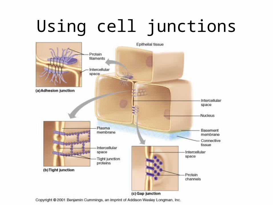
Using cell junctions
Tissues
• Epithelial tissue
• Connective tissue
• Muscle tissue
• Nervous tissue
Epithelial Tissue
• Closely packed cells in continuous sheets connected by many desmosomes & tight junctions
• Apical and Basal surfaces
• Cells rest on Basement membrane
• No blood supply, depend on diffusion from capillaries for food and oxygen
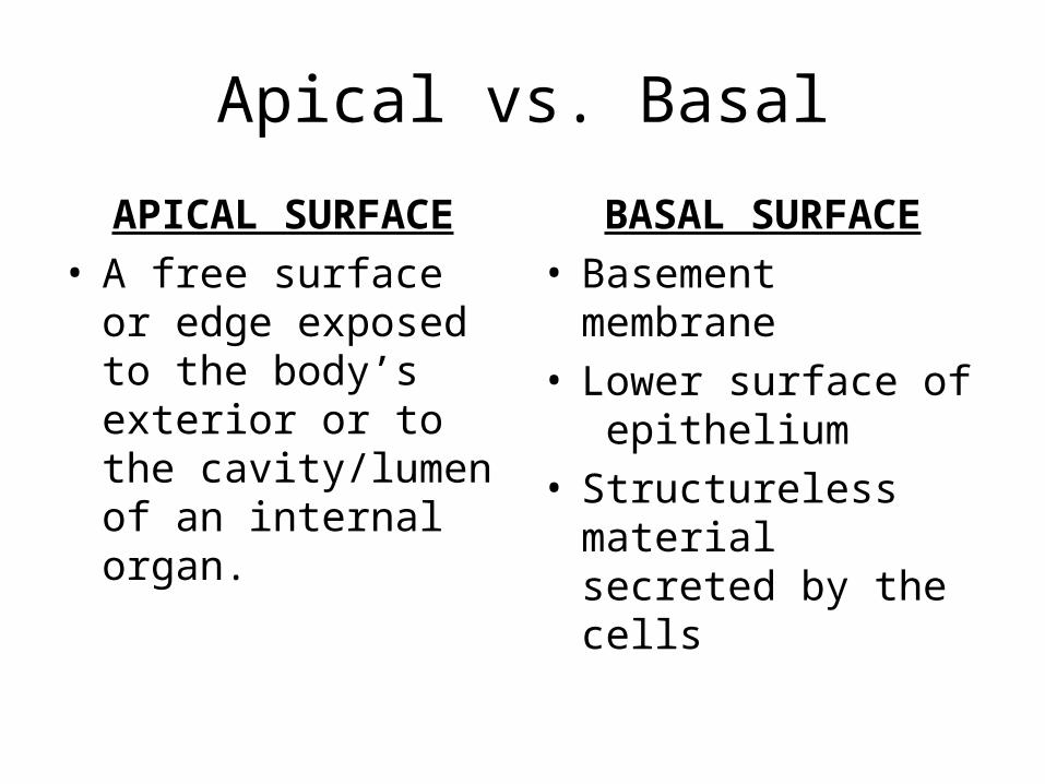
Apical vs. Basal
APICAL SURFACE• A free surface or
edge exposed to the body’s exterior or to the cavity/lumen of an internal organ.
BASAL SURFACE• Basement membrane• Lower surface of
epithelium• Structureless material
secreted by the cells

Types of Epithelial Tissue
SIMPLE• Squamous• Cuboidal• Columnar• Pseudostratified
STRATIFIED• Squamous• Cuboidal• Columnar• Transitional
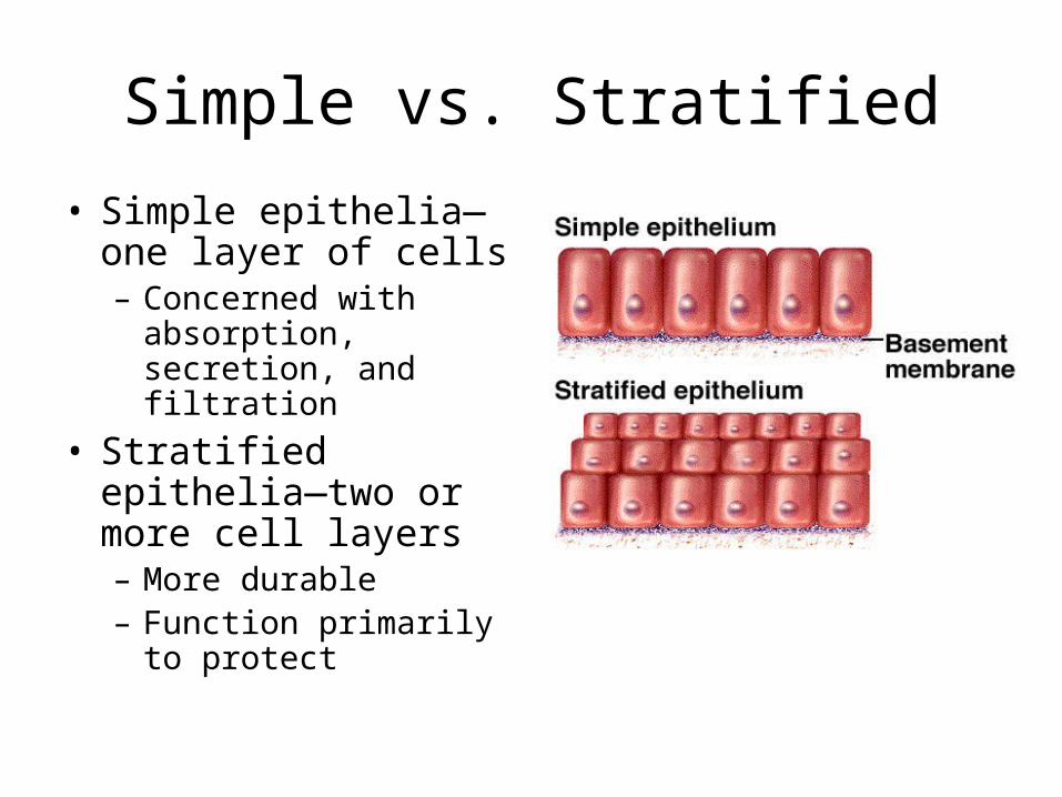
Simple vs. Stratified
• Simple epithelia—one layer of cells– Concerned with
absorption, secretion, and filtration
• Stratified epithelia—two or more cell layers– More durable– Function primarily to
protect
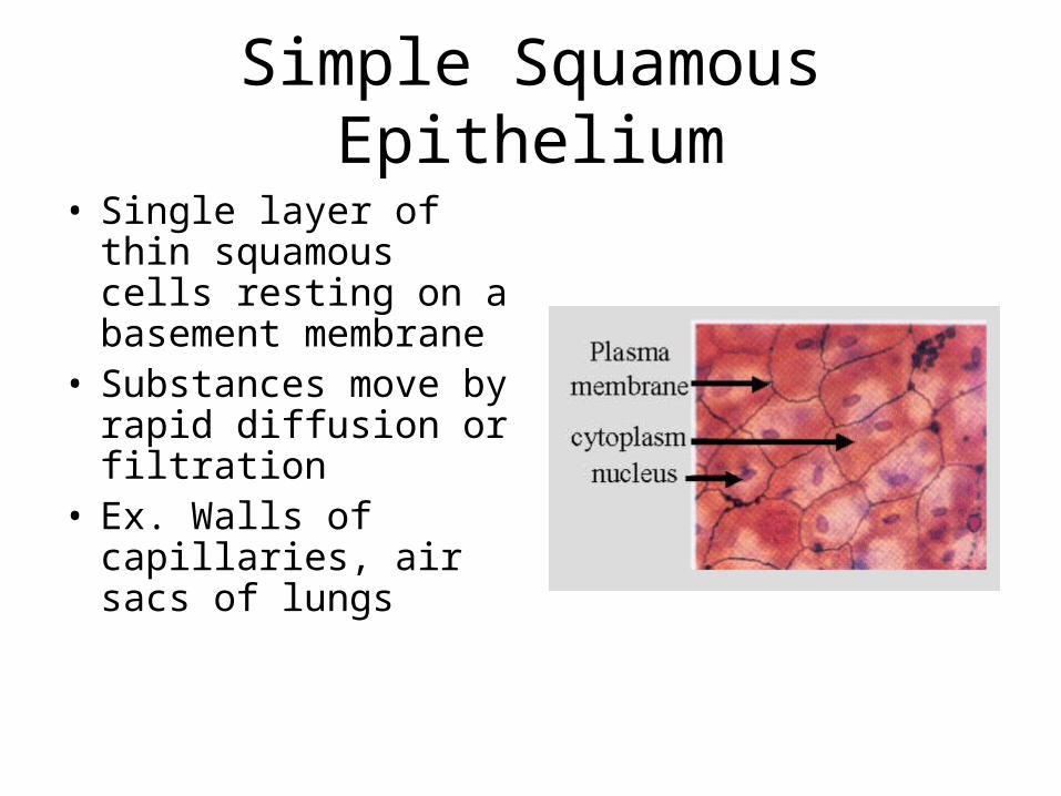
Simple Squamous Epithelium
• Single layer of thin squamous cells resting on a basement membrane
• Substances move by rapid diffusion or filtration
• Ex. Walls of capillaries, air sacs of lungs
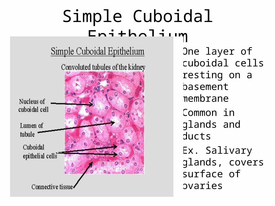
Simple Cuboidal Epithelium• One layer of
cuboidal cells resting on a basement membrane
• Common in glands and ducts
• Ex. Salivary glands, covers surface of ovaries
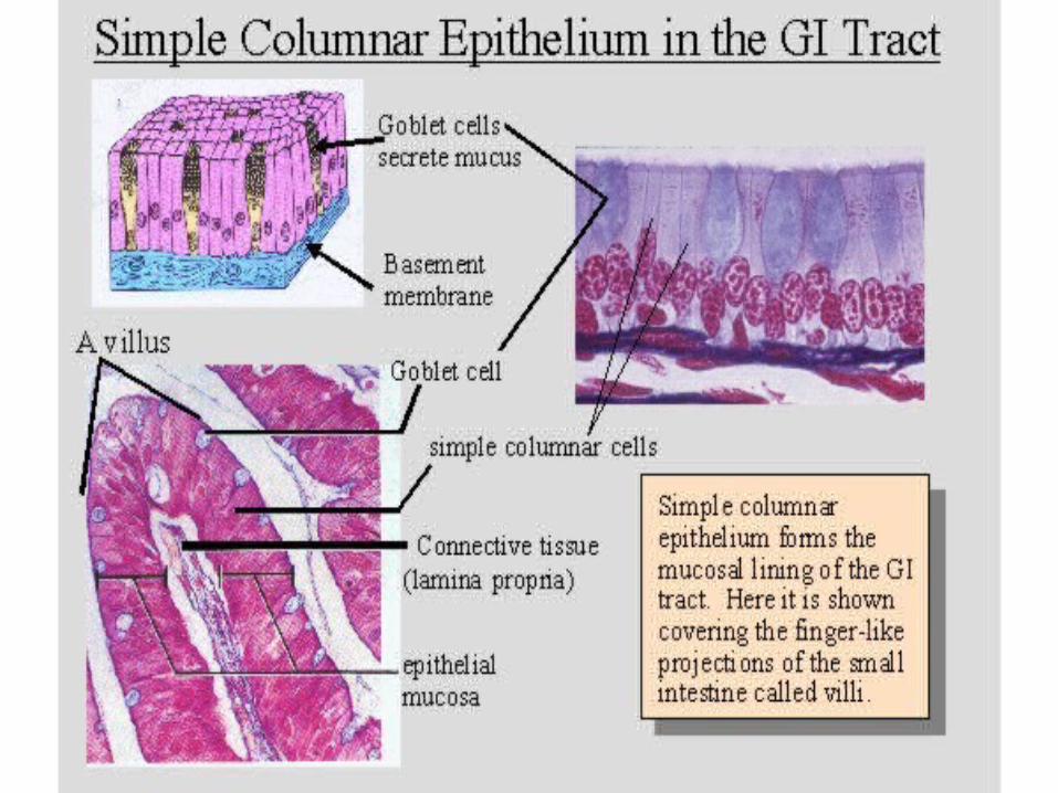
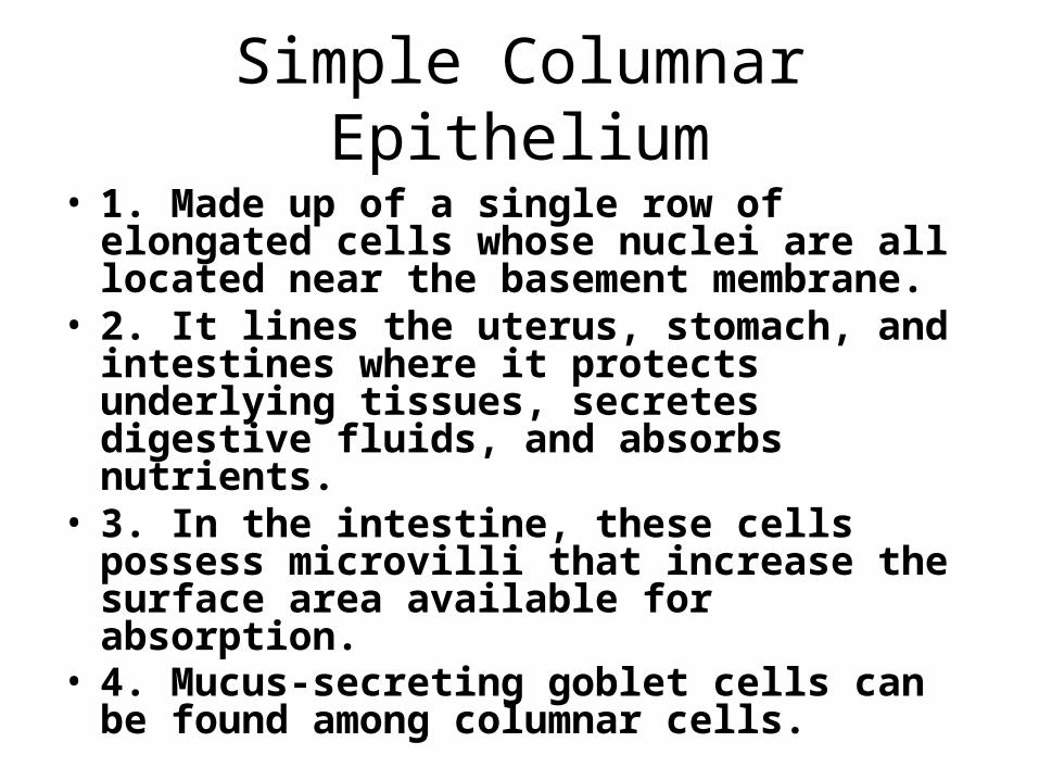
Simple Columnar Epithelium
• 1. Made up of a single row of elongated cells whose nuclei are all located near the basement membrane.
• 2. It lines the uterus, stomach, and intestines where it protects underlying tissues, secretes digestive fluids, and absorbs nutrients.
• 3. In the intestine, these cells possess microvilli that increase the surface area available for absorption.
• 4. Mucus-secreting goblet cells can be found among columnar cells.
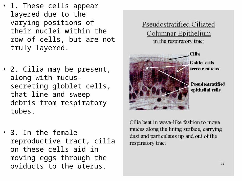
• 1. These cells appear layered due to the varying positions of their nuclei within the row of cells, but are not truly layered.
• 2. Cilia may be present, along with mucus-secreting globlet cells, that line and sweep debris from respiratory tubes.
• 3. In the female reproductive tract, cilia on these cells aid in moving eggs through the oviducts to the uterus.
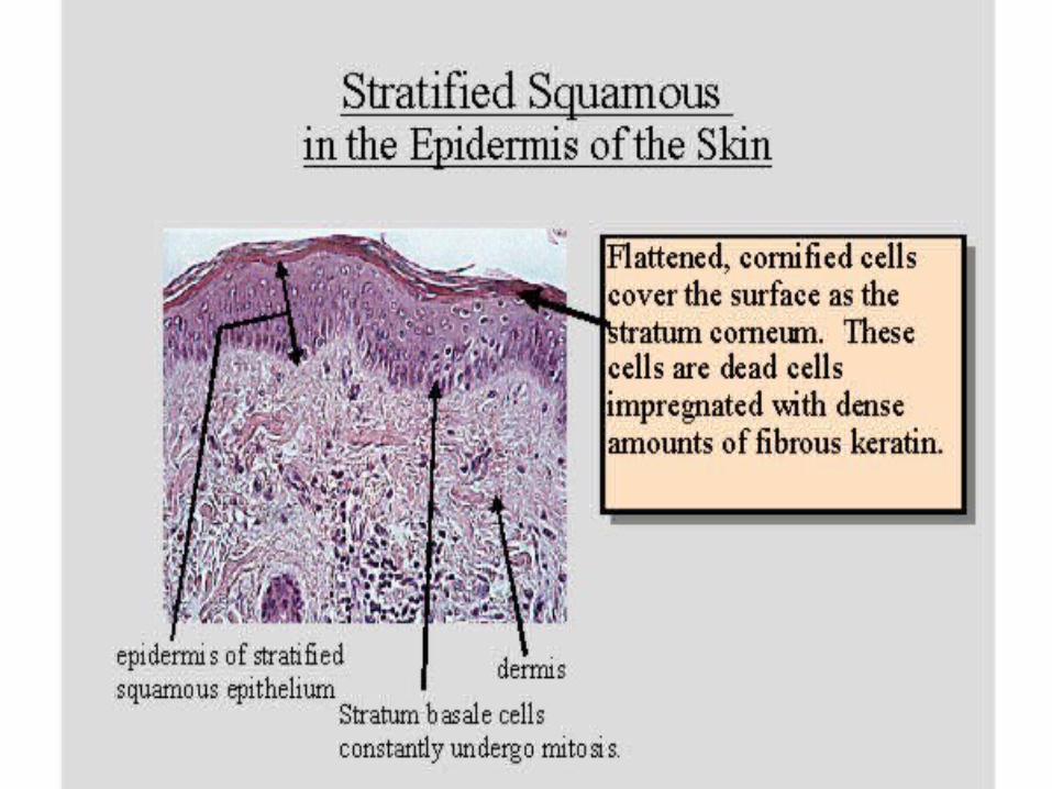
Stratified Squamous
• 1. This type of tissue is made up of layers of flattened cells that are designed to protect underlying layers.
• 2. It makes up the outer layer of skin, and lines the mouth, throat, vagina, and anal canal.
• 3. In the skin, outer layers of cells undergo keratinization; however, this process does not occur where tissues remain moist in the throat, vagina, or anal canal.
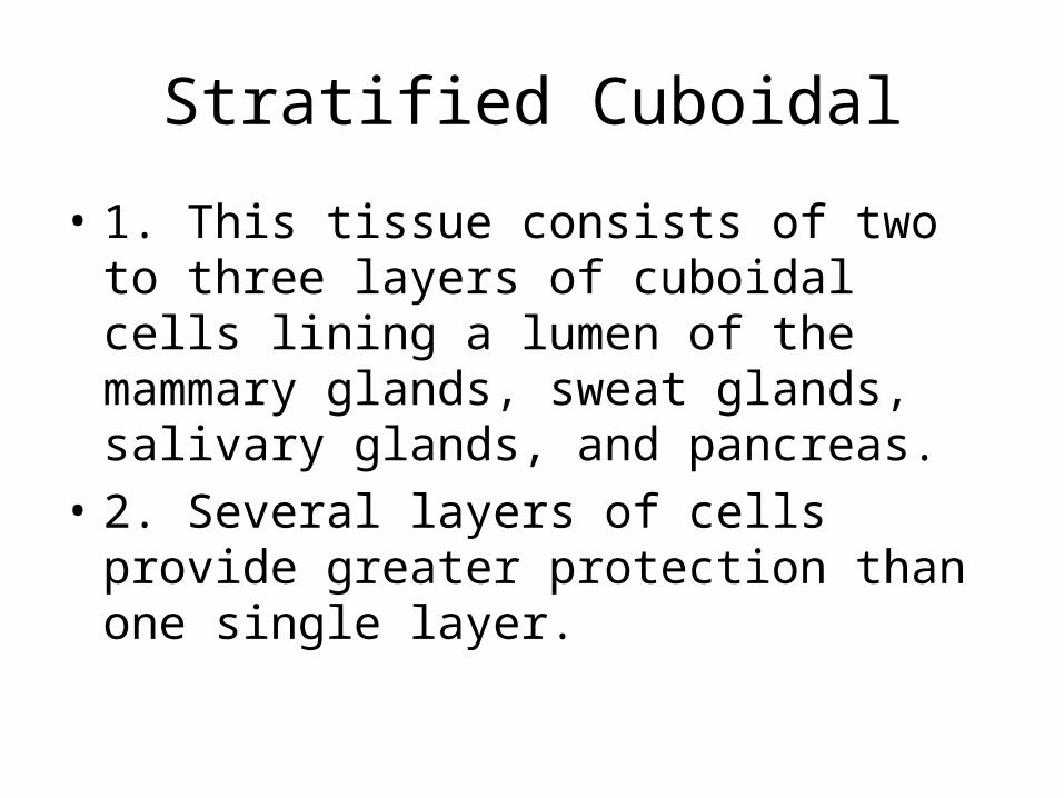
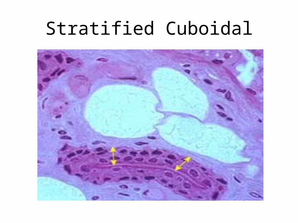
Stratified Cuboidal
• 1. This tissue consists of two to three layers of cuboidal cells lining a lumen of the mammary glands, sweat glands, salivary glands, and pancreas.
• 2. Several layers of cells provide greater protection than one single layer.
Stratified Cuboidal
Stratified Columnar
• 1. This tissue consists of several layers of cells and is found in the vas deferens, part of the male urethra, and parts of the pharynx.
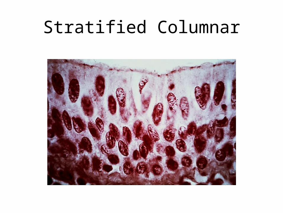
Stratified Columnar
Transitional
• 1. Transitional epithelium is designed to distend and return to its normal size, as it does in the lining of the urinary bladder.
• 2. This design provides distensibility and keeps urine from diffusing back into the internal cavity.

Glandular Epithelium
Endocrine Glands• Lose their connection
to the surface (ductless glands)
• Secretions (all hormones) diffuse directly into blood vessels
• Ex. Thyroid, adrenals, pituitary
Exocrine Glands• Retain their ducts,
secretions empty through ducts to epithelial surface
• Ex. Sweat and oil glands, liver, pancreas
What are glands?
• Glands--Consist of one or more cells that make and secrete a particular product.
• Secretion—typically contain protein molecules in an aqueous (water-based) fluid– Glandular cells obtain needed materials from
the blood and use them to make their secretion, which then get discharge

Connective Tissue
Characteristics of Connective Tissue
• Function primarily in protecting, supporting, and binding together other body tissues
• Most are well vascularized except tendons, ligaments and cartilage.
• Extracellular matrix—produced by connective tissue cells and secreted to the exterior– Liquid, semisolid (gel-like), or very hard– Weight bearing, withstand stretching, and other
abuses
Characteristics Continued…
• Fibers—made by CT cells and secreted– Collagen fibers (white)– Elastic fibers (yellow)– Reticular fibers (fine collagen)
Types of Connective Tissue
From most rigid to softest:
• Bone
• Cartilage
• Dense connective tissue
• Loose connective tissue
• Blood
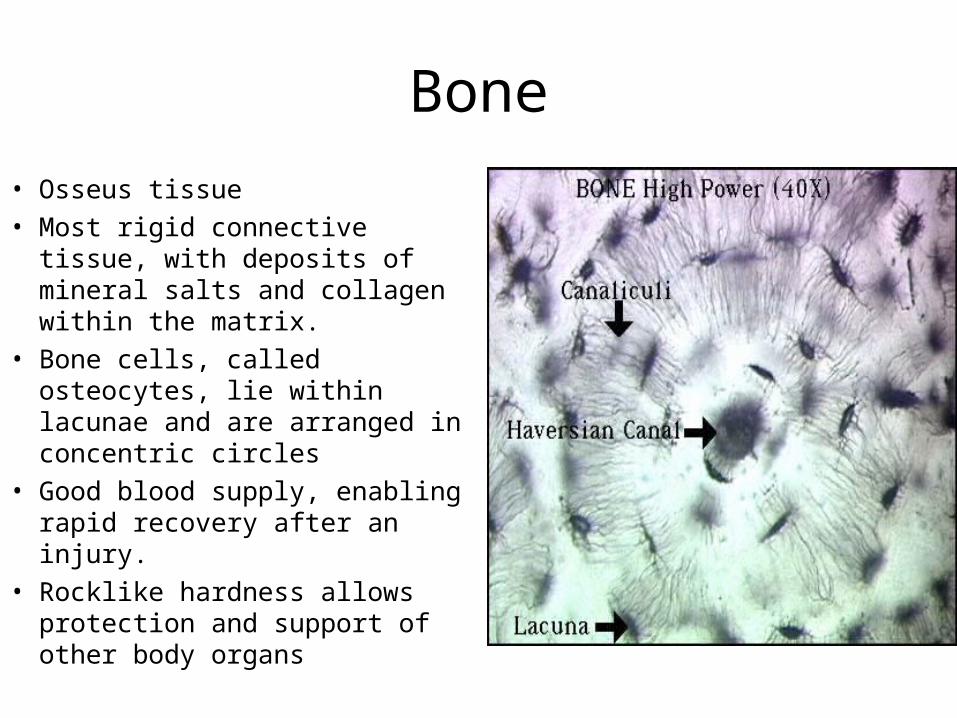
Bone
• Osseus tissue• Most rigid connective tissue,
with deposits of mineral salts and collagen within the matrix.
• Bone cells, called osteocytes, lie within lacunae and are arranged in concentric circles
• Good blood supply, enabling rapid recovery after an injury.
• Rocklike hardness allows protection and support of other body organs

Cartilage
• Provides a supportive framework for various structures.
• Cartilage cells (chondrocytes) lie within lacunae in the gel-like fluid matrix.
Types of Cartilage
• Hyaline cartilage is white with abundant fine collagen fibers, is found at the ends of bones, and supports respiratory passages.
• Elastic cartilage, with elastic fibers, provides a framework for the external ears and parts of the larynx.
• Fibrocartilage is a tough tissue that provides a shock-absorbing function in intervertebral disks and in the knees and pelvic girdle.
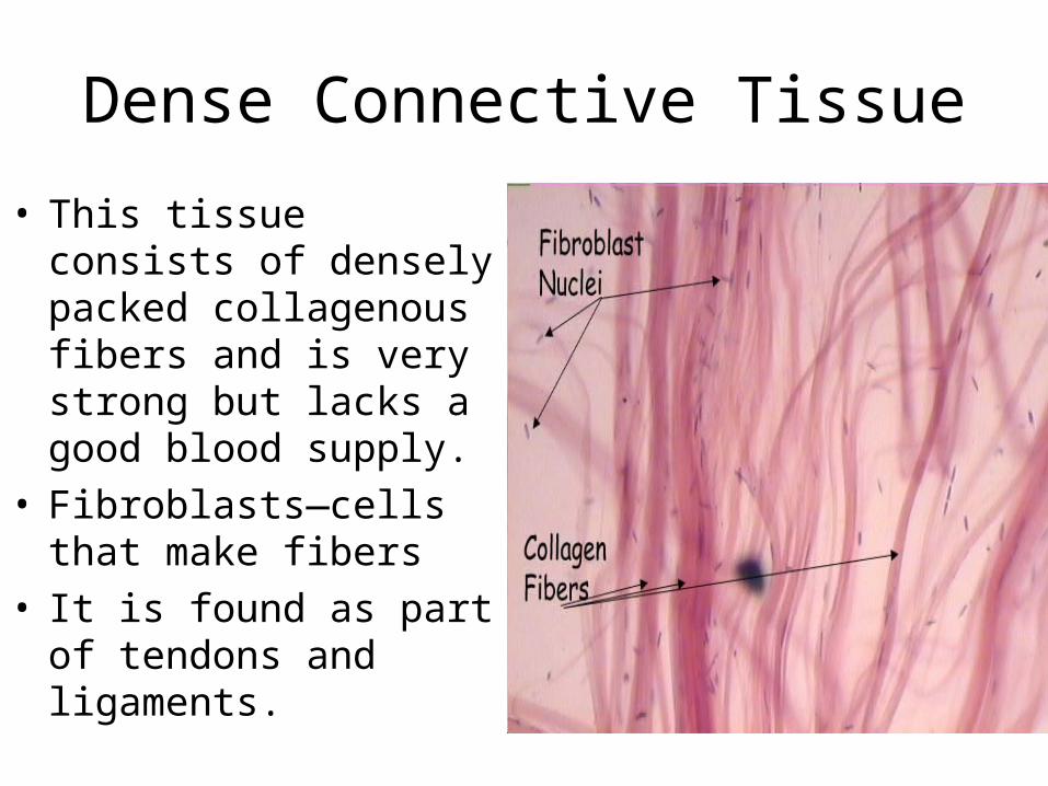
Dense Connective Tissue
• This tissue consists of densely packed collagenous fibers and is very strong but lacks a good blood supply.
• Fibroblasts—cells that make fibers
• It is found as part of tendons and ligaments.
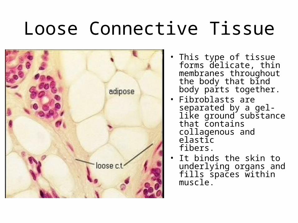
Loose Connective Tissue
• This type of tissue forms delicate, thin membranes throughout the body that bind body parts together.
• Fibroblasts are separated by a gel-like ground substance that contains collagenous and elastic fibers.
• It binds the skin to underlying organs and fills spaces within muscle.
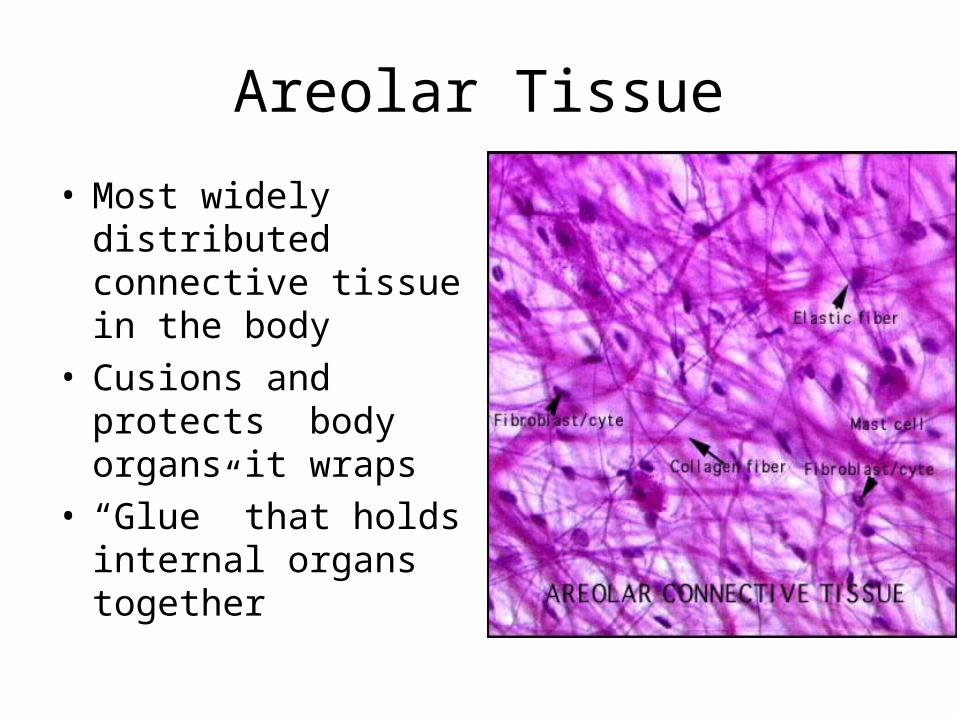
Areolar Tissue
• Most widely distributed connective tissue in the body
• Cusions and protects body organs it wraps
• “Glue” that holds internal organs together
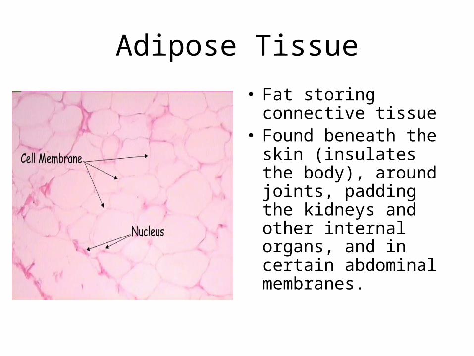
Adipose Tissue
• Fat storing connective tissue
• Found beneath the skin (insulates the body), around joints, padding the kidneys and other internal organs, and in certain abdominal membranes.
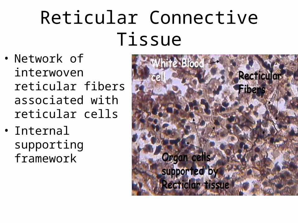
Reticular Connective Tissue
• Network of interwoven reticular fibers associated with reticular cells
• Internal supporting framework
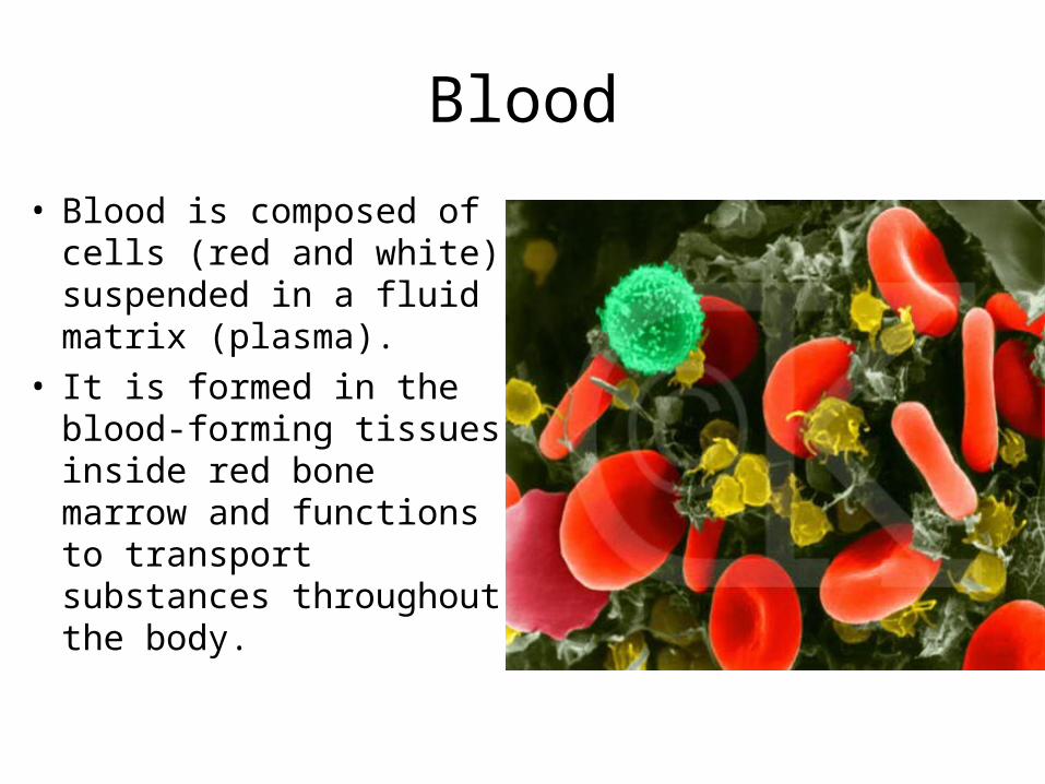
Blood
• Blood is composed of cells (red and white) suspended in a fluid matrix (plasma).
• It is formed in the blood-forming tissues inside red bone marrow and functions to transport substances throughout the body.