how do the plague pathogen and its host interact ... · s&tr march 2002 lague is potentially a...
TRANSCRIPT

S&TR March 2002
LAGUE is potentially a deadly
agent of bioterrorism. Unlike
anthrax, which has been so much in the
news lately, plague is highly infectious
and can be readily passed from one
person to another. The bite of a plague-
infected flea or the inhalation of just a
few cells of plague bacterium can kill.
Like smallpox, plague can spread and
kill large numbers of people very
quickly. Fortunately, it can usually be
treated with antibiotics.
History tells us how devastating a
plague epidemic can be. In what is
known as the Justinian epidemic, from
540 to 590 AD, plague spread from
Lower Egypt to Alexandria to Palestine
and on to the Middle East and Asia.
At its peak, 10,000 deaths occurred
every day in Byzantium. Eight hundred
years later, in 1347, plague came to
Italy from Asia or Africa, probably by
ship. By 1351, fully one-third of
Europe’s population had died from
bubonic plague.
This European epidemic is known
as the Black Death or the Great
Pestilence. In 1894, when Andre
Yersin identified the tiny bacterium
that causes plague, he named it pestisafter the Great Pestilence. He tried to
4
Lawrence Livermore National Laboratory
P
How do the plague pathogen and its hostinteract? Scientists will apply the answer tounderstanding a larger set of possible agentsof biological terrorism.

5Plague VirulenceS&TR March 2002
(See the box on p. 6 for more
information on functional genomics.)
Besides building better detectors,
work on the Y. pestis genome will also
lead to a better understanding of
pathogenicity and better vaccines and
treatments for the disease. The ultimate
goal, says Fitch, “is to produce a
computer model that simulates the
workings of a cell so that we can better
manage exposure to pathogens.”
Plague as PrototypeThe highly contagious Y. pestis is
an excellent model for studying the
interactions of a pathogen and its host.
In the case of Y. pestis, the host may
be a flea, a rodent, or a human. Fleas
carry plague bacteria and help transmit
the disease. Once an infected flea bites
a rodent or human, the bacteria begin
to multiply in the new host, and their
virulence shifts into high gear. Y. pestiscircumvents the host’s defenses by
injecting into host cells a series of
virulence factors that inhibit the
response of the immune system.
Earlier research has shown that
when Y. pestis is grown at the body
temperature of a flea (26°C), its cells
divide, but it does not express (turn on)
many of the genes that make it virulent
in rodents and humans. When the
Lawrence Livermore National Laboratory
growing the suspected bacteria in a
laboratory, take 36 to 48 hours.
For a plague detector to be truly
effective, it must do more than simply
indicate the presence of a specific
organism known to cause plague, says
Pat Fitch, who leads Livermore’s
Chemical and Biological National
Security Program. The detector also
must be able to identify the specific
traits found in atypical plague-causing
organisms. Scientists know of several
hundred strains (or isolates) of
Y. pestis, and they do not all behave in
precisely the same way. A few strains
are believed to have been genetically
modified or engineered to be more
deadly. There have also been two
clinical cases of naturally occurring
antibiotic-resistant plague. Knowing
the precise identity of a strain of
plague—or of any infectious disease,
for that matter—could help physicians
treat a patient properly.
Plague research at Livermore
currently is focusing on what makes
Y. pestis so virulent and able to
overcome the defenses of a host
organism. Fitch is leading the Pathogen
Pathway Project, using plague as a
prototype for the functional genomics
of a larger set of pathogenic agents that
could be used in biological terrorism.
name the genus Pasteurella after his
mentor, Louis Pasteur. But Yersinia,
after its discoverer, is the name
that stuck.
Today, Yersinia pestis is one of
several infectious diseases and agents
of bioterrorism that researchers across
the Department of Energy complex are
studying as part of the Chemical and
Biological National Security Program.
This program comes under the purview
of DOE’s National Nuclear Security
Administration (NNSA). At Livermore,
the work on Y. pestis also receives
support from Laboratory Directed
Research and Development.
Scientists at Livermore have
developed DNA signatures for Y. pestisthat can be used to quickly detect and
identify plague outbreaks. (See
“Uncovering Bioterrorism,” S&TR,
May 2000, pp. 4–12.) Signatures for
nine strains of the disease have been
submitted to the Centers for Disease
Control and Prevention in Atlanta,
Georgia, where they are undergoing
a rigorous validation process.
Livermore’s DNA-based detection
method proved its mettle in northern
Arizona last June when it was used to
identify a plague outbreak in prairie
dogs in just four hours. Standard
detection processes, which require
(a) Yersinia pestis, which causesplague, is a pathogen likely to beused by terrorists. (b) Its DNAforms loops, unlike human DNA,which forms strands. Scientistsstudying it screen between twoto five million of its nucleic acidbases to find unique regions(circled). Using polymerasechain reaction technology, theunique regions can be amplifiedthousands of times andprocessed to identify andcharacterize Y. pestis.
(a) (b)
S&TR March 2002
temperature increases to 37°C (human
or rodent body temperature), the
bacterium begins to produce the
proteins essential to its virulence. This
virulence mechanism can be induced in
the laboratory, making plague relatively
easy to study.
Examination of the Y. pestis genome
before and after virulence has been
induced shows what genes have been
turned on. But that information is not
enough to show precisely which genes
are responsible for various aspects
of virulence.
For comparative purposes, a
Livermore team led by microbiologist
Emilio Garcia collaborated with the
Institut Pasteur in France to sequence
Y. pseudotuberculosis, the parent
organism of Y. pestis. Although
their DNA sequences are about
95 percent identical, Y. pestis and
Y. pseudotuberculosis behave
differently. Y. pseudotuberculosis lodges
in the intestine and causes flulike
intestinal distress. Y. pestis is also
closely related to the mild-mannered
Y. enterocolitica, an intestinal bug
that is itself very much like
Y. pseudotuberculosis. Y. enterocoliticais currently being sequenced by the
Sanger Center in Great Britain.
“Bacteria evolve very efficiently and
make use of about 80 percent of their
DNA,” says Fitch. By comparison,
humans use only about 30 percent of
their DNA. Aiding speedy evolution are
the many insertion sequences in a
bacterial genome. Insertion sequences
are bits of DNA that allow large
6
Lawrence Livermore National Laboratory
Plague Virulence
regions of DNA to replicate
themselves and move around the
genome, relocating themselves
somewhere else. When an insertion
sequence lands within a gene, it
deactivates that gene. These transfers
can also occur across species, and it
is not difficult for a bacterium to grab
DNA from another bacterium.
Y. pestis evolved from
Y. pseudotuberculosis within the past
15,000 years, a rapid evolution even
for bacteria. “Something happened
then to cause Y. pestis to learn how to
live in a flea,” says Garcia.
In addition to their normal
chromosomal DNA, bacteria may have
smaller circles of DNA known as
plasmids. Plasmids replicate separately
from chromosomal DNA and often
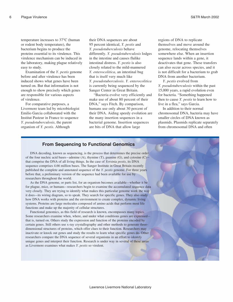
From Sequencing to Functional Genomics
DNA decoding, known as sequencing, is the process that determines the precise order
of the four nucleic acid bases—adenine (A), thymine (T), guanine (G), and cytosine (C)—
that comprise the DNA of all living things. In the case of Yersinia pestis, its DNA
sequence comprises 4.66 million bases. The Sanger Institute in Great Britain recently
published the complete and annotated sequence of the Y. pestis genome. For three years
before that, a preliminary version of the sequence had been available for use by
researchers throughout the world.
As the DNA genome, or parts list, for an organism becomes available—whether it be
for plague, mice, or humans—researchers begin to examine the accumulated sequence data
very closely. They are trying to identify what makes this particular genome work the way
it does—its wiring diagram, so to speak. They search for specific genes. They also study
how DNA works with proteins and the environment to create complex, dynamic living
systems. Proteins are large molecules composed of amino acids that perform most life
functions and make up the majority of cellular structures.
Functional genomics, as this field of research is known, encompasses many topics.
Some researchers examine when, where, and under what conditions genes are expressed—
that is, turned on. Others study the expression and function of the proteins encoded by
certain genes. Still others use x-ray crystallography and other methods to generate three-
dimensional structures of proteins, which offer clues to their function. Researchers may
inactivate or knock out genes and study the results to learn what specific genes do. Other
researchers compare the DNA sequence of several organisms in an effort to identify
unique genes and interpret their function. Research is under way in several of these areas
as Livermore examines what makes Y. pestis so virulent.

house genes that encode enzymes
critical to the host cell or organism. For
example, when a bacterium has become
resistant to antibiotic drugs, it is usually
because the bacterium has acquired a
new plasmid.
One Y. pestis plasmid encodes at
least two genes that allow Y. pestis to
survive in fleas. Another plasmid is
home to the gene that activates the
disease’s invasiveness. Researchers have
found that Salmonella has a similar
plasmid, which one bacterium probably
obtained from the other.
“The interesting thing is that if
you insert the three pestis plasmids
into Y. pseudotuberculosis or
Y. enterocolitica, you don’t get pestis,”
says Garcia. “So something else is going
on. Unfortunately, it’s never simple.”
Once its virulence genes have been
turned on, plague infects its host using
what is known as Type III secretion, an
injection mechanism more colorfully
called “Yersinia’s deadly kiss.”
Salmonella typhi, enteropathogenic
Escherichia coli, Chlamydia psittaci,various species of Bordetella, and other
pathogenic bacteria appear to share this
syringelike injection mechanism. This
common trait may indicate another area
of transferred genomic material.
Before Livermore’s research on
plague started, many of the genes
critical for virulence had been identified
but were poorly understood. The same
was true for the underlying mechanisms
of virulence. There was also little
understanding of the gene and protein
interactions that take place between the
pathogenic bacteria and its host.
The Pathogen Pathway Project is
using functional genomics tools to
identify genes important to virulence
and understand the pathways of
virulence. The team’s hypothesized
pathway, from DNA to the host
organism, is shown in the bottom
figure at right.
Expression and FunctionAn early task for Livermore
bioscientists and computations experts
was to develop a relational database of
the DNA sequence of Y. pestis. In
collaboration with the DOE Genome
Consortium at Oak Ridge National
Laboratory, these data were used to
computationally predict where the
4,500 genes in Y. pestis are located and
which genes might be associated
with virulence.
Next, Livermore bioscientist Vladimir
Motin and colleagues designed chemical
reagents for extracting over 300 genes
from Y. pestis DNA, including all known
virulence-associated genes on the
plasmids. In an initial test, they extracted
85 genes associated with virulence and
spotted them on a glass microscope slide
alongside 11 control spots, making up a
96-spot microarray.
A microarray permits scientists to
study the response of thousands of
genes or other pieces of DNA quickly
and efficiently in a process known as
transcript profiling. In the process, each
gene receives some kind of stimulus,
causing it to turn on and produce
messenger RNA (mRNA). In the case
of plague, the stimuli are changes in
temperature and calcium concentration.
The production of mRNA leads, in turn,
to the synthesis of unique proteins. The
level of mRNA can be measured for
each individual gene. The more active
or expressed genes there are, the more
mRNA will be present.
For the 96-spot microarray, the team
developed a protocol to study the
response of Y. pestis genes under
conditions that mimic the infection
process: at both flea and human/rodent
body temperatures, 26°C and 37°C, and
7
Lawrence Livermore National Laboratory
Plague VirulenceS&TR March 2002
Bacterial cell
Host cell
Genes Message Proteins Function/ Host(DNA) (RNA) environment interactions
Regulation
A schematic diagram of information that is hypothesized to describe the pathways of virulencein a pathogen. The regulatory (feedback) loop is often nonlinear, and there can be multiplefeedback paths with complex interactions.
The Type III secretion, asyringelike injection mechanismmore colorfully called “Yersinia’sdeadly kiss,” which is how plagueinfects a host once its virulencegenes have been turned on.

at calcium levels that correspond to
those of blood (higher level) and
organs (lower level), the latter location
being where more virulence genes
are expressed.
More recently, they developed a
microarray for all 4,500 Y. pestisgenes. All of the genes are being
mapped at six time intervals as
temperature rises and calcium
concentration drops. The team is thus
beginning to establish a timeline for
how and when genes change and are
expressed while the plague bacterium
is infecting a human host. Some genes
are expressed early, while others are
late-onset genes. A detailed picture of
how the bacterium behaves during the
infection process will provide useful
information for the development
of diagnostic techniques and
treatment methods.
Garcia and other researchers also
completed a detailed analysis of three
Y. pestis plasmids, which allowed them
to confirm the location of several
known virulence genes and to uncover
four novel ones believed to contribute
to virulence. Computerized
comparisons with other genomic
databases indicated the presence of a
large number of virulence-related genes
that are similar in both closely related
bacteria such as Y. pseudotuberculosisand distantly related bacteria such as E.coli. The team also found numerous
gene coding regions whose function
they could not determine.
Using a proteomic approach of
protein separation techniques and mass
spectrometry (MS), Livermore
researchers led by Sandra McCutchen-
Maloney are analyzing complex
mixtures of proteins isolated from
8
Lawrence Livermore National Laboratory
Plague Virulence S&TR March 2002
A
B
C
D
E
F
G
H
A A BRCA XRCC PKC ypkA yscC yscO lcrV PKC XRCC BRCA A B yopE caf1A sodA envZ ompR yenl rpoE yopJ yscB lcrH sycE caf1R C tpx rfaH phoP yenR g6pd rpoN yopH lcrF lcrE yopB sycH luxS D oxyR psaA glts rpoS lcrQ yscW tyeA yopD yadA katY hemF toxR E rstB nqrA dha4 rpoH yscL yscU sycN yopM pst hns cbl flhC F nhaB nadB rpoD yscH lcrD sycT pla catA gcvA copR araC nhaC G dam yscG lcrR yopT ymt sodC lysR ilvY tsaA caf1 tuf yscD H yscP lcrG yopK caf1 sodB crp nhaR fliA h5 RAD CDC2 Empt
1 2 3 4 5 6 7 8 9 10 11 12
A 96-spot microarray fortranscript profiling of85 genes of Yersiniapestis; 11 of the spots arecontrols. Genes taggedwith fluorescent dyeschange color in responseto various stimuli.
Y. pestis. By comparing samples grown
at the two physiological conditions
mimicking the flea and the human (at
26°C and 37°C, respectively) and at low
calcium concentration to induce
virulence, the team is detecting
differential protein expression to
identify candidate proteins important for
Y. pestis pathogenicity. Comparisons are
also being made between human cells
that have and have not been exposed to
Y. pestis in order to understand the host
immune response. Because it is the
proteins that are actually responsible for
virulence effects, the group is also
working to correlate their proteomic
data with genomic data obtained from
microarray experiments. To learn more
about the individual proteins responsible
for virulence, the team is using various
biochemical assays to test functional
models of the candidate virulence

factors. In addition, McCutchen-
Maloney’s group is looking at
host–pathogen interactions by using
surface-enhanced laser desorption
ionization (SELDI) MS to study various
protein–DNA and protein–protein
interactions within Y. pestis and
between Y. pestis and the human host.
For example, regulatory proteins that
bind to genes and control differential
expression are under investigation, as
are the specific protein–protein
interactions of suspected virulence
factors. These molecular interactions
are key to the genetic feedback that
occurs as a pathogen infects its host,
as shown in the bottom figure on p. 7.
Differences Are KeyBefore Garcia’s team completed
its comparative sequencing of
Y. pseudotuberculosis, microbiologists
Gary Andersen, Lyndsay Radnedge,
and others examined the differences
between Y. pseudotuberculosis and
Y. pestis using a different technique.
This process, developed in Russia, is
known as suppression subtractive
hybridization (SSH). SSH identifies
regions of DNA that are present in one
species but absent in another.
SSH has the advantage of requiring
only small amounts of genomic DNA.
It can be used with any genome, even
one that has not yet been characterized.
It is especially useful for identifying the
large genomic differences typically
found between bacterial genomes. For
example, SSH identified the genetic
material that causes Kaposi’s sarcoma,
a skin lesion associated with HIV and
AIDS. At Livermore, SSH has been
useful for finding differences among
anthrax strains and other potential
agents of bioterrorism.
Comparison of Y. pestis and
Y. pseudotuberculosis revealed seven
DNA regions in Y. pestis that do not
occur in Y. pseudotuberculosis. Four
of them occur very closely to one
another on the Y. pestis genome. “It
is fair to assume that pestis acquired
this region during its evolution
from Y. pseudotuberculosis,” says
Radnedge.
To learn more about the function of
genes in these areas, Garcia and others
are beginning “knock-out” studies.
They will inactivate, or knock out, one
gene at a time and test the resulting
bacterium on an animal to see how the
host and its genes respond. This is
slow, laborious work, but it will help to
determine what the function of each
Y. pestis gene is, if any, and what gene
or genes in the host are expressed as a
response. This detailed examination of
pathogen–host interaction for plague
will be the first of its kind.
Being PreparedResearch to date on plague lays the
groundwork for additional work
planned at Livermore in the areas of
9
Lawrence Livermore National Laboratory
Plague VirulenceS&TR March 2002
microbiology, proteomics (the global
study of proteins), bioinformatics (the
integration and analysis of biological
data), and biological modeling for the
NNSA’s Chemical and Biological
National Security Program. Some of the
research will elaborate on plague, some
will examine a broader spectrum of
human pathogens, and some will further
the development and use of biodetectors,
mass spectrometry, and other
technologies.
In the U.S. today, plague pops out of
the rodent population and into the human
populace occasionally in the desert
Southwest. It is a larger problem in a few
other countries. But the real fear is that
plague could be used as an agent of mass
destruction. At least in industrialized
countries, it is unlikely that plague
would cause the huge number of deaths
that occurred during earlier epidemics.
Better sanitation, a more educated
populace, and a far superior medical
system would likely prevent that. But the
world needs to be prepared.
—Katie Walter
Key Words: bioterrorism agents, Chemicaland Biological National Security Program,plague, surface-enhanced laser desorptionionization mass spectrometry (SELDI-MS),suppression subtractive hybridization (SSH),virulence, Yersinia pestis, Yersiniapseudotuberculosis.
For further information, contact Pat Fitch(925) 422-3276 ([email protected]).