how far would you go to address diabetic microvascular complications?
TRANSCRIPT


How Far Would You Go To Address Diabetic Microvascular
Complications?

Diabetes is a Significant Healthcare Problem in the United States• Over 18 million Americans have diabetes
• Up to 30% of diabetes cases have not been diagnosed
• 1.3 million new cases are diagnosed each year in the US
• Economic burden of $132 billion per year (2002 healthcare costs)
– Approximately $7333 per patient
American Diabetes Association. Available at: http://www.diabetes.org/diabetes-statistics/national-diabetes-fact-sheet.jsp.Hogan P, et al. Diabetes Care. 2003;26:917-932.World Health Organization. Available at: http://www.wpro.who.int/pdf/rcm51/rd/bhcp-4b.pdf. Accessed November 13, 2003.

Diabetes is a Growing Healthcare Epidemic
0
5
10
15
20
25
1995 2025
Hogan P, et al. Diabetes Care. 2003;26:917-932.King H, et al. Diabetes Care. 1998;21:1414-1431.
Pa
tien
ts (
mill
ion
s)
13.9 million
21.9 million

Long-term Diabetic Complications are Devastating
• Diabetic Macrovascular complications– Coronary artery disease– Cerebrovascular disease– Peripheral vascular disease
• Diabetic Microvascular complications– Diabetic Nephropathy– Diabetic Neuropathy– Diabetic Retinopathy (including Diabetic Macular Edema)
Rousch JEB. J Clin Invest. 2003;112:986-988.Sheetz MJ, King GL. JAMA. 2002;288:2579-2588.Williams R, et al. Diabetologia. 2002;45:S13-S17.
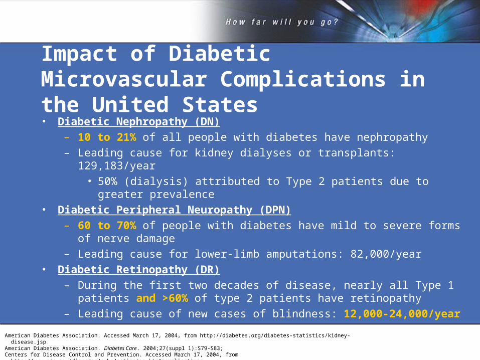
Impact of Diabetic Microvascular Complications in the United States• Diabetic Nephropathy (DN)
– 10 to 21% of all people with diabetes have nephropathy
– Leading cause for kidney dialyses or transplants: 129,183/year
• 50% (dialysis) attributed to Type 2 patients due to greater prevalence
• Diabetic Peripheral Neuropathy (DPN)
– 60 to 70% of people with diabetes have mild to severe forms of nerve damage
– Leading cause for lower-limb amputations: 82,000/year
• Diabetic Retinopathy (DR)
– During the first two decades of disease, nearly all Type 1 patients and >60% of type 2 patients have retinopathy
– Leading cause of new cases of blindness: 12,000-24,000/year
American Diabetes Association. Accessed March 17, 2004, from http://diabetes.org/diabetes-statistics/kidney-disease.jspAmerican Diabetes Association. Diabetes Care. 2004;27(suppl 1):S79-S83; Centers for Disease Control and Prevention. Accessed March 17, 2004, from http://www.cdc.gov/diabetes/pubs/estimates.htm#complicationsFong DS, et al. Diabetes Care. 2004;27(suppl 1): S84-87.

Diabetic Nephropathy

Progression of Diabetic Nephropathy
Present at diagnosis of diabetes
Increased kidney and glomerular size
Mean arterial BP normal
Within first 5 yearsBasement membrane
thickening
Normal BP or slight elevation (1 mm
Hg/year)
After 6-15 years (~35% patients)
Further basement membrane thickening, mesangial expansion
UAE = 20-200 µg/day
BP >3 mm Hg/year
After 15-25 years
(~35% of patients)
Clear, pronounced abnormalities
proteinuria
GFR decline ~10 mL/min/year
BP >5 mm Hg/year
ESRD after 25-30 yearsGlomerular closure,
advanced glomerulopathy
GFR <10 mL/min
BP >5 mm Hg/year
Stage 1
Stage 2
Stage 3
Stage 4
Stage 5
UAE = Urinary albumin excretion
Mogensen CE. Diabetologia. 1999;42:263-285.
Chronology PathologyDiagnosis
and Screening

Diabetic Peripheral Neuropathy

Microvascular Damage Leads to Diabetic Peripheral Neuropathy (DPN)
• Examination of tissues from patients with diabetes reveals capillary damage, including occlusion in the vasa nervorum• Reduced blood supply to the neural tissue results in impairments in nerve signaling that affect both sensory and motor
function
Dyck PJ, Giannini C. J Neuropathol Exp Neurol. 1996;55:1181-1193.Sheetz MJ, King GL. JAMA. 2002;288:2579-2588.
Normal nerve Damaged nerve
Occluded vasa nervorum
Damage to myelinated and unmyelinated
nerve fibers

Diabetic Peripheral Neuropathy Can Progress Over Time
Symptoms (numbness, prickling, pain)
Reflexes
Pressure Sensation (Monofilament)
Vibratory Sensation
Nerve Conduction Abnormalities
Subclinical Clinical
Time
Sig
ns
Onset ofClinical Diseases
Adapted from ADA. Diabetes Care. 2003;26:S33-S50; Abbott CA, et al. Diabetes Care. 1998;21:1071-1075; Armstrong DG, et al. Arch Intern Med. 1998;158:289-292; Armstrong DG, et al. Ostomy Wound Manage. 1998;44:70-76; Carrington AL, et al. Diabetes Care. 2002;25:2010-2015; Feldman EL, et al. Diabetes Care. 1994;17:1281-1289; Shearer A, et al. Diabetes Care. 2003;26:2305-2310; Veves A, et al. Diabet Med. 1991;8:917-921.
• Symptoms may occur any time and intermittently
• Patients may or may not have symptoms of diabetic peripheral neuropathy
• Patients frequently do not report symptoms to their physicians until the symptoms are severe
• The majority of signs of diabetic peripheral neuropathy are not evident at the onset of diabetes

Symptoms and Signs ofDiabetic Peripheral Neuropathy
Symptoms• Numbness or loss of feeling
(asleep or “bunched up sock under toes” sensation)
• Prickling/Tingling• Aching Pain• Burning Pain• Lancinating Pain• Unusual sensitivity or
tenderness when feet are touched (allodynia)
Signs• Diminished vibratory perception• Decreased knee and ankle reflexes• Reduced protective sensation such
as pressure, hot and cold, pain• Diminished ability to sense position
of toes and feet
Symptoms and signsprogress from distal to proximal over time
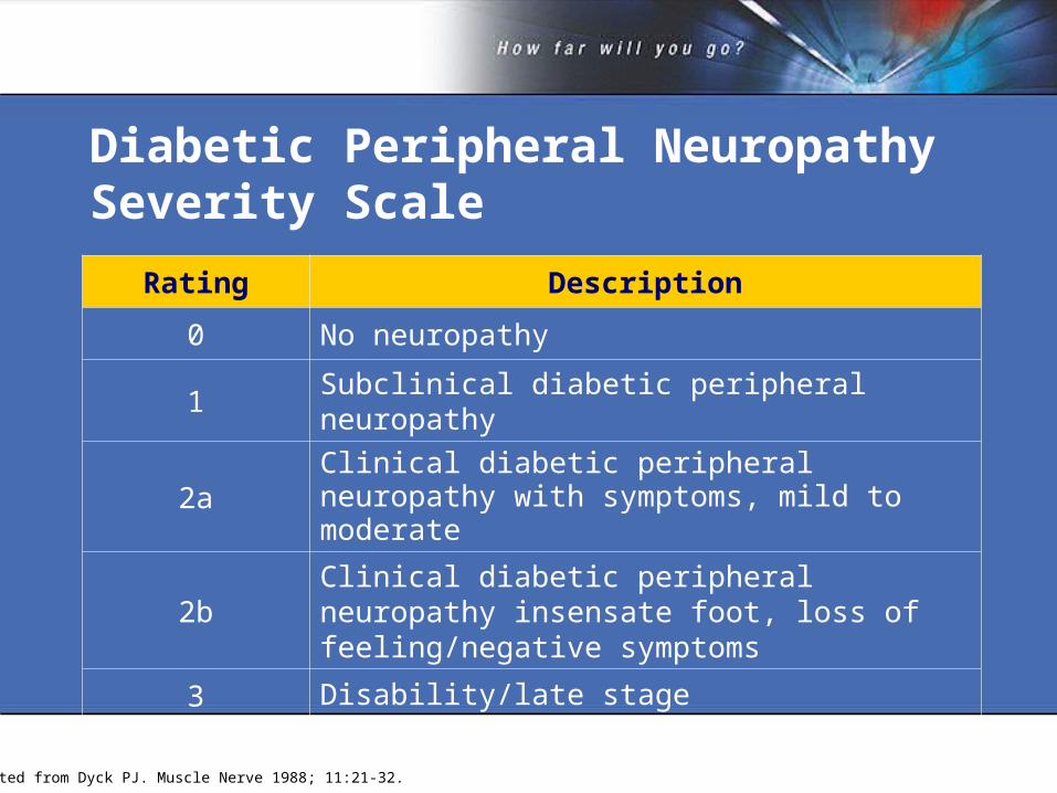
Diabetic Peripheral Neuropathy Severity Scale
Adapted from Dyck PJ. Muscle Nerve 1988; 11:21-32.
Rating Description
0 No neuropathy
1 Subclinical diabetic peripheral neuropathy
2aClinical diabetic peripheral neuropathy with symptoms, mild to moderate
2bClinical diabetic peripheral neuropathy insensate foot, loss of feeling/negative symptoms
3 Disability/late stage

Effects of Diabetic Peripheral Neuropathy
Images: 1,4Edward J Bastyr, III, MD; 2,3Rayaz A Malik, MBChB, PhD, MRCP.
Diabetic Retinopathy (Including Diabetic Macular Edema)

Diabetic Retinopathy: A Progressive Disease
Flynn HW, Smiddy WE, eds. Diabetes and Ocular Disease: Past, Present, and Future Therapies. AAO Monograph No. 14. San Francisco: The Foundation of the American Academy of Ophthalmology; 2000.
Preclinical Nonproliferative Diabetic
Retinopathy
Proliferative Diabetic
Retinopathy
Diabetic Macular Edema
Symptoms None None, or blurred vision and glare
None, or reduced vision or floaters
None, or blurred vision
Clinical signs indicating need for referral
• Normal appearing retina
• Retinal vasodilation
• Microaneurysms• Nerve fiber layer
infarcts• Intraretinal
hemorrhages• IRMAs• Venous bleeding
• Retinal vasodilation
• Beading• IRMAs• Neovascularizatio
n of optic disc, retina, and/or iris
• Swelling of retina due to leaky capillaries
• Increased capillary leakage
• Fluid accumulation in retinal layers

American Academy of Ophthalmology (AAO): Staging of Diabetic Retinopathy
American Academy of Ophthalmology, October, 2002.
Disease Severity Level Observable (Dilated Ophthalmoscope)
No apparent retinopathy No abnormalities
Mild Non-Proliferative Diabetic Retinopathy
Microaneurysms only
Moderate Non-Proliferative Diabetic Retinopathy
More than just microaneurysms but less than severe nonproliferative diabetic retinopathy
Severe Non-Proliferative Diabetic Retinopathy
Any of the following- More than 20 intraretinal hemorrhages in each of 4 quadrants- Definite venous beading in 2+ quadrants- Prominent IRMA in 1+ quadrant and no signs of proliferative diabetic retinopathy
Proliferative Diabetic Retinopathy
One or more of the following- Neovascularization- Vitreous/peretinal hemorrhage

AAO Staging of Diabetic Macular Edema
American Academy of Ophthalmology, October, 2002.
Disease Severity Level Observable (Dilated Ophthalmoscope)
No diabetic macular edema present
No retinal thickening or hard exudates in posterior pole
Diabetic macular edema present
Mild Diabetic Macular Edema
Some retinal thickening or hard exudates in posterior pole but distant from the center of the macula
Moderate Diabetic Macular Edema
Retinal thickening or hard exudates approaching the center of the macula but not involving the center
Severe Diabetic Macular Edema
Retinal thickening or hard exudates involving the center of the macula

Types of Diabetic Retinopathy
• Diabetic macular edema may coexist with either nonproliferative or proliferative diabetic retinopathy of any severity
• The retina is the one place where the microvasculature can be viewed
Images: 1,2Diabetic Retinopathy Study Research Group; 3Phototake.
Normal retinaNonproliferative diabetic
retinopathyProliferative diabetic
retinopathy
Diabetic macular edema

Treatment

Current Treatment Options for Diabetic Microvascular ComplicationsDisease Direct Treatment Indirect Treatment
Diabetic Nephropathy
None BP Control
Diabetic Neuropathy
None Analgesic relief for pain only
Diabetic Retinopathy
Laser (late stage) BP/GC Control
Any Diabetic Microvascular Complications
None BP/GC Control
Therapies that target the underlying process are needed

Until new therapies are available, early detection is the only way to predict the
development and progression of Diabetic Microvascular Complications
(DMCs)

Clinical Guidelines for Early Detection of Diabetic NephropathyTest When Normal Range
Blood pressure
Each office visit <130/80 mm Hg
Urinary albumin
Type 2: Annually beginning at diagnosis
Type 1: Annually, 5 years post-diagnosis
<30 µg/mg creatinine(random spot collection)
American Diabetes Association: Nephropathy in Diabetes (Position Statement). Diabetes Care. 2004; 27(suppl 1):S79-S83.
Equivalent to:
<30 mg/day urinary albumin excretion
<20 µg/min urinary albumin excretion(timed specimen)

Clinical Guidelines for Early Detection of Diabetic Peripheral Neuropathy
Adapted from Boulton AJM, et al. Diabet Med. 1998; 15(6):508-514.Adapted from Dyck PJ. Muscle Nerve 1988; 11:21-32
Stages Characteristics
Stages 0/1: No clinical neuropathy
• No symptoms or signs
Stage 2a: Clinical neuropathy
• Positive symptomology (increasing pains at night): burning, shooting, stabbing pains, “pins & needles”; absent sensation to several modalities and reduced or absent reflexes
• Less common–diabetes poorly controlled, weight loss; diffuse (trunk); minor sensory signs
Stage 2b: Clinical neuropathy• No symptoms or numbness of feet; reduced thermal
sensitivity; painless injury
Stage 3: Disability/late stage• Foot lesions (eg, ulcers); neuropathic deformity
(eg, Charcot joint); non-traumatic amputation

Clinical Guidelines for Management of Diabetic Peripheral Neuropathy
Stages Objectives Referral
Stage 0/1: No clinical neuropathy
Education to reduce risk of progression; glycemic control; annual assessment
As required
Stage 2a: Clinical neuropathyStable glycemic control; symptomatic treatment
Diabetologist, neurologist
Stage 2b: Clinical neuropathyEducation, especially foot care; glycemic control according to needs
Foot care team
Stage 3: Disability/late stage
Prevention or new/ recurrent lesions and amputation; emergency referral if lesions present; otherwise referral within 4 weeks
Diabetologist, neurologist, chiropodist, podiatrist, diabetes specialist nurse, diabetic foot clinic if available
Adapted from Boulton AJM, et al. Diabet Med. 1998; 15(6):508-514.Adapted from Dyck PJ. Muscle Nerve 1988; 11:21-32

Clinical Guidelines for Early Detection of Diabetic Retinopathy and Diabetic Macular Edema
Fong DS et al. Diabetes Care. 2004;27 (suppl 1): S84-S87.
*Eye exam should be performed through dilated pupils by qualified eye specialist†Abnormal findings necessitate more frequent follow-up
Patient group Recommended first examination*
Minimum routine follow-up†
Type 1 diabetes Within 3–5 years after diagnosis of diabetes once patient is age 10 years or older
Yearly
Type 2 diabetes At time of diagnosis of diabetes
Yearly
Pregnancy in preexisting diabetes
Prior to conception and during first trimester
Physician discretion pending results of first trimester exam

Conclusions
• As the incidence and prevalence of diabetes continues to increase globally, more effective risk assessment and diagnostic procedures should be employed to identify patients with DMC
• Tight control of glucose, blood pressure, and lipids can slow progression, but not always prevent DMC
• Additional treatment options could provide further benefits for patients with DMC