how to kill an enzyme (in more ways than one)
TRANSCRIPT
Structure, Vol. 12, 1117–1128, July, 2004, 2004 Elsevier Ltd. All rights reserved. DOI 10.1016/j .str .2004.06.003
Previews
comparatively large propeptide actually plays a dualHow to Kill an Enzymerole, both assisting in the folding of the enzyme and(In More Ways Than One) protecting it from being prematurely activated. Similar,although shorter, propeptides are also found in subtil-isins and in many other proteases, such as asparticproteases that belong to the pepsin family. Although a
The crystal structures of a zymogen and two mutants number of structures of the zymogen forms of proteasesof the serine-carboxyl protease kumamolisin beauti- have been reported, crystallizing these proteins is notfully describe the mode of inhibition and activation of trivial because they are very often unstable under thethe proenzyme, while reminding us that our under- conditions needed to obtain crystals (i.e., preventingstanding of the enzymatic mechanisms is far from activation during crystallization is not always possible).complete. For instance, a structure of the intact zymogen of subtil-
isin has yet to be reported, despite many ingeniousProteolytic enzymes are ubiquitous (almost 500 have attempts in that direction. An extensively mutated subtil-been identified in the human genome, for example) and isin BPN� was crystallized as a complex with a sepa-very important, since almost all life and death processes rately expressed 77-residue prosegment, which alloweddepend on their presence and controlled activity. These through model building an analysis of the proenzymeenzymes have been investigated for so long and in such structure at various stages of activation (Gallagher etdetail that it might be assumed that not much new infor- al., 1995). That work was followed by a study of anmation could be gleaned from them. However, as shown autoprocessed Ser221Cys mutant of subtilisin E (Jainby W. Bode and his collaborators in this issue of Struc- et al., 1998). Although mutation of the catalytic residueture (Comellas-Bigler et al., 2004), unexpected results diminished the activity of the enzyme, it did not abolishare still the norm rather than the exception. it entirely, allowing the initial propeptide cleavage but
For almost 20 years K. Oda and his team have been not further degradation. The complex consisted of twostudying an obscure group of proteases that are active polypeptide chains, one corresponding to the propep-at low pH and often at high temperature (Oda et al., tide and the other to the mature enzyme. The C-terminal1987). While they could show that the activity of these part of the propeptide was bound to the enzyme in aenzymes relied on the presence of multiple side chain manner resembling that of a substrate, as judged bycarboxylates, their exact classification remained uncer- comparison with the structures of the inhibitor com-tain and thus they were named pepstatin-insensitive plexes of subtilisin. However, the distance between thecarboxyl proteases. They were later reclassified as po- C terminus of the propeptide and the N terminus of thetential serine proteases (Rawlings and Barrett, 1999; Lin mature enzyme, both well ordered, is �28 A, indicatinget al., 2001); however, a structural fold or the details of that a large conformational reorganization must havethe catalytic site were not proposed. A postulate that followed the cleavage of the peptide bond.they might represent an unusual family of serine prote- The new structure of an intact kumamolisin precursorases (Rawlings and Barrett, 1999) was proven by the (Comellas-Bigler et al., 2004) shows in atomic-level de-structure of the first-identified family member, now tail what happens before the autocatalytic cleavagecalled sedolisin (Wlodawer et al., 2001), which was soon takes place in sedolisins. The enzyme used in this studyfollowed by the structure of kumamolisin (Comellas- has been mutated by replacement of the nucleophilicBigler et al., 2002). These structures, which included Ser278 by an alanine and is thus completely inactive.both apoenzymes and inhibitor complexes, proved that The peptide bond between His171 and Phe172 of thethe fold of sedolisins is a superset of the well-studied propeptide is placed in the active site of the enzyme,family of subtilisins (Wlodawer et al., 2003), but although with the preceding and following amino acids occupyingboth subtilisins and sedolisins utilize an identical serine the substrate binding subsites. This conclusion isresidue as the principal nucleophile, other members of strongly supported by a comparison with the structurethe triad are different. A histidine, the second member of sedolisin in which, by serendipity, two inhibitor mole-of the triad in subtilisin, is substituted by a topologically cules were found to occupy both the nonprimed andequivalent glutamic acid in sedolisin, while the third resi- primed sites (Wlodawer et al., 2004). As seen in Figure 1,due, although aspartic acid is in both of them, is contrib- the main chains and the side chains of the kumamolisinuted by topologically different parts of the structure. propeptide and the two inhibitors of sedolisin are almostHowever, the overall similarity of these two protease exactly superimposed in the S3 through S3� pockets offamilies is remarkable, with practically all secondary the enzyme. In the zymogen of kumamolisin, the propep-structure elements found in the smaller subtilisins (�275 tide continues uninterrupted until it reaches the well-residues) also found in sedolisins (�375 residues), al- ordered sequence Gln-Ser*Ala-Ala, where another cutthough the converse is not true, obviously. must be made later to expose the N terminus of the
Like many other proteases that have to be expressed mature enzyme. It is quite likely that this second cut isas inactive zymogens in order to control their activity, not autocatalytic and involves other proteases, althoughsedolisins are also synthesized as inactive precursors no unambiguous data are available at this time.
Although the propeptide of kumamolisin is muchthat include a �200 amino acid long propeptide. This

Structure1118
found in the structure lacking Trp129, but it appearsthat the loss of activity is mostly due to the switchingof nucleophilic Ser278 between two conformations, oneof them inactive. These structures show that the loweractivity of the mutants is not a simple result of the re-placement of these side chains, but is also a conse-quence of local structural changes affecting the othercatalytic residues. Thus, the differences between thecatalytic machinery in sedolisin and kumamolisin stillneed to be examined further. Actually, no detailed analy-ses of how a Ser-Glu-Asp triad (with an additional Aspin the oxyanion hole) functions are available in the firstplace, and it can only be hoped that other experimentalapproaches and computational investigations will followstructure determination of this family of enzymes.
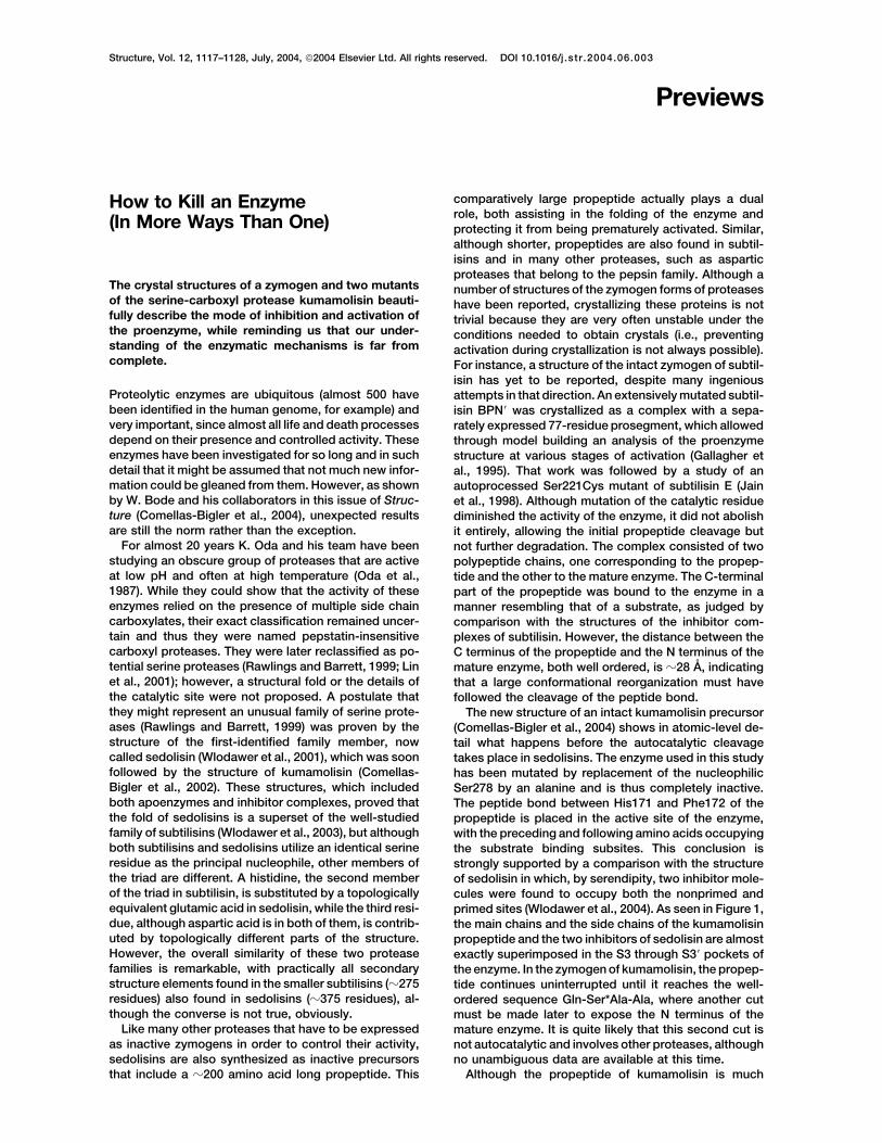
One other important question that still needs to beFigure 1. Inhibitors of Sedolisin and the Propeptide of Kumamolisin answered is the overall significance of this family ofOverlay of two inhibitors of sedolisin (yellow) and seven residues proteases. Analyses of the completed genomic se-(Ala168-Leu174, subsites P4-P3�) of the propeptide of kumamolisin quences of various organisms have shown that sedolis-(carbon atoms, green; oxygens, red; and nitrogens, blue), based
ins are present in many, but by no means in all of themon the superposition of the complete molecules of both enzymes.(Wlodawer et al., 2003). Either a sedolisin or a kumamoli-Excellent agreement between the conformations of the main chainssin is found in many bacteria (with a curious exceptionand the side chains (with the exception that the P3 side chain of
the inhibitor follows the main chain of the propeptide) reinforces of several species of Burkholderia that seem to containthe assumption that both types of ligands properly mimic substrate both sedolisins and kumamolisins), archaea, fungi, andbinding. many higher organisms. It can be safely assumed that
all readers of this preview have a functioning proteasebelonging to this family, since it has been shown that
longer than the corresponding propeptide of subtilisin, mutations leading to the loss of the human enzymethe fold of the latter is a subset of the former, again CLN2, a prominent member of the sedolisin family, resultmimicking the situation found in the mature proteins. in a fatal neurodegenerative disease, classical late-Thus, it can be concluded that serine-carboxyl prote- infantile neuronal ceroid lipofuscinosis (Sleat et al.,ases are autoinhibited by a peptide that perfectly mimics 1997). Although structural studies of sedolisins havea substrate and does not lead to substantial modifica- not firmly established their biological roles, they havetions of the mature enzyme (with an interesting excep- contributed significantly to our understanding of thetion that a positively charged arginine found in the P3 mechanism of protection and activation of proteases.position of the propeptide seems to reorient Asp164, a One may wonder if perhaps the most important functionresidue that normally functions as an oxyanion hole dur- of sedolisins and kumamolisins is to yield crystals thating catalysis, perhaps explaining in part the requirement diffract to atomic resolution (a number of structures ofof low pH for the activity of these enzymes). This is in both proteases have been solved with data extendingcontrast with other known modes of autoinhibition, such to 1.0–1.2 A)! This optimistic assessment is somehowas those reported for zymogens of aspartic proteases. tempered by the failure to obtain diffracting crystals ofThese proenzymes are inactivated by insertion of a ly- CLN2 after years of effort by several laboratories.sine side chain into the vicinity of the catalytic aspartates Clearly, the last word has not yet been said about this(pepsin or phytepsin) or by large domain movement that fascinating family of enzymes.completely rearranges the catalytic site in proplasmep-sin II (Bernstein and James, 1999).
Two other high-resolution structures of the kumamoli- Alexander Wlodawersin mutants were presented by Bode and coworkers Protein Structure Section(Comellas-Bigler et al., 2004) in order to evaluate the Macromolecular Crystallography Laboratorydifferences between the catalytic machinery of meso- National Cancer Institute at Frederickphilic sedolisin and thermophilic kumamolisin. Hydro- Frederick, Maryland 21702gen-bonded interactions in the catalytic triad of the for-mer enzyme involve only Ser, Glu, and Asp, whereas
Selected Readingin kumamolisin they are extended through two moreresidues, Glu32 and Trp129. It was postulated that these Bernstein, N.K., and James, M.N. (1999). Curr. Opin. Struct. Biol. 9,additional interactions might facilitate proton delocaliza- 684–689.tion during a nucleophilic attack at high temperature Comellas-Bigler, M., Fuentes-Prior, P., Maskos, K., Huber, R.,(Comellas-Bigler et al., 2002). Individual mutation of Oyama, H., Uchida, K., Dunn, B.M., Oda, K., and Bode, W. (2002).
Structure 10, 865–876.these residues to alanines decreased the catalytic activ-ity of kumamolisin �20-fold. Removal of the side chain Comellas-Bigler, M., Maskos, K., Huber, R., Oyama, H., Oda, K., and
Bode, W. (2004). Structure 12, this issue, 1313–1323.of Glu32 did not lead to significant structural changesand a single water molecule partially assumed the role Gallagher, T., Gilliland, G., Wang, L., and Bryan, P. (1995). Structure
3, 907–914.of the carboxylate moiety. Much larger changes were

Previews1119
Jain, S.C., Shinde, U., Li, Y., Inouye, M., and Berman, H.M. (1998). Sleat, D.E., Donnelly, R.J., Lackland, H., Liu, C.G., Sohar, I., Pullarkat,R.K., and Lobel, P. (1997). Science 277, 1802–1805.J. Mol. Biol. 284, 137–144.
Wlodawer, A., Li, M., Dauter, Z., Gustchina, A., Uchida, K., Oyama,Lin, L., Sohar, I., Lackland, H., and Lobel, P. (2001). J. Biol. Chem.H., Dunn, B.M., and Oda, K. (2001). Nat. Struct. Biol. 8, 442–446.276, 2249–2255.Wlodawer, A., Li, M., Gustchina, A., Oyama, H., Dunn, B.M., and
Oda, K., Sugitani, M., Fukuhara, K., and Murao, S. (1987). Biochim. Oda, K. (2003). Acta Biochim. Polon. 50, 81–102.Biophys. Acta 923, 463–469.
Wlodawer, A., Li, M., Gustchina, A., Oyama, H., Oda, K., Beyer,Rawlings, N.D., and Barrett, A.J. (1999). Biochim. Biophys. Acta B.B., Clemente, J., and Dunn, B.M. (2004). Biochem. Biophys. Res.
Commun. 314, 638–645.1429, 496–500.
Structure, Vol. 12, July, 2004, 2004 Elsevier Ltd. All rights reserved. DOI 10.1016/j .str .2004.06.009
suggested that the flavivirus capsid does not share theVisualizing the Houseorganization of the surface proteins. The absence of afrom the Brick structure for the flavivirus capsid makes the interpreta-tion of the capsid protein structure akin to inferring theorganization of a house from a brick. Nevertheless, suchhypotheses are the only route toward understandingthese important structures.
Ma et al. (2004) used an elegant NMR method to deter-In this issue of Structure, Dokland et al. (2004) present mine the structure of a dimer of the Dengue virus capsidthe crystal structure of West Nile Virus capsid protein protein. They demonstrated that the flavivirus capsidand open a new perspective on the possible structure protein is a novel fold. The monomer comprises a coreof the flavivirus capsid. of three helices, �1–�3, with a fourth, �4, extending from
the core. The dimer showed an uneven distribution ofcharge. They proposed that the dimer would be oriented
The structures of virus capsid proteins were once fairly in the virus with its positively charged regions extendingpredictable. The occurrence of a common fold in the centrally to interact with the RNA and the hydrophobicfirst virus structures solved by X-ray crystallography led region interacting with the membrane (Figure 1).to the inference that it might be a consistent feature of Dokland et al. (2004) determined the crystal structurevirus structures. Membrane viruses, as is so often the of the capsid protein of the Kunjin strain of West Nilecase, proved exceptions to this generalization. The pre- virus. Their structure confirms the dimer structure pre-diction that the alphavirus capsid protein would be simi- sented by Ma et al. (2004) and revealed flexibility in thelar to the nonenveloped ones (Fuller and Argos, 1987) �1 helix. The structure also revealed that the dimers formwas proven false by the Sindbis capsid protein structure tetramers in the crystal. This shields the hydrophobic(Choi et al., 1991). Indeed, a serine protease proved a regions during crystal formation and provides a highlybetter model for the alphavirus capsid. positively charged surface that could interact with the
Alphavirus and flaviviruses are two icosahedral mem- viral genome (Figure 1). They point out that the tetramersbrane viruses bearing type two fusion proteins with the form filaments reminiscent of those formed by the HEATsame fold (Lescar et al., 2001; Rey et al., 1995). The motifs in many nucleic acid interacting proteins. Thecapsid proteins of the alphaviruses form their icosahe- ribbons formed in the crystal illustrate the types of inter-dral shells prior to the interaction with the envelope actions that could be used to build the more complexproteins. This interaction and lateral interactions be- network required to construct a capsid. An order-disor-tween spikes drives virus budding from the plasma der transition of the �1 helix appears to modulate thesemembrane. The nucleocapsid can be isolated from in- interactions as is common in virus capsid assembly.fected cells and from virions. Image reconstructionsfrom cryo-electron micrographs established the organi-zation of the complementary T � 4 nucleocapsid andenvelope protein layers (Mancini et al., 2000). The capsidprotein contains a positively charged tail that interactswith the viral RNA and is toward the center of the nucleo-capsid (Choi et al., 1991).
The similarity between the fusion proteins does notextend to the capsid of the flaviviruses. Icosahedral cap-sids are difficult to isolate from virions or infected cells. Figure 1. Surface Potential of Flavivirus Capsid OligomersImage reconstruction from cryo-electron micrographs of
The GRASP (Nicholls et al., 1991) representation of the surfacemature or immature viruses show well-ordered envelope potential for the dengue virus capsid protein dimer (left) and theproteins (Kuhn et al., 2002; Zhang et al., 2003) but reveal West Nile virus capsid protein tetramer (right). The face proposed
to be against the membrane is facing the viewer in both images.no ordered density for the capsid. Kuhn et al. (2002)