http:// quality control in radiation therapy, a new concept: dosimetry check wendel dean renner math...
TRANSCRIPT
http://www.MathResolutions.com
1
Quality Control in
Radiation Therapy,A New Concept: Dosimetry Check
Wendel Dean RennerMath Resolutions,
LLCFDA Cleared April 27, 2001 US Patent 6,853,702
http://www.MathResolutions.com
2
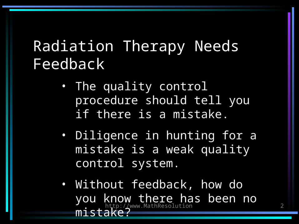
Radiation Therapy Needs Feedback
• The quality control procedure should tell you if there is a mistake.
• Diligence in hunting for a mistake is a weak quality control system.
• Without feedback, how do you know there has been no mistake?
http://www.MathResolutions.com
3
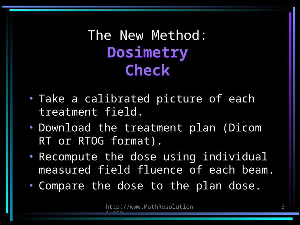
The New Method:Dosimetry
Check
• Take a calibrated picture of each treatment field.
• Download the treatment plan (Dicom RT or RTOG format).
• Recompute the dose using individual measured field fluence of each beam.
• Compare the dose to the plan dose.
http://www.MathResolutions.com
4
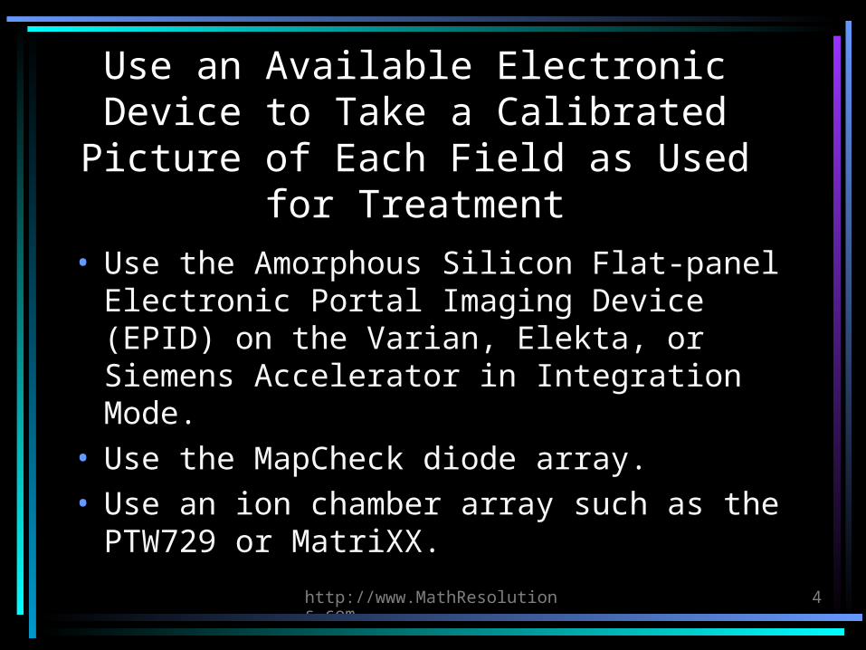
Use an Available Electronic Device to Take a Calibrated Picture of Each
Field as Used for Treatment
• Use the Amorphous Silicon Flat-panel Electronic Portal Imaging Device (EPID) on the Varian, Elekta, or Siemens Accelerator in Integration Mode.
• Use the MapCheck diode array.• Use an ion chamber array such as the
PTW729 or MatriXX.
http://www.MathResolutions.com
5
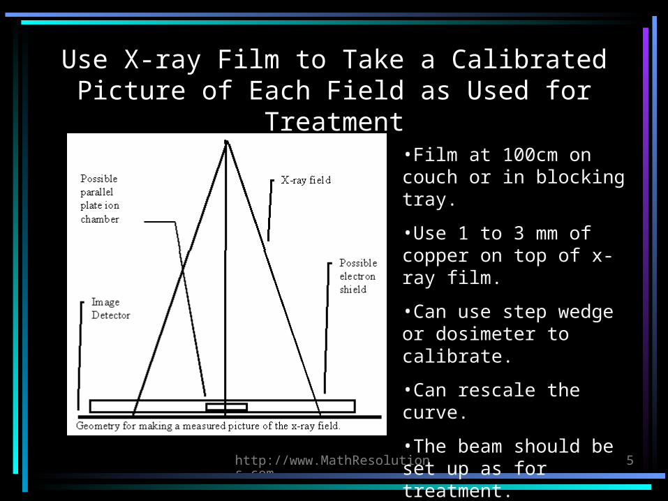
Use X-ray Film to Take a Calibrated Picture of Each Field as Used for
Treatment•Film at 100cm on couch or in blocking tray.
•Use 1 to 3 mm of copper on top of x-ray film.
•Can use step wedge or dosimeter to calibrate.
•Can rescale the curve.
•The beam should be set up as for treatment.
•1 percent accuracy is achievable for confidence.
http://www.MathResolutions.com
6
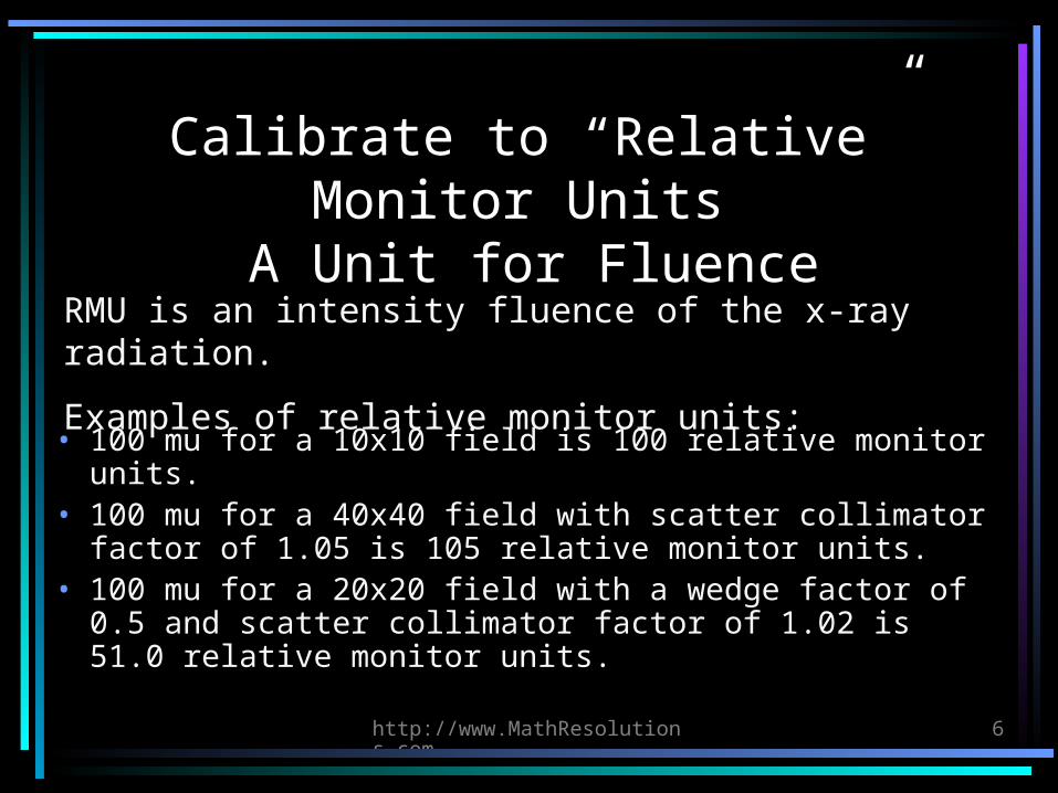
Calibrate to “Relative” Monitor Units
A Unit for Fluence
• 100 mu for a 10x10 field is 100 relative monitor units.
• 100 mu for a 40x40 field with scatter collimator factor of 1.05 is 105 relative monitor units.
• 100 mu for a 20x20 field with a wedge factor of 0.5 and scatter collimator factor of 1.02 is 51.0 relative monitor units.
RMU is an intensity fluence of the x-ray radiation.
Examples of relative monitor units:
http://www.MathResolutions.com
7
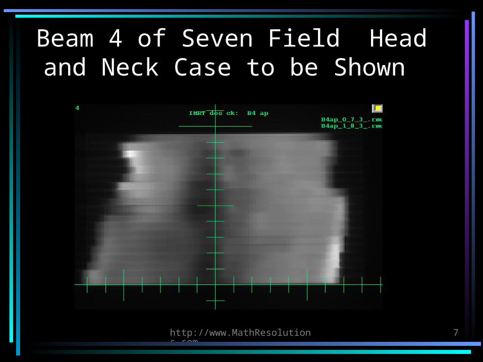
Beam 4 of Seven Field Head and Neck Case to be Shown
http://www.MathResolutions.com
8
Compute the Dose
• Download the plan in Dicom RT or RTOG format. Need CT scans, beam positions, 3D dose matrix, outlined regions of interest.
• Otherwise need the CT scan files and must position the beams to the same isocenter and angles.
• Must associate each field with the measured field picture.
http://www.MathResolutions.com
9
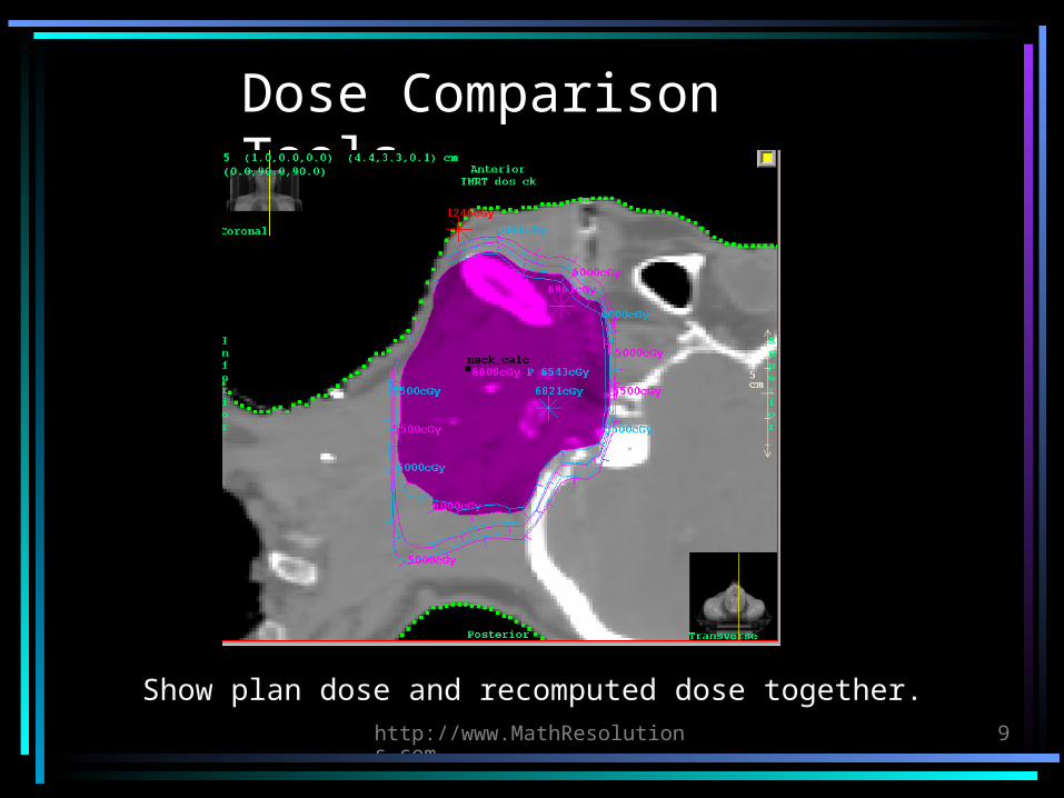
Dose Comparison Tools
Show plan dose and recomputed dose together.
http://www.MathResolutions.com
10
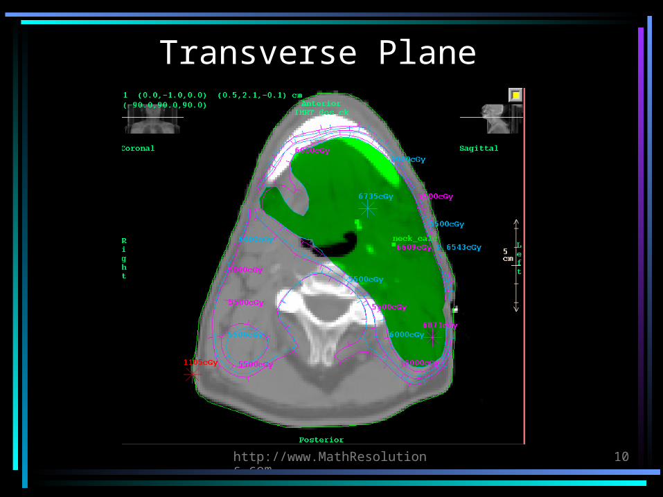
Transverse Plane

http://www.MathResolutions.com
11
Dose Comparison Tools: Gamma Method
3% - 3 mm criteria. Red tinted area is >= criteria.
http://www.MathResolutions.com
12
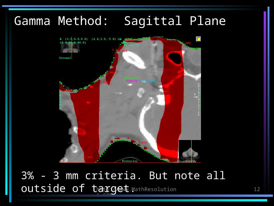
Gamma Method: Sagittal Plane
3% - 3 mm criteria. But note all outside of target.
http://www.MathResolutions.com
13
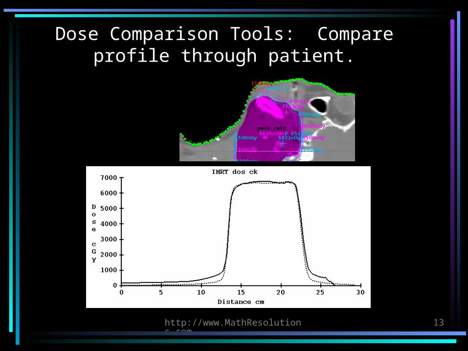
Dose Comparison Tools: Compare profile through patient.
http://www.MathResolutions.com
14
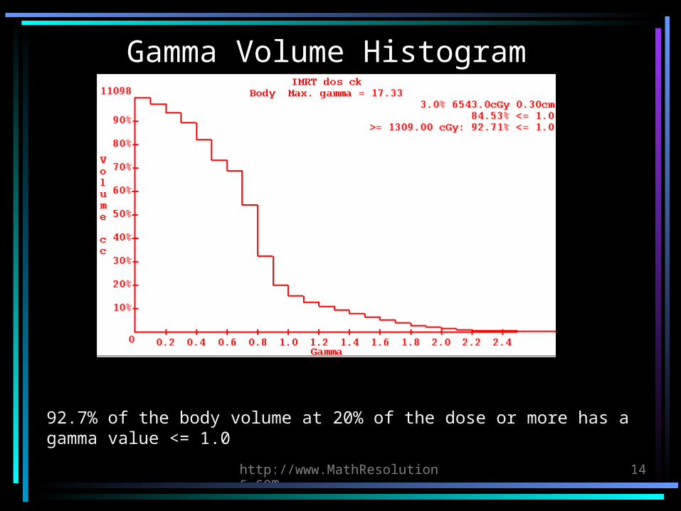
Gamma Volume Histogram
92.7% of the body volume at 20% of the dose or more has a gamma value <= 1.0
http://www.MathResolutions.com
15
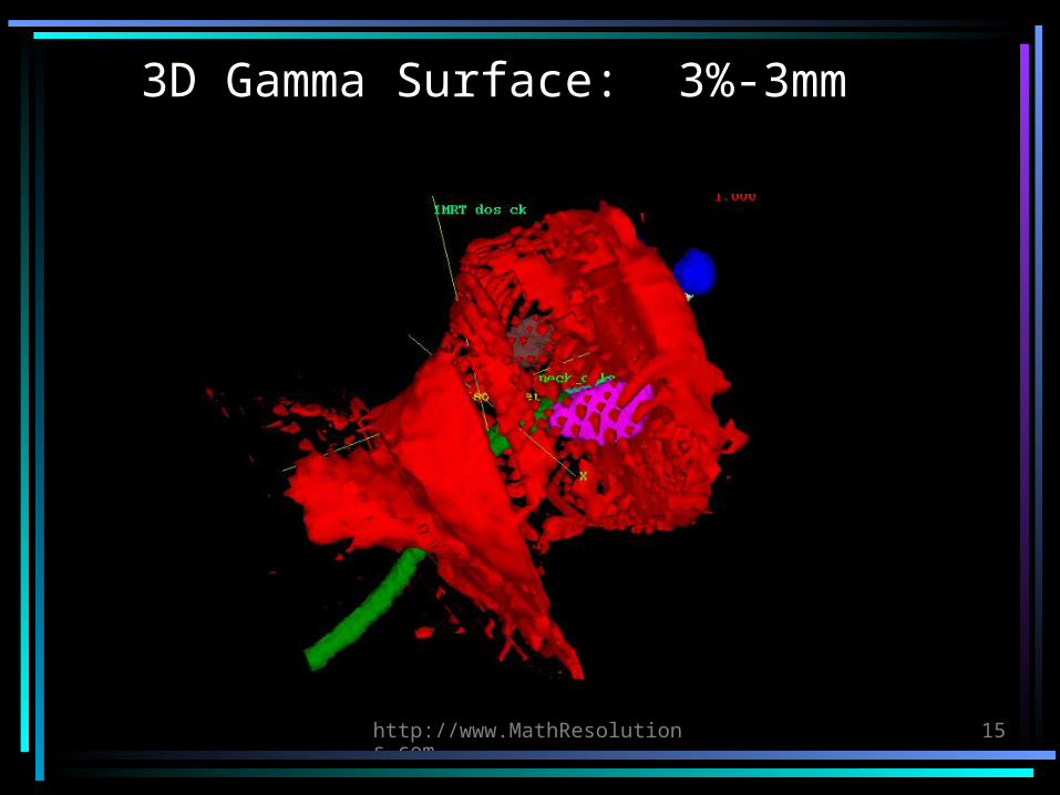
3D Gamma Surface: 3%-3mm
http://www.MathResolutions.com
16
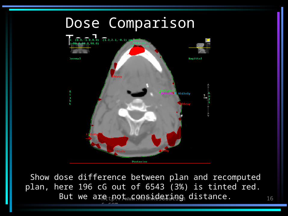
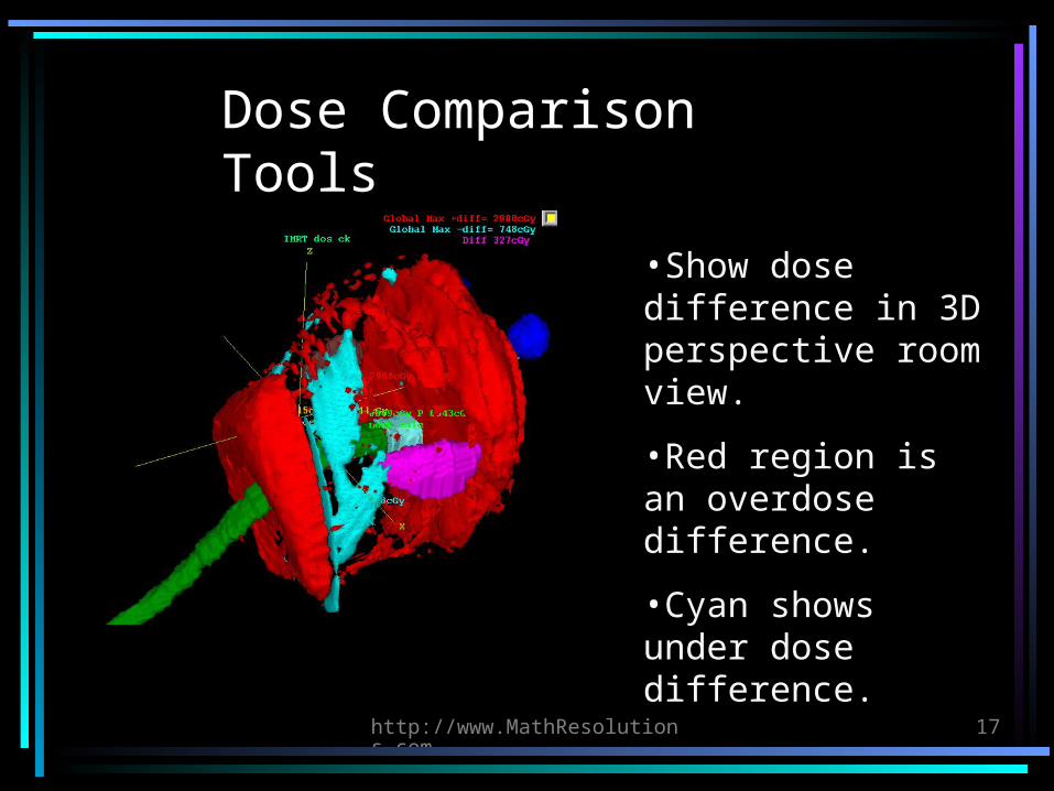
Dose Comparison Tools
Show dose difference between plan and recomputed plan, here 196 cG out of 6543 (3%) is tinted red. But we are not
considering distance.
http://www.MathResolutions.com
17
Dose Comparison Tools
•Show dose difference in 3D perspective room view.
•Red region is an overdose difference.
•Cyan shows under dose difference.
http://www.MathResolutions.com
18
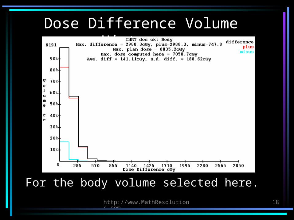
Dose Difference Volume Histogram
For the body volume selected here.
http://www.MathResolutions.com
19
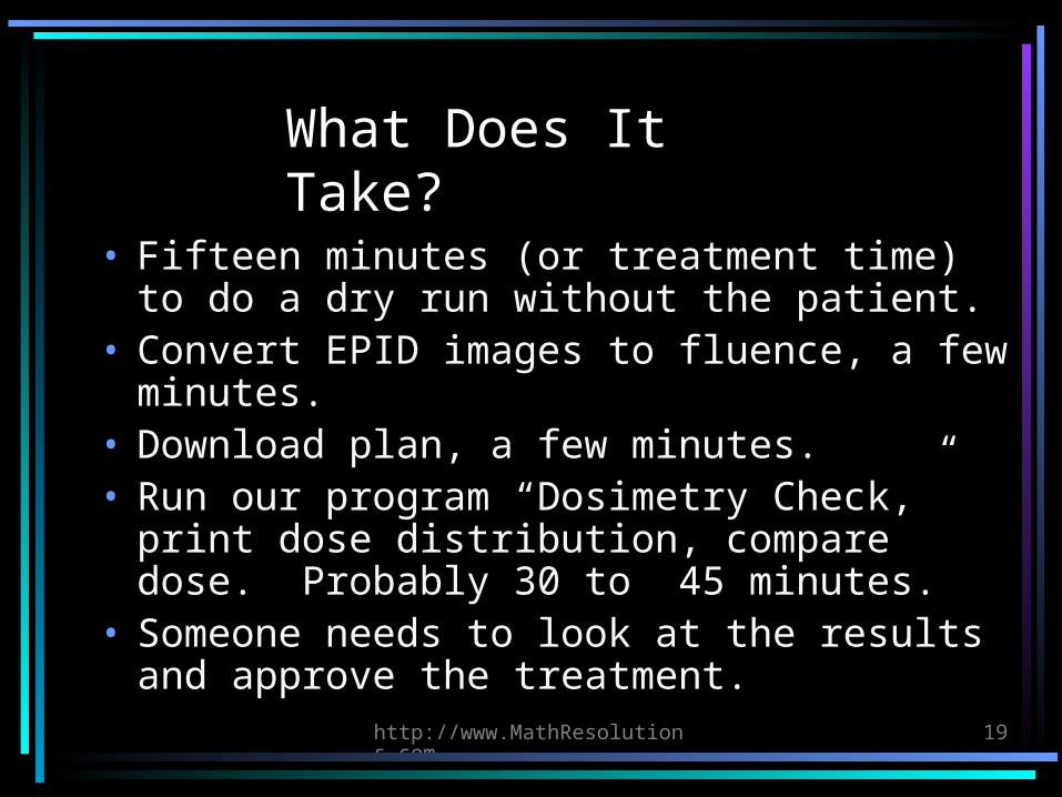
What Does It Take?
• Fifteen minutes (or treatment time) to do a dry run without the patient.
• Convert EPID images to fluence, a few minutes.
• Download plan, a few minutes.• Run our program “Dosimetry Check,” print
dose distribution, compare dose. Probably 30 to 45 minutes.
• Someone needs to look at the results and approve the treatment.
http://www.MathResolutions.com
20
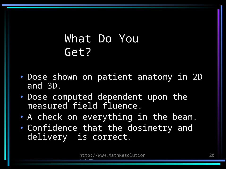
What Do You Get?
• Dose shown on patient anatomy in 2D and 3D.
• Dose computed dependent upon the measured field fluence.
• A check on everything in the beam.• Confidence that the dosimetry and
delivery is correct.
http://www.MathResolutions.com
21
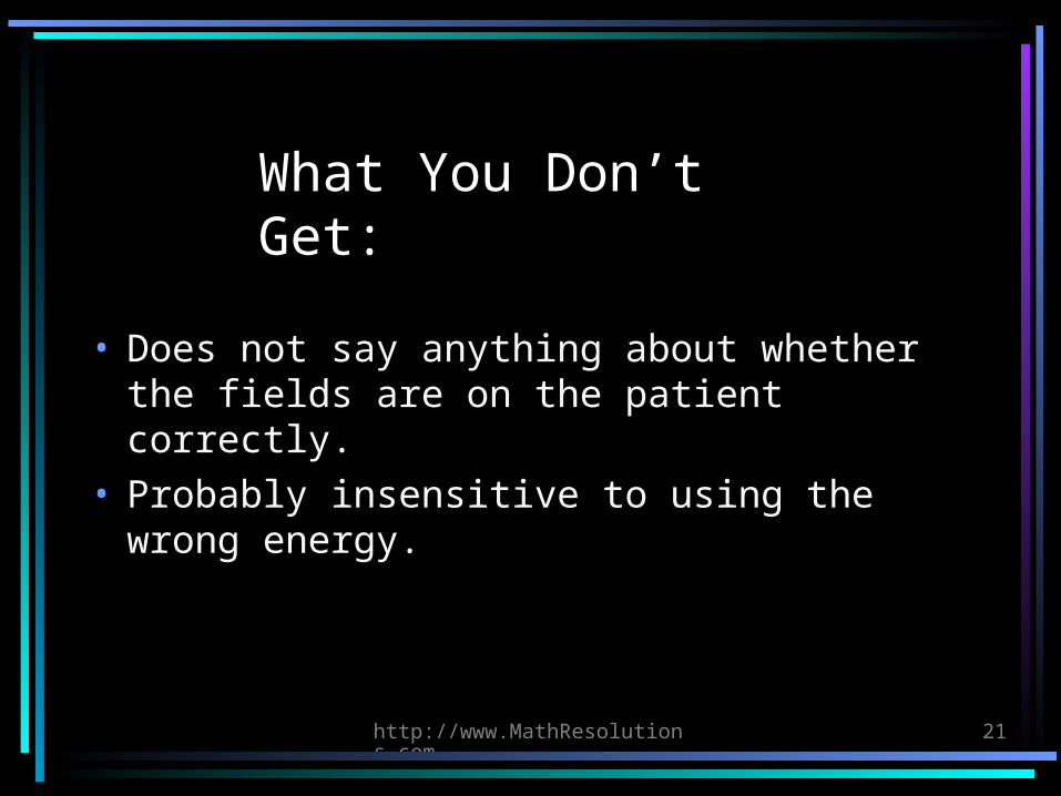
What You Don’t Get:
• Does not say anything about whether the fields are on the patient correctly.
• Probably insensitive to using the wrong energy.
http://www.MathResolutions.com
22
Why?
• Above all else, do no harm.
http://www.MathResolutions.com
23
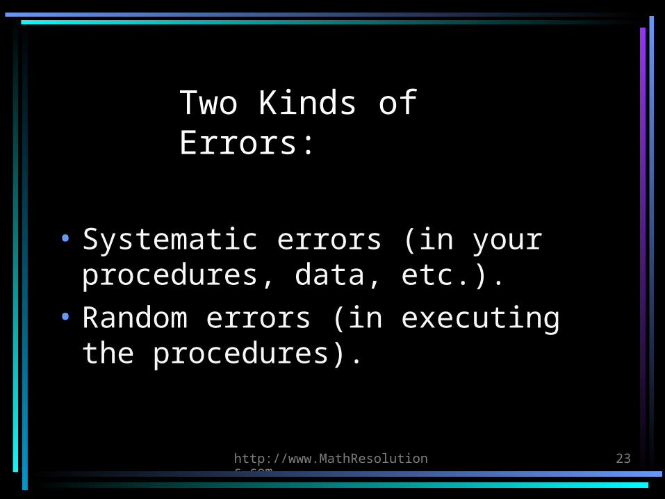
Two Kinds of Errors:
• Systematic errors (in your procedures, data, etc.).
• Random errors (in executing the procedures).
http://www.MathResolutions.com
24
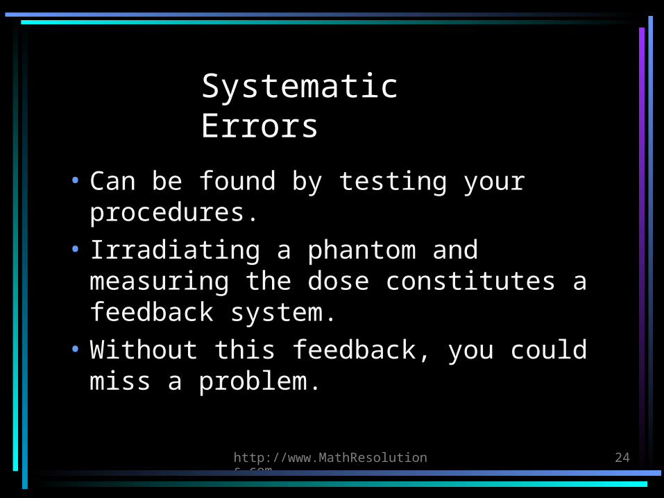
Systematic Errors
• Can be found by testing your procedures.
• Irradiating a phantom and measuring the dose constitutes a feedback system.
• Without this feedback, you could miss a problem.
http://www.MathResolutions.com
25
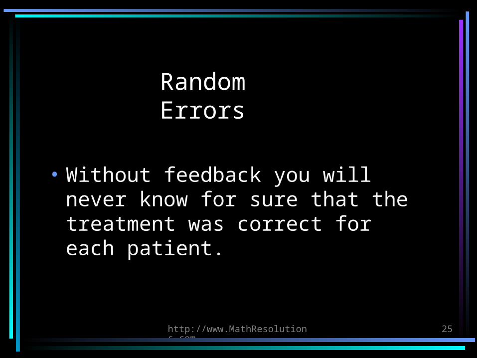
Random Errors
• Without feedback you will never know for sure that the treatment was correct for each patient.
http://www.MathResolutions.com
26
Overdose
• The patient reaction will tell you too late!
http://www.MathResolutions.com
27
Under Dose
• Might never be noticed.• Results in lost opportunity to
effectively treat the cancer.
http://www.MathResolutions.com
28
Do we really knowthe extent of mistakes that occur in radiation
therapy?
http://www.MathResolutions.com
29
Effective Quality Assurance
• We want to maximize feedback.• We want to maximize the amount of
information that is measured in the treatment room.
• We want to minimize the amount of information we use that is NOT measured in the room.
http://www.MathResolutions.com
30
Monitor Unit Check
• Redundancy is good.• But there is no feedback.
http://www.MathResolutions.com
31
Present Methods with Feedback
• Diode surface measurement at one point on each field to be compared to computed dose.
• Irradiate a cylindrical or square phantom using the plan, recompute the plan to same phantom to compare.
http://www.MathResolutions.com
32
Diode Surface Measurement
• This is a simple QA procedure, yet a surprising number of centers do not do it. But this only checks the dose at one point.
• Wedge could still be in wrong direction.• Compensator or IMRT could be wrong.• Blocks and/or field shape could be wrong
(should also pick up from film review, but some redundancy would be good).
• Dose distribution could still be wrong.
http://www.MathResolutions.com
33
Phantom
• Quality controls how the beams add up.• Maximizes the information measured in
room.• Can only show dose where measured in
the phantom.• Comparison of dose is somewhat abstract
in relation to the patient.• Assumption that dose in patient will be
right might not hold.• Does not tell you if beams are on the
patient correctly.
http://www.MathResolutions.com
34
Our new method:Dosimetry Check
• Shows the dose on the patient anatomy. • Evaluation of dose is not abstract.• Can handle arc therapy (but we presently
don’t have a device to measure a field that changes shape during gantry rotation).
• Can show dose in any plane or 3D dose cloud.• Same limitation of only checking dosimetry.• Easy and fast to do.
http://www.MathResolutions.com
35
Dosimetry Check Adds a Powerful Tool for Quality Control
• May replace some less effective procedures.
• May compliment others.
http://www.MathResolutions.com
36
Math Resolutions, LLC, is offering software to accomplish the new
method.
• FDA 510(k) K010225.• Software, manuals,and information
available at our website: http://MathResolutions.com.
• U.S. Patent 6,853,702 on this process.
http://www.MathResolutions.com
37
Measuring the field with X-Ray
film
• One could use a device instead of film.• Optimally the field should be measured
during treatment. That would require a device that the patient could be treated through.
• Film must be digitized.
http://www.MathResolutions.com
38
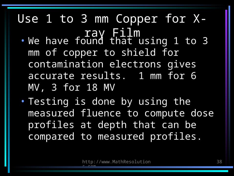
Use 1 to 3 mm Copper for X-ray Film
• We have found that using 1 to 3 mm of copper to shield for contamination electrons gives accurate results. 1 mm for 6 MV, 3 for 18 MV
• Testing is done by using the measured fluence to compute dose profiles at depth that can be compared to measured profiles.
http://www.MathResolutions.com
39
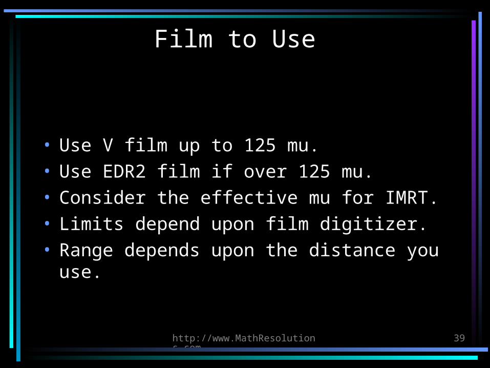
Film to Use
• Use V film up to 125 mu.• Use EDR2 film if over 125 mu.• Consider the effective mu for IMRT.• Limits depend upon film digitizer.• Range depends upon the distance you
use.
http://www.MathResolutions.com
40
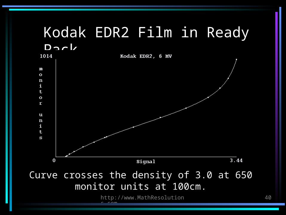
Kodak EDR2 Film in Ready Pack
Curve crosses the density of 3.0 at 650 monitor units at 100cm.
http://www.MathResolutions.com
41
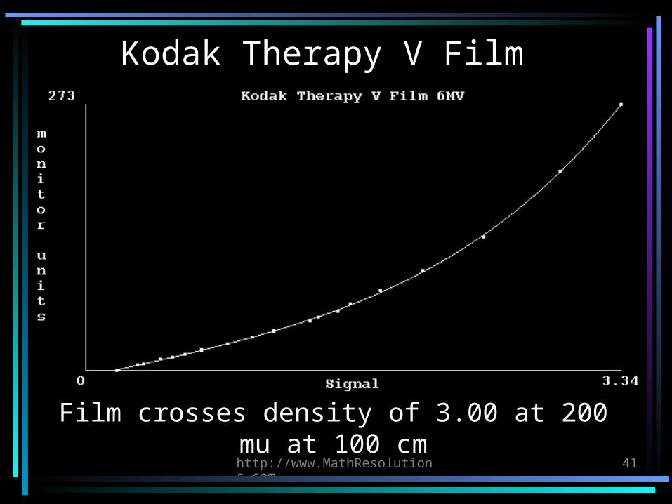
Kodak Therapy V Film
Film crosses density of 3.00 at 200 mu at 100 cm
http://www.MathResolutions.com
42
Calibration Procedures for X-Ray Film
• Include ion chamber or diode in bolus stack.• Or use a step wedge.• Run curve to calibrate the step wedge once.• Thereafter use the step wedge with each
case.• Or consider rescaling the calibration curve
from a single test exposure.• The goal is accuracy to 1 percent for
measuring relative mu
http://www.MathResolutions.com
43
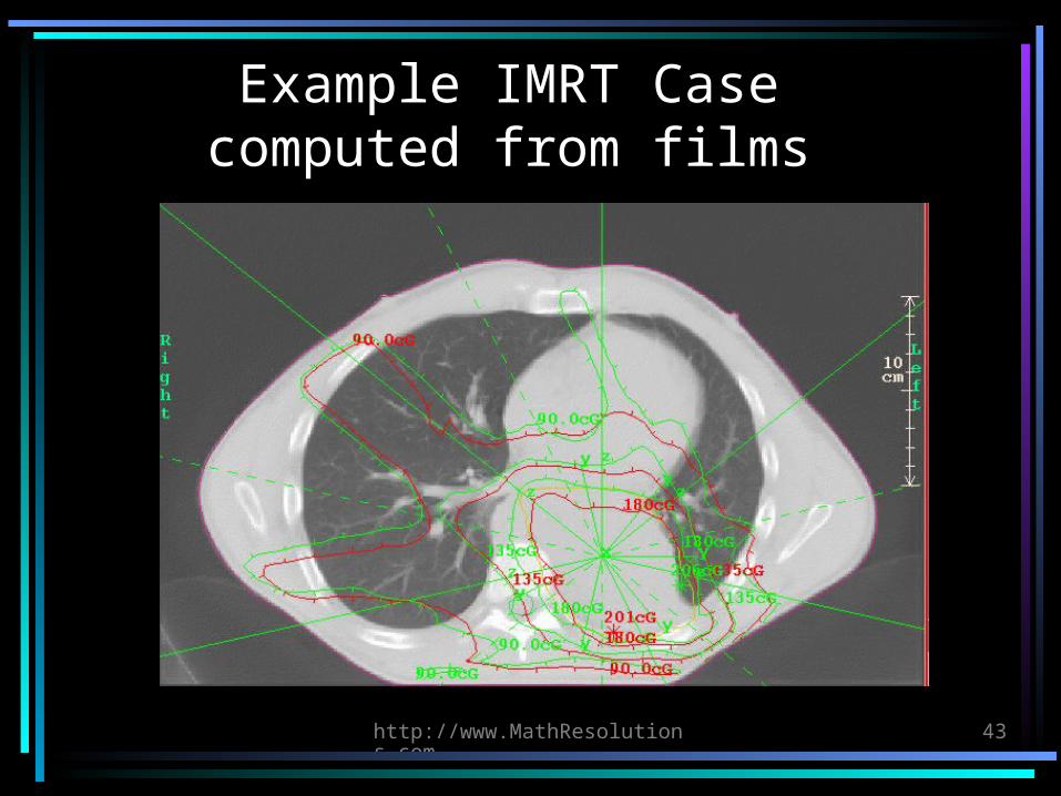
Example IMRT Casecomputed from films
http://www.MathResolutions.com
44
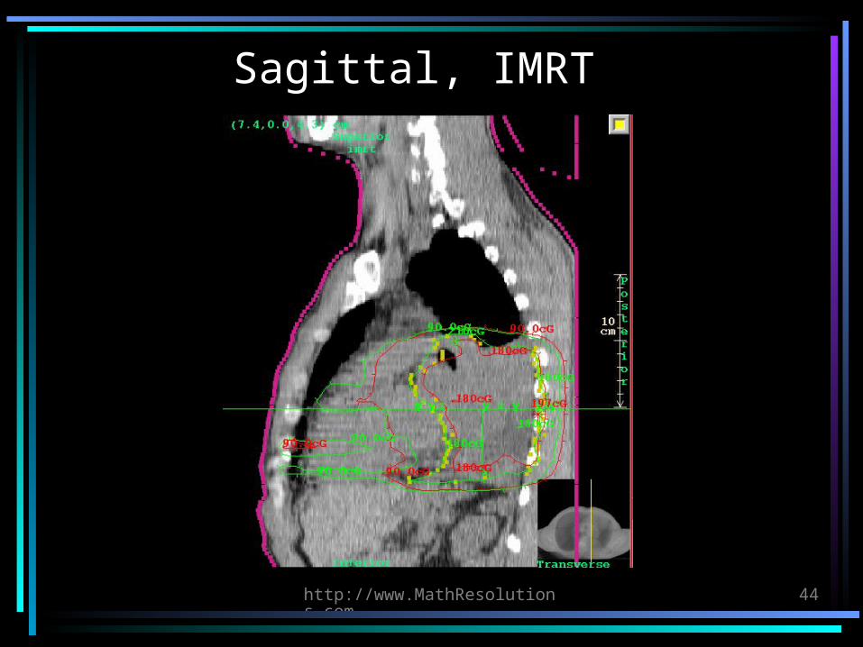
Sagittal, IMRT
http://www.MathResolutions.com
45
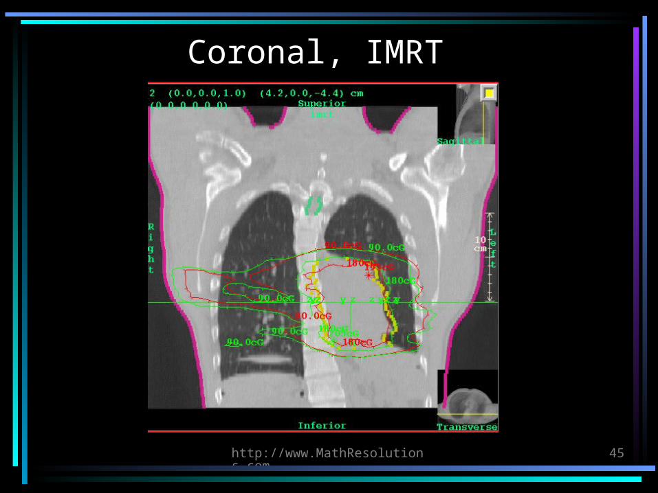
Coronal, IMRT