http:// ?v=bxttynf8zv0. bleomycin a2: anibiotic, antineoplastic; cell cycle arrest in g2 and in...
TRANSCRIPT

http://www.youtube.com/watch?v=BXttyNf8zv0


Bleomycin A2: anibiotic, antineoplastic; cell cycle arrest in G2 and in mitosis.
3-[[2-[2-[2-[[(2S,3R)-2-[[(2S,3S,4R)-4-[[(2S,3R)-2-[[6-Amino-2-[(1S)-3-amino-1-[[(2S)-2,3-diamino-3 oxopropyl]amino]-3-oxopropyl]-5-methylpyrimidine-4-carbonyl]amino]-3-[(2R,3S,4S,5S,6S)-3-[(2R,3S,4S,5R,6R)-4-carbamoyloxy-3,5-dihydroxy-6-(hydroxymethyl)oxan-2-yl]oxy-4,5-dihydroxy-6-(hydroxymethyl)oxan-2-yl]oxy-3-(1H-imidazol-5-yl)propanoyl]amino]-3-hydroxy-2-methylpentanoyl]amino]-3-hydroxybutanoyl]amino]ethyl]-1,3-thiazol-4-yl]-1,3-thiazole-4-carbonyl]amino]propyl-dimethylsulfanium
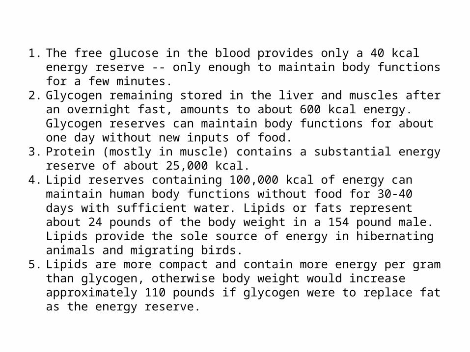
1. The free glucose in the blood provides only a 40 kcal energy reserve -- only enough to maintain body functions for a few minutes.
2. Glycogen remaining stored in the liver and muscles after an overnight fast, amounts to about 600 kcal energy. Glycogen reserves can maintain body functions for about one day without new inputs of food.
3. Protein (mostly in muscle) contains a substantial energy reserve of about 25,000 kcal.
4. Lipid reserves containing 100,000 kcal of energy can maintain human body functions without food for 30-40 days with sufficient water. Lipids or fats represent about 24 pounds of the body weight in a 154 pound male. Lipids provide the sole source of energy in hibernating animals and migrating birds.
5. Lipids are more compact and contain more energy per gram than glycogen, otherwise body weight would increase approximately 110 pounds if glycogen were to replace fat as the energy reserve.

Outline• How are carbohydrates named?• What is the structure and chemistry of
monosaccharides? • What is the structure and chemistry of
oligosaccharides?• What is the structure and chemistry of
polysaccharides? • What are glycoproteins, and how do they function in
cells?• How do proteoglycans modulate processes in cells
and organisms?• Do carbohydrates provide a structural code?

7.1 How Are Carbohydrates Named?
Carbohydrates are hydrates of carbonWith the general formula (CH2O)n
• Carbohydrates are classified in three groups:• Monosaccharides (simple sugars) - cannot be broken
down into simpler sugars under mild conditions • Oligosaccharides (oligo = "a few“) - usually 2 to 10
simple sugar residues• Polysaccharides - are polymers of the simple sugars

7.2 What Is the Structure and Chemistry of Monsaccharides?
Figure 7.1 Structure of a simple aldose and a simple ketose.

7.2 What Is the Structure and Chemistry of Monsaccharides?
Figure 7.2 - Detail

7.2 What Is the Structure and Chemistry of Monsaccharides?
Figure 7.3 - Detail

Stereochemistry Review
Read text on p. 183-185 carefully:• D,L designation refers to the configuration of the
highest-numbered asymmetric center • D,L only refers the stereocenter of interest back to D-
and L-glyceraldehyde• D,L do not specify the sign of rotation of plane-
polarized light • All structures in Figures 7.2 and 7.3 are D-sugars • D-sugars predominate in nature

Stereochemistry ReviewStereoisomers that are mirror images of each other are enantiomers Pairs of isomers that have opposite configurations at one or more chiral centers but are NOT mirror images are diastereomers Any 2 sugars in a row in Figure 7.2 and 7.3 are diastereomers Two sugars that differ in configuration at only one chiral center are epimers

Cyclic monsaccharide structures and anomeric forms
Glucose (an aldose) can cyclize to form a cyclic hemiacetal
• Fructose (a ketose) can cyclize to form a cyclic hemiketal
• When hemiacetals and hemiketals are formed, the carbon atom that carried the carbonyl function becomes an asymmetric carbon
• Isomers of monosaccharides that differ only in their configuration about that asymmetric carbon are called anomers
• Cyclic form of glucose is a pyranose • Cyclic form of fructose is a furanose

Monosaccharides Exist in Cyclic, Anomeric Forms
Figure 7.5 The linear form of D-glucose undergoes an intramolecular reaction to form a cyclic hemiacetal – with an anomeric carbon. Note α and β designations.

7.2 What Is the Structure and Chemistry of Monsaccharides?
Figure 7.5 The linear form of D-fructose undergoes an intramolecular reaction to form a cyclic hemiketal – with an anomeric carbon. Note α and β designations.

Cyclic monsaccharide structures and anomeric forms
• Isomers of monosaccharides that differ only in their configuration about the asymmetric carbon are called anomers
• When the hydroxyl group at the anomeric carbon is on the same side of a Fischer projection as the oxygen atom at the highest numbered asymmetric carbon, the configuration at the anomeric carbon is α, as in α-D-glucose
• A simpler rule:• For D-sugars, alpha has OH down, beta up* • For L-sugars, the reverse is true• *In the customary Haworth drawing

7.2 What Is the Structure and Chemistry of Monsaccharides?
Figure 7.8 (a) Chair and boat conformations of a pyranose sugar.(b) Two possible chair conformations of β-D-glucose. Note the axial and equatorial substituents.

Monosaccharides Can Be Converted to Several Derivative Forms
• A variety of chemical and enzymatic reactions produce derivatives of the simple sugars
• Some of the most common are:– Sugar acids– Sugar alcohols– Deoxy sugars– Sugar esters– Amino sugars– Acetals, ketals, and glycosides

Monosaccharides Can Be Converted to Several Derivative Forms
• A variety of chemical and enzymatic reactions produce derivatives of the simple sugars
• Some of the most common are:– Sugar acids– Sugar alcohols– Deoxy sugars– Sugar esters– Amino sugars– Acetals, ketals, and glycosides

Monosaccharide Derivatives
Reducing sugars are sugars with free anomeric carbons - they will reduce oxidizing agents, such as peroxide, ferricyanide and some metals (Cu2+ and Ag+) These redox reactions convert the sugar to a sugar acid Glucose is a reducing sugar - so these reactions are the basis for diagnostic tests for blood sugar

More Monosaccharide Derivatives
• Sugar alcohols are formed by mild reduction of sugars
• Deoxy sugars: constituents of DNA, etc. • Sugar esters: phosphate esters like ATP are important • Amino sugars contain an amino group in place of a
hydroxyl group • Acetals, ketals and glycosides: basis for oligo- and
polysaccharides

Sugar Alcohols
Figure 7.10 Structures of some sugar alcohols.

Amino Sugars
Figure 7.14
• Sugars with an amino group at C-2 are amino sugars.• They are found in many oligosaccharides and polysaccharides.

Acetals and Ketals
Figure 7.16 Acetals and ketals can be formed from hemiacetals and hemiketals, respectively. These reactions are examples of dehydration syntheses. In this sense, they are similar to the reactions of amino acids to form peptides.

Glycosides
Figure 7.17 The anomeric forms of methyl-D-glucoside.
The pyranose and furanose forms of monosaccharides react with alcohols in dehydration synthesis reactions to form glycosides, with retention of the α- or β-configuration at the C-1 carbon. The new bond formed is called a glycosidic bond.

7.3 What is the Structure and Chemistry of Oligosaccharides?
Figure 7.18 The structures of several important disaccharides. Note the configurations at the anomeric carbons. Note also that sucrose is not a reducing sugar.

7.3 What is the Structure and Chemistry of Oligosaccharides?
It’s not important to memorize structures, but you should know the important features
• Be able to identify anomeric carbons and reducing and nonreducing ends
• Note that sucrose is NOT a reducing sugar • Browse the structures in Figure 7.18 and Figure
7.19 • Note carefully the nomenclature of links. Be able
to recognize alpha(1,4), beta(1,4) linkages, etc., in structures.

A Variety of Higher Oligosaccharides Occur in Nature
Oligosaccharides occur widely as components of antibiotics (derived from various sources).
Figure 7.19 Erythromycin is an antibiotic produced by a strain of Streptomyces erythreus.

A Variety of Higher Oligosaccharides Occur in Nature
Figure 7.19 Streptomycin is an oligosaccharide produced by Stretomyces griseus.
Oligosaccharides occur widely as components of antibiotics (derived from various sources).

StarchA plant storage polysaccharide
• Two forms: amylose and amylopectin • Most starch is 10-30% amylose and 70-90%
amylopectin • Branches in amylopectin every 12-30 residues • Amylose has alpha(1,4) links, one reducing end • The branches in amylopectin are α(1→6).

Amylose and Amylopectin are energy storage molecules in plants
Figure 7.20 Amylose and amylopectin are two forms of starch. Amylopectin is highly branched, with branches occurring every 12 to 30 residues.

Glycogen
The glucose storage device in animals
• Glycogen constitutes up to 10% of liver mass and 1-2% of muscle mass
• Glycogen is stored energy for the organism • Only difference from amylopectin: the frequency of
branching • Alpha(1,6) branches every 8-12 residues • Like amylopectin, glycogen gives a red-violet color
with iodine

Dextrans
A small but significant difference from starch and glycogen
• If you change the main linkages between glucose from alpha(1,4) to alpha(1,6), you get a new family of polysaccharides - dextrans
• Branches can be (1,2), (1,3), or (1,4)

Roles for Dextrans
• Dextrans formed by bacteria are components of dental plaque
• Dextrans in plaque presumably provide protection for oral bacteria
• Cross-linked dextrans are used as "Sephadex" gels in column chromatography
• These gels, used to separate biomolecules on the basis of size, are up to 98% water!

Structural Polysaccharides
• The composition of structural polysaccharides is similar to storage polysaccharides
• But small structural differences greatly influence properties
• Starch and glycogen linkages consist primarily of α(1→4) linkages.
• Cellulose consists of β(1→4) linkages

Cellulose Provides Physical Structure and Strength to Plants
• Cellulose is a structural polysaccharide• It is the most abundant natural polymer in the world• It is found in the cell walls of nearly all plants• The wood and bark of trees are insoluble, highly
organized structures formed from cellulose and lignin• Cotton is almost pure cellulose• Cotton acetates, made from the action of acetic
anhydride on cellulose, are used in dresses, lingerie, and other clothing
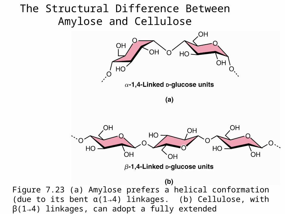
The Structural Difference Between Amylose and Cellulose
Figure 7.23 (a) Amylose prefers a helical conformation (due to its bent α(1→4) linkages. (b) Cellulose, with β(1→4) linkages, can adopt a fully extended conformation.

The Structure of Cellulose
Figure 7.24 The structure of cellulose, showing the hydrogen bonds (blue) between the sheets, which strengthen the structure.
Intrachain H-bonds in red; interchain H-bonds in green.

How Do Ruminant Animals Digest Cellulose?
Figure 7.25 Giraffes, cattle, deer, and camels are ruminant animals that are able to metabolize cellulose, thanks to bacterial cellulase in the rumen, a large first compartment in the stomach of a ruminant.

Structures of cellulose, chitin and mannan
Figure 7.26 Like cellulose, chitin and mannan form extended ribbons and pack together efficiently, taking advantage of multiple hydrogen bonds.

The Structure of Agarose in Solution
Figure 7.27 The favored conformation of agarose in water is a double helix with a threefold screw axis.
• Agarose, a component of agar obtained from marine red algae, is a chain of alternating D-galactose and 3,6-anhydro-L-galactose.
• Agarose gels are used in laboratories to separate biomolecules on the basis of size

Bacterial Cell Walls
Composed of 1 or 2 bilayers and peptidoglycan shell
• Gram-positive: One bilayer and thick peptidoglycan outer shell
• Gram-negative: Two bilayers with thin peptidoglycan shell in between
• Gram-positive: pentaglycine bridge connects tetrapeptides
• Gram-negative: direct amide bond between tetrapeptides

The Structure of Peptidoglycan
Figure 7.29 The tetrapeptides linking adjacent backbone chains contain an unusual γ-carboxyl linkage.
Note the isoglutamate linkage in the tetrapeptide chain

The Structure of Gram-positive Peptidoglycan
Figure 7.29 The crosslink between tetrapeptides in Gram-positive cell walls is a pentaglycine bridge.

The Structure of Gram-negative Peptidoglycan
Note the direct crosslinks between tetrapeptide units
Figure 7.29 In Gram-negative cell walls, the linkage between tetrapeptide segments involves a direct amide bond.