human anatomy & physiology i lab 5 microscopy 2 …...human anatomy & physiology i lab 5...
TRANSCRIPT
Human Anatomy & Physiology I Lab 5 Microscopy 2 – The Microscopic Features of Tissues
Learning Outcomes Set up a microscope to view slides quickly, properly, efficiently, and without damaging the
microscope or slide.
Assessment: Exercises 5.1
Identify accurately the location and type of epithelial tissue within a biological specimen
Assessments: Exercise 5.2
Classify a histological specimen according to which category of connective tissue it belongs to,
based on the types of cells present and the characteristics of the extracellular matrix.
Assessment: Exercises 5.3, 5.4
Identify the main structural features in photomicrographs of cartilage and bone.
Assessments: Exercise 5.5
Understand the visual differences between the three types of muscular tissue.
Assessments: Exercise 5.6
Find the neurons within a smear of nervous tissue and be able to identify the parts of the neural
cell.
Assessments: Exercise 5.7
Review of basic handling of the microscope. Information
Remember, a microscope is an expensive, complex machine with many moving parts. You need
to take care when setting it up with your slides and treat it gently.
Lab exercises 5.1 There is a compound microscope for each student.
1. Obtain one of the slides you will need to day for one of the Lab exercises that follow this one.
2. Perform the following checklist of activities, checking each one off as you carry it out. Stop
when you have your specimen in focus with the second objective (usually the 40x objective, with
most of the lab microscopes.)

Checklist for setting up a microscope and beginning with a new slide.
1. Plug in microscope & turn on light source.
2. Pick up microscope by carrying arm, position it so it is accessible to your seat, with
open side of the stage facing you.
3. Rotate the objectives so that the lowest power objective (smallest in size) clicks into
place.
4. Look at slide with your naked eye and find the location of the specimen.
5. Clip slide into place with stage clips. The cover slip on the slide must be face up. Find
the stage controls and make sure that, when they are turned, the slide moves smoothly
left & ride or up & down, depending on knob.
6. Use stage controls to move slide so that light source is shining directly on to the
specimen to be magnified.
7. Find the coarse and fine focus knobs. Watching the stage and objective, use the coarse
focus knob to bring the low power objective as close to the slide as it will go.
8. Put your eye to the eyepiece (or eyepieces, if the microscope is binocular) and rotate
the coarse focus knob in the lowering direction until some aspect of the specimen
comes into focus.
9. Move your hand to the fine focus knob and get the specimen into perfect focus for your
eyes. Do NOT touch the coarse focus knob again.
10. Use the stage control knobs to move you specimen to close to the exact center of your
field of view.
11. Move to the next highest power objective (do not skip individual objectives) and use
only the fine focus to get your image into perfect focus for your eyes.
12. If you need further magnification, move to the next highest power objective and use
only the fine focus to get your image into perfect focus for your eyes.
13. Do not use the 100x objective (if you have one) in this course. It must be used with
immersion oil and we won’t have students doing that.
Examining epithelial tissue under the microscope Information
Epithelial tissue serves two main functions in the body.
1. It provides linings for external and internal surfaces that face harsh environments. The outer
layer of the skin is epithelial tissue, as are the innermost layers of the digestive tract, the
respiratory tract, and blood vessels.
2. It forms glands that secrete materials onto epithelial surfaces or into the blood. Sweat
glands, salivary glands, mammary glands, adrenal glands, and pituitary glands are examples
of glands made of epithelial tissue.
Epithelial tissue is often classified according to numbers of layers of cells present, and by the
shape of the cells. See Figure 5.1.
A simple epithelium is only one layer of cells thick. A stratified epithelium is more than
one layer of cells thick. A pseudostratified epithelium is really a specialized form of a simple
epithelium in which there appears at first glance to be more than one layer of epithelial cells, but a
closer inspection reveals that each cell in the layer actually extends to the basolateral surface of the
epithelium.
There are three basic shapes used to classify epithelial cells. A squamous epithelial cell looks
flat under a microscope. A cuboidal epithelial cell looks close to a square. A columnar epithelial
cell looks like a column or a tall rectangle. A few epithelial layers are constructed from cells that are
said to have a transitional shape. Transitional epithelial cells are epithelial cells specialized to
change shape if they are stretched laterally. They can transition from columnar- and cuboidal-
looking shapes in their unstretched state to more squamous-looking shapes in their stretched state.
When classifying a stratified epithelial sheet, the sheet is named for the shape of the cells in its
most superficial layers. So a stratified squamous epithelium only necessarily has squamous-shaped
cells in its highest layers and might have a different-shaped cell in its lower layers.
Under a microscope, epithelial cells are readily distinguished by the following features:
The cells will usually be one of the three basic cell shapes – squamous, cuboidal, or
columnar.
The cells will be closely attached to one another, in either a single layer or in multiple layers,
and usually will not have room for extracellular material between the attached cells.
The epithelial layer on one side will face an empty space (or, in some organs, it will face a
secreted substance like mucus) and on the other side will usually be attached to connective
tissue proper.
Usually, a slide will have a section of tissue cut out of a larger organ. Slides with epithelial
tissues usually have some of the underlying tissue found beneath the epithelial tissue with them.
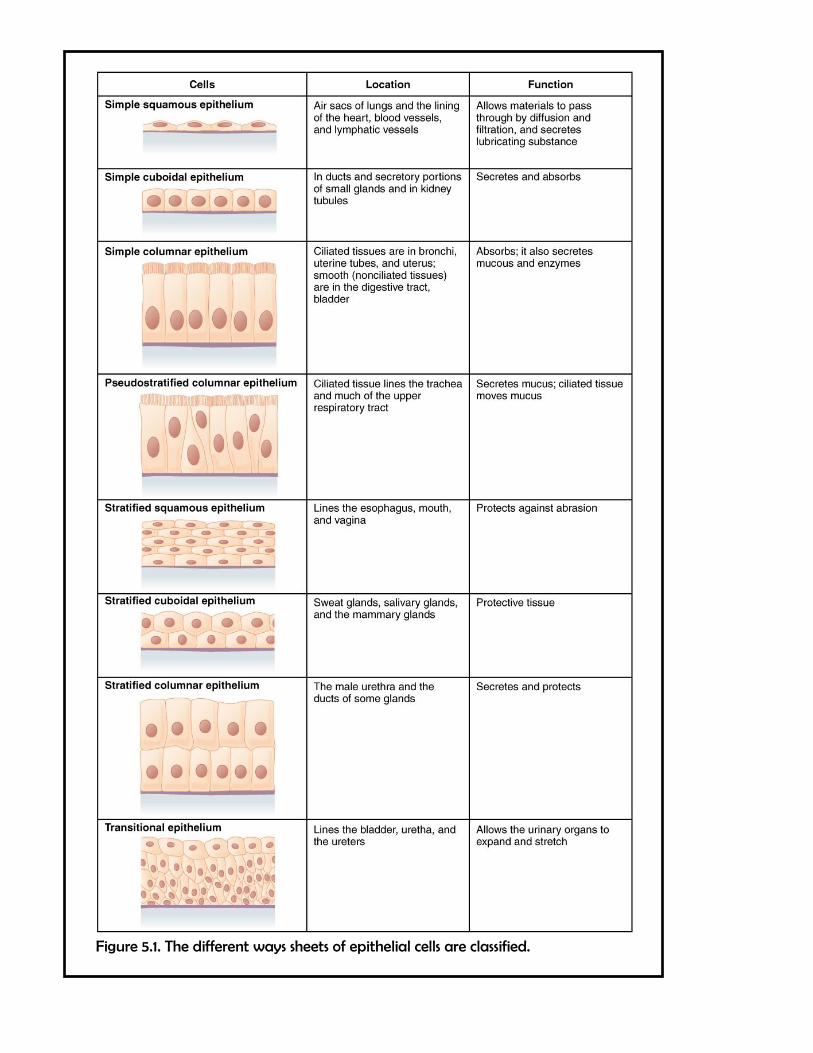
Figure 5.1. The different ways sheets of epithelial cells are classified.
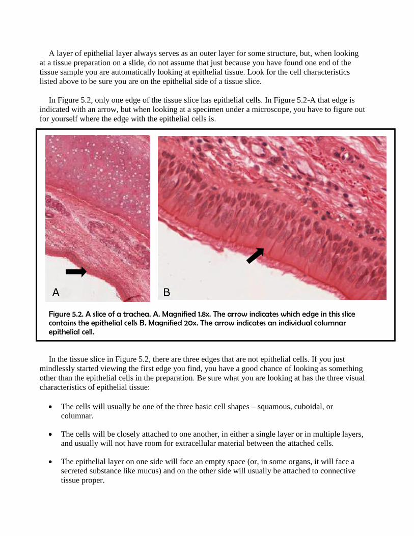
A layer of epithelial layer always serves as an outer layer for some structure, but, when looking
at a tissue preparation on a slide, do not assume that just because you have found one end of the
tissue sample you are automatically looking at epithelial tissue. Look for the cell characteristics
listed above to be sure you are on the epithelial side of a tissue slice.
In Figure 5.2, only one edge of the tissue slice has epithelial cells. In Figure 5.2-A that edge is
indicated with an arrow, but when looking at a specimen under a microscope, you have to figure out
for yourself where the edge with the epithelial cells is.
Figure 5.2. A slice of a trachea. A. Magnified 1.8x. The arrow indicates which edge in this slice contains the epithelial cells B. Magnified 20x. The arrow indicates an individual columnar epithelial cell.
In the tissue slice in Figure 5.2, there are three edges that are not epithelial cells. If you just
mindlessly started viewing the first edge you find, you have a good chance of looking as something
other than the epithelial cells in the preparation. Be sure what you are looking at has the three visual
characteristics of epithelial tissue:
The cells will usually be one of the three basic cell shapes – squamous, cuboidal, or
columnar.
The cells will be closely attached to one another, in either a single layer or in multiple layers,
and usually will not have room for extracellular material between the attached cells.
The epithelial layer on one side will face an empty space (or, in some organs, it will face a
secreted substance like mucus) and on the other side will usually be attached to connective
tissue proper.
A B
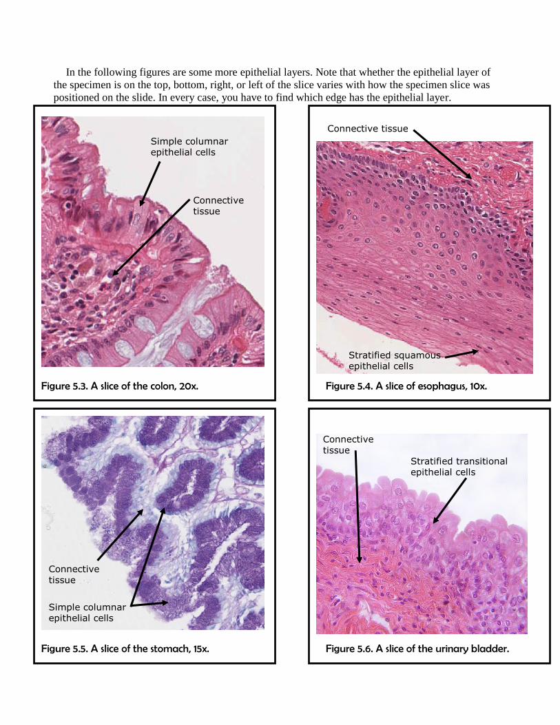
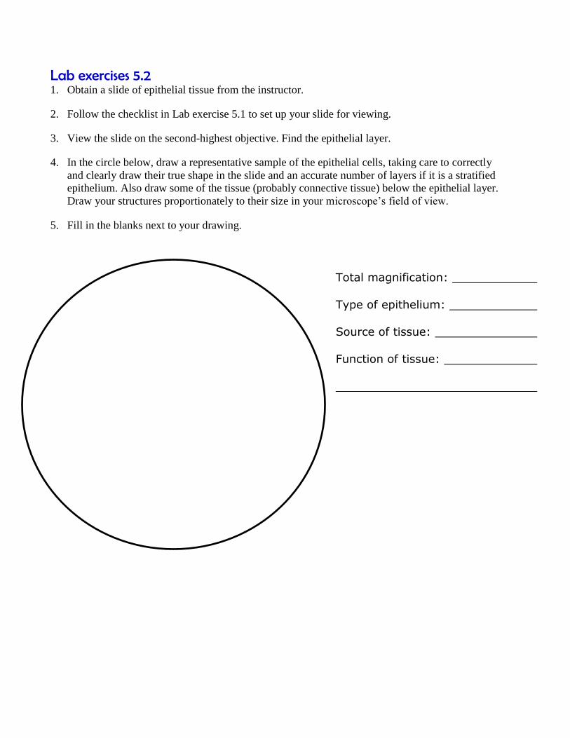
In the following figures are some more epithelial layers. Note that whether the epithelial layer of
the specimen is on the top, bottom, right, or left of the slice varies with how the specimen slice was
positioned on the slide. In every case, you have to find which edge has the epithelial layer.
Figure 5.3. A slice of the colon, 20x. Figure 5.4. A slice of esophagus, 10x.
Figure 5.5. A slice of the stomach, 15x. Figure 5.6. A slice of the urinary bladder.
Simple columnar epithelial cells
Connective tissue
Stratified squamous epithelial cells
Connective tissue
Connective tissue
Simple columnar epithelial cells
Connective tissue
Stratified transitional epithelial cells
Lab exercises 5.2 1. Obtain a slide of epithelial tissue from the instructor.
2. Follow the checklist in Lab exercise 5.1 to set up your slide for viewing.
3. View the slide on the second-highest objective. Find the epithelial layer.
4. In the circle below, draw a representative sample of the epithelial cells, taking care to correctly
and clearly draw their true shape in the slide and an accurate number of layers if it is a stratified
epithelium. Also draw some of the tissue (probably connective tissue) below the epithelial layer.
Draw your structures proportionately to their size in your microscope’s field of view.
5. Fill in the blanks next to your drawing.
Total magnification:
Type of epithelium:
Source of tissue:
Function of tissue:

Examining connective tissue under the microscope Information
Connective tissue is found throughout the body, usually in association with other tissues. As its
name indicates, it often serves to connect different tissues together, but it also can serve as a wrapper
(in locations where a tough epithelial wrapping is not required), a structural support, cushioning, a
storage repository, a protective layer, or a transport medium.
Connective tissue has the most types of subcategories and the most varied functions of all the
four major tissue types (epithelial, muscular, nervous, and connective tissues.) Bone and cartilage
are connective tissues, as are blood and lymph, fat, ligaments, and tendons. Epimysium, the
connective tissue wrapping around skeletal muscles, and periosteum, the connective tissue wrapping
around bones, are both connective tissues.
The different types of connective tissue are so diverse, there is no one set of characteristics that
encompasses all the different types. However, there are three characteristics that we consider
diagnostic of most connective tissue types.
1. The cells are dispersed. Connective tissues generally have cells that are not tightly connected
to each other, the way the cells in epithelial and muscular tissues usually are. There is usually a
fair amount of space between the connective tissue cells. An exception to this is adipose tissue
(also known as fat), the rare type of connective tissue in which the cells are packed tightly
together.
2. The tissue has more extracellular material than cells. Most connective tissues are solid
(blood and lymph are the exceptions) because all the volume between the dispersed cells is filled
with an extracellular matrix of viscous ground substance and protein fibers.
3. An extensive network of protein fibers is found in the extracellular matrix. Protein
fibers are complexes of millions of individual proteins threaded into long fibrous structures that
provide strength and elasticity to the tissue as a whole. The protein fibers are so large, they are
longer than the cells they surround and enmesh. Blood and lymph, being liquid connective
tissues, do not have these enmeshing protein fibers, but they still have an extensive liquid
extracellular matrix.
A common way of classifying the many different types of connective tissue is to subdivide it into
three main sub-categories, and further divide those subcategories into specific types of connective
tissue. The three main sub-categories of connective tissue are:
1. Connective tissue proper.
These are the types of connective tissue that typically have all three of the defining
characteristics listed above. It is further subdivided into dense connective tissue proper, in which
the extracellular protein fibers predominate, and loose connective tissue proper, in which the
extracellular protein fibers are not so densely woven.

2. Supporting connective tissue.
Bones and cartilage are the two types of connective tissue in this sub-category. They both have
all three of the defining characteristics listed above, but their extracellular matrix is tougher,
denser, and more solid than the various types of connective tissue proper.
3. Fluid connective tissue.
Blood and lymph are the two types of connective tissue in this sub-category. Both are fluid,
rather than solid, and both lack the network of extracellular protein fibers found in the other
types of connective tissue.
Connective Tissue Proper Information
Connective tissue proper encompasses the types of connective tissue that usually show all three
of the defining cellular characteristics of connective tissue with the fewest deviations from those
characteristics.
Dispersed cells
More extracellular material than cells.
Extensive protein fibers in the extracellular matrix.
Nonetheless there is still a great variety among the subcategories of connective tissue proper.
Some are classified as dense connective tissue proper and have a dense arrangement of
extracellular protein fibers that give the tissue strength and toughness. Tendons connecting muscles
to bone and ligaments connecting bone to bone are examples of dense connective tissue proper.
Other tissues are classified loose connective tissue proper and have fewer extracellular protein
fibers and more ground substance (the extracellular material surrounding the protein fibers), making
the tissues spongier but more fragile. Areolar tissue, found in the hypodermis of the skin and below
the epithelial layers of the digestive, respiratory, and urinary tracts, is a loose connective tissue
proper, as is adipose tissue, also known as fat.
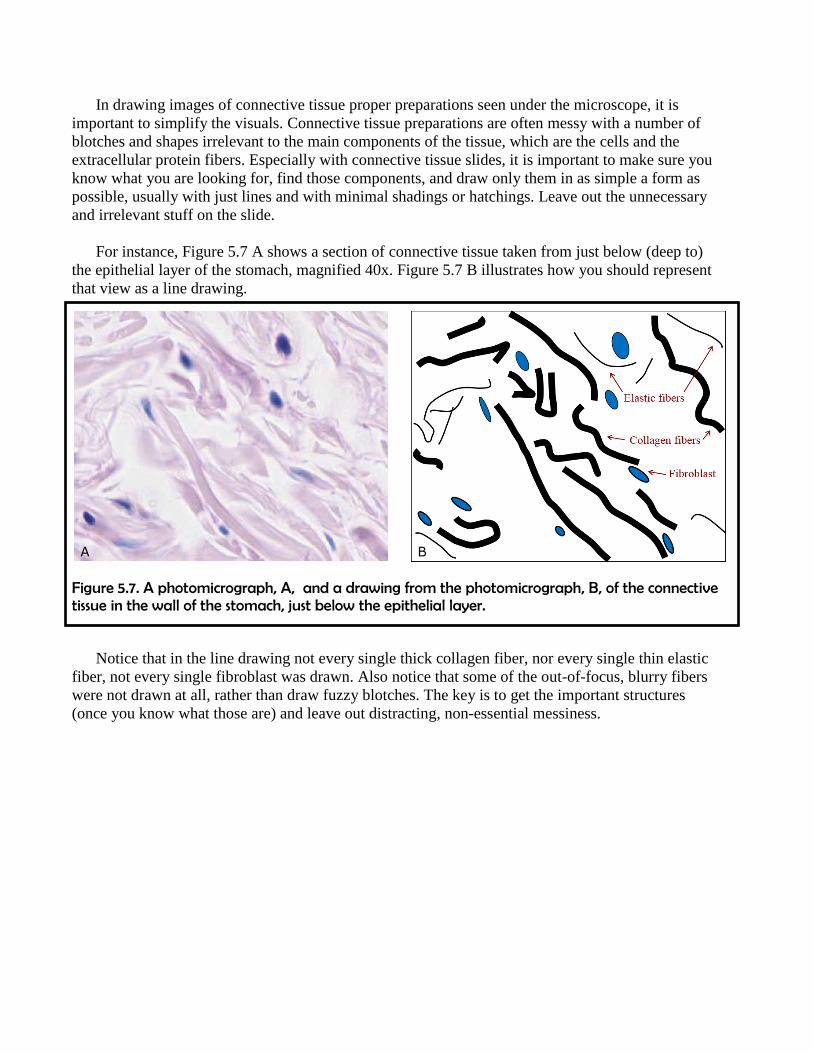
Table 5.1 lists some of the subcategories of connective tissue proper, along with some of their
characteristics and properties.
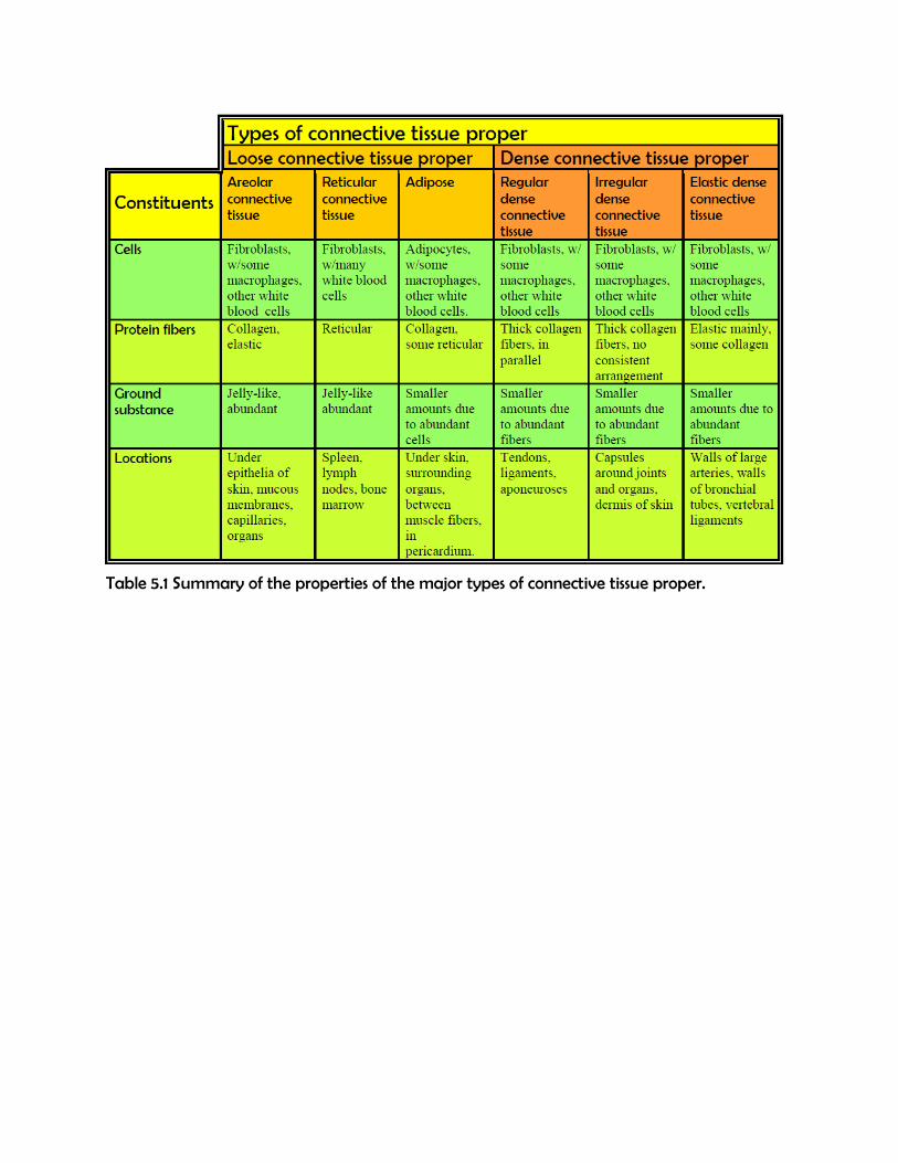
Table 5.1 Summary of the properties of the major types of connective tissue proper.
In drawing images of connective tissue proper preparations seen under the microscope, it is
important to simplify the visuals. Connective tissue preparations are often messy with a number of
blotches and shapes irrelevant to the main components of the tissue, which are the cells and the
extracellular protein fibers. Especially with connective tissue slides, it is important to make sure you
know what you are looking for, find those components, and draw only them in as simple a form as
possible, usually with just lines and with minimal shadings or hatchings. Leave out the unnecessary
and irrelevant stuff on the slide.
For instance, Figure 5.7 A shows a section of connective tissue taken from just below (deep to)
the epithelial layer of the stomach, magnified 40x. Figure 5.7 B illustrates how you should represent
that view as a line drawing.
Figure 5.7. A photomicrograph, A, and a drawing from the photomicrograph, B, of the connective tissue in the wall of the stomach, just below the epithelial layer.
Notice that in the line drawing not every single thick collagen fiber, nor every single thin elastic
fiber, not every single fibroblast was drawn. Also notice that some of the out-of-focus, blurry fibers
were not drawn at all, rather than draw fuzzy blotches. The key is to get the important structures
(once you know what those are) and leave out distracting, non-essential messiness.
A B
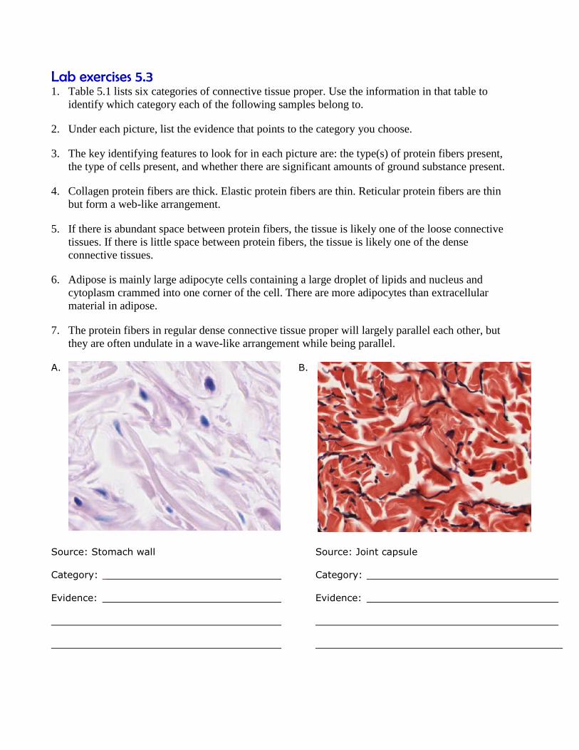
Lab exercises 5.3 1. Table 5.1 lists six categories of connective tissue proper. Use the information in that table to
identify which category each of the following samples belong to.
2. Under each picture, list the evidence that points to the category you choose.
3. The key identifying features to look for in each picture are: the type(s) of protein fibers present,
the type of cells present, and whether there are significant amounts of ground substance present.
4. Collagen protein fibers are thick. Elastic protein fibers are thin. Reticular protein fibers are thin
but form a web-like arrangement.
5. If there is abundant space between protein fibers, the tissue is likely one of the loose connective
tissues. If there is little space between protein fibers, the tissue is likely one of the dense
connective tissues.
6. Adipose is mainly large adipocyte cells containing a large droplet of lipids and nucleus and
cytoplasm crammed into one corner of the cell. There are more adipocytes than extracellular
material in adipose.
7. The protein fibers in regular dense connective tissue proper will largely parallel each other, but
they are often undulate in a wave-like arrangement while being parallel.
A. B.
Source: Stomach wall Source: Joint capsule
Category: Category:
Evidence: Evidence:

C. D.
Source: Lymph node Source: Fat
Category: Category:
Evidence: Evidence:
E. F.
Source: Aorta wall Source: Tendon
Category: Category:
Evidence: Evidence:
Lab exercises 5.4 1. Obtain a slide of connective tissue proper from the instructor.
2. Follow the checklist in Lab exercise 5.1 to set up your slide for viewing.
3. View the slide on the second-highest objective.
4. Fil out the blanks next to your drawing.
5. In the circle below, draw a representative sample of key features you identified, taking care to
correctly and clearly draw their true shapes and directions. Draw your structures proportionately
to their size in your microscope’s field of view.
Total magnification:
Type of connective tissue:
Source:
Function of tissue:
Key features to find and draw:
Supporting connective tissue Information
Supporting connective tissue comprises bone and cartilage. We will examine those tissues in
greater detail in Lab 6 Bones & The Axial Skeleton.
In both bone and cartilage, as in the different types of connective tissue proper, there are
extracellular protein fibers embedded in a viscous ground substance. However, in bone and cartilage,
the ground substance is so viscous as to be very hard and tough solids. Both bone and cartilage use
mainly collagen and elastic protein fibers in their extracellular matrix, but cartilage uses a ground
substance rich in the carbohydrate hyaluronan and bone uses a ground substance rich in a
mineralized calcium phosphate compound known as hydroxyapatite.
The carbohydrate hyaluronan (sometimes known as hyaluronic acid or hyaluronate) binds up
huge numbers of water molecules in the extracellular matrix of cartilage. This helps solidify the
ground substance around the collagen and elastic fibers of cartilage. As a result, it is often difficult to
see the protein fibers in cartilage when viewing preparations under the microscope.
They hydroxyapatite that surrounds the mostly collagen protein fibers in the ground substance of
bone is not soluble in water and forms a mineral solid in which both the bone cells and the collagen
fibers are embedded. As with cartilage, it is usually difficult to see the collagen fibers in the
extracellular matrix of bone due to the density of the ground substance that surrounds them.
There is only one type of cell in cartilage, chondrocytes. They secrete and maintain the
extracellular matrix of the tissue. Chondrocytes arise from mesenchymal stem cells, just like the
fibroblasts of connective tissue proper do, but chondrocytes are specialized to produce just cartilage.
The extracellular matrix produced by the chondrocytes is so tough and durable, the chondrocytes are
in danger of being crushed by it. This is why chondrocytes always leave a region around themselves
free of the cartilaginous extracellular matrix that makes up the rest of the tissue. These non-
cartilaginous pockets around each chondrocyte are called lacunae and are clearly visible when
examining cartilage under the microscope.
There are four types of bone cells, osteoprogenitor cells, osteoblasts, osteoclasts, and osteocytes,
but the osteocytes are the most abundant and the only ones found throughout the bone. Osteocytes
are found in concentric circles of mineralized extracellular matrix. Each circle is called a lamella
(plural: lamellae) and the osteocytes are found along the edges of each lamellae. In compact bone,
groups of lamellae and osteocytes are arranged into individual osteons, the cylindrical arrangement
of material that makes up the fundamental building block of the compact bone. Each osteon has a
hollow central canal in its center that blood vessels and nerves can travel through. In spongy bone,
groups of lamellae are arranged into trabeculae (singular: trabecula), which are the individual
projections of spongy bone. Trabeculae do not have central canals.
Osteocytes, like chondrocytes, are protected from the extracellular matrix that surrounds them by
being housed in lacunae, which are spaces free of mineralized extracellular matrix. Osteocytes,
unlike chondrocytes, have numerous cytoplasmic extensions that project off of the main cell body.
These extension connect up with the extensions from other near-by osteocytes. These projections,
like the osteocyte cell body, are in tiny spaces free of the mineralized extracellular matrix. These
spaces (but not the cytoplasmic projections themselves) are called canaliculi (singular: canaliculum)
because, under the microscope, they look like tiny little canals.
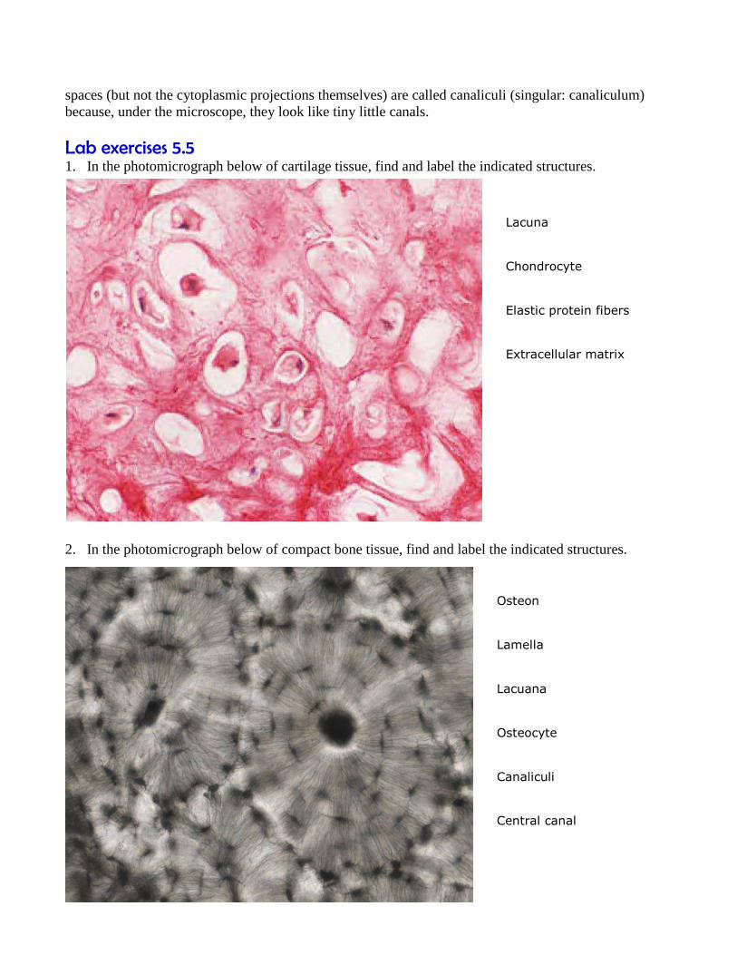
Lab exercises 5.5 1. In the photomicrograph below of cartilage tissue, find and label the indicated structures.
2. In the photomicrograph below of compact bone tissue, find and label the indicated structures.
Osteon
Lamella
Lacuana
Osteocyte
Canaliculi
Central canal
Lacuna
Chondrocyte
Elastic protein fibers
Extracellular matrix

Distinguishing between the three types of muscle tissue Information
Muscular tissue is the third of the four major categories of animal tissue. Muscle tissue is
subdivided into three broad categories: skeletal muscle, cardiac muscle, and smooth muscle. The
three types of muscle can be distinguished by both their locations and their microscopic features.
Skeletal muscle is found attached to bones. It consists of long multinucleate fibers. The fibers
run the entire length of the muscle they come from and so are usually too long to have their ends
visible when viewed under the microscope. The fibers are relatively wide and very long, but
unbranched. Fibers are not individual cells, but are formed from the fusion of thousands of
precursor cells. This is why they are so long and why individual fibers are multinucleate (a single
fiber has many nuclei). The nuclei are usually up against the edge of the fiber. There are striations
in skeletal muscle. These are alternating dark and light bands perpendicular to the edge of the fiber
that are present all along the fiber.
Cardiac muscle is only found in the heart. Its fibers are longer than they are wide, and they are
striated, like skeletal muscle fibers. But, unlike skeletal muscle fibers, cardiac muscle fibers have
distinct ends to them, called intercalated discs. These are dark lines that run from one side of the
fiber to the other. The intercalated discs are not much thicker than the striations, but they are usually
darker and so distinct for that reason. One cardiac muscle fiber is the material between two
intercalated discs. Cardiac muscle fibers are mononucleate, with only one nucleus per fiber, and
they can sometimes be branched.
Smooth muscle is found in the walls of internal organs, such as the organs of the digestive tract,
blood vessels, and others. It consists of mononucleate fibers with tapered edges. No striations
are visible in smooth muscle under the microscope. Because smooth muscle often is wrapping
around the organ it is associated with, it can be hard to find an entire smooth muscle fiber in profile
in a tissue slice on a microscope slide. Most of the fibers will be sectioned at angles or will be
difficult to get into a single plane of focus, but a little bit of searching can usually turn up some with
all of the defining characteristics visible.

Lab exercises 5.6 1. In each of the three photomicrographs below, identify which type of muscle is present. List the
defining visual characteristics of that type of muscle, and draw arrows to features on the
photograph that illustrate each characteristic.
A. Muscle type:
Visual characteristics:
1.
2.
3.
4.
B. Muscle type:
Visual characteristics:
1.
2.
3.
4.
C. Mucscle type:
Visual characteristics:
1.
2.
3.

Identifying neurons within nervous tissue Information
Nervous tissue is the last of the four major categories of animal tissue. Nervous tissue comprises
neurons, the cells specialized for the propagation of electrochemical signals, and neuroglia, the
so-called “supporting cells” of nervous tissue. There are multiple types of neuroglia cells (sometimes
called just glial cells), and most of them do assist neurons, but recent research has discovered that
some of the neuroglia cells play an active role by themselves in the proper functioning of the central
and peripheral nervous systems. It is turning out that neuroglia do more than just support neurons.
We will leave investigation of the neuroglia for later labs. In this lab, we will just focus on the
structural features of neurons as they are visible under microscopic examination.
A neuron is typically represented as having the following features. A large cell body
(sometimes known as the soma) in which the nucleus and other major organelles are found.
Dendrites, which are usually represented as numerous small projections extending from the cell
body. A single axon, which is usually represented as a large single projection extending from the
cell body, much longer than any of the dendrites. Multiple axon terminals that branch off at the
end of the axon. Figure 5.8 shows a typical drawn neuron showing all these features.
Figure 5.8. A typical diagrammatic representation of neuron
All of these features are important to the functioning of neurons. The cell body is where most of
the cellular processes of the neuron take place: protein synthesis, metabolism, etc. The dendrites
contact and pick up signals coming in from other cells (other neurons or sensory cells). The axon
propagates the neuron’s electrical signal to its various targets. And the axon terminals actually
contact (synapse with) those targets and send chemical signals to the targets to trigger changes in
them. However, most neurons in the body do not look as clear-cut as the neuron in Figure 5.8. Some
neurons have short axons. Others have dendrites that are almost as large as the axon. Others have
two axons connected to the same cell body. Many have dendrites and axon terminals that are too thin
and small too see clearly. Cell bodies tend to come in all sorts of sizes and shapes, and are not
always bulbous lumps readily distinguished from the rest of the cell.
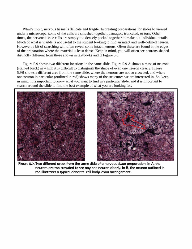
What’s more, nervous tissue is delicate and fragile. In creating preparations for slides to viewed
under a microscope, some of the cells are smushed together, damaged, truncated, or torn. Other
times, the nervous tissue cells are simply too densely packed together to make out individual details.
Much of what is visible is not useful to the student looking to find an intact and well-defined neuron.
However, a bit of searching will often reveal some intact neurons. Often these are found at the edges
of the preparation where the material is least dense. Keep in mind, you will often see neurons shaped
distinctly different from those shown in textbooks and if Figure 5.8.
Figure 5.9 shows two different locations in the same slide. Figure 5.9 A shows a mass of neurons
(stained black) in which it is difficult to distinguish the shape of even one neuron clearly. Figure
5.9B shows a different area from the same slide, where the neurons are not so crowded, and where
one neuron in particular (outlined in red) shows many of the structures we are interested in. So, keep
in mind, it is important to know what you want to find in a particular slide, and it is important to
search around the slide to find the best example of what you are looking for.
Figure 5.9. Two different areas from the same slide of a nervous tissue preparation. In A, the neurons are too crowded to see any one neuron clearly. In B, the neuron outlined in red illustrates a typical dendrite-cell body-axon arrangement.
A B
Lab exercises 5.7 1. Obtain a slide of nervous tissue from the instructor.
2. Follow the checklist in Lab exercise 5.1 to set up your slide for viewing.
3. View the slide on the second-highest objective. Search carefully until you find a clear,
representative neuron in your field of view.
4. In the circle below, draw the neuron you found. Only draw the single neuron. Do not draw any of
the other material. Draw your structures proportionately to their size in your microscope’s field
of view.
5. Label any neural parts you can clearly recognize.
Total magnification:

Licenses and attributions. Unless otherwise noted, all figures
Figure 5.1 Source: modified from:
https://upload.wikimedia.org/wikipedia/commons/6/64/423_Table_04_02_Summary_of_Epitheli
al_Tissue_CellsN.jpg
Figure 5.2 Source: modified from:
http://141.214.65.171/Histology/Basic%20Tissues/Epithelium%20and%20CT/020_HISTO_20X.
svs/view.apml
Figure 5.3 Source: modified from:
http://141.214.65.171/Histology/Basic%20Tissues/Epithelium%20and%20CT/176_HISTO_20X.
svs/view.apml
Figure 5.4 Source: modified from:
http://141.214.65.171/Histology/Basic%20Tissues/Epithelium%20and%20CT/153_HISTO_20X.
svs/view.apml
Figure 5.5 Source: modified from:
http://141.214.65.171/Histology/Basic%20Tissues/Epithelium%20and%20CT/160_HISTO_40X.
svs/view.apml
Figure 5.6 Source: modified from:
http://141.214.65.171/Histology/Urinary%20System/212_HISTO_40X.svs/view.apml
Table 5.1 Source: created by Ross Whitwam for this work.
Figure 5.7 A Source: modified from:
http://141.214.65.171/Histology/Basic%20Tissues/Epithelium%20and%20CT/160_HISTO_40X.
svs/view.apml
Figure 5.7 B Source: created by Ross Whitwam for this work.
Lab Exercise 5.3 Question A Source: modified from:
http://141.214.65.171/Histology/Basic%20Tissues/Epithelium%20and%20CT/160_HISTO_40X.
svs/view.apml
Lab Exercise 5.3 Question B Source: modified from:
http://141.214.65.171/Histology/Basic%20Tissues/Epithelium%20and%20CT/033_HISTO_20X.
svs/view.apml
Lab Exercise 5.3 Question C Source: modified from:
http://141.214.65.171/Histology/Basic%20Tissues/Epithelium%20and%20CT/028-
2_HISTO_40X.svs/view.apml

Lab Exercise 5.3 Question D Source: modified from:
http://141.214.65.171/Histology/Basic%20Tissues/Epithelium%20and%20CT/019-
2_HISTO_20X.svs/view.apml
Lab Exercise 5.3 Question E Source: modified from:
http://141.214.65.171/Histology/Cardiovascular%20System/036_HISTO_20X.svs/view.apml
Lab Exercise 5.3 Question F Source: modified from:
http://141.214.65.171/Histology/Basic%20Tissues/Epithelium%20and%20CT/74.svs/view.apml
Lab Exercise 5.5 Question 1 Source: modified from:
http://141.214.65.171/Histology/Basic%20Tissues/Cartilage%20and%20Bone/044H_HISTO_20
X.svs/view.apml
Lab Exercise 5.5 Question 2 Source: modified from:
http://141.214.65.171/Histology/Basic%20Tissues/Cartilage%20and%20Bone/051xc_HISTO_4
0X.svs/view.apml
Lab Exercise 5.6 Question A Source: modified from:
http://141.214.65.171/Histology/Cardiovascular%20System/098HE_HISTO_40X.svs/view.apml
Lab Exercise 5.6 Question B Source: modified from:
http://141.214.65.171/Histology/Basic%20Tissues/Muscle/058L_HISTO_40X.svs/view.apml
Lab Exercise 5.6 Question C Source: modified from:
http://141.214.65.171/Histology/Basic%20Tissues/Muscle/169_HISTO_40X.svs/view.apml
Figure 5.8 Source: modified from:
https://commons.wikimedia.org/w/index.php?curid=18271454
Figure 5.9 A Source: modified from:
http://141.214.65.171/Histology/Central%20Nervous%20System/13270.svs/view.apml
Figure 5.9 B Source: modified from:
http://141.214.65.171/Histology/Central%20Nervous%20System/13270.svs/view.apml