human biology enabling course module 2 cellular … college of natural health page 3 enabling...
TRANSCRIPT

Human Biology Enabling Course –
Module 2
Cellular Level of Organisation
This document is the property of Endeavour College of Natural Health and contains confidential information of Endeavour
College of Natural Health.
Copyright in the whole and every part of this document belongs to Endeavour College of Natural Health and may not be
used, sold, transferred, adapted or modified or reproduced in whole or in part in any manner or form or in any media, to
any persons other than in agreement with Endeavour College of Natural Health.
This document remains the confidential information of Endeavour College of Natural Health and should not be used for any
other purpose other than that expressly approved by Endeavour College of Natural Health at the time the document was
provided by Endeavour College of Natural Health.
May 2011

Endeavour College of Natural Health Page 2 Enabling Course: Human Biology Module 2
Contents
Module 2 – Cellular Level of Organisation
1 Introduction
1.1 Background
2 Anatomy and physiology of the human cell
2.1 The plasma membrane
2.2 Plasma membrane proteins
2.3 Diffusion
2.4 Cytoplasm
2.5 Cytoskeleton
2.6 Endoplasmic reticulum (ER)
2.7 Ribosomes
2.8 The Golgi complex
2.9 Lysosomes
2.10 Mitochondria
2.11 Nucleus
2.12 Cell Division
2.13 Mitosis
2.14 Prophase
2.15 Metaphase
2.16 Anaphase
2.17 Telophase and Cytokinesis
2.18 Meiosis
2.19 Activity
3 References

Endeavour College of Natural Health Page 3 Enabling Course: Human Biology Module 2
Module 2 – Cellular Level of Organisation
1 Introduction
1.1 Background
As briefly mentioned in module 1, there are various levels of organisation that make up a
human body. The first level is the chemical level and contains the smallest units known as
atoms. Over millennia atoms formed molecules through a multitude of chemical reactions
and eventually gave rise to the living cell.
The human body is made up of trillions of cells. There are many different types of cells, for
example, liver cells, brain cells and muscle cells. Every cell is programmed by human DNA
to carry out a specific function (physiology) such as the liver cell’s role in detoxifying
substances taken into the body in the form of liquids, food or airborne pollutants. When
viewed under the microscope cells vary in shape and size (anatomy) but they all essentially
share common features and it is these similarities that this module will focus on using a
generic model/picture of a cell as a reference point such as Figure 1.
Figure 1. Human cell

Endeavour College of Natural Health Page 4 Enabling Course: Human Biology Module 2
2 Anatomy and physiology of the human cell
2.1 The plasma membrane
Figure 2. Plasma Membrane
As mentioned in Module 1 (cellular level) each cell is a complex factory of important parts
that work integrally to maintain the life and function of the cell. To contain and protect the
inner components of the cell a protective membrane formed known as the plasma
membrane Figure 2. The term plasma refers to the contents of the cell.
The plasma membrane is made up of a phospholipid (phospho = containing phosphorus;
lipid = fat) double layer known as a bilayer. Facing the extracellular (outside) and
intracellular (inside) environments are phosphorous polar (charged) heads (see Figures 2 &
3.) that attract water and known to be hydrophilic (hydro = water; philic = attract).
Between the layers of polar heads are the lipid tails (non-polar) that repel water making them
hydrophobic (phobic = repel). Together this arrangement makes up the cell (plasma)
membrane (Figure 3). It is important to note that plasma membrane and cell membrane are
terms used interchangeably.
Figure 3. (from http://www.prism.gatech.edu)

Endeavour College of Natural Health Page 5 Enabling Course: Human Biology Module 2
The plasma membrane separates the contents of the cell (intracellular) from the outside
(extracellular) environment. It functions like a gatekeeper allowing some substances such as
nutrients like glucose to pass into the cell for metabolism while keeping other harmful
substances out. This protective role can break down in the face of some viruses and bacteria
that have developed ways to trick the cell into allowing them to pass.
Figure 4. Cell membrane proteins
2.2 Plasma membrane proteins
The plasma membrane is littered with various shaped bodies some of which penetrate
through the double layer to the inside of the cell (Figures 2 & 4). These structures are protein
molecules that may function as locks and gatekeepers.
This arrangement of surface proteins provide the mechanism of entry for life giving
substances into the cell needed for metabolism and the exit points for waste products that
are produced as a result of metabolism. It’s essentially the same as when you eat food and
your digestive system extracts the nutrients required for health and expels the waste in the
form of urine or faeces.

Endeavour College of Natural Health Page 6 Enabling Course: Human Biology Module 2
2.3 Diffusion
Some substances such as oxygen and carbon dioxide enter and exit the cell membrane with
ease a process referred to as simple diffusion (see Figure 5). Diffusion occurs when a
substance moves from an area of high concentration to an area of lower concentration
known as the concentration gradient.
In the example of oxygen and carbon dioxide, the concentration of oxygen is greater on the
outside of the cell as fresh oxygen arrives from the lungs, while on the inside of the cell the
process of metabolism has created a higher concentration of carbon dioxide (waste product).
Therefore according to simple diffusion laws oxygen will naturally flow into the cell where it’s
concentration is lower and carbon dioxide will flow toward the outside of the cell where it’s
concentration is lower. This process ensures that body cells always have a fresh supply of
life giving oxygen and also ensures that waste products are removed from the cell to
maintain homeostasis.
Figure 5. Simple Diffusion
Endeavour College of Natural Health Page 7 Enabling Course: Human Biology Module 2
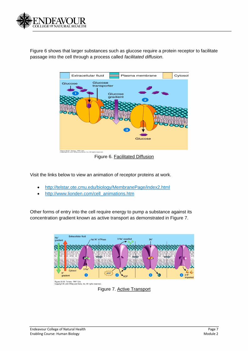
Figure 6 shows that larger substances such as glucose require a protein receptor to facilitate
passage into the cell through a process called facilitated diffusion.
Figure 6. Facilitated Diffusion
Visit the links below to view an animation of receptor proteins at work.
http://telstar.ote.cmu.edu/biology/MembranePage/index2.html
http://www.lionden.com/cell_animations.htm
Other forms of entry into the cell require energy to pump a substance against its
concentration gradient known as active transport as demonstrated in Figure 7.
Figure 7. Active Transport
Endeavour College of Natural Health Page 8 Enabling Course: Human Biology Module 2
Visit the links below to view an animation of passive (simple) and facilitated diffusion.
http://www.youtube.com/watch?v=s0p1ztrbXPY
http://www.youtube.com/watch?v=PkmF7yoWiXU&feature=related
The various functions of membrane proteins will be covered in more detail in classes but it is
important that you view the animation links provided to gain a conceptual overview of the
important role these proteins play in the life and maintenance of human body cells.
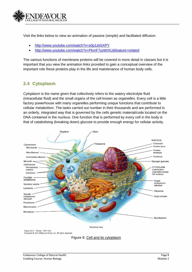
2.4 Cytoplasm
Cytoplasm is the name given that collectively refers to the watery electrolyte fluid
(intracellular fluid) and the small organs of the cell known as organelles. Every cell is a little
factory powerhouse with many organelles performing unique functions that contribute to
cellular metabolism. The tasks carried out number in their thousands and are performed in
an orderly, integrated way that is governed by the cells genetic material/code located on the
DNA contained in the nucleus. One function that is performed by every cell in the body is
that of catabolising (breaking down) glucose to provide enough energy for cellular activity.
Figure 8. Cell and its cytoplasm
Endeavour College of Natural Health Page 9 Enabling Course: Human Biology Module 2
Refer to Figure 8 (above) when you read through the following list of organelles to help you
connect the structure with its function.
2.5 Cytoskeleton
The cytoskeleton comprises proteins called microfilaments, intermediate filaments and
microtubules forming a network of interconnecting structures acting as a framework for the
integrity of the cell, essentially forming the cell’s skeleton.
2.6 Endoplasmic reticulum (ER)
Functionally the ER is involved in synthesising (making), transporting and storing newly
made molecules/substances that are involved in cellular metabolism, for example, protein.
Figure 8 shows two parts to the ER, a rough section called the rough endoplasmic reticulum
(RER) studded with ribosomes (see explanation below) and a smooth section (SER) which
in the picture is coloured pink sitting in front of the RER.
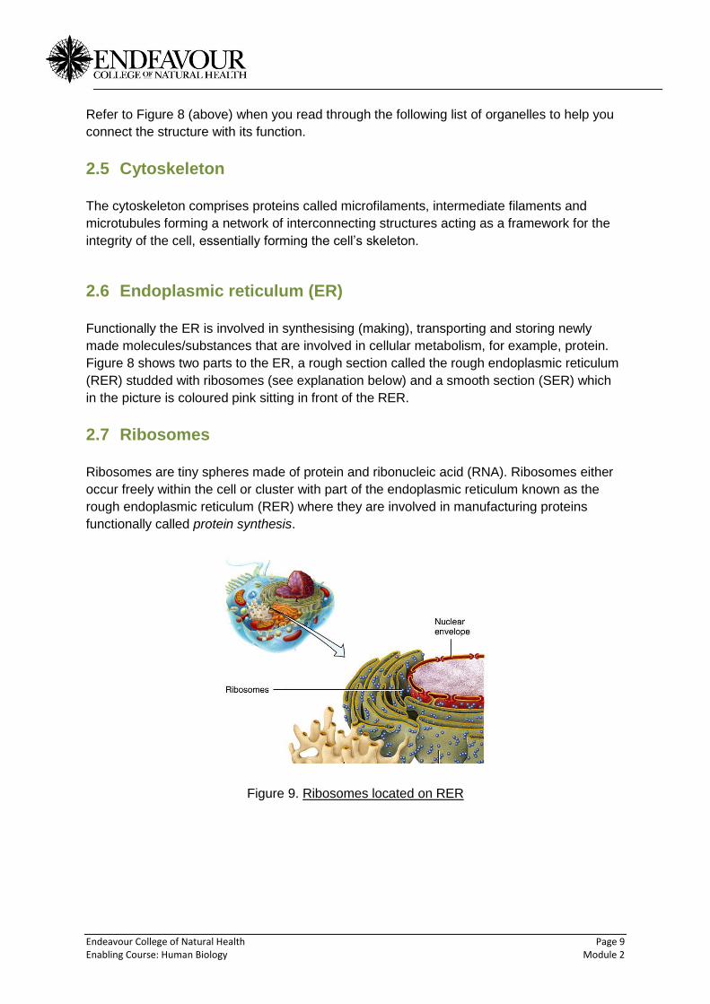
2.7 Ribosomes
Ribosomes are tiny spheres made of protein and ribonucleic acid (RNA). Ribosomes either
occur freely within the cell or cluster with part of the endoplasmic reticulum known as the
rough endoplasmic reticulum (RER) where they are involved in manufacturing proteins
functionally called protein synthesis.
Figure 9. Ribosomes located on RER

Endeavour College of Natural Health Page 10 Enabling Course: Human Biology Module 2
2.8 The Golgi complex
The main function of the Golgi complex (Figure 10) is to process, sort and deliver newly
manufactured proteins (from the RER) and lipids to the cell membrane, to lysosomes (for
digestion) and to secretory vesicles that enable contents within the cell to be released
outside the cell, for example, certain glandular secretions. Note in Figure 10 the green
spheres entering the golgi complex, they are newly created proteins from the RER.
Figure 10. Golgi Complex

Endeavour College of Natural Health Page 11 Enabling Course: Human Biology Module 2
2.9 Lysosomes
Figure 11. Lysosome
Lysosomes are membrane enclosed spheres or vesicles that contain powerful digestive
enzymes. They function to digest foreign substances and worn out organelles. They play an
important role in the homeostasis of the cell.
2.10 Mitochondria
Mitochondria (Figure 12) are the energy powerhouses of the cell. These organelles utilise
the energy molecules consumed from food such as glucose and fat and produce energy in
the form of ATP (adenosine triphosphate) for cellular metabolism.
Figure 12. Mitochondria

Endeavour College of Natural Health Page 12 Enabling Course: Human Biology Module 2
2.11 Nucleus
Figure 13. Nucleus
When viewing a cell under the microscope such as the bottom picture in Figure 13, the
nucleus is usually the most prominent feature. Most body cells have a single nucleus
whereas muscle cells, for example, have several. Some have no nucleus such as red blood
cells. The main function of the nucleus is to house the genetic code found on the DNA.
View the link below for a tour of the cell.
http://people.eku.edu/ritchisong/301notes1.htm
2.12 Cell Division
Especially important during growth and development is the cell’s ability to divide or make
copies of itself. The human lifespan is a dynamic process involving many changes in the
body over time. Some cells, such as neurons (nervous system cells) last a lifetime, whereas
other cells, such as skin cells are sloughed off daily, making up the majority of the dust in
your home.
There are two types of cell division in the human body known as mitosis, which is replication
of body cells and meiosis, which involves only the production cells known as sperm and
oocytes (gametes).

Endeavour College of Natural Health Page 13 Enabling Course: Human Biology Module 2
2.13 Mitosis
Mitosis is somatic (all body cells except gametes) cell division and comprises several steps
or phases. Just prior to the mitotic phase (actual cell division), the cells undergo a growth
and replication phase called interphase. During interphase the cell’s cytoplasm increases
and the DNA made up of 46 chromosomes (23 from mum and 23 from dad) make copies of
themselves (replicate) so they can divide neatly into two new cells. Figure 14 shows the
cycle of cell division including a description of interphase.
Figure 14. Mitotic cycle
Once interphase is complete, the cell enters the mitotic or dividing phase. There are four
stages of mitosis.

Endeavour College of Natural Health Page 14 Enabling Course: Human Biology Module 2
2.14 Prophase
Figure 15. Prophase (from http://www.chuck16.wordpress.com)
During prophase chromosomes not normally visible under the microscope become visible as
they condense in preparation for division. The nucleolus and nuclear envelope disappear.
The centrosomes responsible for pulling apart (dividing) the chromosomes move to opposite
ends of the cell.
2.15 Metaphase
Figure 16. (from http://www.staff.jccc.net)
The chromosomes which comprise two identical parts called chromatids (having replicated
during interphase) are held together by a centromere. These line up in the centre of the cell
along what is known as the equatorial plane. Later, when the chromatids are separated, this
separation starts from the centromeres.
Endeavour College of Natural Health Page 15 Enabling Course: Human Biology Module 2
2.16 Anaphase
Figure 17. (from http://www.staff.jccc.net)
Anaphase results is the actual splitting and separation of the chromatids at the centromere.
Long protein spindle ‘fingers’ extend form the centrosome and grab hold of the centromeres.
Once this occurs, the protein spindles start pulling on the centromeres, ultimately separating
the pairs of sister chromatids from each other. As soon as the chromatids are separated,
they are no longer called sister chromatids, they are now known as individual chromosomes.
In late anaphase a cleavage furrow is present and is the beginning of complete division of
the cell.
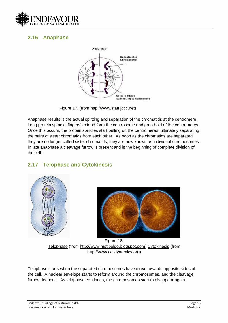
2.17 Telophase and Cytokinesis
Figure 18.
Telophase (from http://www.mstiboldo.blogspot.com) Cytokinesis (from
http://www.celldynamics.org)
Telophase starts when the separated chromosomes have move towards opposite sides of
the cell. A nuclear envelope starts to reform around the chromosomes, and the cleavage
furrow deepens. As telophase continues, the chromosomes start to disappear again.

Endeavour College of Natural Health Page 16 Enabling Course: Human Biology Module 2
The final step in Mitosis is Cytokinesis. This is the process by which the cleavage furrow is
so big that it meets in the middle of the cell. Ultimately this will pinch off the two newly
created cells from one another, each with their own nucleus and their own organelles.
Visit http://www.youtube.com/watch?v=D1_-mQS_FZ0&feature=related to view mitosis.
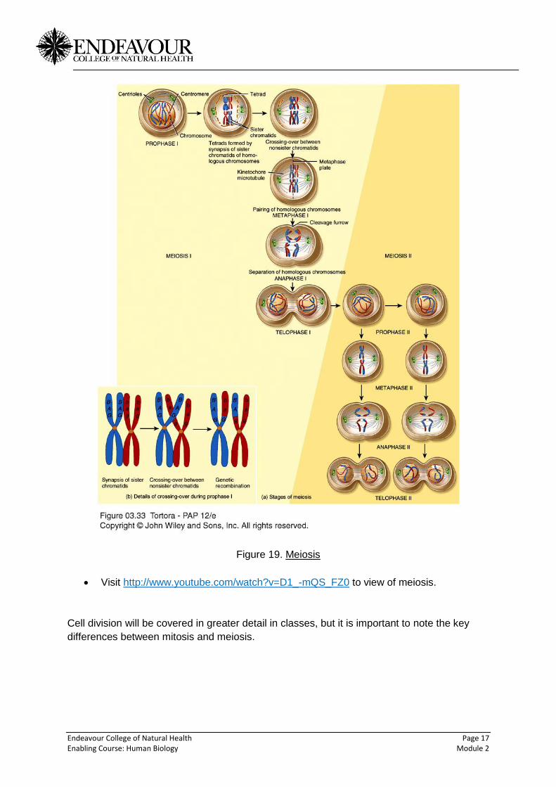
2.18 Meiosis
Meiosis occurs in the production of gametes (sperm and oocytes) only. Like mitosis, meiosis
also involves the stages of prophase, metaphase, anaphase and telophase, however the
original cell undergoes two rounds of division, therefore, meiosis consists of prophase 1 & 2,
metaphase 1 & 2 etc. The critical event that occurs during meiosis that accounts for the
individual differences in humans is that during prophase 1, chromosomes cross over (see
Figure 19), which results in a sharing of genes and creation of new combinations of
characteristics in DNA within the gametes/cells.
Endeavour College of Natural Health Page 17 Enabling Course: Human Biology Module 2
Figure 19. Meiosis
Visit http://www.youtube.com/watch?v=D1_-mQS_FZ0 to view of meiosis.
Cell division will be covered in greater detail in classes, but it is important to note the key
differences between mitosis and meiosis.
Endeavour College of Natural Health Page 18 Enabling Course: Human Biology Module 2
2.19 Activity
View the links below to explore different varieties of cell shapes and sizes
http://www.lennartnilsson.com/human_body.html
http://www.cellsalive.com/
3 References
Tortora, G.J., Derrickson, B., 2012. Principles of Anatomy and Physiology, 13th edn, John
Wiley & Sons, Inc, USA.
Winston, R., 2004. Human, DK Publishing, London.