human blastocyst culture in ivf: current laboratory ... · human blastocyst culture in ivf: current...
TRANSCRIPT
Romanian Journal of Morphology and Embryology 2010, 51(3):441–445
RREEVVIIEEWW
Human blastocyst culture in IVF: current laboratory applications in reproductive
medicine practice E. S. SILLS1), G. D. PALERMO2)
1)Division of Reproductive Research, Sims IVF/Department of Obstetrics and Gynecology,
School of Medicine, Royal College of Surgeons in Ireland, Dublin, Ireland 2)Center for Reproductive Medicine and Infertility,
Weill Medical College of Cornell University, New York, USA
Abstract For fertility patients undergoing in vitro fertilization (IVF), blastocyst culture brings a number of potential advantages over laboratory techniques leading to traditional cleavage-stage embryo transfer. Because day 2–3 embryos normally should transit the oviduct only, their direct exposure to an intrauterine microenvironment is physiologically inappropriate. This mismatch is obviated by blastocyst transfer. Moreover, the nutritional milieu inside the fallopian tube is not the same as within the endometrial compartment, a feature possibly antagonistic to implantation when a day 2–3 embryo is placed directly within the uterus. Delaying transfer to day 5–6 may also improve reproductive outcome by reducing risk of embryo expulsion, given increased myometrial pulsatility measured at day 2–3. However, rigid reliance on a blastocyst culture approach will more often result in treatment cancellation due to embryo loss (no transfer), or having fewer embryos for cryopreservation. The development of sequential media to support embryos in extended in vitro culture was a significant laboratory refinement, since it enabled direct observation of embryos to improve transfer selection bias. This approach, in tandem with blastocyst cryopreservation, leads to fewer embryos being transferred and reducing multiple gestation rate. This review discusses key features of human blastocyst culture and its application in clinical reproductive medicine practice. Keywords: human reproduction, IVF, blastocyst transfer, embryo culture.
Introduction
Extended in vitro embryo culture and blastocyst transfer (BT) have emerged as essential components of the advanced reproductive technology armamentarium, permitting selection of more advanced embryos considered best suited for transfer. Since the first IVF birth in 1978, the optimal time to perform embryo transfer (ET) has remained controversial. Cleavage stage (day 2 or 3) ET was generally used in IVF and became established as the usual laboratory approach. This intentional placement of a day 2 or 3 embryo directly into the uterine cavity was recognized as non-physiologic, but there was little to offer as an alternative due to the inability to sustain human embryos in culture to the blastocyst stage. And, unlike embryos derived from other primates, the human embryo is reasonably tolerant of being prematurely placed inside the uterus [1].
The “choice” to perform day 2 or 3 ET was therefore merely a fortuitous default response to contemporary technical challenges associated with extended in vitro culture. However, with the advent of more sophisticated sequential media, BT became a reality. A central tenet in embryo culture began to be challenged by the 1990’s, as traditional thinking assumed that mere blastocyst formation was sufficient to signal development of viable blastocysts [2, 3]. To validate this hypothesis it was necessary to follow under direct observation the
developmental progress of many embryos. Early blasto-cyst culture media was pioneered by incorporating amino acids as an energy substrate, a discovery that was perhaps the most significant laboratory reagent achievement since the introduction of human tubal fluid [4]. While the “sequential media” approach has not necessarily resulted in higher blastocyst yield, it enabled cultivation of blastocysts with improved implantation potential [5].
As the benefits of BT continue to be widely debated [6–8], the reality of clinical practice is that not all patients are good candidates for BT. This is because it is possible that after five days in culture, no embryo will survive to the blastocyst stage and the IVF cycle will be cancelled. Moreover, selection criteria for BT are variable and there is no consensus on the appropriate-ness of BT protocols applied specifically to patients with multiple unsuccessful IVF cycles. In exceptional cases, an IVF patient may even undergo both day 3 transfer and BT in the same cycle [9].
IVF: Patients and Techniques
For many IVF patients, pituitary downregulation is achieved with oral contraceptives and GnRH agonist, followed by daily administration of gonadotropins. We typically use a combined FSH+hMG protocol, with dosing influenced by periodic ultrasound and serum estradiol data. Treatment continues until adequate

E. S. Sills, G. D. Palermo
442
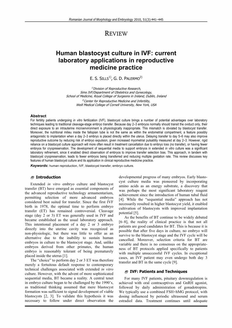
ovarian response is attained, defined as the maximum potential number of follicles with mean diameter of 17 mm. Transvaginal sonogram-guided oocyte retrieval is performed 36 hours after subcutaneous administration of hCG. Immediately after retrieval oocyte-cumulus complexes are placed into Universal IVF medium (MediCult, Jyllinge, Denmark). Conventional insemi-nation or intracytoplasmic sperm injection (Figure 1) is carried out using this reagent under washed liquid paraffin oil (MediCult, Jyllinge, Denmark). Fertilization is assessed after 16–18 hours and is considered normal when two distinct pronuclei are noted; embryos demon-strating additional pronuclei are considered abnormal and are not transferred (Figure 2). Culture is maintained to day five in microdrops of BlastAssist media I and II (MediCult, Jyllinge, Denmark) under washed paraffin oil in a 5% CO2 + 5% O2 atmosphere at 95% humidity. Embryos are assessed daily for cell number, fragment-ation and compaction (Figure 3). Day five blastocysts selected for in utero transfer should demonstrate a well-defined inner cell mass and highly cellular, expanding trophoectoderm (Figure 4). Blastocysts are loaded into an embryo transfer catheter (K–Soft–5000 Catheter, Cook Medical Inc., Spencer, Indiana, USA), for in utero transfer no sooner than 120 hours post-fertilization. At our institution, BT is done under direct transabdo-minal sonogram guidance by a physician.
Figure 1 – Intracytoplasmic sperm injection (ICSI) can enable fertilization even in the setting of severe impairments in semen parameters. In this image, the mature (metaphase II) oocyte is indicated by the presence of the first polar body (PB) just within the zona pellucida (ZP).
Figure 2 – Three pronuclei (3pn) are noted within the ooplasm after conventional (microdroplet) inse-mination as a trapped sperm (S) is seen at the zona pellucida.
Figure 3 – Well-developed day 2 embryo comprised of four symmetric blastomeres and minimal frag-mentation.
Figure 4 – Human blastocyst observed five days after fertilization demonstrating appropriate zona thin-ning, a well-defined inner cell mass (ICM), and even trophectoderm (T).
Discussion
Fertility patients not conceiving after several IVF attempts typically face a difficult prognosis. The impact of increased refractoriness to IVF on reproductive outcome following BT has been studied among patients with a history of repetitive failed day three embryo transfers [10]. How best to guide medical decisions after such multiple IVF failures is unclear [11] but in such settings maternal endocrine, anatomic, immunologic, infectious, and genetic parameters are usually investi-gated [12]. Embryology and oocyte quality are known to be central to recurrent IVF failure [13], yet how specific follicular recruitment protocols influence reproductive outcome remains difficult to verify. For example, it has been proposed that adjustments to controlled hyperstimulation regimens in IVF might reduce embryo fragmentation and optimize gamete quality [14] but there are no controlled studies to prove this [15]. Indeed, comparisons of different IVF stimu-lation regimens have not revealed any significant impact on pregnancy rate [16, 17]. Selecting embryos for transfer based on day 3 morphological criteria alone may be inadequate, while extended in vitro culture can help identify viable human embryos. Accordingly, growing embryos out to blastocyst stage has been advocated as one way to enhance implantation and improve reproductive outcome [18].

Human blastocyst culture in IVF: current laboratory applications in reproductive medicine practice
443
Attention has been focused on embryo genetics, since the frequency of chromosomal abnormality is probably higher in embryos from patients experiencing multiple IVF failures [19]. Impaired implantation associated with embryo aneuploidy renders implantation failure as the most frequent cause of unsuccessful IVF and places blastocyst nidation as a key rate-limiting step in overall reproductive outcome [20, 21]. For example, when pre-implantation genetic diagnosis (PGD) is performed on embryos obtained from patients with recurrent IVF failure, aneuploidy is more frequently observed in the cycle that followed the first failure [22]. This has implicated a reduced capacity to produce “high quality” embryos among patients with recurrent IVF failure [23]. Given the paucity of robust evidence supporting a beneficial effect of embryo biopsy in the setting of multiple IVF failures [24, 25], BT has emerged as an alternative to human embryo biopsy and PGD [26].
To study this, data from blastocyst transfers were reviewed to identify two fairly homogenous populations, clinically similar except for the number of failed IVF cycles at baseline (before BT treatment). Comparing fertility patients between 2002 and 2007 (with no patient ever having had BT before), a nearly six-fold increase in BT utilization was noted. This was achieved with fewer embryos being transferred per patient, on average, from 2002 to 2007. Clinical pregnancy rates were not signi-ficantly different between the two patient groups, yet the number of prior failed IVF cycles at baseline was higher in the 2007 group [10]. While it cannot be known what proportion of these patients would have conceived if they had undergone another day 3 ET, they nevertheless declined another day 3 ET perhaps because this approach was identified with earlier IVF failure [10]. Another report on reproductive outcomes among “good prognosis” patients who had embryo transfers either at cleavage-stage or blastocyst stage found a significant difference in live-birth rate in favor of the blastocyst group [27]. Again, patients with high numbers of eight-cell embryos on day 3 may have achieved pregnancy regardless of their embryo transfer day. The meta-analysis noted that maintaining embryos in laboratory culture until the blastocyst stage has not been shown to lead to more pregnancies than regular IVF (i.e., no blastocyst culture) [27].
Day 3 embryos or blastocysts have also been plated above a layer of autologous endometrial cells to supply growth factors as well as remove metabolic toxins, imparting an in vitro environment in closer alignment with actual physiologic conditions compared to (synthetic) sequential media [28]. Interleukin-6 (IL-6) is a crucial protein secreted by endometrial cells, and seems to favor in vitro blastocyst development [29]. Observations of hormonal and embryonic regulation of specific endometrial chemokines have suggested various mechanisms inducing production of chemokines by endometrial cells, thus contributing to the attraction of specific leukocyte populations during the peri-implantation phase [30].
These technical advances in human embryo culture notwithstanding, an essential practical matter remains: when to deploy extended embryo culture with a view to perform blastocyst transfer. Even though some patients, particularly those with a difficult prognosis, may specifically seek blastocyst transfer, such refractory cases are usually the least likely to have embryos of sufficient robustness to permit extended culture and blastocyst transfer. A minimum number of viable embryos at day 2 or 3 may be set as a threshold for considering extended culture, so that patients with limited embryo numbers (i.e., <6) are scheduled for day 3 transfer. While such guidelines can be useful, a rigid formulaic approach to blastocyst transfer is difficult to follow in clinical practice. For example, some patients will accept the possibility of culture arrest and “no embryo for transfer” if this provides insights about prior IVF failure where day 3 transfer was offered. In this circumstance, an unsuccessful blastocyst transfer attempt can at least yield some “closure” which may, at a personal level, be necessary before some patients can consider further fertility treatment incorpo-rating donor gametes [31].
One potentially negative aspect of human blastocyst culture has been the observation that monozygotic (MZ) twinning may occur at a higher rate with extended in vitro embryo culture [32], compared to traditional day 3 ET. However, because MZ twins are an un-common outcome both in assisted and natural con-ceptions, the phenomenon presents important metho-dological challenges for accurate study. Considerable speculation has been offered to explain why MZ twins might occur more often in assisted reproduction in general, and more specifically, in extended culture for blastocyst transfer. Some investigators have theorized that prolonged in vitro culture could have a detrimental impact on human embryos, particularly if glucose [33] or calcium [34] disturbances affect the inner cell mass. In contrast, more recent research has concluded that concerns about MZ twinning should not be a factor to discourage extended embryo culture for blastocyst transfer, considering the higher pregnancy rate and lower number of transferred embryos in BT cycles compared to embryo transfers performed at earlier developmental stages [35]. Indeed, findings derived exclusively from single blastocyst transfer cycles supported the opinion that blastocyst transfer does not increase the probability for MZ twins [36].
In conclusion, BT can be helpful for younger patients with multiple failed IVF cycles where day 3 ET had been performed previously with no success. Blastocyst culture has been one of the dramatic advances in reproductive biology (along with impro-vements in ovulation induction and embryo transfer techniques) that have enabled sharp increases in pregnancy/embryo transfer between 1994 and 2003 [37]. A comparative investigation of day 3 ET vs. BT in similar patients (where other conditions are controlled), while ideal, is extremely difficult to implement. It will

E. S. Sills, G. D. Palermo
444
be important to undertake further research emphasizing embryo and blastocyst morphology to better define which patients are best suited for BT, and how in vitro culture conditions may be optimized.
References [1] MARSTON JH, PENN R, SIVELLE PC, Successful autotransfer
of tubal eggs in the rhesus monkey (Macaca mulatta), J Reprod Fertil, 1977, 49(1):175–176.
[2] GARDNER DK, LANE M, SPITZER A, BATT PA, Enhanced rates of cleavage and development for sheep zygotes cultured to the blastocyst stage in vitro in the absence of serum and somatic cells: amino acids, vitamins and culturing embryos in groups stimulate development, Biol Reprod, 1994, 50(2):390–400.
[3] GARDNER DK, LANE M, Culture and selection of viable blastocysts: a feasible proposition for human IVF?, Hum Reprod Update, 1997, 3(4):367–382.
[4] QUINN P, KERIN JF, WARNES GM, Improved pregnancy rate in human in vitro fertilization with the use of a medium based on the composition of human tubal fluid, Fertil Steril, 1985, 44(4):493–498.
[5] BEHR B, MILKI AA, GIUDICE LC, High yield blastocyst culture and transfer: a new approach using P1 and blastocyst medium in a reduced O2 environment, Proceed-ings of the American Society for Reproductive Medicine Annual Meeting, Fertil Steril, 1998, 70(Suppl 1):S98, Abstract 262.
[6] EDWARDS RG, BEARD HK, Is the success of human IVF more a matter of genetics and evolution than growing blastocysts?, Hum Reprod, 1999, 14(1):1–4.
[7] ALPER MM, BRINSDEN P, FISCHER R, WIKLAND M, To blastocyst or not to blastocyst? That is the question, Hum Reprod, 2001, 16(4):617–619.
[8] HARTSHORNE GM, LILFORD RJ, Different perspectives of patients and health care professionals on the potential benefits and risks of blastocyst culture and multiple embryo transfer, Hum Reprod, 2002, 17(4):1023–1030.
[9] HAYRINEN LH, SILLS ES, FOGARTY AO, WALSH DJ, LUTSYK AD, WALSH AP, First Irish delivery following sequential, two-stage embryo and blastocyst transfer, Ir J Med Sci, 2009, Oct 8 [Epub ahead of print].
[10] WALSH AP, SHKROBOT LV, COULL GD, PEIRCE KL, WALSH DJ, SALMA U, SILLS ES, Blastocyst transfer for multiple prior IVF failure: a five year descriptive study, Ir Med J, 2009, 102(9):282–285.
[11] TAN BK, VANDEKERCKHOVE P, KENNEDY R, KEAY SD, Investi-gation and current management of recurrent IVF treatment failure in the UK, BJOG, 2005, 112(6):773–780.
[12] CHRISTIANSEN OB, NIELSEN HS, KOLTE AM, Future directions of failed implantation and recurrent miscarriage research, Reprod Biomed Online, 2006, 13(1):71–83.
[13] LEVI SETTI PE, COLOMBO GV, SAVASI V, BULLETTI C, ALBANI E, FERRAZZI E, Implantation failure in assisted reproduction technology and a critical approach to treatment, Ann N Y Acad Sci, 2004, 1034:184–199.
[14] SCOTT L, Embryological strategies for overcoming recurrent assisted reproductive technology treatment failure, Hum Fertil (Camb), 2002, 5(4):206–214.
[15] MARGALIOTH EJ, BEN-CHETRIT A, GAL M, ELDAR-GEVA T, Investigation and treatment of repeated implantation failure following IVF–ET, Hum Reprod, 2006, 21(12):3036–3043.
[16] SILLS ES, SCHATTMAN GL, VEECK LL, LIU HC, PRASAD M, ROSENWAKS Z, Characteristics of consecutive in vitro fertilization cycles among patients treated with follicle-stimulating hormone (FSH) and human menopausal gonadotropin versus FSH alone, Fertil Steril, 1998, 69(5):831–835.
[17] SILLS ES, LEVY DP, MOOMJY M, MCGEE M, ROSENWAKS Z, A prospective, randomized comparison of ovulation induction using highly purified follicle-stimulating hormone alone and with recombinant human luteinizing hormone in in-vitro fertilization, Hum Reprod, 1999, 14(9):2230–2235.
[18] GRAHAM J, HAN T, PORTER R, LEVY M, STILLMAN R, TUCKER MJ, Day 3 morphology is a poor predictor of blastocyst quality in extended culture, Fertil Steril, 2000, 74(3):495–497.
[19] VOULLAIRE L, COLLINS V, CALLAGHAN T, MCBAIN J, WILLIAMSON R, WILTON L, High incidence of complex chromosome abnormality in cleavage embryos from patients with repeated implantation failure, Fertil Steril, 2007, 87(5):1053–1058.
[20] BOOMSMA CM, MACKLON NS, Does glucocorticoid therapy in the peri-implantation period have an impact on IVF outcomes?, Curr Opin Obstet Gynecol, 2008, 20(3):249–256.
[21] GOODMAN C, JEYENDRAN RS, COULAM CB, Vascular endothelial growth factor gene polymorphism and implantation failure, Reprod Biomed Online, 2008, 16(5):720–723.
[22] PAGIDAS K, YING Y, KEEFE D, Predictive value of pre-implantation genetic diagnosis for aneuploidy screening in repeated IVF–ET cycles among women with recurrent implantation failure, J Assist Reprod Genet, 2008, 25(2–3):103–106.
[23] FARHI J, BEN-HAROUSH A, DRESLER H, PINKAS H, SAPIR O, FISCH B, Male factor infertility, low fertilisation rate following ICSI and low number of high-quality embryos are associated with high order recurrent implantation failure in young IVF patients, Acta Obstet Gynecol Scand, 2008, 87(1):76–80.
[24] SOINI S, Preimplantation genetic diagnosis (PGD) in Europe: diversity of legislation a challenge to the community and its citizens, Med Law, 2007, 26(2):309–323.
[25] ANDERSON RA, PICKERING S, The current status of pre-implantation genetic screening: British Fertility Society Policy and Practice Guidelines, Hum Fertil (Camb), 2008, 11(2):71–75.
[26] BARRENETXEA G, LÓPEZ DE LARRUZEA A, GANZABAL T, JIMÉNEZ R, CARBONERO K, MANDIOLA M, Blastocyst culture after repeated failure of cleavage-stage embryo transfers: a comparison of day 5 and day 6 transfers, Fertil Steril, 2005, 83(1):49–53.
[27] BLAKE DA, FARQUHAR CM, JOHNSON N, PROCTOR M, Cleavage stage versus blastocyst stage embryo transfer in assisted conception, Cochrane Database Syst Rev, 2007, (4):CD002118.
[28] KATTAL N, COHEN J, BARMAT LI, Role of coculture in human in vitro fertilization: a meta-analysis, Fertil Steril, 2008, 90(4):1069–1076.
[29] DOMINGUEZ F, GADEA B, MERCADER A, ESTEBAN FJ, PELLICER A, SIMÓN C, Embryologic outcome and secretome profile of implanted blastocysts obtained after coculture in human endometrial epithelial cells versus the sequential system, Fertil Steril, 2010, 93(3):774–782.e1.
[30] CABALLERO-CAMPO P, DOMÍNGUEZ F, COLOMA J, MESEGUER M, REMOHÍ J, PELLICER A, SIMÓN C, Hormonal and embryonic regulation of chemokines IL-8, MCP-1 and RANTES in the human endometrium during the window of implantation, Mol Hum Reprod, 2002, 8(4):375–384.
[31] SANTIAGO-DELEFOSSE M, CAHEN F, COEFFIN-DRIOL C, The analysis of physicians’ work: announcing the end of attempts at in vitro fertilization, Encephale, 2003, 29(4 Pt 1):293–305.
[32] VITTHALA S, GELBAYA TA, BRISON DR, FITZGERALD CT, NARDO LG, The risk of monozygotic twins after assisted reproductive technology: a systematic review and meta-analysis, Hum Reprod Update, 2009, 15(1):45–55.
[33] CASSUTO G, CHAVRIER M, MENEZO Y, Culture conditions and not prolonged culture time are responsible for monozygotic twinning in human in vitro fertilization, Fertil Steril, 2003, 80(2):462–463.
[34] STEINMAN G, Mechanisms of twinning. II. Laterality and intercellular bonding in monozygotic twinning, J Reprod Med, 2001, 46(5):473–479.
[35] SHARARA FI, ABDO G, Incidence of monozygotic twins in blastocyst and cleavage stage assisted reproductive tech-nology cycles, Fertil Steril, 2010, 93(2):642–645.

Human blastocyst culture in IVF: current laboratory applications in reproductive medicine practice
445[36] PAPANIKOLAOU EG, FATEMI H, VENETIS C, DONOSO P,
KOLIBIANAKIS E, TOURNAYE H, TARLATZIS B, DEVROEY P, Monozygotic twinning is not increased after single blasto-cyst transfer compared with single cleavage-stage embryo transfer, Fertil Steril, 2010, 93(2):592–597.
[37] WATERS AM, DEAN JH, SULLIVAN EA, Assisted reproduction technology in Australia and New Zealand 2003, AIHW Cat. No. PER 31, Syndey Assisted Reproduction/Series 9, AIHW National Perinatal Statistics Unit, 2006.
Corresponding author Eric Scott Sills, MD, Division of Reproductive Research, Sims IVF, Department of Obstetrics and Gynecology, School of Medicine, Royal College of Surgeons in Ireland, Rosemount Hall, Dundrum Road, Dublin 14, Ireland; Fax +353 1(0) 296–8512, email: [email protected] Received: April 10th, 2010
Accepted: June 28th, 2010