human dermal fibroblasts in tissue engineering
TRANSCRIPT
Linköping University Medical Dissertations No. 1133
Human Dermal Fibroblasts In Tissue Engineering
Johan PE Junker
Division of Surgery
Department of Clinical And Experimental Medicine Faculty of Health Sciences SE-581 85 Linköping, Sweden
2009

© Johan PE Junker All rights reserved. Published papers are reprinted with the kind permission of the publisher Paper I © S. Karger AG, Basel Paper II © Elsevier Ltd and ISBI Paper IV © Elsevier Ltd and ISBI ISBN 978-91-7393-618-7 ISSN 0345-0082 Printed in Sweden by LiU-Tryck, Linköping, 2009

“Jag ska göra några sista kalkyler och studera förloppet i ett experiment.
Om förändringarna stöder teorin så kan vatten förvandlas till vin”
Genesarets Sjö, Kjell Höglund, 1984
To Myself

Supervisor
Gunnar Kratz, Professor Laboratory for Reconstructive Plastic Surgery, Department of Plastic and Hand Surgery Department of Clinical and Experimental Medicine Linköping University, Sweden
Co-supervisor
Hans Johnson, Professor Department of Surgery Section of Plastic Surgery and Burn Centre Haukeland University Hospital Bergen, Norway
Opponent
Anders Lindahl, Professor Institute of Biomedicine Department of Clinical Chemistry and Transfusion Medicine Sahlgrenska Academy, Gothenburg, Sweden
Committee Board
Henry Svensson, Professor Department of Plastic and Reconstructive Surgery Malmö University Hospital Lund University, Sweden
Jan-Ingvar Jönsson, Professor Division of Experimental Hematology Department of Clinical and Experimental Medicine Linköping University, Sweden
Pia Forsberg, Professor Division of Infectious Diseases Department of Clinical and Experimental Medicine Linköping University, Sweden
Abstract
he loss or failure of tissues and/or organs is one of the most frequent problems in modern healthcare. The field of tissue engineering applies the principles of biology and engineering in order to develop functional
substitutes for damaged tissues. Tissue engineering contains elements of medicine, material science and engineering with major components in focus being cells, biomaterials and soluble factors. All three components may be required for the development of clinical treatments.
The usage of autologous tissue specific cells for clinical treatment is often not feasible due to poor growth kinetics or unstable phenotypes of the cells. Furthermore, lack of availability of healthy tissue that can be biopsied is a major problem in many applications. One approach to overcome this problem is to use adult stem cells which have the capacity to give rise to several different cell types. Although promising, adult stem cells have major impediments for use in several tissue engineering applications. The difficulties associated with harvest, culture and storage render problems in the development of clinically relevant procedures.
During the last years, the inherent plasticity of differentiated somatic cells has been demonstrated. One of the easiest human cell types to obtain, expand and store is the dermal fibroblast. Recent reports indicate that dermal fibroblasts can be induced to differentiate towards several distinct mesenchymal lineages in vitro.
The main aim of this thesis was to investigate the inherent stem cell plasticity of human dermal fibroblasts and explore their possible usefulness in tissue engineering applications. The papers included in this thesis employ routine and immunohistochemical staining, enzyme activity assay, analysis of low density lipoprotein incorporation, capillary-like network formation assay and full expression micro array analysis.
Fibroblasts were shown to differentiate towards adipocyte, chondrocyte, endothelial and osteoblast-like cell types in vitro. The differentiation from fibroblasts to myofibroblasts in burn scar tissue upon stimulation by mechanical tension was also demonstrated. Adipogenic, chondrogenic and osteogenic induced fibroblasts display the upregulation of several genes associated with adipocytes, chondrocytes and osteoblasts.
T

Table of Contents
INTRODUCTION ............................................................................................. 1 TISSUE ENGINEERING .................................................................................................. 1 CELL SOURCES IN TISSUE ENGINEERING ....................................................................... 2
Donor cells ........................................................................................................... 2 Autologous cells ................................................................................................... 2
STEM CELLS ................................................................................................................ 3 Embryonic stem cells ........................................................................................... 3 Adult stem cells .................................................................................................... 4
STEM CELL PLASTICITY IN “NORMAL” ADULT CELLS ...................................................... 5 DERMAL FIBROBLASTS IN TISSUE ENGINEERING ........................................................... 6
Surface markers in stem cell populations ........................................................... 6 SINGLE CELL CLONING ................................................................................................ 7 DIFFERENTIATION OF CELLS IN VITRO .......................................................................... 7
Soluble factors used for induction of differentiation .......................................... 8 Adipogenic induction .................................................................................................... 8 Chondrogenic induction ............................................................................................... 9 Endothelial induction .................................................................................................... 9 Osteogenic induction ................................................................................................... 10
Mechanical tension ............................................................................................. 11 Myogenic induction ....................................................................................................... 11
CONFIRMATION OF DIFFERENTIATION ....................................................................... 11 Adipogenesis ....................................................................................................... 11
Oil red O ......................................................................................................................... 11 Perilipin........................................................................................................................... 11
Chondrogenesis ................................................................................................... 12 Alcian blue ..................................................................................................................... 12 Aggrecan ........................................................................................................................ 12 Collagen II ..................................................................................................................... 12
Endotheliogenesis ............................................................................................... 12 B2 bradykinin receptor.................................................................................................. 12 Endothelial nitric oxide synthase ................................................................................ 12 Platelet endothelial cell adhesion molecule-1 ............................................................. 13 Vascular endothelial cadherin ...................................................................................... 13 Von Willebrand factor ................................................................................................... 13 Low-density lipoprotein uptake ................................................................................... 13 Capillary-like network formation ................................................................................. 13
Myogenesis ......................................................................................................... 14 α-smooth muscle actin ................................................................................................. 14
Osteogenesis ...................................................................................................... 14 Von Kossa ...................................................................................................................... 14 Osteocalcin .................................................................................................................... 14 Osteonectin ................................................................................................................... 14 Alkaline phosphatase assay .......................................................................................... 14
Full expression micro array ............................................................................... 14 AIMS .............................................................................................................. 17 MATERIALS AND METHODS ........................................................................ 19
FIBROBLASTS ............................................................................................................ 19 PREADIPOCYTES ....................................................................................................... 19 ENDOTHELIAL CELLS ................................................................................................ 19 HYPERTROPHIC BURN SCAR TISSUE ........................................................................... 20 SINGLE CELL CLONING .............................................................................................. 20 FLOW CYTOMETRY ................................................................................................... 20 INDUCTION OF DIFFERENTIATION ............................................................................. 22 CONFIRMATION OF DIFFERENTIATION ...................................................................... 22
Routine staining ................................................................................................. 22 Immunohistochemisty ....................................................................................... 22 Uptake of Fluorochrome-labelled LDL .............................................................. 23 Capillary-like network formation ...................................................................... 23 Alkaline phosphatase assay ............................................................................... 23 Full expression micro array ............................................................................... 25
RESULTS ....................................................................................................... 29 PAPER I .................................................................................................................... 29 PAPER II ................................................................................................................... 29 PAPER III AND IV ..................................................................................................... 29 PAPER V ................................................................................................................... 30
DISCUSSION ................................................................................................. 31 FUTURE PERSPECTIVES.............................................................................................. 34
CONCLUSIONS .............................................................................................. 35 ACKNOWLEDGEMENTS ............................................................................... 37 REFERENCES ................................................................................................. 41 PAPER I ......................................................................................................... 57 PAPER II ........................................................................................................ 73 PAPER III ....................................................................................................... 81 PAPER IV ...................................................................................................... 101 PAPER V ....................................................................................................... 115
Abbreviations
A2P ascorbate-2-phosphate ALP alkaline phosphatase ASC adult stem cell BGP beta-glycerophosphate C/EBP CCAAT/enhancer-binding protein cAMP cyclic adenosine mono phosphate CD cluster of differentiation COX-2 inducible cyclooxygenase DEX dexamethasone ECM extracellular matrix eNOS endothelial nitric oxide synthase ESC embryonal stem cell FB fibroblast FCS fetal calf serum HSL hormone sensitive lipase IBMX 3-isobutyl-1-methylxanthine IGF-1R insulin growth factor receptor 1 IHC immunohistochemistry LDL low-density lipoprotein LDLR LDL receptor NO nitric oxide PDE phosphodiesterase PDGF platelet-derived growth factor PECAM-1 platelet endothelial cell adhesion molecule-1 PKA protein kinase A PPARγ peroxisome proliferator-activated receptor-γ SC stem cell SCC single cell clone SMA α smooth muscle actin TGFβ1 transforming growth factor β1 VE-cadherin vascular endothelial cadherin VEGF vascular endothelial growth factor vWf von Willebrand factor
List of Papers
his thesis is based on the following papers, which are referred to in the text by their Roman numerals.
I. Johan PE Junker, Pehr Sommar, Mårten Skog, Hans Johnson, Gunnar Kratz
Adipogenic, Chondrogenic and Osteogenic Differentiation of Human Dermal Fibroblasts In press, Cells, Tissues, Organs, 2009
II. Johan PE Junker, Camilla Kratz, Anna Tollbäck, Gunnar Kratz
Mechanical Tension Stimulates the Transdifferentiation of Fibroblasts into Myofibroblasts in Human Burn Scars Burns, 2008 Nov;34(7):942-6
III. Lisa K Karlsson, Johan PE Junker, Magnus Grenegård, Gunnar Kratz
Human Dermal Fibroblasts and Single-Cell Clone Fibroblasts Have the Capacity to Alter Their Phenotype Towards an Endothelial-Like Cell type Under revision, European Cells and Materials Journal, June 2009
IV. Lisa K Karlsson, Johan PE Junker, Magnus Grenegård, Gunnar Kratz
Human Dermal Fibroblasts: a Potential Cell Source for Endothelialization of Vascular Grafts In press, Annuals of Vascular Surgery May 2009
V. Johan PE Junker, Jonathan Rakar, Hans Johnson, Gunnar Kratz
Gene Expression Analysis of Adipogenic, Chondrogenic and Osteogenic Induced Human Dermal Fibroblasts Manuscript
T

Introduction | 1
Introduction
he loss or failure of tissues and/or organs is one of the most frequent problems in modern healthcare. Reasons for this loss or failure include trauma, congenital anomalies and conditions related to clinical
treatment (i.e. tissue loss after tumor resection). Current regimes for treatment of these conditions often rely on transplantation of healthy tissue to the site of injury. If possible, autologous tissue is the preferred source for transplantation, the reason being the absence of immune system modulated rejection. In many cases autologous transplantation is not a plausible option due to the fact that no functional tissue can be obtained from the patient. When using allogenous donor tissue several difficulties arise. First and foremost there are currently more than 100,000 candidates awaiting donor organs and tissues in the U.S. alone (http://www.unos.org, May 2009). Furthermore, the required immunological matching of donor and recipient limits transplantation options even more.
Tissue Engineering
Tissue engineering, which is one of the fastest growing areas in medicine today, applies the principles of biology and engineering to the development of functional substitutes for damaged tissues. The term tissue engineering was coined at a National Science Foundation workshop in 1988. Later, Langer and Vacanti published a paper in the journal Science defining the field:
“Tissue engineering is an interdisciplinary field that applies the principles of engineering and the life sciences toward the development of biological substitutes that restore, maintain, or improve tissue function”
(LANGER and VACANTI 1993) During the last decades the area has gained a lot of interest and a number of
clinically relevant therapies have been developed. Cultured autologous autografts has been used extensively when treating severe burns (GALLICO, O'CONNOR et al. 1984; GUSTAFSON and KRATZ 1999). Autologous melanocyte transplantation has been employed in the treatment of vitiligo (OLSSON and JUHLIN 1992). In vitro expanded chondrocytes transplanted to damaged knee joints has been shown to lead to substantial long-term improvements for patients (BRITTBERG, LINDAHL et al. 1994; PETERSON, MINAS et al. 2000). A tissue engineered airway has been employed in clinical treatment with successful results (MACCHIARINI, JUNGEBLUTH et al. 2008). Artificial grafts seeded with autologous cells that promote regeneration of a functional cornea have been used in clinical
T

2 | Introduction
treatments (MCLAUGHLIN, TSAI et al. 2009). Several ongoing trials regarding the engineering of urinary tissue have reported promising results (GUSTAFSON, ELDH et al. 1998; FOSSUM, NORDENSKJOLD et al. 2004; ATALA 2008; LIU, BHARADWAJ et al. 2009).
Tissue engineering contains elements of medicine, material science and engineering, thus requiring interdisciplinary colla-borations. The major components in focus of tissue engineering research include cells, biomaterials and soluble factors (fig 1). In many cases all three components are required for the development of clinical treatments.
This thesis will focus on possible cell sources for use in tissue engine-ering applications, and the means of in vitro modification of cell cultures that can render viable alternatives to allogenic transplantation. Paper IV also incorporates the use of a biomaterial in order study the possible generation of vascular tissue constructs.
Cell sources in tissue engineering
Donor cells The availability of donor cells is a limiting factor in the treatment of many
patients (SHIMAZONO 2007). Furthermore, patients receiving grafted cells require treatment with immunosuppressive drugs, which leads to a reduced quality of life and imbues heavy costs on healthcare systems (GUTIERREZ-DALMAU and CAMPISTOL 2007).
Autologous cells The problem of immunosuppression can largely be avoided by using
autologous tissue as a source for cell transplantation. Harvested cells can often be expanded in vitro prior to engraftment, generating a higher number of cells than what was obtained originally. Several procedures widely used in clinical settings incorporate the use of autologous cells, for example cultured epidermal autografts in the treatment of large burns (GUSTAFSON and KRATZ 1999).
In many cases, this approach may not be feasible due to poor growth kinetics or unstable phenotypes of the autologous cells. Furthermore, lack of
Fig 1. The three main components in tissue engineering

Introduction | 3
availability of healthy tissue that can be biopsied is a major problem in many applications.
Stem Cells
The term stem cell (SC) was first used 1908 by Alexander Maximov at a congress in Berlin. Since then, the term has come to include several different cell types with different properties. SCs are characterised by their ability to undergo asymmetric division, thus renewing themselves and giving rise to differentiated daughter cells.
The term potency specifies the differentiation potential of a certain SC. Totipotent stem cells can differentiate into embryonic as well as extraembryonic cell types, and are therefore capable of generating a complete organism. Pluripotent SCs are the descendants of totipotent cells and can differentiate into nearly all cells derived from all three germ layers. Multipotent SCs can differentiate into several different cells, but are considered to be less potent than pluripotent SCs. Oligopotent SCs can differentiate into only a few cell types, and are often limited to cells of a certain tissue. Unipotent cells can produce only one cell type, but have the property of self-renewal which distinguishes them from non-SCs.
SCs can be divided in to two groups based on their origin. Embryonic SCs (ESCs), which are present in the developing embryo, and adult SCs (ASCs), which are present in the adult organism.
Embryonic stem cells The earliest ESCs are derived from the morula and are totipotent, thus able
to form all cells and tissues of an organism. ESCs derived at a later developmental stage, from the inner cell mass of the blastocyst are pluripotent, giving rise to all cells required for the generation of a complete organism except the placenta.
With the extensive potency of ESCs in mind, the development of cell-based therapies using ESCs is appealing at first, but many considerations must be taken in to account. The problem with rejection of grafted cells and/or tissues has to be overcome, as there is no plausible scheme for acquiring autologous ESCs in most cases. There is also an evident risk of terratoma formation, i.e. transplanted cells leading to uncontrollable growth and tumor formation. Furthermore, many ethical issues exist when harvesting cells from human embryos, including the moral status of the embryo, the sanctity of life and the possible use of savior siblings as a source of ESCs (SPRIGGS and SAVULESCU 2002; TOWNS and JONES 2004).
4 | Introduction
Adult stem cells The existence of SCs in the adult organism was reported in the 1950s, and
their potential for use in tissue engineering applications have been debated extensively (CAPLAN 1991; CAPLAN 2007; TOGEL and WESTENFELDER 2007). Several studies have reported the successful differentiation of human ASC populations from a wide array of tissues towards mesenchymal lineages in vitro (PITTENGER, MACKAY et al. 1999; JAISWAL, JAISWAL et al. 2000; ASAKURA, KOMAKI et al. 2001; DEASY, JANKOWSKI et al. 2001; ALESSANDRI, PAGANO et al. 2004; SCHULTZ and LUCAS 2006; GAO, YAO et al. 2007; PASSIER, VAN LAAKE et al. 2008). The bone marrow derived stem cell was one of the first ASCs shown to have multilineage potential as shown by experiments performed in vitro (JAISWAL, HAYNESWORTH et al. 1997; YOO, BARTHEL et al. 1998). Reports of several ASC populations derived from other human tissues were published during the following years (YOUNG, STEELE et al. 2001; ZUK, ZHU et al. 2002).
During the last decade several reports indicating that SCs transplanted into a new environment resulted in detectable contribution in several lineages distinct from their tissue of origin have been published (BRUDER, KURTH et al. 1998; ARINZEH, PETER et al. 2003; YOUNG, DUPLAA et al. 2004; YOUNG, DUPLAA et al. 2005; CANCEDDA, GIANNONI et al. 2007; DRAGOO, CARLSON et al. 2007; BAJADA, MAZAKOVA et al. 2008; HEYDARKHAN-HAGVALL, SCHENKE-LAYLAND et al. 2008; SVENDSEN 2008).
Although promising, ASCs have major impediments for use in several tissue engineering applications. The difficulties associated with harvest, culture and storage render problems in the development of clinically relevant procedures (GARDNER 2007; FU and LI 2009). Currently, there is a lack of standardisation procedures for SC preparations. It has been demonstrated that procedures need to be carefully monitored in order to obtain comparable stem cell populations (SEEGER, TONN et al. 2007). One major concern about cell therapy in general is the potential consequences associated with a treatment that result in long-term or permanent presence of foreign cells in the recipient. Administered cells remain in the body and although many studies have shown only limited or transient engraftment (LANGE, TOGEL et al. 2005; VACANTI 2006), it cannot be excluded that there is long term engraftment. ASCs are not as prone to form tumors as compared to reports on implantation of ESCs (BLUM and BENVENISTY 2008), but there is a risk present.
There has been an intense discussion about the mechanisms underlying differentiation of ASCs and there are a variety of different hypothesis on the subject (EISENBERG and EISENBERG 2003; JIN and GREENBERG 2003). For instance, the possible fusion of different cell types giving rise to new phenotypes has been proposed (CAMARGO, CHAMBERS et al. 2004; O'MALLEY and SCOTT
Introduction | 5
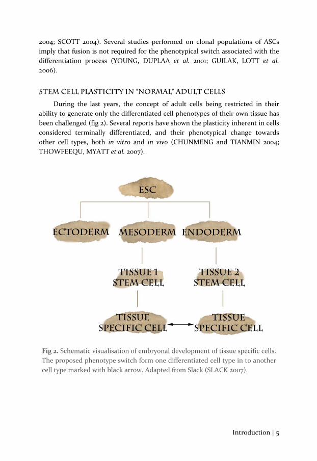
Fig 2. Schematic visualisation of embryonal development of tissue specific cells. The proposed phenotype switch form one differentiated cell type in to another cell type marked with black arrow. Adapted from Slack (SLACK 2007).
2004; SCOTT 2004). Several studies performed on clonal populations of ASCs imply that fusion is not required for the phenotypical switch associated with the differentiation process (YOUNG, DUPLAA et al. 2001; GUILAK, LOTT et al. 2006).
Stem cell plasticity in “normal” adult cells
During the last years, the concept of adult cells being restricted in their ability to generate only the differentiated cell phenotypes of their own tissue has been challenged (fig 2). Several reports have shown the plasticity inherent in cells considered terminally differentiated, and their phenotypical change towards other cell types, both in vitro and in vivo (CHUNMENG and TIANMIN 2004; THOWFEEQU, MYATT et al. 2007).
6 | Introduction
The regeneration of liver and pancreatic tissues by the means of transdifferentiation has been proposed, and shown in model organisms as reviewed by Zaret and Grompe (ZARET and GROMPE 2008). Although this phenomenon of transdifferentiation might be rarely occurring in nature, the possibility of using reprogrammed cells for therapeutic purposes has been suggested (SLACK 2007).
Dermal fibroblasts in Tissue Engineering
Several publications describe the presence of cells exhibiting SC plasticity in the dermis. Experiments performed in vitro have shown the possible osteogenic differentiation of these so called skin-derived precursor cells (TOMA, AKHAVAN et al. 2001; BURANASINSUP, SILA-ASNA et al. 2006). Reports indicate that these cells may reside in close proximity to the dermal hair follicles (JAHODA, WHITEHOUSE et al. 2003; FERNANDES, MCKENZIE et al. 2004; RICHARDSON, ARNOTT et al. 2005). Fibroblasts (FBs) derived from adult dermis display stem cell properties when genetically modified both in vitro (PHILLIPS, GULDBERG et al. 2007; TAKAHASHI, TANABE et al. 2007) and in vivo (RUTHERFORD, MOALLI et al. 2002). Furthermore, adult dermal FBs cultured in demineralised bone powder have shown promising results in regard to chondrogenic induction (MIZUNO and GLOWACKI 1996; MIZUNO and GLOWACKI 2005).
The inherent plasticity of FBs has been proposed, suggesting that they are able to differentiate towards several mesenchymal lineages (FRENCH, ROSE et al. 2004; BARTSCH, YOO et al. 2005; CHEN, ZHANG et al. 2007; HIRATA, MIZUNO et al. 2007; LORENZ, SICKER et al. 2008; LOWRY, RICHTER et al. 2008; SOMMAR, PETTERSSON et al. 2009). The dermal FB exists in large numbers throughout the connective tissue in the adult human and primary cultures can be obtained through minimally invasive biopsies. The dermal FB is one of the easiest human cell types to process and expand in vitro (WONG, MCGRATH et al. 2007). The usage of this cell in cell-based therapies may facilitate the in vitro production of autologous tissues in a most dramatic way and thus open up for new regimes in the reconstruction of damaged tissues and organs.
Surface markers in stem cell populations
The cluster of differentiation (CD) is a protocol that can be used for the identification and investigation of cell surface molecules present on various SC populations (GRONTHOS, FRANKLIN et al. 2001; WOGNUM, EAVES et al. 2003; YOUNG and BLACK 2004). CD molecules have numerous cellular functions, and are often acting as receptors or ligands. The number of CD markers is constantly increasing and there are currently more than 350 unique markers identified

Introduction | 7
(www.hcdm.or, May 2009). The analysis of CD markers present in a cell population is most often performed using flow cytometry. This method relies on the labeling of CD marker specific antibodies with fluorochromes. In the flow cytometer cells are interrogated using laser beams directed onto a hydrodynamically focused stream of fluid. A number of detectors are aimed at the interrogation point and each cell passing through is measured in regard to size, granularity and fluorescent signal, indicating the expression of labelled surface markers.
Single cell cloning
The method of single cell cloning relies on the production of separate cultures which originate from one single cell. There are several methods available for producing single cell clones (SCCs). Serial dilution is a process where a cell suspension is diluted to the point where the expected number of cells in a certain volume is below one. The solution is then aliquoted in cell culture wells. After giving the cells time to attach to the bottom of the wells, visual examination is used to locate wells containing one single cell. This method is very time consuming and an evident risk is the presence of more than one cell in a culture well. If the observer does not realise this, the single cell origin of the population is compromised.
Another possible method for creating SCCs is by employing flow cytometric cell sorting. The main problem with this method is the low survival rates of cells being trypsinised and passed through the cytometer. Furthermore, proper setup of the instrument is needed to ensure that only one cell is dispersed in every well.
In the present study (paper I) single FBs were transferred to cell culture wells by using a micro manipulator fitted with a micro pipette. The single cells were then allowed to divide during several weeks, rendering cell populations with identical clones. This method is very robust, although time consuming.
Differentiation of cells in vitro
Commonly, differentiation can be induced by modifying the medium in which cells are cultured. A plethora of soluble factors are known to modulate the transcription of target genes, thus leading to a phenotypical change of the cultured cells. ASC populations have been subjected to these media modifications, and the effect on differentiation has been extensively studied (JAISWAL, HAYNESWORTH et al. 1997; PITTENGER, MACKAY et al. 1999; ZUK, ZHU et al. 2001; ZUK, ZHU et al. 2002).
8 | Introduction
Soluble factors used for induction of differentiation Adipogenic induction
To induce adipogenic differentiation cells have been subjected to treatment with 3-isobutyl-1-methylxanthine (IBMX), insulin, indomethacin and dexameth-asone (DEX) (STUDENT, HSU et al. 1980; HAUNER and LOFFLER 1987; HAUNER, SCHMID et al. 1987; KAWAI, NAMBA et al. 2007).
IBMX is a phosphodiesterase (PDE) antagonist mediating an increase in intracellular cyclic adenosine mono phosphate (cAMP) levels. Increased cAMP levels leads to protein kinase A (PKA) activation which causes phosphorylation of hormone sensitive lipase (HSL), which increases lipolysis. Insulin, however, inhibits HSL by both increasing PDE activity, thus decreasing cAMP levels, and by non-cAMP dependent mechanisms involving phosphatase activation leading to deactivation of HSL (STRALFORS and HONNOR 1989). These effects may not be important as such for the differentiation of adipocytes, but in concert with DEX and insulin treatment, the increased cAMP level seems to play an important role. DEX and IBMX are have been shown crucial for the differentiation of the 3T3 fibroblast cell line to adipocytes (GREGOIRE, SMAS et al. 1998). Insulin has been shown to increase peroxisome proliferator-activated receptor-γ (PPARγ) transcriptional activity (KERSTEN, SEYDOUX et al. 1999). PPARγ is a central adipogenic transcription factor (LING, NURCOMBE et al. 2009) and is required for adipogenic differentiation, as determined by murine chimeric knock-out models (ROSEN, SARRAF et al. 1999). DEX is a potent synthetic glucocorticoid that stimulates a range of transcription factors, including some from the CCAAT/enhancer-binding protein (C/EBP) family, members of which are important for the transcription of PPARγ (ROSEN, SARRAF et al. 1999; KAWAI, NAMBA et al. 2007). C/EBPα is also vital for adipocyte differentiation as it transactivates promotors for genes important for adipocyte function and differentiation such as leptin, adipocyte fatty-acid binding protein and insulin receptors (GREGOIRE, SMAS et al. 1998). PPARγ and C/EBP are necessary for the establishment of insulin-sensitive glucose uptake (DESVERGNE and WAHLI 1999).
Indomethacin is a non-steroid anti-inflammatory drug that inhibits the function of inducible cyclooxygenase (COX-2). While acting to modulate the presence of neutral lipids, indomethacin is primarily used at higher concentrations as an agonist to PPARγ (DESVERGNE and WAHLI 1999; RIVAL, STENNEVIN et al. 2004). Indomethacin binds directly to the PPARγ ligand binding domain (LEHMANN, LENHARD et al. 1997). After transcription of PPARγ during adipogenic differentiation, its activity is boosted by the presence of indomethacin, leading to increased expression of PPARγ gene targets.
Introduction | 9
Chondrogenic induction To induce chondrogenic differentiation cells have been cultured in medium
containing low amounts of fetal calf serum (FCS), supplemented with insulin, ascorbate-2-phosphate (A2P) and transforming growth factor β1 (TGFβ1) (IWASAKI, NAKATA et al. 1993; MACKAY, BECK et al. 1998; AWAD, HALVORSEN et al. 2003; GAO, YAO et al. 2007). The low concentration of FCS is thought to mimic the low perfusion of nutrients in cartilage tissue in vivo.
TGFβ promotes chondrocyte differentiation and causes expression of collagen. It has been shown that TGFβ leads to the formation of precartilaginous mesenchyme (MOSES and SERRA 1996). TGFβ signaling is thought to co-operate with insulin signaling to promote chondrocyte differentiation (MCMAHON, PRENDERGAST et al. 2008).
Insulin binds to insulin growth factor receptor 1 (IGF-1R). Intracellular mediators coupled to IGF-R1 include PI3K-Akt/PKB, ERK1/2-MAPK and p38-MAPK pathways which all has been proposed to be important for chondrogenesis (MCMAHON, PRENDERGAST et al. 2008).
A2P has been shown to increase collagen content in the extracellular matrix (ECM) of cultured FBs (HATA and SENOO 1989). This increase reflects the upregulation of pro-alpha 1 and 2 collagen gene transcriptions in chondrocytes (KURATA, SENOO et al. 1993). The ECM–cytoskeleton interactions involve many intracellular mediators of chondrogenesis and provide both positive and negative feed-back. Dense culture with high amounts of cells and matrix is required for the maintenance of a chondrocyte phenotype in vitro (TACCHETTI, TAVELLA et al. 1992; WOODS, WANG et al. 2007). Therefore, high density micro mass culture is commonly used when culturing cartilage tissue (DENKER, NICOLL et al. 1995; HANDSCHEL, DEPPRICH et al. 2007).
Endothelial induction
Human serum contains numerous growth factors and bioactive molecules including vascular endothelial growth factor (VEGF), TGFβ1 and platelet-derived growth factor (PDGF) amongst others. There are several proangiogenetic and proendothelial mechanisms attributed to these factors. Previous studies have shown the endothelial differentiation of cells derived from adipose tissue upon subjection to serum (BALWIERZ, CZECH et al. 2008).
VEGF acts by binding to tyrosine kinase receptors (the VEGFR family) on cell surfaces, causing them to dimerize and become activated through transphosphorylation. The VEGF receptors have an extracellular portion consisting of 7 immunoglobulin-like domains, a single transmembrane spanning region and an intracellular portion containing a split tyrosine-kinase domain. VEGFR-2 appears to mediate most known cellular responses to VEGF. Several
10 | Introduction
studies show the generation of endothelial-like cells from adult stem cells after VEGF treatment in vitro (REYES, DUDEK et al. 2002; OSWALD, BOXBERGER et al. 2004). VEGF and its receptors have important functions in the differentiation of endothelial progenitor cells, angiogenesis and endothelial cell proliferation (KUBO and ALITALO 2003; THURSTON and GALE 2004).
TGFβ1 has been shown to promote capillary formation related apoptosis in endothelial cells (FERRARI, COOK et al. 2009). Furthermore, TGFβ1 regulates endothelial cell attachment, migration and differentiation in vitro (ZHU, YING et al. 2005).
PDGF is a dimeric glycoprotein that binds to tyrosine kinase receptors (the PDGFR family). PDGF has been shown to play a significant role in embryonic development of the vascular system as well as postnatal angiogenesis, as reviewed by Andrae et al (ANDRAE, GALLINI et al. 2008).
Osteogenic induction
To induce osteogenic differentiation cells have been subjected to treatment with DEX, A2P and beta-glycerophosphate (BGP) (CHENG, YANG et al. 1994; ZUK, ZHU et al. 2002).
A2P increases the collagen production of the fibroblasts. It has been extensively used in differentiation of osteogenic precursors, and is even said to be essential for osteoblast differentiation (TAKAMIZAWA, MAEHATA et al. 2004). A2P has also been shown to stimulate ALP activity, an early marker for osteoblast differentiation (TAKAMIZAWA, MAEHATA et al. 2004).
BGP increases collagen deposition and calcification of the ECM of FB cultures (KWIATKOWSKI, MELCHIOR et al. 2008). Type I collagen, stimulated by both BGP and A2P, acts as a ligand to osteopontin in bone, which promotes osteogenesis (SHIN, KIM et al. 2008). Osteopontin is postulated to be presented on the progenitor cell surface during osteogenesis, but the mechanisms regulating the presence of osteopontin, or indeed the determination of cell fate, has yet to be elucidated (SHIN, KIM et al. 2008).
DEX has been shown to promote osteogenesis of mesenchymal stem cells, specifically by regulating the expression of osteocalcin, osteopontin and alkaline phosphatase (ALP) (BERESFORD, GALLAGHER et al. 1984; GRIGORIADIS, HEERSCHE et al. 1988; SUBRAMANIAM, COLVARD et al. 1992; CHENG, YANG et al. 1994). DEX upregulates the parathyroid hormone receptor, and increases the basal level of cAMP by activation of adenylate cyclase (OGSTON, HARRISON et al. 2002). The mechanisms that promote osteogenesis on behalf of DEX are poorly understood. The DEX-mediated inhibition of collagenases has been shown to be a potent differentiation stimulus (HAYAMI, ZHANG et al. 2007).
Introduction | 11
Mechanical tension Myogenic induction
Several reports describe the induction of FB to myofibroblast transdifferentiation by mechanical tension (DARBY, SKALLI et al. 1990; HINZ, MASTRANGELO et al. 2001; DESMOULIERE, CHAPONNIER et al. 2005; HINZ 2007).
Upon mechanical tension the first phenotypic change occurs in response to changes in the composition, organisation, and mechanical property of the extracellular matrix (HINZ and GABBIANI 2003). With increasing stress in the ECM resulting from their own remodeling activity, the myofibroblasts further differentiate and express α smooth muscle actin (SMA), the most widely used myofibroblast marker. Expression of SMA is precisely controlled by the joint action of growth factors like TGFβ1, specialised ECM proteins like fibronectin and of the mechanical microenvironment (TOMASEK, GABBIANI et al. 2002). Incorporation of SMA into stress fibers significantly augments the contractile activity of fibroblastic cells and hallmarks the contraction phase of connective tissue remodeling (HINZ, MASTRANGELO et al. 2001).
Confirmation of Differentiation
Several different methods can be used to detect the genotypic and phenotypic changes associated with the differentiation process of a cell population. The papers included in this thesis employ routine and immunohistochemical (IHC) stainings, enzyme activity assay, analysis of low density lipoprotein incorporation, capillary-like network formation assay and full expression micro array analysis.
Adipogenesis Oil red O
Oil red O is a non-polar dye that stains triglycerides, lipids and some lipoproteins. Although not specific for adipocytes, it is frequently used to visualise intracellular lipid accumulation (KUTT and TSALTAS 1959; HAUSMAN 1981; KOOPMAN, SCHAART et al. 2001). Perilipin
Perilipin is a phosphorylated protein localised on the surface of intracellular lipid droplets in adipocytes and acts as protective coating against endogenous lipases (BLANCHETTE-MACKIE, DWYER et al. 1995). The expression of perilipin is strongly associated with mature adipocytes (GREENBERG, EGAN et al. 1991).
12 | Introduction
There is also evidence that perilipin is a vital component in the regulation of fat metabolism (WANG, SONI et al. 2008).
Chondrogenesis Alcian blue
Alcian blue is a copper containing phthalocyanine based dye that binds to negatively charged macromolecules. It is widely used to stain glucose-aminoglycans in cartilage tissue (LEV and SPICER 1964; GOLDSTEIN and HOROBIN 1974; JOHNSTONE, HERING et al. 1998; WOODS, KHAN et al. 2007).
Aggrecan
Aggrecan is a chondroitin sulphate proteoglycan and one of the two major components of cartilage. Aggrecan is highly specific for cartilage produced by chondrocytes (WILSON, BELLUOCCIO et al. 2008). Aggrecan binds to hyaluronan and Link protein, forming aggregates. These aggregates lead to a hydrated gel-like structure of cartilage and resistibility to compression and deformation in joints (WATANABE, YAMADA et al. 1998).
Collagen II
Collagen exists in several different forms in the human body. Collagen II is the other major component of hyaline cartilage (together with aggrecan) and provides strength to the cartilage matrix. The expression of collagen II is specific for chondrocytes in vivo.
Endotheliogenesis B2 bradykinin receptor
The B2 bradykinin receptor is a G protein-coupled receptor that is expressed in various tissues. When activated it leads to an increased intracellular calcium concentration mediated by phospholipase C. It is highly expressed in endothelial cells, and causes vasodilatation when activated by bradykinin.
Endothelial nitric oxide synthase
Endothelial nitric oxide synthase (eNOS) is an enzyme expressed in endothelial cells that generates nitric oxide by catalyzing an oxidation of nitrogen on L-arginine. NO diffuses into adjacent smooth muscle cells and activates guanylate cyclase, thus leading to blood vessel dilation via smooth muscle cell relaxation.
Introduction | 13
Platelet endothelial cell adhesion molecule-1 Platelet endothelial cell adhesion molecule-1 (PECAM-1) (also referred to as
CD31) is most abundant in endothelial cells, but also expressed in leukocytes, lymphocytes and osteoclasts. It has been has been demonstrated to be involved in the initial formation and stabilisation of cell-cell contacts at lateral junctions of endothelial cells, the maintenance of a vascular permeability barrier, modulation of cell migration, transendothelial migration of monocytes and neutrophils and formation of new blood vessels in angiogenesis (JACKSON 2003).
Vascular endothelial cadherin
Vascular endothelial (VE) cadherin is a calcium-dependent cell-cell adhesion glycoprotein comprised of five extracellular cadherin repeats, a transmembrane region and a cytoplasmic tail. It plays an important role in endothelial cell biology through control of the cohesion and organisation of the intercellular junctions.
VE cadherin is crucial for vascular development (CARMELIET, LAMPUGNANI et al. 1999; GORY-FAURE, PRANDINI et al. 1999). VE cadherin has important functions in maintaining newly formed vessels (CROSBY, FLEMING et al. 2005).
Von Willebrand factor
Von Willebrand factor (vWf) is a multimeric glycoprotein produced in endothelial cells. It is stored in granules in the Weibel-Palade bodies. vWf binds to proteins and is a vital component in the blood coagulation process (SADLER 1998). Low-density lipoprotein uptake
Low-density lipoprotein (LDL) is a lipoprotein that transports cholesterol and triglycerides from the liver to the peripheral tissues. Endothelial cells have the LDL receptor (LDLR), which enables endocytosis of LDL (VOYTA, VIA et al. 1984). LDLR is a chimeric protein and has also been proposed to be involved in intracellular signaling (MAY, BOCK et al. 2003).
Capillary-like network formation
The process of angiogenesis can be studied by using in vitro culture models where endothelial cells are seeded on gel substrates (i. e. ECMatrixTM, MatrigelTM etc). The gel substrates contain a number of proteins and resemble the extracellular matrix associated with endothelial cells. One major limitation to these culture systems includes the lack of other cell types.
14 | Introduction
Myogenesis α-smooth muscle actin
The expression of SMA is considered to be a marker for the transdifferentiation of FBs into myofibroblasts in contractile tissues (SKALLI, ROPRAZ et al. 1986; DARBY, SKALLI et al. 1990; SERINI and GABBIANI 1999; HINZ 2007). The expression of α-smooth muscle actin in vivo is regulated by various growth factors, including the TGFβ family (WANG, SONI et al. 2008).
Osteogenesis Von Kossa
Von Kossa staining uses a silver nitrate solution to detect calcified tissues like bone. Silver ions replace calcium ions present in a sample and are visible as a dark brown/black percipitate. Positive von Kossa staining is generally considered to be indicative of mineralised bone-like matrix (BILLS, EISENBERG et al. 1974).
Osteocalcin
Osteocalcin is a noncollagenous protein secreted by osteoblasts in bone tissue. It is the most abundant non-collagenous protein in bone. Osteocalcin can be used as a biochemical marker for the bone formation process (PAGANI, FRANCUCCI et al. 2005).
Osteonectin
Osteonectin is a calcium binding glycoprotein present in bone tissue. It is a high affinity calcium binding protein secreted by osteoblasts during bone formation, initiating mineralisation and mineral crystal formation (YAN and SAGE 1999).
Alkaline phosphatase assay
ALP is a hydrolase enzyme with three closely spaced metal ions present at the active center. It is most effective in an alkaline environment. ALP is expressed in several human tissues, but is most prominent in bone, liver and kidney (COLEMAN 1992).
Full expression micro array The development of micro array methods has lead to the possibility of
analyzing the expression of the complete genome in a sample of interest. Arrays can be purchased from commercial vendors and are highly standardised.
A microarray is a nano scale probe matrix, where each probe is present multiple times at different positions on the array surface. Each probe is
Introduction | 15
complementary to a specific sequence of a gene transcript. Multiple probes are used for different parts of each gene to increase the accuracy of the hybridisation assay.
The Affymetrix Full Expression Micro Array relies on the hybridisation of cDNA to short (~25 mer) oligonucleotides probes attached to a hydrophobic surface. The sequences are derived from sequence databases, and each gene or mRNA is represented by multiple different anti-sense probes.
The sense-strand cDNA sample is fragmented at predictable sites which are taken into account when constructing the probes. The fragments of sense cDNA are fluorescently labelled and the signal at each probe-spot on the array is measured by a micro array scanner. The resulting intensity data is transformed to a quantification of the hybridisation at specific spots, which are grouped and translated to the level of expression of each gene in the sample. Numerous control spots are present to enable comparison of two or more arrays by utilizing quality control analyses.
There are three layers of replicates in the microarray experiment: biological replicates, technical replicates and duplicate spots (KIM, RHA et al. 2004). Biological replicates are similar samples that are grouped and display biological variance. Technical replicates display variance inherent to the method protocol and include multiple array controls. Duplicate spots serve to average out stochastic differences in the hybridisation kinetics.
Statistical analyses used to interpret micro array data are numerous, and there is no consensus on which analytical method that is the most suitable (ALLISON, CUI et al. 2006). The collected data is first pre-processed in order to normalise the data to minimise systematic variation. Following normalisation, the data is filtered and transformed to exclude non-relevant differences in between the samples in the experiment. This filtration is highly subjective and depends on the criteria defined when designing the experiment. The obtained data is then be classified and differences between samples can be interpreted. Methods to classify data include clustering according to gene ontologies (i.e. grouping genes according to recognised functions or pathways), which requires access to bioinformatic databases. Clustering can also be performed using similarity-matching algorithms for gene expression in hierarchical fashions or using theoretical “expression spaces” to visualise differential expression (MOCELLIN and ROSSI 2007).

16 |
Aims | 17
Aims
he main aim of this thesis was to investigate the inherent stem cell plasticity of human dermal FBs and explore their possible usefulness in tissue engineering applications. This was achieved by studying
differentiation induced by media supplements and mechanical tension in vitro.
The specific aims for this thesis were:
• To investigate the possibility of adipogenic, chondrogenic and osteogenic differentiation of dermal FBs mediated by culture in media supplemented with soluble factors previously used for the differentiation of stem cell populations.
• To attempt to identify whether subpopulations are present in the dermal FB
culture, and assess their possible contribution to multilineage differentiation of the culture.
• To study the differentiation from FB to myofibroblast upon mechanical
tension in burn scars by using a tissue culture model. • To explore possible methods for achieving differentiation towards an
endothelial cell-like phenotype of dermal FBs and their possible use for the endothelialisation of a biomaterial surface.
• To study the full genome expression of adipogenic, chondrogenic and
osteogenic induced human dermal FBs, and identify differentially expressed genes as compared to normal uninduced FBs.
T

18 |

Materials and Methods | 19
Materials and Methods
ll in vitro culture of cells used for experiments described in this thesis were performed on cell culture grade polystyrene culture flasks unless stated otherwise. Cells were incubated at 37° C, 95 % humidity, 21 %
oxygen and 5 % carbon dioxide. When reaching subconfluence cells were enzymatically detached using Trypsin/EDTA and reseeded in new culture flasks at a 1:3 ratio.
Fibroblasts
Primary cultures of FBs were obtained from skin taken from healthy patients undergoing routine reduction mammoplastic or abdomenoplastic procedures at the university hospital in Linköping, Sweden. The dermal component of the skin was dissected into 1 mm3 fragments and enzymatically digested using collagenase and dispase. After centrifugation and resuspension, cells were seeded in FB growth medium. Several identical or highly similar procedures have been widely used for preparing dermal FBs, and are generally considered to give primary cultures with high purity and proliferative capacity. FBs were cultured in Dulbecco’s minimum essential medium (DMEM) with 10 % fetal calf serum (FCS), 50 U/mL penicillin and 50 µg/mL streptomycin.
Preadipocytes
Primary cultures of preadipocytes were obtained from adipose tissue taken from patients undergoing routine reduction abdomenoplastic procedures at the university hospital in Linköping, Sweden using an adapted protocol described by Entenmann et al (ENTENMANN and HAUNER 1996). Visible blood vessels were removed and the remaining tissue was dissected into 1 mm3 pieces before enzymatic digestion. After centrifugation and resuspension, cells were seeded in culture flasks and cultured in Ham’s F-12/DMEM with 10 % FCS, 50 U/mL penicillin and 50 µg/mL streptomycin.
Endothelial Cells
Primary culture of endothelial cells were obtained from umbilical cords taken from newborns at the university hospital in Linköping, Sweden using previously described protocol (JAFFE, NACHMAN et al. 1973). The veins of the umbilical cords were cannulated and injected with PBS containing collagenase. After 20 minutes, vessels were massaged gently and perfused. The cell solution
A
20 | Materials and Methods
obtained was seeded in gelatin coated culture flasks and cultured in DMEM containing 30 % human serum, 50 U/mL penicillin and 50 µg/mL streptomycin.
Hypertrophic burn scar tissue
Hypertrophic burn scar tissue was obtained from burn patients with deep partial or full thickness injuries. At the time of biopsy the scars were at least 12 months old. The tissue was incubated in DMEM containing 10 % human serum, 50 U/mL penicillin and 50 µg/mL streptomycin during the experiments.
Single cell cloning
SCCFBs were prepared by using a TransferMan micromanipulator fitted with a CellTramAir micropipette (Eppendorff, Germany) fitted to an IX51 inverse light/fluorescence microscope (Olympus, Sweden). Capillary tips were custom ordered and had an inner diameter of 30 μm. Precautions were taken to ensure that only one cell was transferred from the primary culture to each of the culture wells used. SCCFBs were cultured in DMEM with 10 % fetal calf serum (FCS), 50 U/mL penicillin and 50 µg/mL streptomycin.
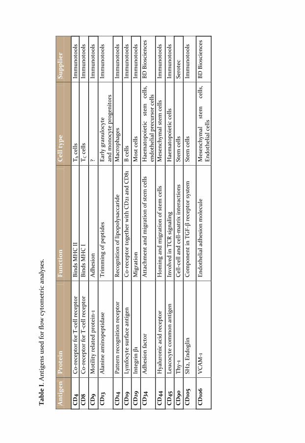
Flow cytometry
Primary culture FBs and SCCFBs were analysed by flow cytometry using antibodies towards several CD markers previously used in studies of populations exhibiting stem cell plasticity (table I) (PITTENGER, MACKAY et al. 1999; ZUK, ZHU et al. 2002; LIU, AKIYAMA et al. 2008; LORENZ, SICKER et al. 2008). 5 x 10⁵ to 106 cells from FB or SCCFB cultures were used in each antibody labeling. Cells were detached from the culture flasks using trypsin/EDTA, filtered to remove clusters and kept on ice throughout the staining procedure. 10 µl of FITC-conjugated antibodies were added to each reaction. Antibodies directed towards CD4, CD9, CD14, CD19, CD34, CD45 and CD106 did not stain the FBs or SCCFBs and were pooled in one sample. Controls included omission of antibodies and matched isotype controls. Analyses were performed using a BD LSR flow cytometer (Becton Dickinson, Brondby, Denmark). The mean fluorescence intensity was compared between CD-marker and the corresponding isotype control and the fold increase was calculated. Furthermore, the percentages of positively labelled FBs and SCCFBs were compared. Normal FBs and three SCCFB populations from separate donors were analysed at 8 different occasions. Each cell population was divided into several samples and each reaction only contained one fluorescently labelled antibody. All experiments where performed in duplicates. Statistical analysis was performed using a non-parametric Mann-Whitney test. A p value of less than 0.05 was considered significant.
Tabl
e I.
Ant
igen
s us
ed fo
r flo
w c
ytom
etri
c an
alys
es.
A
nti
gen
Pro
tein
Fu
nct
ion
Cel
l typ
e Su
ppli
er
CD
4 C
o-re
cept
or fo
r T-c
ell r
ecep
tor
Bind
s M
HC
II
T h c
ells
Im
mun
otoo
ls
CD
8 C
o-re
cept
or fo
r T-c
ell r
ecep
tor
Bind
s M
HC
I T C
cel
ls
Imm
unot
ools
CD
9 M
otili
ty r
elat
ed p
rote
in-1
A
dhes
ion
? Im
mun
otoo
ls
CD
13
Ala
nine
am
inop
eptid
ase
Trim
min
g of
pep
tides
Ea
rly
gran
uloc
yte
and
mon
ocyt
e pr
ogen
itors
Im
mun
otoo
ls
CD
14
Patt
ern
reco
gniti
on r
ecep
tor
Reco
gniti
on o
f lip
opol
ysac
cari
de
Mac
roph
ages
Im
mun
otoo
ls
CD
19
Lym
focy
te s
urfa
ce a
ntig
en
Co-
rece
ptor
toge
ther
with
CD
21 a
nd C
D81
B
cells
Im
mun
otoo
ls
CD
29
Inte
grin
β1
Mig
ratio
n M
ost c
ells
Im
mun
otoo
ls
CD
34
Adh
esio
n fa
ctor
A
ttac
hmen
t and
mig
ratio
n of
ste
m c
ells
H
aem
atop
oiet
ic
stem
ce
lls,
endo
thel
ial p
recu
rsor
cel
ls
BD B
iosc
ienc
es
CD
44
Hya
luro
nic
acid
rec
epto
r H
omin
g an
d m
igra
tion
of s
tem
cel
ls
Mes
ench
ymal
ste
m c
ells
Im
mun
otoo
ls
CD
45
Leuc
ocyt
e co
mm
on a
ntig
en
Invo
lved
in T
CR
sign
alin
g H
aem
atop
oiet
ic c
ells
Im
mun
otoo
ls
CD
90
Thy-
1 C
ell-
cell
and
cell-
mat
rix
inte
ract
ions
St
em c
ells
Se
rote
c C
D10
5 SH
2, E
ndog
lin
Com
pone
nt in
TG
F-β
rece
ptor
sys
tem
St
em c
ells
Im
mun
otoo
ls
CD
106
VC
AM
-1
Endo
thel
ial a
dhes
ion
mol
ecul
e M
esen
chym
al
stem
ce
lls,
Endo
thel
ial c
ells
BD
Bio
scie
nces
22 | Materials and Methods
Induction of differentiation
In order to induce the phenotypic shift of FBs, PAs and SCCFBs towards adipocyte, chondrocyte, endothelial and osteoblast-like cell types specific induction media were used (paper I, III-V). The specific induction factors and concentrations used can be reviewed in respective papers. To induce myogenic differentiation in burn scar tissue, mechanical tension was applied (paper II).
Confirmation of Differentiation
Uninduced FBs were used as control samples in experiments included in paper I, III, IV and V. In paper I, uninduced PAs were also included as controls. Unstretched burn scar tissue was used as control samples in paper II.
When staining 3D cultures and tissue (paper I, II and IV) samples were dehydrated through ethanol-xylene series, embedded in paraffin, sectioned using a microtome (Leica, Stockholm, Sweden), mounted on microscopy slides and rehydrated before analysis.
Routine staining All routine stainings were performed at room temperature. Cells were
fixated using 4 % neutral buffered paraformaldehyde (Apoteket, Stockholm, Sweden). All routine stainings were performed using previously described protocols (KUTT and TSALTAS 1959; LEV and SPICER 1964; MASON 1971; BILLS, EISENBERG et al. 1974; HAUSMAN 1981; WOODS, KHAN et al. 2007). Oil red O staining in paper I was quantified by examining randomly selected high power fields and counting stained and unstained cells. Alcian blue and von Kossa stainings in paper I were scored by independent blinded observers judging the staining intensity on a four degree scale. All quantifications were statistically compared using a non-parametric Kruskal-Wallis test with a Dunn’s post test.
Immunohistochemisty IHC were performed using primary antibodies towards a number of
antigens. Primary antibodies were localised using peroxidase or fluorochrome labelled secondary antibodies. For a list of all antibodies used see table II. To exclude unspecific binding of secondary antibodies control samples where primary antibodies were omitted were included in all experiments. Matched isotype non-specific immunoglobulins were employed in paper II to control for unspecific binding of the primary antibodies. When staining intracellular antigens in paper III and IV 0.01 % Triton-X was added to the blocking buffer to permeabilise cell membranes.
Materials and Methods | 23
In paper II, immunostaining was scored by independent observers. The scoring was compared using a non-parametric Kruskal-Wallis test with a Dunn’s post test. A two-tailed Spearman correlation analysis was performed to examine if the observers’ scoring of the groups were similar.
Uptake of Fluorochrome-labelled LDL
Acetylated LDL labelled with 1, 1´-dioctadecyl-3, 3, 3´, 3´-tetramethyl-indocarbocyanine perchlorate (Ac-DiI-LDL) was used to label FBs differentiated towards endothelial cell-like phenotype. Cells were visualised using a fluorescence microscope with an excitation wave length maximum of 488 nm.
Capillary-like network formation The ability of endothelial induced FBs to form capillary-like networks was
evaluated by light microscopy after 6 hours of culture on an in vitro angiogenesis assay kit, ECMatrixTM. The gel used in this kit consists of various extracellular matrix proteins (e.g. laminin, collagen type IV, heparin sulphate, and proteoglycans) and several growth factors and proteolytic enzymes.
Alkaline phosphatase assay Alkaline phosphatase activity was measured by adding paranitro-
phenylphosphate (pNPP) to cell cultures of osteogenic induced cells. pNPP is a substrate for alkaline phosphatase and is cleaved into the yellow colored para-nitrophenol (MAGNUSSON and FARLEY 2002)S. The reaction can be quantified by using a spectrophotometer measuring absorbance at a wavelength of 405 nm. The results obtained were statistically compared using linear regression analysis.

Tabl
e II
. Pri
mar
y an
d se
cond
ary
antib
odie
s us
ed in
this
thes
is.
A
nti
gen
Supp
lier
D
ilut
ion
Seco
ndar
y an
tibo
dy
Supp
lier
D
ilut
ion
Use
d in
pap
er
Agg
reca
n A
bcam
1:5
0 A
LEX
A48
8 M
olec
ular
Pro
bes
1:500
I,
V
B2 r
ecep
tor
Sigm
a-A
ldri
ch
5 µg
/mL
Vec
tast
ain
ABC
Im
mun
kem
i Re
ady
to u
se
IV
Col
lage
n II
A
bcam
1:1
00
ALE
XA
488
Mol
ecul
ar P
robe
s 1:5
00
I, V
eN
OS
Sant
a C
ruz
Biot
ech
1:50
Vec
tast
ain
ABC
Im
mun
kem
i Re
ady
to u
se
IV
Ost
eoca
lcin
C
hem
icon
1:5
00
ALE
XA
546
Mol
ecul
ar P
robe
s 1:5
00
I, V
O
steo
nec
tin
Che
mic
on
1:100
0 A
LEX
A54
6 M
olec
ular
Pro
bes
1:500
I,
V
PEC
AM
-1
Abc
am
1:50
Vec
tast
ain
ABC
Im
mun
kem
i Re
ady
to u
se
III,
IV
PEC
AM
-1
Abc
am
1:50
ALE
XA
546
Mol
ecul
ar P
robe
s 1:5
00
III,
IV
Per
ilip
in
Rese
arch
Dia
g In
c 1:2
00
ALE
XA
488
Mol
ecul
ar P
robe
s 1:5
00
I, V
SM
A
Dak
o C
ytom
atio
n 1:5
0 V
ecta
stai
n A
BC
Imm
unke
mi
Read
y to
use
II
V
E C
adhe
rin
Sant
a C
ruz
Biot
ech
1:50
Vec
tast
ain
ABC
Im
mun
kem
i Re
ady
to u
se
IV
vWF
Dak
o C
ytom
atio
n 1:2
00
ALE
XA
546
Mol
ecul
ar P
robe
s 1:5
00
III
vWF
Dak
o C
ytom
atio
n 1:2
0 V
ecta
stai
n A
BC
Imm
unke
mi
Read
y to
use
IV

Materials and Methods | 25
Full expression micro array RNA was extracted from cultured cells using the NucleoSpin RNA II kit
(Macherey-Nagel, Germany). In accordance with the provided protocol, a lysis buffer (RA1) and β-mercaptoethanol were added to each sample. After lysation of cells each sample was passed through a clean-up filter column by centrifugation. Ethanol (70 %) was added to the filtrate to precipitate the nucleic acids which, after centrifugation were bound to a filter-column. The populated filter-column was treated with a membrane desalting buffer to minimise the risk of salts interfering with subsequent DNAse treatment. The DNAse degrades the DNA which is then removed from the filter, leaving only the RNA in the filter-column. The RNA was eluted in RNAse-free water.
1 μl of the RNA suspension was used to spectrophotometrically analyse the purity of the RNA using a NanoDrop instrument (Thermo Scientific, Wilmington, USA). Nucleic acid content was measured at set wavelengths of 260 nm for nucleic acids, 230 nm for phenols and other contaminants, and 280 nm for proteins. Purity was indicated by 260/280 and 260/230 ratios, and a measure of yield was also given.
Furthermore, 1 μl of the RNA suspension was used to analyse the purity and degree of degradation in the Agilent Bioanalyzer 2100 (Agilent Technologies Inc, Santa Clara, USA). Samples were transferred to separate wells in a microfabricated 12-well chip (RNA 6000 LabChip) and analysed using a gel-electrophoretic technique. The banding is scanned and then analysed by the accompanying software to calculate a RIN value (RNA integrity number). The RIN value is set between 1 and 10, where 1 is very degraded and 10 is very intact.
For micro array analysis, the Affymetrix Human Gene 1.0ST full expression array was used. The method was performed according to the supplied protocol for 300 ng RNA starting material (Affymetix GeneChip WT ST Labeling Assay Manual rev 4, P/N 701880, with addendum rev 1, P/N 702577).
Poly-A RNA controls and primers with N-terminal T7-promoters were added to the 300 ng of total RNA. Samples were run in a first-cycle first-strand cDNA synthesis with a reverse transcription master-mix containing RNAse inhibitors, dNTPs and the Superscript II enzyme. The product was mixed with a second master-mix containing MgCl2, dNTPs, RNAse H and DNA polymerase I for a first-cycle second-strand cDNA synthesis step. In the first-cycle cRNA synthesis and clean-up a master-mix containing IVT 10X Buffer, IVT NTP mix and IVT enzyme mix was used. During the following cRNA clean-up steps the samples were diluted with RNAse-free water, cRNA Binding Buffer and EtOH (99.5 %). Subsequently, samples were transferred to an IVT cRNA Clean-up Spin Column and centrifuged. Flow-through was discarded and the filter column was moved into a new collection tube.

26 | Materials and Methods
Each filter column was washed in cRNA Wash Buffer and centrifuged, again discarding the flow-through. Another wash was performed with EtOH (80 %) and samples were dried by centrifugation. cRNA was eluted in RNAse-free water and the yield measured in the NanoDrop.
The second cycle first-strand cDNA synthesis was commenced by the addition of random primers to samples. Reverse transcription components were added to each sample (5X 1st strand buffer, 0.1 M DTT, 10 mM dNTP+dUTP, Superscript II enzyme). The following hydrolysis of cRNA and cleanup of ssDNA, were performed by adding RNAse H to each sample to remove all RNA. RNAse-free water and cDNA Binding Buffer was added and each sample was transferred to a cDNA Spin column. The samples were centrifuged and the flow-through discarded. cDNA Wash Buffer was added to the filters, samples centrifuged and flow-through discarded again. The filter columns were dried by centrifugation, samples eluted using cDNA elution buffer and yield determined using the NanoDrop.
The obtained ssDNA was fragmented by adding 10X fragmentation buffer, UDG, APE1 and RNAse-free water. The fragmented ssDNA was labelled by adding 5X TdT buffer, TdT and DNA labeling reagent.
A hybridisation cocktail was prepared containing Control oligonucleotide B2, 20X Eukaryotic Hybridisation Controls (1.5 pM bioB, 5 pM bioC, 25 pM bioD and 100 pM cre), 2X Hybridisation Mix, 7 % DMSO and RNAse-free water. The arrays were allowed to reach room temperature before use and marked with sample designations. Each sample was pipetted into the designated array through the septa on the back of the array. The arrays were then placed in a hybridisation oven at 45° C for 17 ± 1 hours.
The Fluidics Station 450 was prepared (with bottles of dH2O, Wash Buffer A and Wash Buffer B), initiated and primed in preparation for the array washing and staining steps. The experiment information was entered in the computer and the sample in each array was removed and replaced with Wash Buffer A. Three vials were prepared with Stain Cocktail 1, Stain Cocktail 2 and Array Holding Buffer for each array. The vials and the arrays were placed in the Fluidics Station and the run started with four arrays at a time, while the other four were still in the oven.
The fluidics run included a number of steps which were performed and monitored by the Fluidics Station itself. The arrays were washed, stained with SAPE solution at 35° C, and washed again. Second stain was performed in antibody solution and third stain with SAPE solution again. After the final wash the array was filled with array holding buffer. After completion the fluidics station wash shut down according to the protocol. All arrays were inspected visually to ensure that the wash and stain had not left any bubbles or other
Materials and Methods | 27
obstructions. The arrays were scanned, generating .CEL-files, containing raw scanning-data. The eight .CEL-files were imported into the GeneSpring GX 10.0 (Agilent) microarray data analysis program and grouped according to induction or growth media. GeneSpring GX10 by default uses the RMA16 (Robust Multichip Averaging) algorithm to perform normalisation, background correction and probe summarisation of each array and also performs a median transform to baseline on all samples.
Entity lists were created based on further filtering and analysis selected by the user. Filtering included probe sets that gave signals between 20th to 100th percentile, and avoiding ambiguous signals below the recommended 20th percentile threshold to eliminate background noise. Approximately 28810 entities out of 28869 were retained by the procedure recommended by Agilent.
The filtered entity list was analysed for fold change between the induced and uninduced FBs, in three separate steps to avoid multi-group correction stringencies. Fold change cut-off was set to ≥2 and statistical significance was calculated using an unpaired t-test with p <0.01. Benjamin-Hochberg corrections were performed on the probe set p-values to lower the number of expected false positives. The fold-change entity lists were separated into upregulated and downregulated for induced group relative to uninduced control FBs. Venn diagrams were constructed to visualise and select the distribution of genes that were differentially regulated for each cell type. Regulated genes were checked manually for phenotype relevance.

28 |

Results | 29
Results
etailed descriptions of all results obtained can be reviewed in respective paper. The most significant findings are presented below.
Paper I
FBs can be induced to differentiate towards several mesenchymal lineages in vitro. Indications of adipogenic differentiation include intracellular lipid accumulation and the expression of perilipin localised to droplet surfaces. Chondrogenic differentiation causes FBs to produce cartilage-resembling extracellular matrix and produce proteins specific for mature chondrocytes. Osteogenic differentiation induced a phenotypical change regarding matrix composition, alkaline phosphatase activity and protein expression.
The experiments using clonal populations presented in paper I also exclude the presence of contaminant cells in primary FB cultures giving rise to differentiated phenotypes.
Paper II
The in vitro transdifferentiation from FB to myoFB mediated by mechanical tension in burn scars were shown in paper II. The FBs in the scar tissue expressed SMA in a time dependent manner. This has a profound effect on the healing process of burn wounds, and may prove to be a significant factor when treating large burn wounds.
Paper III and IV
When subjected to high amounts of human serum, FBs alter their phenotype into an endothelial cell-like state. FBs start to produce several proteins relevant for endothelial functions and form capillary-like networks in vitro. The acquired ability of LDL uptake also reflects an important function in endothelial induced FBs. Furthermore, FBs can be seeded onto a gelatin scaffold and be induced to differentiate towards an endothelial phenotype on site.
D

30 | Results
Paper V
Adipogenic, chondrogenic and osteogenic induced FBs display the upregulation of several genes associated with adipocytes, chondrocytes and osteoblasts. Genes related to the differentiation process of progenitor cells into the above mentioned cell types were also identified (BENAYAHU, KLETTER et al. 1989; HUNG, CHANG et al. 2004; MEHLHORN, NIEMEYER et al. 2006; DJOUAD, DELORME et al. 2007; PERRIEN, AKEL et al. 2007).
Discussion | 31
Discussion
n recent years, autologous ASCs have been proposed as a cell source for tissue engineering and reconstructive surgery. Several difficulties associated with the use of ASCs still remain, and the search for alternative
cell sources is ongoing. The results presented in this thesis demonstrate that human dermal FBs possess stem cell plasticity, and can be induced to differentiate into cells with phenotypes resembling adipocytes, chondrocytes, endothelial cells, smooth muscle cells and osteoblasts.
Several reports published during the last years have supported the identification of differentiation of ASCs towards adipocyte-, chondrocyte- and osteoblast-like cells by using a variety of histological routine staining and immunohistochemical methods similar or identical to protocols used in paper I and V in this thesis (ZUK, ZHU et al. 2002; BARTSCH, YOO et al. 2005; YOUNG, DUPLAA et al. 2005; MASUOKA, ASAZUMA et al. 2006; CHEN, ZHANG et al. 2007; SOMMAR, PETTERSSON et al. 2009).
The results presented in paper I confirm the differentiation of FBs towards adipocyte-, chondrocyte- and osteoblast-like cell types. The results obtained from this study combined with present knowledge renders the FB a possible source for multiple autologous cell types for use in tissue engineering and reconstructive surgery.
Scar contracture is a common complication of deep and partial thickness burn wounds which can lead to severe disability for affected patients. Thus, it is of great importance to limit this pathological process in the scar during the rehabilitation of a burn wound. The contraction of the scar is largely dependent on cells generating mechanical forces in the granulation tissue. Current regimes for treating burn scar contraction involve active stretching of the burn area. Paper II demonstrates the effects of mechanical tension applied on burn scars in vitro, and that a continuous stretching of a burn scar in human skin increases the transdifferentiation of fibroblasts into myofibroblasts. This implies that current protocols used for the treatment of burn scars are counterproductive in regard to contraction of the tissue. The in vitro model used in paper II is limited by the exclusion of systemic inflammatory responses. However, all local components including extracellular matrix and viable cells are present in the tissue, thus the culture model represents a standardised way to investigate the responses to mechanical tension.
In paper III and IV, the phenotypical shift of FBs towards an endothelial-like cell type was studied. The results presented showed that FBs cultured in human serum expressed bradykinin B2 receptor, eNOS, VE-cadherin and vWf,
I
32 | Discussion
incorporated fluorochrome labelled LDL, and formed capillary-like structures in an endothelial cell-like fashion. These findings are, to our knowledge, the first that describe a phenotypic change of human dermal FBs towards an endothelial-like cell type. The methods used for confirmation of differentiation have been employed to characterise endothelial cells in previous studies (VOYTA, VIA et al. 1984; RISAU 1995; BACHETTI and MORBIDELLI 2000; HANNAH, WILLIAMS et al. 2002). The results showed that the presence of human serum has a decisive role in the phenotypic change of FBs towards an endothelial cell-like phenotype. Human serum contains numerous growth factors that are crucial for the differentiation and proliferation of endothelial cells, some of which may be essential for inducing the phenotypic shift of FBs. VEGF plays an important part in angiogenesis, and TGFβ1 is an important regulator of differentiation of endothelial cells and a major modulator of angiogenesis (YANG and MOSES 1990; SANKAR, MAHOOTI-BROOKS et al. 1996). It has also been postulated that VEGF is necessary for the in vitro differentiation of both endothelial precursor cells and hematopoietic stem cells into mature endothelial cells (ASAHARA, MUROHARA et al. 1997). However, neutralizing VEGF or TGFβ1 did not affect the phenotypic shift. We therefore concluded that neither VEGF nor TGF-β1, at least not alone, is responsible for the induction of differentiation of FBs towards an endothelial-like cell type.
Endothelial differentiated FBs formed a confluent monolayer similar to that of endothelial cells, when cultured on a gelatin scaffold in vitro. This further strengthens the claim that FBs can be induced towards an endothelial cell phenotype. A confluent, anti-coagulative, and anti-thrombogenic cell layer is crucial for the function of a vascular graft. Previous studies have shown a correlation between the production of nitric oxide (NO) and the patency rates of vascular grafts used in bypass surgery (AMIEL, KOMURA et al. 2006). eNOS and bradykinin B2 receptor are both vital for NO production, hence the presence of these proteins an important feature of FBs differentiated towards an endothelial cell-like state (GOVERS and RABELINK 2001; DUDZINSKI, IGARASHI et al. 2006). Results presented in paper IV indicate that FBs differentiated towards endothelial-like cells can serve as a functional cell layer substitute to endothelial cells. The results of these studies may have an impact on future cell sourcing in vascular tissue engineering. Endothelial differentiated FBs might function as a cell source for the formation of vessel networks in engineered three-dimensional tissues in vitro and facilitate the creation of tissue engineered grafts. Results obtained in paper II as well as previous published reports have suggested that fibroblasts can differentiate into smooth muscle cells (GABBIANI 2003; DESMOULIERE, CHAPONNIER et al. 2005). This implies the possibility of FBs giving rise to all three cell types required for the generation of a complete blood
Discussion | 33
vessel, which could facilitate the in vitro production of autologous vascular grafts and lead to improved possibilities of replacing damaged blood vessels with autologous tissue.
With the advancement of gene expression analysis and by employing micro array technology a greater understanding of the genetic basis for cellular differentiation can be achieved. In paper V adipogenic, chondrogenic and osteogenic FBs were studied in regard to gene expression using micro array analysis. Several genes shown up-regulated have important function in regard to differentiation of adult stem cells and normal function of terminally differentiated adipocytes, chondrocytes or osteoblasts. The three induction media used in paper I and V modified the cultures to different degrees, and while an overall shift of histology and morphology was seen, the heterogeneity of the induced cultures was apparent. Taking this into account, the gene expression analysis was performed on RNA obtained from heterogeneous cells along the entire spectrum of differentiation. This consideration becomes relevant in the choice of fold-change cut-off, which greatly affects the results of the micro array analysis. In paper V, a fold-change cut-off of 1.5 instead 0f 2.0 would have increased the number of identified genes 5-fold in all different groups. Chances are that more relevant genes are found in such extended lists than using a 2-fold cut-off, but at the same time the chances of random differences supporting false assumptions may overshadow the increased data output.
The presence of contaminant stem or precursor cells in the primary culture of FBs could possibly render false positive results in regard to differentiation capacity. This issue was addressed in several ways. Differentiation experiments in paper I, III and IV were performed using SCCFBs, which minimises the risk of contamination. Furthermore, flow cytometric analysis of SCCFBs in paper I confirmed them to not to be circulating bone marrow derived stem cells. The flow cytometric analysis of the FBs and SCCFBs revealed a set of CD markers upregulated on the SCCFBs. These markers have been employed in other studies focusing on adult stem cells from various tissues. The CD marker profile can be used in order to further characterise and purify the SCCFB populations.
The results obtained from our studies on SCCFBs also indicate that fusion between different cell types does not play a significant role in the phenotypical shift of FBs, as suggested by previous publications (CAMARGO, CHAMBERS et al. 2004). This is in line with publications exploring the inherent plasticity of FBs derived from dermis (BARTSCH, YOO et al. 2005). The results presented in this thesis suggest that the multilineage potential of dermal FBs is not necessarily dependant on the presence of subpopulations in the dermis.
34 | Discussion
Future Perspectives
Further studies investigating additional features of adipogenic, chondrogenic, endothelial, myogenic and osteogenic differentiated FBs are needed before the employment of these cells in clinical applications. Studies which focus on the stability of the phenotypic shift after induction, both in vitro and in vivo, are of great importance in order to develop beneficial treatment regimes.
Future studies focusing on other lineages of differentiation might show how extensive the differentiation potential of FBs is. Current ongoing work is exploring differentiation spanning across germ layers. If possible, this feature may alter the current perception of the differentiation process inherent in adult human cells.
The in vivo differentiation of fibroblasts is an appealing scenario, which could lead to new regimens of treating diseases without the need of harvest and in vitro modification of cells.

Conclusions | 35
Conclusions
n summary, the findings presented in this thesis suggest the use of human dermal FBs in tissue engineering applications. Given the apparent possibility of a phenotypic shift of FBs into cells with the characteristic of
adipocytes, chondrocytes, endothelial cells, osteoblasts and smooth muscle cells, the generation of autologous cells for transplantation purposes is evident. This might render a plethora of clinically relevant treatment procedures using dermal FBs.
I

36 |

Acknowledgements | 37
Acknowledgements
would like to express my sincere gratitude to a number of people involved in the making of this thesis and, perhaps more importantly, my personal development during the last years. This section is going to be long, so
brace yourselves. First and foremost, I wish to thank my supervisor Gunnar Kratz. Your
enthusiasm and ability to make everything seem interesting and fun is very valuable! It has been a true pleasure working with you. Thank you also all the barbeques on your patio, and that exquisite 14 year old Oban.
Many thanks to my co-supervisor and friend Hans Johnson, with whom I
have had many productive evenings and weekends. Thank you for all dissociated conversations (and by conversations I mean beer and flipper). I look forward to working with you in the future, though perhaps not in an underdeveloped country neighbouring Sweden. Maybe there is a need for a “Dr Junkers garden” at the foot of Ulriken? THIS MEANS WAR!!
Pehr Sommar. We started off as colleagues and now, some years later, I
consider you one of my closest friends. Many are the adventures we have experienced together, and I am sure there is many more coming. Many thanks also to your wonderful wife Marie for always welcoming me to breakfast, lunch and dinner.
Everyone working at the laboratory for experimental plastic surgery. Lisa
Karlsson, for all collaborations and endless friendship. FINISH YOUR THESIS! I know you have it in you! Sofia Pettersson, for many late nights with kung fu, drinking and everything in between. Camilla Fredriksson, for all the get-togethers at your house. Fredrik Huss, for being the senior researcher that has been taking care of the PhD students at the lab. Call me if you are in the mood for a beer. Anita Lönn, for always keeping an eye on me and making sure I didn’t waste too much time and money… Kristina Briheim, thank you for making work a cheerful place to be, and for all refreshing beverages that you have been supplying me with during the years.
Jonathan Rakar, for being a true friend that always helps out whatever I ask
of you, and for teaching an old dog some new tricks. I have a feeling that the student will master the mentor.
I

38 | Acknowledgements
Mårten Skog, for all the good times in front of the cytometer or in front of a cup of coffee/glass of beer.
Robert Cimermancic, for always taking the time to call me and ask how
everything’s going, and for all moments spent together. I hope there will be many more during the rest of our lives. By the way, I’m sorry about your pectoralis.
Charlotte Ness, for all the help with our joint projects and all the long days
at the labs in Linköping and in Bergen. All the students that I supervised during the years! It has been a pleasure
seeing you develop and enjoy medical research. Many thanks to the former and present inhabitants of the Men´s Room.
Anders Carlsson, for being a true friend for a long time, and for all the great weekends in Falköping and Hunnebostrand. Femke Lutgendorff, for all the philosophical discussions during late nights at work. I miss you in da club! Henrik Blomqvist, for all the funny video clips and twisted humour. Åsa Keita and her husband Alpha, for coping with the rule of “writing to rock music” and poker nights in Linghem.
Thank you everyone at the KEF staff. Kerstin Hagersten, for always helping
me with practical issues at work, whether being related to illegal gambling or not. Håkan Wiktander, for everything, and I do mean everything. There are no limits to your kindness. I will never forget the fantastic times at Sörstad.
Florence Sjögren, for all the help at “flödesenheten” and for always being
positive and cheerful. Jan-Ingvar Jönsson, for discussions concerning my project and shared
laughter concerning “Frau am Steuer”. Magnus Grenegård, for all the entertaining discussions and your insight in
the wonderful world of medical science. Jonas Ungerbäck my dive buddy no 1, for being a true friend in all ways
imaginable. I look forward to exploring many more countries and dive sites with you in the future.

Acknowledgements | 39
Sa’ad Salim and Faisal Omar for being the caring friends you are. I hope we will meet both in Egypt and Boston!
Tina Bendrik, for always brightening up my day with a smile! Monday
morning at work is better with you there. Joakim Henricsson and Johan Ruud, for the occasional dinner, beer and
many great memories of semi-sober discussions. Sebastian Wolfgang Schultz and Pernilla Eliasson for all good times
spent arranging or attending various festivities. Alexander Persson, for all the poker nights in the dungeon. BLP is dead,
long live BLP! Patiyan and Lauren Andersson, for all the good times spent together in
Linköping, Darwin and everywhere in between. I know you will be in my life forever.
Henrik Åberg and Ulrika Nilsson, for being wonderful friends and for all
warm hugs and laughter. I know we will continue our friendship for many more years to come, and finally settle down with friends and families in the countryside. Keep the dream alive!
Johan Olausson and Annica Damm, for all the nights out and for being
there whenever I needed you. For sharing so many moments together in front of Kjell Höglund, live or in your living room.
Calle Jonson and Cissi Johansson, for all dinners and time spent in
Mellösa. JFBB/Misfrits (Cissi, Petter, Micke C, Patrik, Katarina, Thor, Sa’ad,
Micke P and Jonathan), for all the great music in the basement. If the science career goes down the drain, I know at least we can all become rock stars.
Per Eneqvist, for giving me a chance to momentarily escape the late hours
at the lab and debate the abstract issues of life on your balcony.

40 | Acknowledgements
My old friends from Nyköping; Tommie Gustafsson, Lina Lindfors, Dan Eklund Martin Arnöman, Daniel Herkner, Chewbacca Åhlund, Andreas Fyrlundh, David Källströmer, Roger Ring and all of your families. I am truly sorry for being terribly bad at keeping in touch, but coming home to you guys and feeling like no time passed since we last met marks true lifelong friendship. My door is always open for you, wherever in the world it may be.
The Gewertz family, for all the wonderful times spent together. I know that
I am always welcome to Slängan, and look forward to spending many more Christmases with all of you.
Lotta Lindquist and Görgen. My favorite aunt (although I only have one!),
always welcoming me to your home when I’m I the capital. Solvig Nilsson and family, for always making me feel at home when visiting
you, and for giving me the possibility to relax and keep my mind off work. My father. I know how proud you would have been if you were here and
experienced this with me. You are always on my mind. My mother. I would have been nowhere without you and the endless
support you have given me throughout life. I really owe everything to you. I love you!
Klara Unosson and family. Even if we have been in separate corners of the
world, I have always felt that you were near me. Thank you for making me feel the way you do. I look forward to spending the rest of my life with you.
To everyone I forgot, thank you from the bottom of my heart!
References | 41
References
ALESSANDRI, G., S. PAGANO, A. BEZ, A. BENETTI, S. POZZI, G. IANNOLO, M. BARONIO, G. INVERNICI, A. CARUSO, C. MUNERETTO, G. BISLERI and E. PARATI (2004). "Isolation and culture of human muscle-derived stem cells able to differentiate into myogenic and neurogenic cell lineages." Lancet 364(9448): 1872-83. ALLISON, D. B., X. CUI, G. P. PAGE and M. SABRIPOUR (2006). "Microarray data analysis: from disarray to consolidation and consensus." Nat Rev Genet 7(1): 55-65. AMIEL, G. E., M. KOMURA, O. SHAPIRA, J. J. YOO, S. YAZDANI, J. BERRY, S. KAUSHAL, J. BISCHOFF, A. ATALA and S. SOKER (2006). "Engineering of blood vessels from acellular collagen matrices coated with human endothelial cells." Tissue Eng 12(8): 2355-65. ANDRAE, J., R. GALLINI and C. BETSHOLTZ (2008). "Role of platelet-derived growth factors in physiology and medicine." Genes Dev 22(10): 1276-312. ARINZEH, T. L., S. J. PETER, M. P. ARCHAMBAULT, C. VAN DEN BOS, S. GORDON, K. KRAUS, A. SMITH and S. KADIYALA (2003). "Allogeneic mesenchymal stem cells regenerate bone in a critical-sized canine segmental defect." J Bone Joint Surg Am 85-A(10): 1927-35. ASAHARA, T., T. MUROHARA, A. SULLIVAN, M. SILVER, R. VAN DER ZEE, T. LI, B. WITZENBICHLER, G. SCHATTEMAN and J. M. ISNER (1997). "Isolation of putative progenitor endothelial cells for angiogenesis." Science 275(5302): 964-7. ASAKURA, A., M. KOMAKI and M. RUDNICKI (2001). "Muscle satellite cells are multipotential stem cells that exhibit myogenic, osteogenic, and adipogenic differentiation." Differentiation 68(4-5): 245-53. ATALA, A. (2008). "Bioengineered tissues for urogenital repair in children." Pediatr Res 63(5): 569-75. AWAD, H. A., Y. D. HALVORSEN, J. M. GIMBLE and F. GUILAK (2003). "Effects of transforming growth factor beta1 and dexamethasone on the growth and chondrogenic differentiation of adipose-derived stromal cells." Tissue Eng 9(6): 1301-12. BACHETTI, T. and L. MORBIDELLI (2000). "Endothelial cells in culture: a model for studying vascular functions." Pharmacol Res 42(1): 9-19.
42 | References
BAJADA, S., I. MAZAKOVA, J. B. RICHARDSON and N. ASHAMMAKHI (2008). "Updates on stem cells and their applications in regenerative medicine." J Tissue Eng Regen Med 2(4): 169-83. BALWIERZ, A., U. CZECH, A. POLUS, R. K. FILIPKOWSKI, B. MIODUSZEWSKA, T. PROSZYNSKI, P. KOLODZIEJCZYK, J. SKRZECZYNSKA-MONCZNIK, W. DUDEK, L. KACZMAREK, J. KULIG, J. PRYJMA and A. DEMBINSKA-KIEC (2008). "Human adipose tissue stromal vascular fraction cells differentiate depending on distinct types of media." Cell Prolif 41(3): 441-59. BARTSCH, G., J. J. YOO, P. DE COPPI, M. M. SIDDIQUI, G. SCHUCH, H. G. POHL, J. FUHR, L. PERIN, S. SOKER and A. ATALA (2005). "Propagation, expansion, and multilineage differentiation of human somatic stem cells from dermal progenitors." Stem Cells Dev 14(3): 337-48. BENAYAHU, D., Y. KLETTER, D. ZIPORI and S. WIENTROUB (1989). "Bone marrow-derived stromal cell line expressing osteoblastic phenotype in vitro and osteogenic capacity in vivo." J Cell Physiol 140(1): 1-7. BERESFORD, J. N., J. A. GALLAGHER, J. W. POSER and R. G. RUSSELL (1984). "Production of osteocalcin by human bone cells in vitro. Effects of 1,25(OH)2D3, 24,25(OH)2D3, parathyroid hormone, and glucocorticoids." Metab Bone Dis Relat Res 5(5): 229-34. BILLS, C. E., H. EISENBERG and S. L. PALLANTE (1974). "Complexes of organic acids with calcium phosphate: the Von Kossa stain as a clue to the composition of bone mineral." Johns Hopkins Med J 128(4): 194-207. BLANCHETTE-MACKIE, E. J., N. K. DWYER, T. BARBER, R. A. COXEY, T. TAKEDA, C. M. RONDINONE, J. L. THEODORAKIS, A. S. GREENBERG and C. LONDOS (1995). "Perilipin is located on the surface layer of intracellular lipid droplets in adipocytes." J Lipid Res 36(6): 1211-26. BLUM, B. and N. BENVENISTY (2008). "The tumorigenicity of human embryonic stem cells." Adv Cancer Res 100: 133-58. BRITTBERG, M., A. LINDAHL, A. NILSSON, C. OHLSSON, O. ISAKSSON and L. PETERSON (1994). "Treatment of deep cartilage defects in the knee with autologous chondrocyte transplantation." N Engl J Med 331(14): 889-95. BRUDER, S. P., A. A. KURTH, M. SHEA, W. C. HAYES, N. JAISWAL and S. KADIYALA (1998). "Bone regeneration by implantation of purified, culture-expanded human mesenchymal stem cells." J Orthop Res 16(2): 155-62.
References | 43
BURANASINSUP, S., M. SILA-ASNA, N. BUNYARATVEJ and A. BUNYARATVEJ (2006). "In vitro osteogenesis from human skin-derived precursor cells." Dev Growth Differ 48(4): 263-9. CAMARGO, F. D., S. M. CHAMBERS and M. A. GOODELL (2004). "Stem cell plasticity: from transdifferentiation to macrophage fusion." Cell Prolif 37(1): 55-65. CANCEDDA, R., P. GIANNONI and M. MASTROGIACOMO (2007). "A tissue engineering approach to bone repair in large animal models and in clinical practice." Biomaterials 28(29): 4240-50. CAPLAN, A. I. (1991). "Mesenchymal stem cells." J Orthop Res 9(5): 641-50. CAPLAN, A. I. (2007). "Adult mesenchymal stem cells for tissue engineering versus regenerative medicine." J Cell Physiol 213(2): 341-7. CARMELIET, P., M. G. LAMPUGNANI, L. MOONS, F. BREVIARIO, V. COMPERNOLLE, F. BONO, G. BALCONI, R. SPAGNUOLO, B. OOSTHUYSE, M. DEWERCHIN, A. ZANETTI, A. ANGELLILO, V. MATTOT, D. NUYENS, E. LUTGENS, F. CLOTMAN, M. C. DE RUITER, A. GITTENBERGER-DE GROOT, R. POELMANN, F. LUPU, J. M. HERBERT, D. COLLEN and E. DEJANA (1999). "Targeted deficiency or cytosolic truncation of the VE-cadherin gene in mice impairs VEGF-mediated endothelial survival and angiogenesis." Cell 98(2): 147-57. CHEN, F. G., W. J. ZHANG, D. BI, W. LIU, X. WEI, F. F. CHEN, L. ZHU, L. CUI and Y. CAO (2007). "Clonal analysis of nestin(-) vimentin(+) multipotent fibroblasts isolated from human dermis." J Cell Sci 120(Pt 16): 2875-83. CHENG, S. L., J. W. YANG, L. RIFAS, S. F. ZHANG and L. V. AVIOLI (1994). "Differentiation of human bone marrow osteogenic stromal cells in vitro: induction of the osteoblast phenotype by dexamethasone." Endocrinology 134(1): 277-86. CHUNMENG, S. and C. TIANMIN (2004). "Skin: a promising reservoir for adult stem cell populations." Med Hypotheses 62(5): 683-8. COLEMAN, J. E. (1992). "Structure and mechanism of alkaline phosphatase." Annu Rev Biophys Biomol Struct 21: 441-83. CROSBY, C. V., P. A. FLEMING, W. S. ARGRAVES, M. CORADA, L. ZANETTA, E. DEJANA and C. J. DRAKE (2005). "VE-cadherin is not required for the formation of nascent blood vessels but acts to prevent their disassembly." Blood 105(7): 2771-6. DARBY, I., O. SKALLI and G. GABBIANI (1990). "Alpha-smooth muscle actin is transiently expressed by myofibroblasts during experimental wound healing." Lab Invest 63(1): 21-9.

44 | References
DEASY, B. M., R. J. JANKOWSKI and J. HUARD (2001). "Muscle-derived stem cells: characterization and potential for cell-mediated therapy." Blood Cells Mol Dis 27(5): 924-33. DENKER, A. E., S. B. NICOLL and R. S. TUAN (1995). "Formation of cartilage-like spheroids by micromass cultures of murine C3H10T1/2 cells upon treatment with transforming growth factor-beta 1." Differentiation 59(1): 25-34. DESMOULIERE, A., C. CHAPONNIER and G. GABBIANI (2005). "Tissue repair, contraction, and the myofibroblast." Wound Repair Regen 13(1): 7-12. DESVERGNE, B. and W. WAHLI (1999). "Peroxisome proliferator-activated receptors: nuclear control of metabolism." Endocr Rev 20(5): 649-88. DJOUAD, F., B. DELORME, M. MAURICE, C. BONY, F. APPARAILLY, P. LOUIS-PLENCE, F. CANOVAS, P. CHARBORD, D. NOEL and C. JORGENSEN (2007). "Microenvironmental changes during differentiation of mesenchymal stem cells towards chondrocytes." Arthritis Res Ther 9(2): R33. DRAGOO, J. L., G. CARLSON, F. MCCORMICK, H. KHAN-FAROOQI, M. ZHU, P. A. ZUK and P. BENHAIM (2007). "Healing full-thickness cartilage defects using adipose-derived stem cells." Tissue Eng 13(7): 1615-21. DUDZINSKI, D. M., J. IGARASHI, D. GREIF and T. MICHEL (2006). "The regulation and pharmacology of endothelial nitric oxide synthase." Annu Rev Pharmacol Toxicol 46: 235-76. EISENBERG, L. M. and C. A. EISENBERG (2003). "Stem cell plasticity, cell fusion, and transdifferentiation." Birth Defects Res C Embryo Today 69(3): 209-18. ENTENMANN, G. and H. HAUNER (1996). "Relationship between replication and differentiation in cultured human adipocyte precursor cells." Am J Physiol 270(4 Pt 1): C1011-6. FERNANDES, K. J., I. A. MCKENZIE, P. MILL, K. M. SMITH, M. AKHAVAN, F. BARNABE-HEIDER, J. BIERNASKIE, A. JUNEK, N. R. KOBAYASHI, J. G. TOMA, D. R. KAPLAN, P. A. LABOSKY, V. RAFUSE, C. C. HUI and F. D. MILLER (2004). "A dermal niche for multipotent adult skin-derived precursor cells." Nat Cell Biol 6(11): 1082-93. FERRARI, G., B. D. COOK, V. TERUSHKIN, G. PINTUCCI and P. MIGNATTI (2009). "Transforming growth factor-beta 1 (TGF-beta1) induces angiogenesis through vascular endothelial growth factor (VEGF)-mediated apoptosis." J Cell Physiol 219(2): 449-58.
References | 45
FOSSUM, M., A. NORDENSKJOLD and G. KRATZ (2004). "Engineering of multilayered urinary tissue in vitro." Tissue Eng 10(1-2): 175-80. FRENCH, M. M., S. ROSE, J. CANSECO and K. A. ATHANASIOU (2004). "Chondrogenic differentiation of adult dermal fibroblasts." Ann Biomed Eng 32(1): 50-6. FU, X. and H. LI (2009). "Mesenchymal stem cells and skin wound repair and regeneration: possibilities and questions." Cell Tissue Res 335(2): 317-21. GABBIANI, G. (2003). "The myofibroblast in wound healing and fibrocontractive diseases." J Pathol 200(4): 500-3. GALLICO, G. G., 3RD, N. E. O'CONNOR, C. C. COMPTON, O. KEHINDE and H. GREEN (1984). "Permanent coverage of large burn wounds with autologous cultured human epithelium." N Engl J Med 311(7): 448-51. GAO, J., J. Q. YAO and A. I. CAPLAN (2007). "Stem cells for tissue engineering of articular cartilage." Proc Inst Mech Eng [H] 221(5): 441-50. GARDNER, R. L. (2007). "Stem cells and regenerative medicine: principles, prospects and problems." C R Biol 330(6-7): 465-73. GOLDSTEIN, D. J. and R. W. HOROBIN (1974). "Surface staining of cartilage by Alcian blue, with reference to the role of microscopic dye aggregates in histological staining." Histochem J 6(2): 175-84. GORY-FAURE, S., M. H. PRANDINI, H. POINTU, V. ROULLOT, I. PIGNOT-PAINTRAND, M. VERNET and P. HUBER (1999). "Role of vascular endothelial-cadherin in vascular morphogenesis." Development 126(10): 2093-102. GOVERS, R. and T. J. RABELINK (2001). "Cellular regulation of endothelial nitric oxide synthase." Am J Physiol Renal Physiol 280(2): F193-206. GREENBERG, A. S., J. J. EGAN, S. A. WEK, N. B. GARTY, E. J. BLANCHETTE-MACKIE and C. LONDOS (1991). "Perilipin, a major hormonally regulated adipocyte-specific phosphoprotein associated with the periphery of lipid storage droplets." J Biol Chem 266(17): 11341-6. GREGOIRE, F. M., C. M. SMAS and H. S. SUL (1998). "Understanding adipocyte differentiation." Physiol Rev 78(3): 783-809. GRIGORIADIS, A. E., J. N. HEERSCHE and J. E. AUBIN (1988). "Differentiation of muscle, fat, cartilage, and bone from progenitor cells present in a bone-derived clonal cell population: effect of dexamethasone." J Cell Biol 106(6): 2139-51.
46 | References
GRONTHOS, S., D. M. FRANKLIN, H. A. LEDDY, P. G. ROBEY, R. W. STORMS and J. M. GIMBLE (2001). "Surface protein characterization of human adipose tissue-derived stromal cells." J Cell Physiol 189(1): 54-63. GUILAK, F., K. E. LOTT, H. A. AWAD, Q. CAO, K. C. HICOK, B. FERMOR and J. M. GIMBLE (2006). "Clonal analysis of the differentiation potential of human adipose-derived adult stem cells." J Cell Physiol 206(1): 229-37. GUSTAFSON, C. J., J. ELDH and G. KRATZ (1998). "Culture of human urothelial cells on a cell-free dermis for autotransplantation." Eur Urol 33(5): 503-6. GUSTAFSON, C. J. and G. KRATZ (1999). "Cultured autologous keratinocytes on a cell-free dermis in the treatment of full-thickness wounds." Burns 25(4): 331-5. GUTIERREZ-DALMAU, A. and J. M. CAMPISTOL (2007). "Immunosuppressive therapy and malignancy in organ transplant recipients: a systematic review." Drugs 67(8): 1167-98. HANDSCHEL, J. G., R. A. DEPPRICH, N. R. KUBLER, H. P. WIESMANN, M. OMMERBORN and U. MEYER (2007). "Prospects of micromass culture technology in tissue engineering." Head Face Med 3: 4. HANNAH, M. J., R. WILLIAMS, J. KAUR, L. J. HEWLETT and D. F. CUTLER (2002). "Biogenesis of Weibel-Palade bodies." Semin Cell Dev Biol 13(4): 313-24. HATA, R. and H. SENOO (1989). "L-ascorbic acid 2-phosphate stimulates collagen accumulation, cell proliferation, and formation of a three-dimensional tissuelike substance by skin fibroblasts." J Cell Physiol 138(1): 8-16. HAUNER, H. and G. LOFFLER (1987). "Adipose tissue development: the role of precursor cells and adipogenic factors. Part I: Adipose tissue development and the role of precursor cells." Klin Wochenschr 65(17): 803-11. HAUNER, H., P. SCHMID and E. F. PFEIFFER (1987). "Glucocorticoids and insulin promote the differentiation of human adipocyte precursor cells into fat cells." J Clin Endocrinol Metab 64(4): 832-5. HAUSMAN, G. J. (1981). "Techniques for studying adipocytes." Stain Technol 56(3): 149-54. HAYAMI, T., Q. ZHANG, Y. KAPILA and S. KAPILA (2007). "Dexamethasone's enhancement of osteoblastic markers in human periodontal ligament cells is associated with inhibition of collagenase expression." Bone 40(1): 93-104.
References | 47
HEYDARKHAN-HAGVALL, S., K. SCHENKE-LAYLAND, J. Q. YANG, S. HEYDARKHAN, Y. XU, P. A. ZUK, W. R. MACLELLAN and R. E. BEYGUI (2008). "Human adipose stem cells: a potential cell source for cardiovascular tissue engineering." Cells Tissues Organs 187(4): 263-74. HINZ, B. (2007). "Formation and function of the myofibroblast during tissue repair." J Invest Dermatol 127(3): 526-37. HINZ, B. and G. GABBIANI (2003). "Mechanisms of force generation and transmission by myofibroblasts." Curr Opin Biotechnol 14(5): 538-46. HINZ, B., D. MASTRANGELO, C. E. ISELIN, C. CHAPONNIER and G. GABBIANI (2001). "Mechanical tension controls granulation tissue contractile activity and myofibroblast differentiation." Am J Pathol 159(3): 1009-20. HIRATA, K., A. MIZUNO and A. YAMAGUCHI (2007). "Transplantation of skin fibroblasts expressing BMP-2 contributes to the healing of critical-sized bone defects." J Bone Miner Metab 25(1): 6-11. HUNG, S. C., C. F. CHANG, H. L. MA, T. H. CHEN and L. LOW-TONE HO (2004). "Gene expression profiles of early adipogenesis in human mesenchymal stem cells." Gene 340(1): 141-50. IWASAKI, M., K. NAKATA, H. NAKAHARA, T. NAKASE, T. KIMURA, K. KIMATA, A. I. CAPLAN and K. ONO (1993). "Transforming growth factor-beta 1 stimulates chondrogenesis and inhibits osteogenesis in high density culture of periosteum-derived cells." Endocrinology 132(4): 1603-8. JACKSON, D. E. (2003). "The unfolding tale of PECAM-1." FEBS Lett 540(1-3): 7-14. JAFFE, E. A., R. L. NACHMAN, C. G. BECKER and C. R. MINICK (1973). "Culture of human endothelial cells derived from umbilical veins. Identification by morphologic and immunologic criteria." J Clin Invest 52(11): 2745-56. JAHODA, C. A., J. WHITEHOUSE, A. J. REYNOLDS and N. HOLE (2003). "Hair follicle dermal cells differentiate into adipogenic and osteogenic lineages." Exp Dermatol 12(6): 849-59. JAISWAL, N., S. E. HAYNESWORTH, A. I. CAPLAN and S. P. BRUDER (1997). "Osteogenic differentiation of purified, culture-expanded human mesenchymal stem cells in vitro." J Cell Biochem 64(2): 295-312. JAISWAL, R. K., N. JAISWAL, S. P. BRUDER, G. MBALAVIELE, D. R. MARSHAK and M. F. PITTENGER (2000). "Adult human mesenchymal stem cell differentiation to the osteogenic or adipogenic lineage is regulated by mitogen-activated protein kinase." J Biol Chem 275(13): 9645-52.
48 | References
JIN, K. and D. A. GREENBERG (2003). "Tales of transdifferentiation." Exp Neurol 183(2): 255-7. JOHNSTONE, B., T. M. HERING, A. I. CAPLAN, V. M. GOLDBERG and J. U. YOO (1998). "In vitro chondrogenesis of bone marrow-derived mesenchymal progenitor cells." Exp Cell Res 238(1): 265-72. KAWAI, M., N. NAMBA, S. MUSHIAKE, Y. ETANI, R. NISHIMURA, M. MAKISHIMA and K. OZONO (2007). "Growth hormone stimulates adipogenesis of 3T3-L1 cells through activation of the Stat5A/5B-PPARgamma pathway." J Mol Endocrinol 38(1-2): 19-34. KERSTEN, S., J. SEYDOUX, J. M. PETERS, F. J. GONZALEZ, B. DESVERGNE and W. WAHLI (1999). "Peroxisome proliferator-activated receptor alpha mediates the adaptive response to fasting." J Clin Invest 103(11): 1489-98. KIM, B. S., S. Y. RHA, G. B. CHO and H. C. CHUNG (2004). "Spearman's footrule as a measure of cDNA microarray reproducibility." Genomics 84(2): 441-8. KOOPMAN, R., G. SCHAART and M. K. HESSELINK (2001). "Optimisation of oil red O staining permits combination with immunofluorescence and automated quantification of lipids." Histochem Cell Biol 116(1): 63-8. KUBO, H. and K. ALITALO (2003). "The bloody fate of endothelial stem cells." Genes Dev 17(3): 322-9. KURATA, S., H. SENOO and R. HATA (1993). "Transcriptional activation of type I collagen genes by ascorbic acid 2-phosphate in human skin fibroblasts and its failure in cells from a patient with alpha 2(I)-chain-defective Ehlers-Danlos syndrome." Exp Cell Res 206(1): 63-71. KUTT, H. and T. T. TSALTAS (1959). "Staining properties of oil red O and a method of partial purification of the commercial product." Clin Chem 5(2): 149-60. KWIATKOWSKI, J. N., A. MELCHIOR, H. ENDT-KNAUER, K. SCHROR, J. W. FISCHER and A. A. WEBER (2008). "Effects of fibroblast extracellular matrix calcification on platelet adhesion in vitro." Platelets 19(6): 467-70. LANGE, C., F. TOGEL, H. ITTRICH, F. CLAYTON, C. NOLTE-ERNSTING, A. R. ZANDER and C. WESTENFELDER (2005). "Administered mesenchymal stem cells enhance recovery from ischemia/reperfusion-induced acute renal failure in rats." Kidney Int 68(4): 1613-7. LANGER, R. and J. P. VACANTI (1993). "Tissue engineering." Science 260(5110): 920-6.
References | 49
LEHMANN, J. M., J. M. LENHARD, B. B. OLIVER, G. M. RINGOLD and S. A. KLIEWER (1997). "Peroxisome proliferator-activated receptors alpha and gamma are activated by indomethacin and other non-steroidal anti-inflammatory drugs." J Biol Chem 272(6): 3406-10. LEV, R. and S. S. SPICER (1964). "Specific Staining of Sulphate Groups with Alcian Blue at Low Ph." J Histochem Cytochem 12: 309. LING, L., V. NURCOMBE and S. M. COOL (2009). "Wnt signaling controls the fate of mesenchymal stem cells." Gene 433(1-2): 1-7. LIU, F., Y. AKIYAMA, S. TAI, K. MARUYAMA, Y. KAWAGUCHI, K. MURAMATSU and K. YAMAGUCHI (2008). "Changes in the expression of CD106, osteogenic genes, and transcription factors involved in the osteogenic differentiation of human bone marrow mesenchymal stem cells." J Bone Miner Metab 26(4): 312-20. LIU, Y., S. BHARADWAJ, S. J. LEE, A. ATALA and Y. ZHANG (2009). "Optimization of a natural collagen scaffold to aid cell-matrix penetration for urologic tissue engineering." Biomaterials 30(23-24): 3865-73. LORENZ, K., M. SICKER, E. SCHMELZER, T. RUPF, J. SALVETTER, M. SCHULZ-SIEGMUND and A. BADER (2008). "Multilineage differentiation potential of human dermal skin-derived fibroblasts." Exp Dermatol 17(11): 925-32. LOWRY, W. E., L. RICHTER, R. YACHECHKO, A. D. PYLE, J. TCHIEU, R. SRIDHARAN, A. T. CLARK and K. PLATH (2008). "Generation of human induced pluripotent stem cells from dermal fibroblasts." Proc Natl Acad Sci U S A 105(8): 2883-8. MACCHIARINI, P., P. JUNGEBLUTH, T. GO, M. A. ASNAGHI, L. E. REES, T. A. COGAN, A. DODSON, J. MARTORELL, S. BELLINI, P. P. PARNIGOTTO, S. C. DICKINSON, A. P. HOLLANDER, S. MANTERO, M. T. CONCONI and M. A. BIRCHALL (2008). "Clinical transplantation of a tissue-engineered airway." Lancet 372(9655): 2023-30. MACKAY, A. M., S. C. BECK, J. M. MURPHY, F. P. BARRY, C. O. CHICHESTER and M. F. PITTENGER (1998). "Chondrogenic differentiation of cultured human mesenchymal stem cells from marrow." Tissue Eng 4(4): 415-28. MAGNUSSON, P. and J. R. FARLEY (2002). "Differences in sialic acid residues among bone alkaline phosphatase isoforms: a physical, biochemical, and immunological characterization." Calcif Tissue Int 71(6): 508-18. MASON, R. M. (1971). "Observations on the glycosaminoglycans of aging bronchial cartilage studied with Alcian Blue." Histochem J 3(6): 421-34.
50 | References
MASUOKA, K., T. ASAZUMA, H. HATTORI, Y. YOSHIHARA, M. SATO, K. MATSUMURA, T. MATSUI, B. TAKASE, K. NEMOTO and M. ISHIHARA (2006). "Tissue engineering of articular cartilage with autologous cultured adipose tissue-derived stromal cells using atelocollagen honeycomb-shaped scaffold with a membrane sealing in rabbits." J Biomed Mater Res B Appl Biomater 79(1): 25-34. MAY, P., H. H. BOCK and J. HERZ (2003). "Integration of endocytosis and signal transduction by lipoprotein receptors." Sci STKE 2003(176): PE12. MCLAUGHLIN, C. R., R. J. TSAI, M. A. LATORRE and M. GRIFFITH (2009). "Bioengineered corneas for transplantation and in vitro toxicology." Front Biosci 14: 3326-37. MCMAHON, L. A., P. J. PRENDERGAST and V. A. CAMPBELL (2008). "A comparison of the involvement of p38, ERK1/2 and PI3K in growth factor-induced chondrogenic differentiation of mesenchymal stem cells." Biochem Biophys Res Commun 368(4): 990-5. MEHLHORN, A. T., P. NIEMEYER, S. KAISER, G. FINKENZELLER, G. B. STARK, N. P. SUDKAMP and H. SCHMAL (2006). "Differential expression pattern of extracellular matrix molecules during chondrogenesis of mesenchymal stem cells from bone marrow and adipose tissue." Tissue Eng 12(10): 2853-62. MIZUNO, S. and J. GLOWACKI (1996). "Chondroinduction of human dermal fibroblasts by demineralized bone in three-dimensional culture." Exp Cell Res 227(1): 89-97. MIZUNO, S. and J. GLOWACKI (2005). "Low oxygen tension enhances chondroinduction by demineralized bone matrix in human dermal fibroblasts in vitro." Cells Tissues Organs 180(3): 151-8. MOCELLIN, S. and C. R. ROSSI (2007). "Principles of gene microarray data analysis." Adv Exp Med Biol 593: 19-30. MOSES, H. L. and R. SERRA (1996). "Regulation of differentiation by TGF-beta." Curr Opin Genet Dev 6(5): 581-6. O'MALLEY, K. and E. W. SCOTT (2004). "Stem cell fusion confusion." Exp Hematol 32(2): 131-4. OGSTON, N., A. J. HARRISON, H. F. CHEUNG, B. A. ASHTON and G. HAMPSON (2002). "Dexamethasone and retinoic acid differentially regulate growth and differentiation in an immortalised human clonal bone marrow stromal cell line with osteoblastic characteristics." Steroids 67(11): 895-906.
References | 51
OLSSON, M. J. and L. JUHLIN (1992). "Melanocyte transplantation in vitiligo." Lancet 340(8825): 981. OSWALD, J., S. BOXBERGER, B. JORGENSEN, S. FELDMANN, G. EHNINGER, M. BORNHAUSER and C. WERNER (2004). "Mesenchymal stem cells can be differentiated into endothelial cells in vitro." Stem Cells 22(3): 377-84. PAGANI, F., C. M. FRANCUCCI and L. MORO (2005). "Markers of bone turnover: biochemical and clinical perspectives." J Endocrinol Invest 28(10 Suppl): 8-13. PASSIER, R., L. W. VAN LAAKE and C. L. MUMMERY (2008). "Stem-cell-based therapy and lessons from the heart." Nature 453(7193): 322-9. PERRIEN, D. S., N. S. AKEL, P. K. EDWARDS, A. A. CARVER, M. S. BENDRE, F. L. SWAIN, R. A. SKINNER, W. R. HOGUE, K. M. NICKS, T. M. PIERSON, L. J. SUVA and D. GADDY (2007). "Inhibin A is an endocrine stimulator of bone mass and strength." Endocrinology 148(4): 1654-65. PETERSON, L., T. MINAS, M. BRITTBERG, A. NILSSON, E. SJOGREN-JANSSON and A. LINDAHL (2000). "Two- to 9-year outcome after autologous chondrocyte transplantation of the knee." Clin Orthop Relat Res(374): 212-34. PHILLIPS, J. E., R. E. GULDBERG and A. J. GARCIA (2007). "Dermal fibroblasts genetically modified to express Runx2/Cbfa1 as a mineralizing cell source for bone tissue engineering." Tissue Eng 13(8): 2029-40. PITTENGER, M. F., A. M. MACKAY, S. C. BECK, R. K. JAISWAL, R. DOUGLAS, J. D. MOSCA, M. A. MOORMAN, D. W. SIMONETTI, S. CRAIG and D. R. MARSHAK (1999). "Multilineage potential of adult human mesenchymal stem cells." Science 284(5411): 143-7. REYES, M., A. DUDEK, B. JAHAGIRDAR, L. KOODIE, P. H. MARKER and C. M. VERFAILLIE (2002). "Origin of endothelial progenitors in human postnatal bone marrow." J Clin Invest 109(3): 337-46. RICHARDSON, G. D., E. C. ARNOTT, C. J. WHITEHOUSE, C. M. LAWRENCE, A. J. REYNOLDS, N. HOLE and C. A. JAHODA (2005). "Plasticity of rodent and human hair follicle dermal cells: implications for cell therapy and tissue engineering." J Investig Dermatol Symp Proc 10(3): 180-3. RISAU, W. (1995). "Differentiation of endothelium." Faseb J 9(10): 926-33.
52 | References
RIVAL, Y., A. STENNEVIN, L. PUECH, A. ROUQUETTE, C. CATHALA, F. LESTIENNE, E. DUPONT-PASSELAIGUE, J. F. PATOISEAU, T. WURCH and D. JUNQUERO (2004). "Human adipocyte fatty acid-binding protein (aP2) gene promoter-driven reporter assay discriminates nonlipogenic peroxisome proliferator-activated receptor gamma ligands." J Pharmacol Exp Ther 311(2): 467-75. ROSEN, E. D., P. SARRAF, A. E. TROY, G. BRADWIN, K. MOORE, D. S. MILSTONE, B. M. SPIEGELMAN and R. M. MORTENSEN (1999). "PPAR gamma is required for the differentiation of adipose tissue in vivo and in vitro." Mol Cell 4(4): 611-7. RUTHERFORD, R. B., M. MOALLI, R. T. FRANCESCHI, D. WANG, K. GU and P. H. KREBSBACH (2002). "Bone morphogenetic protein-transduced human fibroblasts convert to osteoblasts and form bone in vivo." Tissue Eng 8(3): 441-52. SADLER, J. E. (1998). "Biochemistry and genetics of von Willebrand factor." Annu Rev Biochem 67: 395-424. SANKAR, S., N. MAHOOTI-BROOKS, L. BENSEN, T. L. MCCARTHY, M. CENTRELLA and J. A. MADRI (1996). "Modulation of transforming growth factor beta receptor levels on microvascular endothelial cells during in vitro angiogenesis." J Clin Invest 97(6): 1436-46. SCHULTZ, S. S. and P. A. LUCAS (2006). "Human stem cells isolated from adult skeletal muscle differentiate into neural phenotypes." J Neurosci Methods 152(1-2): 144-55. SCOTT, E. W. (2004). "Stem cell plasticity or fusion: two approaches to targeted cell therapy." Blood Cells Mol Dis 32(1): 65-7. SEEGER, F. H., T. TONN, N. KRZOSSOK, A. M. ZEIHER and S. DIMMELER (2007). "Cell isolation procedures matter: a comparison of different isolation protocols of bone marrow mononuclear cells used for cell therapy in patients with acute myocardial infarction." Eur Heart J 28(6): 766-72. SERINI, G. and G. GABBIANI (1999). "Mechanisms of myofibroblast activity and phenotypic modulation." Exp Cell Res 250(2): 273-83. SHIMAZONO, Y. (2007). "The state of the international organ trade: a provisional picture based on integration of available information." Bull World Health Organ 85(12): 955-62.
References | 53
SHIN, M. K., M. K. KIM, Y. S. BAE, I. JO, S. J. LEE, C. P. CHUNG, Y. J. PARK and S. MIN DO (2008). "A novel collagen-binding peptide promotes osteogenic differentiation via Ca2+/calmodulin-dependent protein kinase II/ERK/AP-1 signaling pathway in human bone marrow-derived mesenchymal stem cells." Cell Signal 20(4): 613-24. SKALLI, O., P. ROPRAZ, A. TRZECIAK, G. BENZONANA, D. GILLESSEN and G. GABBIANI (1986). "A monoclonal antibody against alpha-smooth muscle actin: a new probe for smooth muscle differentiation." J Cell Biol 103(6 Pt 2): 2787-96. SLACK, J. M. (2007). "Metaplasia and transdifferentiation: from pure biology to the clinic." Nat Rev Mol Cell Biol 8(5): 369-78. SOMMAR, P., S. PETTERSSON, C. NESS, H. JOHNSON, G. KRATZ and J. P. JUNKER (2009). "Engineering three-dimensional cartilage- and bone-like tissues using human dermal fibroblasts and macroporous gelatine microcarriers." J Plast Reconstr Aesthet Surg. SPRIGGS, M. and J. SAVULESCU (2002). ""Saviour siblings"." J Med Ethics 28(5): 289. STRALFORS, P. and R. C. HONNOR (1989). "Insulin-induced dephosphorylation of hormone-sensitive lipase. Correlation with lipolysis and cAMP-dependent protein kinase activity." Eur J Biochem 182(2): 379-85. STUDENT, A. K., R. Y. HSU and M. D. LANE (1980). "Induction of fatty acid synthetase synthesis in differentiating 3T3-L1 preadipocytes." J Biol Chem 255(10): 4745-50. SUBRAMANIAM, M., D. COLVARD, P. E. KEETING, K. RASMUSSEN, B. L. RIGGS and T. C. SPELSBERG (1992). "Glucocorticoid regulation of alkaline phosphatase, osteocalcin, and proto-oncogenes in normal human osteoblast-like cells." J Cell Biochem 50(4): 411-24. SVENDSEN, C. (2008). "Stem cells and Parkinson's disease: toward a treatment, not a cure." Cell Stem Cell 2(5): 412-3. TACCHETTI, C., S. TAVELLA, B. DOZIN, R. QUARTO, G. ROBINO and R. CANCEDDA (1992). "Cell condensation in chondrogenic differentiation." Exp Cell Res 200(1): 26-33. TAKAHASHI, K., K. TANABE, M. OHNUKI, M. NARITA, T. ICHISAKA, K. TOMODA and S. YAMANAKA (2007). "Induction of pluripotent stem cells from adult human fibroblasts by defined factors." Cell 131(5): 861-72.
54 | References
TAKAMIZAWA, S., Y. MAEHATA, K. IMAI, H. SENOO, S. SATO and R. HATA (2004). "Effects of ascorbic acid and ascorbic acid 2-phosphate, a long-acting vitamin C derivative, on the proliferation and differentiation of human osteoblast-like cells." Cell Biol Int 28(4): 255-65. THOWFEEQU, S., E. J. MYATT and D. TOSH (2007). "Transdifferentiation in developmental biology, disease, and in therapy." Dev Dyn 236(12): 3208-17. THURSTON, G. and N. W. GALE (2004). "Vascular endothelial growth factor and other signaling pathways in developmental and pathologic angiogenesis." Int J Hematol 80(1): 7-20. TOGEL, F. and C. WESTENFELDER (2007). "Adult bone marrow-derived stem cells for organ regeneration and repair." Dev Dyn 236(12): 3321-31. TOMA, J. G., M. AKHAVAN, K. J. FERNANDES, F. BARNABE-HEIDER, A. SADIKOT, D. R. KAPLAN and F. D. MILLER (2001). "Isolation of multipotent adult stem cells from the dermis of mammalian skin." Nat Cell Biol 3(9): 778-84. TOMASEK, J. J., G. GABBIANI, B. HINZ, C. CHAPONNIER and R. A. BROWN (2002). "Myofibroblasts and mechano-regulation of connective tissue remodelling." Nat Rev Mol Cell Biol 3(5): 349-63. TOWNS, C. R. and D. G. JONES (2004). "Stem cells, embryos, and the environment: a context for both science and ethics." J Med Ethics 30(4): 410-3. VACANTI, C. A. (2006). "History of tissue engineering and a glimpse into its future." Tissue Eng 12(5): 1137-42. WANG, S., K. G. SONI, M. SEMACHE, S. CASAVANT, M. FORTIER, L. PAN and G. A. MITCHELL (2008). "Lipolysis and the integrated physiology of lipid energy metabolism." Mol Genet Metab 95(3): 117-26. WATANABE, H., Y. YAMADA and K. KIMATA (1998). "Roles of aggrecan, a large chondroitin sulfate proteoglycan, in cartilage structure and function." J Biochem 124(4): 687-93. WILSON, R., D. BELLUOCCIO and J. F. BATEMAN (2008). "Proteomic analysis of cartilage proteins." Methods 45(1): 22-31. WOGNUM, A. W., A. C. EAVES and T. E. THOMAS (2003). "Identification and isolation of hematopoietic stem cells." Arch Med Res 34(6): 461-75. WONG, T., J. A. MCGRATH and H. NAVSARIA (2007). "The role of fibroblasts in tissue engineering and regeneration." Br J Dermatol 156(6): 1149-55.
References | 55
WOODS, A., S. KHAN and F. BEIER (2007). "C-type natriuretic peptide regulates cellular condensation and glycosaminoglycan synthesis during chondrogenesis." Endocrinology 148(10): 5030-41. WOODS, A., G. WANG and F. BEIER (2007). "Regulation of chondrocyte differentiation by the actin cytoskeleton and adhesive interactions." J Cell Physiol 213(1): 1-8. VOYTA, J. C., D. P. VIA, C. E. BUTTERFIELD and B. R. ZETTER (1984). "Identification and isolation of endothelial cells based on their increased uptake of acetylated-low density lipoprotein." J Cell Biol 99(6): 2034-40. YAN, Q. and E. H. SAGE (1999). "SPARC, a matricellular glycoprotein with important biological functions." J Histochem Cytochem 47(12): 1495-506. YANG, E. Y. and H. L. MOSES (1990). "Transforming growth factor beta 1-induced changes in cell migration, proliferation, and angiogenesis in the chicken chorioallantoic membrane." J Cell Biol 111(2): 731-41. YOO, J. U., T. S. BARTHEL, K. NISHIMURA, L. SOLCHAGA, A. I. CAPLAN, V. M. GOLDBERG and B. JOHNSTONE (1998). "The chondrogenic potential of human bone-marrow-derived mesenchymal progenitor cells." J Bone Joint Surg Am 80(12): 1745-57. YOUNG, H. E. and A. C. BLACK, JR. (2004). "Adult stem cells." Anat Rec A Discov Mol Cell Evol Biol 276(1): 75-102. YOUNG, H. E., C. DUPLAA, R. KATZ, T. THOMPSON, K. C. HAWKINS, A. N. BOEV, N. L. HENSON, M. HEATON, R. SOOD, D. ASHLEY, C. STOUT, J. H. MORGAN, 3RD, P. N. UCHAKIN, M. RIMANDO, G. F. LONG, C. THOMAS, J. I. YOON, J. E. PARK, D. J. HUNT, N. M. WALSH, J. C. DAVIS, J. E. LIGHTNER, A. M. HUTCHINGS, M. L. MURPHY, E. BOSWELL, J. A. MCABEE, B. M. GRAY, J. PISKURICH, L. BLAKE, J. A. COLLINS, C. MOREAU, D. HIXSON, F. P. BOWYER, 3RD and A. C. BLACK, JR. (2005). "Adult-derived stem cells and their potential for use in tissue repair and molecular medicine." J Cell Mol Med 9(3): 753-69. YOUNG, H. E., C. DUPLAA, M. ROMERO-RAMOS, M. F. CHESSELET, P. VOURC'H, M. J. YOST, K. ERICSON, L. TERRACIO, T. ASAHARA, H. MASUDA, S. TAMURA-NINOMIYA, K. DETMER, R. A. BRAY, T. A. STEELE, D. HIXSON, M. EL-KALAY, B. W. TOBIN, R. D. RUSS, M. N. HORST, J. A. FLOYD, N. L. HENSON, K. C. HAWKINS, J. GROOM, A. PARIKH, L. BLAKE, L. J. BLAND, A. J. THOMPSON, A. KIRINCICH, C. MOREAU, J. HUDSON, F. P. BOWYER, 3RD, T. J. LIN and A. C. BLACK, JR. (2004). "Adult reserve stem cells and their potential for tissue engineering." Cell Biochem Biophys 40(1): 1-80.
56 | References
YOUNG, H. E., C. DUPLAA, T. M. YOUNG, J. A. FLOYD, M. L. REEVES, K. H. DAVIS, G. J. MANCINI, M. E. EATON, J. D. HILL, K. THOMAS, T. AUSTIN, C. EDWARDS, J. CUZZOURT, A. PARIKH, J. GROOM, J. HUDSON and A. C. BLACK, JR. (2001). "Clonogenic analysis reveals reserve stem cells in postnatal mammals: I. Pluripotent mesenchymal stem cells." Anat Rec 263(4): 350-60. YOUNG, H. E., T. A. STEELE, R. A. BRAY, J. HUDSON, J. A. FLOYD, K. HAWKINS, K. THOMAS, T. AUSTIN, C. EDWARDS, J. CUZZOURT, M. DUENZL, P. A. LUCAS and A. C. BLACK, JR. (2001). "Human reserve pluripotent mesenchymal stem cells are present in the connective tissues of skeletal muscle and dermis derived from fetal, adult, and geriatric donors." Anat Rec 264(1): 51-62. ZARET, K. S. and M. GROMPE (2008). "Generation and regeneration of cells of the liver and pancreas." Science 322(5907): 1490-4. ZHU, C., D. YING, D. ZHOU, J. MI, W. ZHANG, Q. CHANG and L. LI (2005). "Expression of TGF-beta1 in smooth muscle cells regulates endothelial progenitor cells migration and differentiation." J Surg Res 125(2): 151-6. ZUK, P. A., M. ZHU, P. ASHJIAN, D. A. DE UGARTE, J. I. HUANG, H. MIZUNO, Z. C. ALFONSO, J. K. FRASER, P. BENHAIM and M. H. HEDRICK (2002). "Human adipose tissue is a source of multipotent stem cells." Mol Biol Cell 13(12): 4279-95. ZUK, P. A., M. ZHU, H. MIZUNO, J. HUANG, J. W. FUTRELL, A. J. KATZ, P. BENHAIM, H. P. LORENZ and M. H. HEDRICK (2001). "Multilineage cells from human adipose tissue: implications for cell-based therapies." Tissue Eng 7(2): 211-28.