human homologs of yeast prp16 and prp17 reveal conservation of
TRANSCRIPT

The EMBO Journal Vol.17 No.7 pp.2095–2106, 1998
Human homologs of yeast Prp16 and Prp17 revealconservation of the mechanism for catalytic step IIof pre-mRNA splicing
Zhaolan Zhou and Robin Reed1
Department of Cell Biology, Harvard Medical School, Boston,MA 02115, USA1Corresponding authore-mail: [email protected]
Pre-mRNA splicing takes place in two catalytic steps.The second step is poorly understood, especially inmammals. In yeast, the splicing factors, Prps 16, 17,18 and Slu7 function exclusively in step II. Here wereport the isolation of cDNAs encoding human Prps16 and 17 which are 41 and 36% identical to theiryeast counterparts. The Prp16 gene is essential in yeast,and we show that a chimeric yeast–human Prp16protein rescues a yeast Prp16 knockout strain.Immunodepletion of hPrp16 from splicing extractsspecifically blocks step II, and the activity can be fullyrestored with recombinant hPrp16. Moreover, bothhPrps 16 and 17 associate with the spliceosome late inthe splicing pathway. Mutations at the 39 splice site thatspecifically block step II do not affect the association ofhPrps 16 and 17 with the spliceosome, indicating thatthese factors may function at a stage of step II priorto recognition of the 39 splice site. Recently, the humanhomologs of Prp18 and Slu7 were identified. Theobservation that humans contain homologs of all fourknown step II proteins in yeast indicates that themechanism for catalytic step II is highly conserved.Keywords: catalytic step II/hPrp16/hPrp17/spliceosome/splicing factor
Introduction
Pre-mRNA splicing in vitro requires the formation ofseries of spliceosomal complexes which assemble in theorder E, A, B and C. The two catalytic steps of splicingtake place in the C complex. The spliceosomal complexesand the functions of the five spliceosomal snRNAs (U1,U2, U4, U5 and U6) are conserved fromSaccharomycescerevisiae to humans (for reviews, see Rymond andRosbash, 1992; Mooreet al., 1993; Madhani and Guthrie,1994). More recently, a number of spliceosomal proteinshave also been shown to be conserved from yeast tohumans. In yeast, genetic screens have identified ~50protein-coding genes involved in splicing (for reviews,see Kramer, 1996; Hodgeset al., 1997; Will and Luhrmann,1997). Likewise, ~50 proteins have been identified ontwo-dimensional gels of purified human spliceosomes. Sofar, about half of these have been identified as componentsof U1, U2, U4/U6 and U5 snRNPs (for reviews, seeKramer, 1996; Reed and Palandjian, 1997; Will andLuhrmann, 1997). Most of the human spliceosomalproteins that have counterparts in yeast first associate with
© Oxford University Press 2095
pre-mRNA in the E, A or B spliceosomal complexeswhich assemble prior to catalytic step I (Reed andPalandjian, 1997). The conserved proteins in these com-plexes include three U1 snRNP components, six U2snRNP proteins, four U5 snRNP proteins and two U4/U6snRNP proteins (Lauberet al., 1997; Tanget al., 1997;for reviews, see Kra¨mer, 1996; Will and Lu¨hrmann, 1997).Among the non-snRNP spliceosomal proteins that haveyeast homologs are the E complex components SF1/mBBPand U2AF65 (Zamoreet al., 1992; Abovichet al., 1994;Arning et al., 1996; Abovich and Rosbash, 1997).
In contrast to the progress in identifying proteinsrequired for early stages in spliceosome assembly, com-paratively little is known about the proteins involved incatalytic step II. A general picture that has emerged fromstudies in both humans and yeast is that the spliceosomeundergoes substantial remodeling prior to step II (forreview, see Umen and Guthrie, 1995c). For example, inhumans, 14 new spliceosomal proteins associate with theC complex prior to step II (Gozaniet al., 1994; for review,see Umen and Guthrie, 1995c). Moreover, U5 snRNP,which interacts at the 59 splice site prior to step I (Newmanand Norman, 1992; Wyattet al., 1992; Corteset al.,1993; Sontheimer and Steitz, 1993; Newmanet al., 1995;Teigelkampet al., 1995b; Chiaraet al., 1996; Reyeset al.,1996), binds to the 39 splice site prior to step II, where itis thought to play a key role in 39 splice site recognitionfor step II (Umen and Guthrie, 1995a; Chiaraet al., 1997).Finally, both the U5 snRNP protein, U5220 (Prp8 in yeast)and U5 snRNA contact exon sequences adjacent to the 59splice site prior to step I and then, prior to step II, alsocontact intron and exon sequences next to the 39 splicesite (Sontheimer and Steitz, 1993; Newmanet al., 1995;Teigelkampet al., 1995a; Umen and Guthrie, 1995a,b,1996; O’Keefe et al., 1996; Chiaraet al., 1997; forreviews, see Umen and Guthrie, 1995c; Newman, 1997).These interactions are thought to align the exons forligation during step II.
In order to understand how the spliceosome is remodeledfor step II and how the active site is formed for this stepin splicing, it is necessary to identify and characterizeindividual proteins involved in these processes. Most ofthe progress towards this goal has been made in yeast,where four proteins that function exclusively in catalyticstep II have been identified (for review, see Umen andGuthrie, 1995c). These are Prp16 (Coutoet al., 1987;Burgesset al., 1990; Schwer and Guthrie, 1991, 1992a,b),Prp17 (Vijayraghavanet al., 1989; Joneset al., 1995),Prp18 (Vijayraghavan and Abelson, 1990; Horowitz andAbelson, 1993a,b) and Slu7 (Frank and Guthrie, 1992;Ansari and Schwer, 1995; Joneset al., 1995). Prp16 andSlu7 are essential for viability in yeast, whereas Prps 17and 18 are not (for review, see Umen and Guthrie, 1995c).As an approach for identifying mammalian proteins func-

Z.Zhou and R.Reed
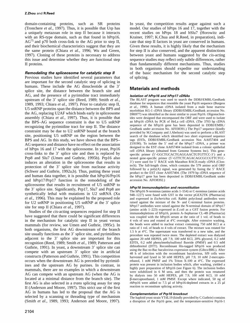
Fig. 1. Characterization of hPrp16 and hPrp17 cDNAs. (A) Predicted amino acid sequence of hPrp16. The 3703 bp cDNA sequence of the hPrp16gene has been deposited in DDBJ/EMBL/GenBank under accession No. AF038391. The N-terminal REDS residues are in bold, conserved motifsidentical to the DEAH box protein family are boxed, and the NTP-binding fold is underlined. (B) Co-migration ofin vitro translated (IVT) hPrp16and hPrp16 detected in nuclear extract by hPrp16 antibody. Twoµl of nuclear extract was mixed with 1µl of [ 35S]methionine-labeled IVT-hPrp16,separated by SDS–PAGE and then proteins were immobilized on a membrane. The membrane was probed with hPrp16 antibodies and bands weredetected using a horseradish perioxidase-conjugated secondary antibody and enhanced chemiluminescence (ECL) (lane 2) followed by detection ofthe 35S-labeled protein by autoradiography (lane 1). Note that the IVT-hPrp16 is below the level of detection of the antibody (data not shown).(C) Predicted amino acid sequence of hPrp17. The 1979 bp cDNA sequence of the hPrp17 gene has been deposited in DDBJ/EMBL/GenBank underaccession No.AF038392. Seven conserved WD repeats (GH-X27–35-WD) identified by the PROSITE Program are in bold. (D) Co-migration of IVT-hPrp17 and hPrp17 detected in nuclear extract by hPrp17 antibodies. Samples were analyzed as in (B). The IVT-hPrp17 is below the level ofdetection of the antibody (data not shown).
tionally important for step II, we sought to identify humanhomologs of these yeast step II proteins. Recently, thehuman homologs of Prp18 and Slu7 were identified andshown to function in step II in humans (Horowitz andKrainer, 1997; K.Chua and R.Reed, in preparation). Wereport here the identification of the human homologs ofPrps 16 and 17. Our data indicate that these proteins areboth structurally and functionally conserved with theiryeast homologs.
Results
Characterization of hPrp16 and hPrp17 cDNAsThe probable human homologs of yeast Prp16 (Burgesset al., 1990) and Prp17 (M.Company and J.Abelson, per-sonal communication) were identified by a BLAST searchof theGenBank database, and full-length cDNAclones wereisolated using PCR strategies (see Materials and methods).The human clones are designated hPrp16 and hPrp17. Theopen reading frame (ORF) of the hPrp16 cDNA predicts aprotein of 1227 amino acids with an expected mol. wt of140.5 kDa and an isoelectric point of 6.35 (Figure 1A). The
2096
hPrp17 cDNA encodes a 579 amino acid protein with acalculated mol. wt of 66 kDa and an isoelectric point of 6.89(Figure 1C). To characterize the proteins encoded by thesecDNAs, we raised rabbit polyclonal antibodies to GSTfusions of each protein (see Materials and methods). Onemain band of 140 kDa is detected by hPrp16 antibodies ona Western blot of nuclear extract (Figure 1B, lane 2). Thisband co-migrates with thein vitro translated (IVT) proteinproduced from the hPrp16 cDNA (Figure 1B, lane 1).Similarly, one main band of ~66 kDa, which co-migrateswith IVT-hPrp17, is detected by hPrp17 antibodies in nuc-lear extract (Figure 1D). These observations, together withthe fact that there is a consensus Kozak sequence (Kozak,1986) upstream of the designated initiator methionine in thehPrp16 and 17 cDNAs, indicate that these cDNAs encodefull-length proteins.
As shown in Figure 1A, hPrp16 contains an NTP-binding motif (underlined) and six RNA helicase motifs(boxed) that are present in yeast Prp16 and all othermembers of the DEAH box family of putative RNAhelicases (for review, see Wassarman and Steitz, 1991;Schmid and Linder, 1992). These motifs are strikingly

Human homologs of Prp16 and Prp17
Fig. 2. (A andB) Amino acid alignment of yeast and human Prp16and Prp17 homologs. Alignments were done by the Clustal method(DNASTAR Inc). Identical residues are shown in white on black. Analignment of hPrp16 with yeast Prps 2, 22 and 43 was done by thesame method (data not shown). The overall identity to hPrp16 forthese proteins is 37, 33 and 34%, respectively, and the C-terminalidentity is 47, 48 and 36%, respectively. (C) Alignment of the sevenWD repeats (GH-X27–35-WD) conserved between hPrp17 and Prp17using the Lipman–Pearson method (DNASTAR Inc). Identical residuesare indicated, and similar residues are shown by dots. GH and WDresidues are underlined.
2097

Z.Zhou and R.Reed
conserved in hPrp16/Prp16 and in the three DEAH boxsplicing factors, Prps 2, 22 and 43 (Burgesset al., 1990;Chen and Lin, 1990; Companyet al., 1991; Onoet al.,1994; Arenas and Abelson, 1997). All of the motifsindicated in the boxes are identical among these differentproteins (Figure 1A and data not shown). The overallidentity between hPrp16 and Prp16 is 41%, and hPrp16is more related to yeast Prp16 than to the other membersof the DEAH box family (Figure 2A and legend). TheC-terminal 400 amino acids of hPrp16 are also highlyconserved in Prp16 (identity 51%) and the other membersof the DEAH box splicing factor family, implying agenerally important function for this domain (Figure 2Aand legend). hPrp16 diverges from its yeast counterpartin the N-terminus, where hPrp16 contains a region richin the amino acids arginine, glutamate, aspartate andserine. These residues (REDS) account for 54% of theamino acids extending from 59 to 400 (shown in bold,Figure 1A). The N-terminus of hPrp16 is also enriched inTP and SP dipeptides which, like RS dipeptides, arepotential phosphorylation sites. A likelyCaenorhabditiseleganshomolog of Prp16, which is 55% identical tohPrp16, is also present in the database (DDBJ/EMBL/GenBank accession No. P34498).
The closest relative to hPrp17 in the database is yeastPrp17, with the identity being 36% (Figure 2B). YeastPrp17 is identical to Cdc40, a protein that plays a role inDNA replication (Vaismanet al., 1995). How the functionsof Prp17 and Cdc40 are related is not known. Homologybetween hPrp17 and Prp17 is the strongest in theC-terminal two-thirds of the proteins (41% identity, 67%similarity), which contains seven potential WD repeats(Seshadriet al., 1996) (shown in bold in Figure 1C andaligned in Figure 2C). WD repeats are found in a widevariety of proteins and are ~40 amino acids long, beginningwith a conserved GH and terminating with a conservedWD (Neeret al., 1994). These repeats function in protein–protein interactions (see Discussion).
Complementation of a yeast Prp16 knockout strainFour DEAH box splicing factors are highly related to oneanother (Prps 2, 16, 22 and 43) (Burgesset al., 1990;Chen and Lin, 1990; Companyet al., 1991; Onoet al.,1994; Arenas and Abelson, 1997; Geeet al., 1997). Todetermine whether the hPrp16 cDNA that we have isolatedis the functional homolog of yeast Prp16, we attemptedto complement a yeast Prp16 knockout strain (kindlyprovided by C.Guthrie) using plasmid shuffling (Sikorskiand Boeke, 1987) (see Materials and methods). Theconstructs that were used for the complementation areshown in Figure 3A. As expected, the wild-type yeaststrain grows on plates containing selective media [5-fluoro-orotic acid (5-FOA)] as does the yeast Prp16 knockoutstrain transformed with yeast Prp16 (Figure 3B). Incontrast, the vector alone, full-length hPrp16 and C- orN-terminal deletions of hPrp16 do not rescue the yeastPrp16 knockout (Figure 3A and B). Significantly, however,a chimeric construct containing the N-terminus of yeastPrp16 (amino acids 1–298) fused to the remainder ofhPrp16 (251–1227) does rescue the knockout (Figure 3Aand B). A chimera lacking the C-terminus (Chimera∆C)or a construct containing the yeast N-terminus alone
2098
Fig. 3. Complementation of yeast Prp16 knockout strain with ahuman–yeast chimeric gene. (A) Schematic representation of Prp16and hPrp16 derivatives used for transformation into the yeast Prp16knockout strain. The yeast and human Prp16 are denoted by light ordark shading, respectively. The REDS and DEAH domains areindicated. The numbers indicate the amino acid positions in eachconstruct. The constructs that rescue (1) or do not rescue (–) thePrp16 knockout are indicated. (B) Growth comparison on a 5-FOAplate of yeast cells transformed with Prp16, vector alone, hPrp16 andthe chimera.
(Prp16/1–298) were non-functional in the assay(Figure 3A).
The observation that the chimeric construct of hPrp16,which contains 80% of the hPrp16 gene, complements theyeast Prp16 knockout indicates that our hPrp16 cDNA isthe likely functional homolog of yeast Prp16. Additionalstudies supporting this conclusion are presented below(Figures 4–6). Our data also indicate that the N-terminusof yeast Prp16 contains a domain essential for functionin yeast, and that the N-terminus of hPrp16, which containsthe REDS-rich region, cannot provide this function. Inaddition, amino acids 911–1227 of hPrp16, which arehighly conserved in DEAH box splicing factors (Figure2A and data not shown), are essential for complementation,indicating that this region contains an important func-tional domain.
hPrp16 and hPrp17 join the spliceosome late in thesplicing pathwayTo obtain further evidence that hPrps 16 and 17 aresplicing factors, we asked whether these proteins arepresent in purified human spliceosomes. A splicing timecourse was carried out, and spliceosomal complexes fromeach time point were isolated (Figure 4). At the 10 mintime point, only unspliced pre-mRNA is detected (Figure4A). The splicing intermediates and products are firstdetected at 25 min and increase in abundance at 40 and60 min (Figure 4A). Western analysis of spliceosomalcomplexes isolated at each of these times shows that the

Human homologs of Prp16 and Prp17
Fig. 4. hPrp16 and hPrp17 associate with the spliceosome late in thesplicing pathway. (A) AdML pre-mRNA (1.92µg) containing biotinwas incubated under splicing conditions (2.4 ml reaction) for the timesindicated. Complexes were fractionated by gel filtration. Total RNAfrom the complexes was prepared and separated on a 15% denaturingpolyacrylamide gel. The bands corresponding to pre-mRNA,intermediates and products are indicated. (B–D) Gel filtration-isolatedcomplexes in (A) were affinity-purified by binding to avidin–agarose.Western analysis of total protein isolated from the purified complexeswas then carried out. The same Western blot was probed with a rabbitpolyclonal antibody to the U2 snRNP protein SAP 130 (B), to hPrp16(C) or to hPrp17 (D). Note that the 10 min time point is underloaded.On long exposures, no hPrp16 or hPrp17 was detected at this timepoint (data not shown).
U2 snRNP protein, SAP 130, is present at all of the timepoints (Figure 4B). This result is consistent with previouswork showing that U2 snRNP first binds stably to thespliceosome in the A complex and remains bound through-out spliceosome assembly (for review, see Kra¨mer, 1996).In contrast, hPrp16 and hPrp17 join the spliceosomelate in the splicing pathway and concomitant with thetransesterification reactions (Figure 4C and D). Interest-ingly, less hPrp16 is detected at 60 min than at 25 and 40min, whereas hPrp17 is present at about the same levelsat all three time points. We conclude that both hPrp16and 17 associate with the spliceosome concomitant with thecatalytic steps. However, hPrp16 appears to be destabilizedfrom the spliceosome late in the splicing pathway whereashPrp17 remains tightly bound. It is difficult to determine
2099
Fig. 5. Association of hPrp16 or hPrp17 with the spliceosomeprecedes recognition of the 39 splice site during step II.(A) Schematics of AdML and mutant derivatives. The BPS–AGdistance for wild-type (WT) and GG-GG is 23 nucleotides. In pyJ andRanA, py20 refers to the length of the pyrimidine tract which isisogenic with WT. Py(29) is a 29 nucleotide insertion of pyrimidines.Ran(29) is an insertion of 29 nucleotides of random sequence (Chiaraet al., 1997). (B) The indicated32P-labeled pre-mRNAs wereincubated under splicing conditions for 40 min, and then total RNAwas fractionated on a 15% denaturing polyacrylamide gel. Splicingintermediates and products are indicated. (C) Western assay of thepurified complexes assembled on the pre-mRNA substrates indicated.The blot was probed with hPrp16 and hPrp17 antibodies, and thepositions of hPrps 16 and 17 are indicated.
the exact point at which hPrp16 is destabilized becausethe complexes at any given time point are heterogeneous.However, if the release occurs at the same stage as inyeast, then it would occur concomitant with step II (Schwerand Guthrie, 1991).
A similar time course experiment was performed usingdifferent pre-mRNA substrates,α-tropomyosin and Fushitarazu (ftz). Again, both hPrps 16 and 17 associatedwith these spliceosomes late in the splicing pathway,concomitant with the catalytic steps (data not shown).The observation that both hPrp16 and hPrp17 join thespliceosome late in the splicing pathway suggests thatthese proteins, like their yeast counterparts, function incatalytic step II of the splicing reaction.
Association of Prps 16 and 17 with thespliceosome precedes functional recognition of the39 splice site for step IIPrevious studies showed that the presence of pyrimidinesnext to the AG at the 39 splice site plays a critical role in

Z.Zhou and R.Reed
Fig. 6. hPrp16 is required for catalytic step II of the splicing reaction.(A) A Western blot probed with hPrp16 antibodies showing normalnuclear extract (lane 1) and the same extract depleted with pre-immune serum (∆PI) (lane 2) or with hPrp16 antibodies (∆hPrp16)(lane 3). The position of hPrp16 is indicated. (B) Coomassie bluestaining of the recombinant hPrp16 protein from baculovirus (2µg ofthe protein was loaded) which was used for complementation. rhPrp16migrates slightly larger than hPrp16 because of the His tag. (C) Timecourse of pre-mRNA splicing in extracts mock depleted using pre-immune sera (∆PI) or hPrp16 depleted with hPrp16 sera (∆hPrp16).AdML pre-mRNA was incubated under standard splicing conditionsusing∆PI nuclear extract (lanes 1–3) or∆hPrp16 extract (lanes 4–6).For lanes 7–9, rhPrp16 was added to the∆hPrp16 extract. Thereactions were incubated for the times indicated. (D and E) Splicing ofβ-globin (D) or ftz (E) pre-mRNAs in∆PI extract (lane 1),∆hPrp16extract (lane 2) or∆hPrp16 extract containing rhPrp16 (lane 3).β-globin and ftz splicing products were separated on 9 or 12%denaturing polyacrylamide gels, respectively. Splicing intermediatesand products are indicated.
catalytic step II in mammals (Chiaraet al., 1997). Inaddition, the distance between the branch point sequence(BPS) and AG also plays an important role in step II(Chiaraet al., 1997). To determine whether either of theseparameters affects the association of hPrps 16 and 17 withthe spliceosome, complexes were assembled on mutantpre-mRNAs and Western analyses were performed (Figure5). The structures of the AdML pre-mRNAs are shown
2100
in Figure 5A. GG-GG pre-mRNA contains GG substitu-tions of the AG at the 39 splice site and the AG sixnucleotides downstream. Catalytic step II is completelyblocked with this pre-mRNA (Figure 5B, lane 2) (Chiaraet al., 1997). PyJ is the same as wild-type except for theinsertion of 29 pyrimidines downstream of the normalpyrimidine tract. The BPS–AG distance is increased from23 nucleotides in wild-type to 49 nucleotides in pyJ. Thislong BPS–AG distance results in an inefficient catalyticstep II (Chiaraet al., 1997) (Figure 5B, lane 3). Finally,RanA contains random sequences adjacent to the AG atthe 39 splice site, and catalytic step II is abolished (Chiaraet al., 1997) (Figure 5B, lane 4).
As shown in Figure 5C, hPrp16 and hPrp17 are detectedin complexes assembled on all of these pre-mRNAs (notethat we could not establish whether the differences in thelevels of these proteins are significant due to the differencesin splicing kinetics of each pre-mRNA and the inherentvariability in the complexes). The observation that bothhPrp16 and hPrp17 are present in spliceosomes assembledon GG-GG pre-mRNA and RanA pre-mRNA, both ofwhich cannot undergo step II, indicates that these proteinsassociate with the spliceosome prior to catalytic step II.Moreover, a specific BPS–AG sequence and distance arenot required for the binding of hPrps 16 and 17 to thespliceosome. The AG itself is also not required for theirbinding. Thus, all three of these mutations impair catalyticstep II at a stage that is subsequent to the binding ofhPrp16 and hPrp17 to the spliceosome.
hPrp16 is essential for catalytic step II in humansTo determine directly whether hPrps 16 and 17 are requiredfor splicing, we attempted to immunodeplete each proteinfrom nuclear extracts (Figure 6A). Only 50% of thehPrp16 was depleted under normal splicing conditions,and little effect on splicing was observed with theseextracts (data not shown). However, when the salt concen-tration of the nuclear extract was raised to 750 mM,hPrp16 was depleted efficiently (Figure 6A, lane 3). NohPrp16 was depleted with pre-immune sera (Figure 6A,lane 2), and no co-depletion of hPrp17 was detected(data not shown). Although hPrp17 antibodies specificallyimmunoprecipitate IVT-hPrp17 from reticulocyte lysates,we were not able to find conditions for immunodepletingthis protein from splicing extracts (data not shown).
To determine whether splicing is affected in the hPrp16-depleted extracts, AdML pre-mRNA was incubated undernormal splicing conditions in hPrp16-depleted (∆hPrp16)extracts or extracts mock-depleted with pre-immune sera(∆PI). A splicing time course using these extracts is shownin Figure 6C. The kinetics and efficiency of catalytic stepI are the same in∆PI and∆hPrp16 extracts (e.g. compare25 min time points, lanes 1 and 4). In contrast, catalyticstep II is severely impaired in∆hPrp16 extracts, relativeto ∆PI extracts (Figure 6C, compare lanes 2 and 3 with 5and 6). The low levels of step II activity in the∆hPrp16extract are most probably due to the incomplete depletionof hPrp16 (Figure 6A, and data not shown). To determinewhether the step II activity depleted from∆hPrp16 extractscan be reconstituted, we produced histidine-tagged recom-binant hPrp16 in baculovirus (rhPrp16, see Materials andmethods, Figure 6B) and added it to∆hPrp16 extractsbefore incubation of splicing reactions for 25, 40 and 60

Human homologs of Prp16 and Prp17
min (Figure 6C, lanes 7–9). Step I was unaffected by theaddition of rhPrp16 (e.g. compare lanes 4 and 7). Incontrast, step II was fully reconstituted by rhPrp16 (com-pare lanes 2 and 3 with 8 and 9).
To determine whether hPrp16 is a general step IIsplicing factor, immunodepletion/add-back studies wereperformed using different pre-mRNA substrates. For bothβ-globin (Figure 6D) and (ftz) (Figure 6E) pre-mRNAs,step II was severely impaired by depletion of hPrp16 andwas restored by addition of rhPrp16. As shown in Figure7A, the restoration of step II by rhPrp16 is also dosedependent. Quantitation reveals a linear increase in activitywith increasing rhPrp16, and maximal activity wasobtained at 100 ng (Figure 7A and data not shown).Together, these studies indicate that hPrp16 is a generalsplicing factor essential for catalytic step II.
To investigate the kinetics of step II when hPrp16 isadded to∆hPrp16 spliceosomes, AdML pre-mRNA wasincubated for 40 min in∆hPrp16 extracts to accumulatethe splicing intermediates (Figure 7B, lane 2), and thenrhPrp16 or buffer alone was added, and incubation wascontinued for 5, 15 or 30 min (Figure 7B, lanes 3–8). With buffer alone, the intermediates continued toaccumulate (Figure 7B, lanes 3–5). When rhPrp16 wasadded, a low level of step II products appeared by 5 minof incubation and accumulated to much higher levels by30 min (Figure 7B, lanes 6–8). Quantitation of the data(Figure 7C) shows that the intermediates are convertedinto products after addition of hPrp16. Thus, we concludethat the∆hPrp16 spliceosome is a functional intermediatein the splicing pathway. As yet, we have not found suitableconditions for detecting the∆hPrp16 spliceosome onnative gels or for purifying this complex.
To obtain additional evidence that the complementationof ∆hPrp16 extracts is due to rhPrp16 (and not a contamin-ant in the preparation), we asked whether rhPrp16 isincorporated into functional spliceosomes. To do this,rhPrp16 was added to∆hPrp16 extracts, and spliceosomeswere assembled for 10 or 40 min on biotinylated AdMLpre-mRNA (Figure 8). Complexes were then isolated bygel filtration and affinity-purified by binding to avidin–agarose. Western analysis of the complexes was performedusing an antibody against the histidine tag in rhPrp16. Asshown in Figure 8A, this antibody efficiently detectspurified rhPrp16 alone (lane 3). Significantly, rhPrp16 isdetected in the 40 min spliceosomal complex, but not inthe 10 min complex (compare lanes 1 and 2). (Note thatit was not possible to affinity-purify the spliceosome bybinding to nickel resin, indicating that the His tag is notexposed in the native spliceosome.) The same blot wasreprobed with antibodies to the U2 snRNP protein, SAP130, which is present throughout spliceosome assembly(Figure 8B). This protein is detected in both the 10 and40 min complexes. Together, these data show that rhPrp16is indeed incorporated into the spliceosome. Furthermore,the association of rhPrp16 is specific as it is only detectedin the 40 min spliceosome, consistent with the observationthat native hPrp16 is present only in late spliceosomalcomplexes (Figure 4).
Discussion
Until recently, very little was known about the proteinsinvolved in catalytic step II of the splicing reaction.
2101
Fig. 7. (A) Reconstitution of splicing activity in∆hPrp16 extracts byrhPrp16 is dose dependent. AdML pre-mRNA was incubated undersplicing conditions in∆PI extract (lane 1) or∆hPrp16 extract (lane 2).rhPrp16 was added at 10, 25, 50, 100 or 200 ng, respectively, to∆hPrp16 extract in a 25µl standard splicing reaction (lanes 3–7).Splicing was carried out for 40 min. Splicing intermediates andproducts are indicated. (B) AdML pre-mRNA was spliced in∆PIextract (lane 1) or∆hPrp16 extract (lanes 2–8). Splicing was carriedout for 40 min followed by addition of buffer alone or rhPrp16, andincubation was continued for 5 (lanes 3 and 6), 15 (lanes 4 and 7) or30 min (lanes 5 and 8). Splicing intermediates and products areindicated. (C) Quantitation of the data shown in (B) (lanes 3–8) byphosphorimager analysis. Each of the RNA species was normalizedagainst the total counts at the start of the chase. Pre-mRNA is shownin white, intermediates in stippled boxes, and products in black. Thetime points are indicated.

Z.Zhou and R.Reed
However, this picture has begun to change dramaticallythrough a combination of genetic and biochemical studiesin yeast and mammals. The known or putative stepII proteins in humans that have yeast counterparts aresummarized in Table I. So far, four step II proteins, Prps16, 17 and 18 and Slu7, have been identified in yeast (forreview, see Umen and Guthrie, 1995c). We now reportthe identification of human homologs of Prps 16 and 17.The human homologs of Prp18 and Slu7 have alsobeen identified (Horowitz and Krainer, 1997; K.Chua andR.Reed, in preparation). All four of these proteins areknown or are likely (hPrp17) to function exclusively in
Fig. 8. Recombinant hPrp16 is present in purified reconstitutedspliceosomal complexes. (A) Biotinylated AdML pre-mRNA wasincubated for 10 or 40 min in∆hPrp16 extracts containing rhPrp16.Splicing reactions were fractionated by gel filtration, and spliceosomalcomplexes were affinity-purified by binding to avidin–agarose. AWestern blot of the purified complexes was probed with antibodies tothe His tag on rhPrp16. rhPrp16 alone (0.5µg) was loaded in lane 3.(B) The same blot was reprobed with antibodies to the U2 snRNPprotein, SAP 130.
Table I. Known or putative human step II proteins that are conserved in yeast
Human Yeast Conserved Step II activity U5 snRNP U5 snRNP interaction (yeast)structural domains component (human)
hPrp16 Prp16 DEAH box motifs 39 ss xlink, ATPase, induces ND synthetic lethalprotection of 39 ss (Y)
hPrp17 Prp17 WD motifs ND synthetic lethalhPrp18 Prp18 none identified no synthetic lethal and weakly associatedhSlu7 Slu7 Zn knuckle 39 ss xlink (Y) ND synthetic lethalU5220 Prp8 none identified 39 ss xlink (Y,H) yes associatedU5100a Prp28 DEAD box motifs 39 ss xlink (H) yes genetic interactionU5116 Snu114 G domain 39 ss xlink (H) yes ND
The structural domains that are conserved between corresponding yeast and human proteins are indicated. Step II activity indicates the main functionidentified for each protein related specifically to step II. Cross-linking to the 39 splice site in the spliceosome prior to step II is designated as 39 ssxlink. Y and H indicate that the activity has been shown in yeast or humans, respectively. ND: not determined. See text for references.aU5100 previously was named U5110 (Bennettet al., 1992; Chiaraet al., 1997) but we will now refer to it as U5100 described in Teigelkampet al.(1997) because the two proteins are the same (Bennettet al., 1992).
2102
step II in both humans and yeast. In yeast, Prps 16, 17,18 and Slu7 all interact genetically with U5 snRNP (Franket al., 1992). However, none of these proteins are actualU5 snRNP components, except Prp18, which is weaklyassociated (Horowitz and Abelson, 1993a,b; for review,see Umen and Guthrie, 1995c). In humans, Prp18 is notassociated with U5 snRNP (Horowitz and Krainer, 1997),and it is not yet known whether hSlu7, hPrp16 and hPrp17are U5 snRNP components (Table I).
In addition to the proteins that function exclusively instep II, some proteins may have roles in both catalyticsteps. One clear example of this is the U5 snRNP protein,Prp8, which was first identified as an essential factor forspliceosome assembly (Whittakeret al., 1990; Brown andBeggs, 1992) and then was found, in both genetic andUV cross-linking studies, to function in recognition of the39 splice site for catalytic step II (Teigelkampet al.,1995a,b; Umen and Guthrie, 1995a,b, 1996). Likewise,the human homolog of Prp8, U5220, as well as two otherU5 snRNP proteins (U5100 and U5116), UV cross-link tothe 39 splice site prior to step II (Chiaraet al., 1997; Liuet al., 1997). In addition, antibody inhibition studiessuggest a role for U5116 in step II (Fabrizioet al., 1997).Essential yeast homologs of U5100 and U5116(Snu114 andPrp28, respectively) have also been identified recently(Strauss and Guthrie, 1991, 1994; Fabrizioet al., 1997;Teigelkampet al., 1997). These proteins have not yetbeen tested for a function in step II. Thus, although manygaps remain to be filled, all of the data point to a highlevel of conservation in the factors involved in step II anda key role for U5 snRNP in step II in both yeast andhumans (Table I).
hPrp16 is an essential step II protein in humansOur data indicate that hPrp16 is both structurally andfunctionally conserved. Prp16 is a member of the DEAHbox family of splicing factors (Burgesset al., 1990). Thisfamily, which includes Prps 2, 22 and 43, is characterizedby the presence of several motifs found in the prototypicfamily member, eIF4α, an ATP-dependent RNA helicase(for review, see Umen and Guthrie, 1995c). Prps 2 and16 are indeed ATPases, but helicase activity for theseproteins has yet to be reported (Schwer and Guthrie, 1991;Kim and Lin, 1996). hPrp16 resembles yeast Prp16 (41%identity) more closely than any of the other DEAH family

Human homologs of Prp16 and Prp17
members. In addition, we show that a chimeric proteinconsisting of the C-terminal 977 amino acids of hPrp16and the N-terminal 298 amino acids of yeast Prp16 canrescue a yeast Prp16 knockout strain. Due to the highsimilarity between Prp16 and the other DEAH familymembers, this rescue alone does not demonstrate thathPrp16 is the Prp16 homolog. However, this conclusionis supported by the observation that hPrp16 is essentialfor step II, like its yeast counterpart, whereas the otherDEAH box splicing proteins are not. Another functionalsimilarity between hPrp16 and Prp16 is that both proteinsassociate with spliceosome late in the splicing pathway(Schwer and Guthrie, 1991; this study). In addition, hPrp16is destabilized from the splicesome late in the splicingpathway, which is most likely related to the ATP-dependentrelease of Prp16 from the yeast splicesome (Schwerand Guthrie, 1992a,b). Finally, hPrp16, like its yeastcounterpart, associates with spliceosomes assembled onpre-mRNAs containing an AG to GG mutation at the 39splice site, a mutation that blocks catalytic step II (Umenand Guthrie, 1995b; this study). Together, these dataprovide compelling evidence that hPrp16 is the functionalhomolog of Prp16.
hPrp16, unlike Prp16, contains a 340 amino acid domainhighly enriched in the residues, REDS. Rescue of theyeast Prp16 knockout strain with hPrp16 required thereplacement of this domain with the yeast N-terminus.Thus, the REDS domain probably plays a role specific tometazoans and the N-terminus of Prp16 contains a domainessential for function in yeast. A REDS domain similarto that in hPrp16 is found in the N-terminus of anotherhuman homolog of a DEAH box family member, Prp22,which is required for the release of spliced mRNA fromthe spliceosome (Companyet al., 1991; Onoet al., 1994;Ohno and Shimura, 1996). This domain in hPrp22 interactswith members of the SR family of splicing factors andalso functions as a nuclear localization signal (Onoet al.,1994; Ohno and Shimura, 1996). The observation thathPrp16 contains a REDS domain suggests that it alsointeracts with SR proteins, and that SR proteins may havea role in catalytic step II.
Evidence that hPrp17 is a step II protein inhumanshPrp17 is 36% identical to yeast Prp17, and the highestlevel of conservation is in the carboxy-terminus whichcontains seven WD motifs (Seshadriet al., 1996;M.Company and J.Abelson, unpublished; this study).These motifs are present in a number of proteins involvedin RNA processing, signal transduction, DNA replication,cytoskeletal assembly and cell cycle control (for review,see Neeret al., 1994). Recently, the crystal structure ofthe prototypic WD family member, theβ subunit of aheterotrimeric G protein, was solved (Wallet al., 1995;Lambrightet al., 1996; Sondeket al., 1996). This proteincontains seven WD repeats and forms a seven-bladedβ-propeller structure. Seven WD repeats are also presentin the polyadenylation factor CstF (Takagaki and Manley,1992) and the splicing factor, Prp4, and its human homolog(Banroques and Abelson, 1989; Bjornet al., 1989;Dalrymple et al., 1989; Lauberet al., 1997). These WDdomains and Prp17/hPRP17 presumably fold into a seven-bladedβ-propeller structure and, like other WD proteins,
2103
the WD motifs in Prp17/hPrp17 may function in protein–protein interactions (Neeret al., 1994; Wallet al., 1995;Lambright et al., 1996; Sondeket al., 1996). Consistentwith the observation that the WD repeats are conservedbetween Prp17 and hPrp17, the repeats in Prp17 areessential for function in yeast (Seshadriet al., 1996).
Studies in yeast showed that Prp16 and Prp17 interactgenetically and function at the same stage during step II(Joneset al., 1995; Umen and Guthrie, 1995b; Seshadriet al., 1996). These observations suggest that Prps 16 and17 may physically interact. However, we have not obtainedevidence for an interaction between the human counter-parts. hPrp16 and hPrp17 antibodies specifically immuno-precipitate the correspondingin vitro translated proteins(data not shown). However, these proteins do not interactin co-immunoprecipitation, GST pull-down or far Westernassays (data not shown).
In addition to the structural similarity between hPrp17and Prp17, functional similarities between the proteinssupport the conclusion that they are functional homologs.Although we were unable to immunodeplete hPrp17 fromnuclear extracts to demonstrate directly its role in step II,we have shown that hPrp17 is present in the spliceosomeand joins it late in the splicing pathway. These observationsare consistent with the step II role of Prp17 in yeast. Ourdata also show that hPrp17 is present in spliceosomesassembled on pre-mRNAs containing mutations that blockor impair step II. Moreover, hPrp16 joins the spliceosomeat about the same time as hPrp17 and is present inthe step II-blocked spliceosomes. These results parallelfunctional studies in yeast showing that Prps 16 and 17act at a similar stage of step II (Joneset al., 1995; Umenand Guthrie, 1995b). Thus, together, the data support theconclusion that hPrp17 is a step II protein in humans andis the functional homolog of yeast Prp17.
In yeast, Prp16 and 17 can be immunodepleted fromextracts under splicing conditions (Schwer and Guthrie,1991; Joneset al., 1995). In constrast, immunodepletionof hPrp16 requires high salt, and we were unable toimmunodeplete hPrp17 even in high salt. Thus, it ispossibile that the human proteins are present in a complex.Further work is necessary to test this possibility and toexplain the differences between the yeast and human data.
Other known or putative step II proteins that have beenidentified in humans include U5200 (Lauberet al., 1996),PSF (Pattonet al., 1993; Gozaniet al., 1994), Urp(Tronchereet al., 1997), AG75 (Chiaraet al., 1996) andp70 (Wu and Green, 1997). U5200 is a human DEADbox family member that has an essential yeast homolog(Snu246/Brr2/Slt22) (Lauberet al., 1996; Noble andGuthrie, 1996; Xuet al., 1996). Interestingly, the ATPaseactivity of the yeast homolog is stimulated by U2 and U6snRNAs, and the protein plays a role in association of U5snRNP with the spliceosome (Xuet al., 1996). Moreover,antibodies to U5200 inhibit catalytic step II in humans(Lauber et al., 1996). Thus, together, these data areconsistent with a role for U2–U5 snRNP interactions instep II (see below). PSF and Urp do not have apparentyeast homologs, and it is not yet known whether thesefactors are general step II splicing factors. Consistent witha general role for PSF, it recently was detected in purifiedU4/U5/U6 snRNP (Teigelkampet al., 1997). Urp containsan RS domain and interacts with U2AF65 and other RS
Z.Zhou and R.Reed
domain-containing proteins, such as SR proteins(Tronchereet al., 1997). Thus, it is possible that Urp hasa uniquely metazoan role in step II because it interactswith an RS-type domain, such as that found in hPrp16.AG75 and p70 both cross-link to the AG prior to step II,and their biochemical characteristics suggest that they arethe same protein (Chiaraet al., 1996; Wu and Green,1997). Cloning of these proteins is necessary to addressthis issue and determine whether they are functional stepII proteins.
Remodeling the spliceosome for catalytic step IIPrevious studies have identified several parameters thatare important for the second catalytic step of splicing inhumans. These include the AG dinucleotide at the 39splice site, the distance between the branch site andAG, and the presence of a pyrimidine tract immediatelyupstream of the 39 splice site (Reed, 1989; Smithet al.,1989, 1993; Chiaraet al., 1997). Prior to catalytic step II,U5 snRNP proteins span the distance between the BPS andthe AG, replacing U2AF which binds early in spliceosomeassembly (Chiaraet al., 1997). Thus, it is possible thatthe BPS–AG sequence constraint is due to U5 snRNPrecognizing the pyrimidine tract for step II. The distanceconstraint may be due to U2 snRNP bound at the branchsite, positioning U5 snRNP on the region between theBPS and AG. In this study, we have shown that the BPS–AG sequence and distance have no effect on the associationof hPrps 16 and 17 with the spliceosome. In yeast, Prp16cross-links to the 39 splice site and then is replaced byPrp8 and Slu7 (Umen and Guthrie, 1995b). Prp16 alsoinduces an alteration in the spliceosome that results inprotection of the 39 splice site from nuclease activity(Schwer and Guthrie, 1992a,b). Thus, putting these yeastand human data together, it is possible that hPrp16/Prp16and hPrp17/Prp17 function in the remodeling of thespliceosome that results in recruitment of U5 snRNP tothe 39 splice site. Significantly, Prp17, Slu7 and Prp8 aresynthetically lethal with mutations in U2 snRNA (Xuet al., 1996). This may be explained by the proposed rolefor U2 snRNP in positioning U5 snRNP at the 39 splicesite for step II (Chiaraet al., 1997).
Studies of thecis-acting sequences required for step IIhave suggested that there could be significant differencesin the mechanism for catalytic step II in yeast versusmammals (for review, see Umen and Guthrie, 1995c). Inboth organisms, the first AG downstream of the branchsite usually functions as the 39 splice site, and pyrimidinesadjacent to the 39 splice site are important for thisrecognition (Reed, 1989; Smithet al., 1989; Patterson andGuthrie, 1991). In yeast, a downstream 39 splice site cancompete with an upstream 39 splice site in artificialconstructs (Patterson and Guthrie, 1991). This competitionoccurs when the downstream AG is preceded by pyrimid-ines and the upstream AG by purines. In contrast, inmammals, there are no examples in which a downstreamAG can compete with an upstream AG (when the AG islocated at a minimal distance from the branch site). Thefirst AG is also selected in atrans splicing assay for stepII (Anderson and Moore, 1997). This strict use of the firstAG in humans has led to the proposal that the AG isselected by a scanning or threading type of mechanism(Smith et al., 1989, 1993; Anderson and Moore, 1997).
2104
In yeast, the competition results argue against such amodel. Our studies of hPrps 16 and 17, together with therecent studies on hPrps 18 and hSlu7 (Horowitz andKrainer, 1997; K.Chua and R.Reed, in preparation), indi-cate that step II factors in yeast are conserved in humans.Given these results, it is highly likely that the mechanismfor step II is also conserved, and the apparent distinctionsbetween yeast and humans suggested by thecis-actingsequence studies may reflect only subtle differences, ratherthan fundamentally different mechanisms. Thus, studiesin both organisms should expedite our understandingof the basic mechanism for the second catalytic stepof splicing.
Materials and methods
Isolation of hPrp16 and hPrp17 cDNAsThe BLAST program was used to search the DDBJ/EMBL/GenBankdatabase for sequences that resemble the yeast Prp16 sequence (Burgesset al., 1990). A human cDNA isolated from a male bone marrowmyeloblast KG-1 cDNA library (DDBJ/EMBL/GenBank accession No.D86977) was identified as the most similar to yeast Prp16. Oligonucleot-ides were designed that encompassed the ORF and were used to isolatean hPrp16 cDNA by PCR of HeLa cell cDNA. (The 3703 bp cDNAsequence of the hPrp16 gene has been deposited in DDBJ/EMBL/GenBank under accession No. AF038391.) The Prp17 sequence (kindlyprovided by M.Company and J.Abelson) was used to perform a BLASTsearch of the database which identified two short expressed sequencetags (ESTs, DDBJ/EMBL/GenBank accession Nos AA057404 andZ19190). To isolate the 59 end of the hPrp17 cDNA, a primer wasdesigned to the EST clone AA057404 isolated from a colonic epithelialcell cDNA library (obtained from Genome Systems Inc). This gene-specific primer (59-GGCTGCACTGAGGAACTGTGTTCC-39) and anested gene-specific primer (59-GTTCTCAGACAGCGCCGTTCTCC-39) were used for 59 RACE with Marathon RACE-ready cDNA (Clon-tech). The full-length clone, which contains the 579 amino acid ORFfollowed by a poly(A) signal, was generated by fusing the 59 RACEproduct to the EST clone AA057404. (The 1979 bp cDNA sequence ofthe hPrp17 gene has been deposited in DDBJ/EMBL/GenBank underaccession No. AF038392.)
hPrp16 immunodepletion and reconstitutionThe hPrp16 N-terminus (amino acids 1–554) or C-terminus (amino acids948–1227) were fused with GST in the vector pGEX2TK (Pharmacia)and expressed inEscherichia coli. Rabbit polyclonal antibodies wereraised against the mixture of the N- and C-terminal fusion proteins.hPrp17 antibodies were raised against the N-terminal domain of hPrp17(amino acids 1–223) expressed inE.coli as a GST fusion protein. Forimmunodepletions of hPrp16, protein A–Sepharose CL-4B (Pharmacia)was coupled with the hPrp16 serum at the ratio of 1 vol. of beads to4 vols of sera and rotated at 4°C overnight. After extensive washing,the beads were added to nuclear extract containing 750 mM KCl at aratio of 1 vol. of beads to 4 vols of extract. The mixture was rotated for1.5 h at 4°C. The supernatant was transferred to a new tube, and theprocedure was repeated twice more. The depleted extract was dialyzedagainst 20 mM HEPES, pH 7.9, 100 mM KCl, 20% glycerol, 0.2 mMEDTA, 0.2 mM phenylmethylsulfonyl fluoride (PMSF) and 0.5 mMdithiothreitol (DTT). Recombinant His-tagged hPrp16 was producedusing the Bac-to-Bac baculovirus expression system (Gibco/BRL). After48 h of infection with the recombinant baculovirus, Sf9 cells wereharvested and lysed in 50 mM HEPES, pH 7.9, 10 mM 2-mercapto-ethanol, 1 mM PMSF and 1% Triton X-100 at 4°C. The expressedprotein was present in inclusion bodies which, after washing, yielded ahighly pure preparation of hPrp16 (see Figure 6). The inclusion bodieswere solubilized in 6 M urea, and then the protein was renaturedby dialysis into 50 mM HEPES, pH 7.9, 100 mM KCl, 10 mMβ-mercaptoethanol, 1 mM PMSF. Except where indicated, 50 ng ofrhPrp16 were added to 7.5µl of hPrp16-depleted extracts in a 25µlreaction to reconstitute splicing activity.
Complementation of Prp16 knockoutThe haploid yeast strain YTdL19 (kindly provided by C.Guthrie) containsa disruption of the Prp16 gene, and the temperature-sensitive Prp16-1

Human homologs of Prp16 and Prp17
gene is carried on a plasmid containing theURA3marker gene (Burgesset al., 1990; Schwer and Guthrie, 1991). The full-length hPrp16 cDNAor the indicated derivatives were cloned downstream of the yeastglyceraldehyde-3-phosphate dehydrogenase promoter in the 2µmplasmid pG1 (containing aTRP marker gene). pG1-Prp16 and theindicated hPrp16 constructs were transformed into the YTdL19 strainand selected at 30°C in medium lacking tryptophan. Expression ofhPrp16 genes was verified by Western blotting. Colonies were thenpatched on 5-FOA plates to select for cells lacking the URA3 plasmid.Those constructs that allow growth are able to rescue the yeast Prp16knockout.
Analysis of spliceosomal complexesWild-type AdML is encoded by pAdML (Michaud and Reed, 1993),and the plasmids encoding GG-GG, PyJ and RanA pre-mRNAs weredescribed in Chiaraet al. (1997). Fushi tarazu (ftz) pre-mRNA wasdescribed in Rio (1988) andα-tropomyosin pre-mRNA was describedin Smith and Nadal-Ginard (1989). pAdML and tropomyosin werelinearized withBamHI, and ftz was linearized withXhoI. AdML and ftzDNAs were transcribed with T7 RNA polymerase and tropomyosin wastranscribed with SP6. The spliceosomal complexes were assembled byincubating 1.92µg of 32P-labeled biotinylated pre-mRNAs in a 2.4 mlreaction for the times indicated. Complexes were isolated by gel filtrationfollowed by avidin affinity chromatography, and then RNA or proteinwere analyzed as described (Reed, 1990; Bennettet al., 1992). Purifiedspliceosomal complexes containing rhPrp16 were assembled by incubat-ing biotinylated AdML pre-mRNA in hPrp16-depleted extract containing4.8 µg of rhPrp16 per 2.4 ml of splicing reaction.
Acknowledgements
We are grateful to M.Company and J.Abelson for providing the sequenceof Prp17, and to C.Collins and C.Guthrie for Prp16 strains and plasmids.We thank R.Das and J.Kim for excellent technical assistance, andB.Graveley, K.Hertel, L.Palandjian and K.Chua for critical comments.We also thanks C.Collins for helpful suggestions and A.Jocuns forassistance in preparation of the manuscript. This work was supportedby a Tobacco Research Council grant and an NIH grant to R.R.
References
Abovich,N. and Rosbash,M. (1997) Cross-intron bridging interactions inthe yeast commitment complex are conserved in mammals.Cell, 89,403–412.
Abovich,N., Liao,X.C. and Rosbash,M. (1994) The yeast MUD2 protein:an interaction with PRP11 defines a bridge between commitmentcomplexes and U2 snRNP addition.Genes Dev., 8, 843–854.
Anderson,K. and Moore,M.J. (1997) Bimolecular exon ligation by thehuman spliceosome.Science, 276, 1712–1716.
Ansari,A. and Schwer,B. (1995) SLU7 and a novel activity, SSF1, actduring the PRP16-dependent step of yeast pre-mRNA splicing.EMBO J., 14, 4001–4009.
Arenas,J.E. and Abelson,J.N. (1997) Prp43: an RNA helicase-like factorinvolved in spliceosome disassembly.Proc. Natl Acad. Sci. USA, 94,11798–11802.
Arning,S., Gruter,P., Bilbe,G. and Kra¨mer,A. (1996) Mammalian splicingfactor SF1 is encoded by variant cDNAs and binds to RNA.RNA, 2,794–810.
Banroques,J. and Abelson,J.N. (1989) PRP4: a protein of the yeast U4/U6small nuclear ribonucleoprotein particle.Mol. Cell. Biol.,9, 3710–3719.
Bennett,M., Michaud,S., Kingston,J. and Reed,R. (1992) Proteincomponents specifically associated with prespliceosome andspliceosome complexes.Genes Dev., 6, 1986–2000.
Bjorn,S.P., Soltyk,A., Beggs,J.D. and Friesen,J.D. (1989) PRP4 (RNA4)from Saccharomyces cerevisiae: its gene product is associated with theU4/U6 small nuclear ribonucleoprotein particle.Mol. Cell. Biol., 9,3698–3709.
Brown,J.D. and Beggs,J.D. (1992) Roles of PRP8 protein in the assemblyof splicing complexes.EMBO J., 11, 3721–3729.
Burgess,S., Couto,J.R. and Guthrie,C. (1990) A putative ATP bindingprotein influences the fidelity of branchpoint recognition in yeastsplicing.Cell, 60, 705–717.
Chen,J.H. and Lin,R.J. (1990) The yeast PRP2 protein, a putative RNA-dependent ATPase, shares extensive sequence homology with two otherpre-mRNA splicing factors.Nucleic Acids Res., 18, 6447.
2105
Chiara,M., Gozani,O., Bennett,M., Champion-Arnaud,P., Palandjian,L.and Reed,R. (1996) Identification of proteins that interact with exonsequences, splice sites, and the branchpoint sequence during each stageof spliceosome assembly.Mol. Cell. Biol., 16, 3317–3326.
Chiara,M.D., Palandjian,L., Feld Kra¨mer,R. and Reed,R. (1997) Evidencethat U5 snRNP recognizes the 39 splice site for catalytic step II inmammals.EMBO J., 16, 4746–4759.
Company,M., Arenas,J. and Abelson,J. (1991) Requirement of the RNAhelicase-like protein PRP22 for release of messenger RNA fromspliceosomes.Nature, 349, 487–493.
Cortes,J.J., Sontheimer,E.J., Seiwert,S.D. and Steitz,J.A. (1993) Mutationsin the conserved loop of human U5 snRNA generate use of novel cryptic59 splice sitein vivo. EMBO J., 12, 5181–5189.
Couto,J.R., Tamm,J., Parker,R. and Guthrie,C. (1987) Atrans-actingsuppressor restores splicing of a yeast intron with a branch pointmutation.Genes Dev., 1, 445–455.
Dalrymple,M.A., Petersen-Bjorn,S., Friesen,J.D. and Beggs,J.D. (1989)The product of the PRP4 gene ofS.cerevisiaeshows homology to betasubunits of G proteins.Cell, 58, 811–812.
Fabrizio,P.,Laggerbauer,B.,Lauber,J.,Lane,W.S.andLu¨hrmann,R. (1997)An evolutionarily conserved U5 snRNP-specific protein is a GTP-binding factor closely related to the ribosomal translocase EF-2.EMBOJ., 16, 4092–4106.
Frank,D. and Guthrie,C. (1992) An essential splicing factor, SLU7,mediates 39 splice site choice in yeast.Genes Dev., 6, 2112–2124.
Frank,D., Patterson,B. and Guthrie,C. (1992) Synthetic lethal mutationssuggest interactions between U5 small nuclear RNA and four proteinsrequired for the second step of splicing.Mol. Cell. Biol.,12, 5197–5205.
Gee,S., Krauss,S.W., Miller,E., Aoyagi,K., Arenas,J. and Conboy,J.G.(1997) Cloning of mDEAH9, a putative RNA helicase and mammalianhomologue ofSaccharomyces cerevisiaesplicing factor prp43.Proc.Natl Acad. Sci. USA, 94, 11803–11807.
Gozani,O., Patton,J.G. and Reed,R. (1994) A novel set of spliceosome-associated proteins and the essential splicing factor PSF bind stably topre-mRNA prior to catalytic step II of the splicing reaction.EMBO J.,13, 3356–3367.
Hodges,P.E., Plumpton,M. and Beggs,J.D. (1997) Pre-mRNA splicingfactors in the yeastSaccharomyces cerevisiae. In Krainer,A.R. (ed.),Eukaryotic mRNA Processing. Oxford University Press, New York, pp.213–241.
Horowitz,D.S. and Abelson,J. (1993a) A U5 small nuclearribonucleoprotein particle protein involved only in the second step ofpre-mRNA splicing inSaccharomyces cerevisiae. Mol. Cell. Biol., 13,2959–2970.
Horowitz,D.S. and Abelson,J. (1993b) Stages in the second reaction ofpre-mRNA splicing: the final step is ATP independent.Genes Dev., 7,320–329.
Horowitz,D.S. and Krainer,A.R. (1997) A human protein required for thesecond step of pre-mRNA splicing is functionally related to a yeastsplicing factor.Genes Dev., 11, 139–151.
Jones,M.H., Frank,D.N. and Guthrie,C. (1995) Characterization andfunctional ordering of Slu7p and Prp17p during the second step of pre-mRNA splicing in yeast.Proc. Natl Acad. Sci. USA, 92, 9687–9691.
Kim,S.H. and Lin,R.J. (1996) Spliceosome activation by PRP2 ATPaseprior to the first transesterification reaction of pre-mRNA splicing.Mol.Cell. Biol., 16, 6810–6819.
Kozak,M. (1986) Point mutations define a sequence flanking the AUGinitiator codon that modulates translation by eukaryotic ribosomes.Cell,44, 283–292.
Kramer,A. (1996) The structure and function of proteins involved inmammalian pre-mRNA splicing.Annu. Rev. Biochem., 65, 367–409.
Lambright,D.G., Sondek,J., Bohm,A., Skiba,N.P., Hamm,H.E. andSigler,P.B. (1996) The 2.0 Å crystal structure of a heterotrimeric Gprotein.Nature, 379, 311–319.
Lauber,J., Fabrizio,P., Teigelkamp,S., Lane,W.S., Hartmann,E. andLuhrmann,R. (1996) The HeLa 200 kDa U5 snRNP-specific protein andits homologue inSaccharomyces cerevisiaeare members of the DEXH-box protein family of putative RNA helicases.EMBO J.,15, 4001–4015.
Lauber,J., Plessel,G., Prehn,S., Will,C.L., Fabrizio,P., Groning,K.,Lane,W.S. and Lu¨hrmann,R. (1997) The human U4/U6 snRNP contains60 and 90kD proteins that are structurally homologous to the yeastsplicing factors Prp4p and Prp3p.RNA, 3, 926–941.
Liu,Z.-R., Laggerbauer,B., Lu¨hrmann,R. and Smith,C. (1997)Crosslinkingof theU5snRNP-specific116-kDaprotein toRNAhairpinsthat block step 2 of splicing.RNA, 3, 1207–1219.
Madhani,H.D. and Guthrie,C. (1994) Dynamic RNA–RNA interactions inthe spliceosome.Annu. Rev. Genet., 28, 1–26.

Z.Zhou and R.Reed
Michaud,S. and Reed,R. (1993) A functional association between the 59and 39 splice site is established in the earliest prespliceosome complex(E) in mammals.Genes Dev., 7, 1008–1020.
Moore,M.J., Query,C.C. and Sharp,P.A. (1993) Splicing of precursors tomessenger RNAs by the spliceosome. In Gesteland,R.F. and Atkins,J.F.(eds),The RNA World. Cold Spring Harbor Laboratory Press, ColdSpring Harbor, NY, pp. 303–357.
Neer,E.J., Schmidt,C.J., Nambudripad,R. and Smith,T.F. (1994) Theancient regulatory-protein family of WD-repeat proteins.Nature, 371,297–300.
Newman,A.J. (1997) The role of U5 snRNP in pre-mRNA splicing.EMBOJ., 16, 5797–5800.
Newman,A.J. and Norman,C. (1992) U5 snRNA interacts with exonsequences at 59 and 39 splice sites.Cell, 68, 743–754.
Newman,A.J., Teigelkamp,S. and Beggs,J.D. (1995) snRNA interactionsat 59 and 39 splice sites monitored by photoactivated crosslinking inyeast spliceosomes.RNA, 1, 968–980.
Noble,S.M. and Guthrie,C. (1996) Identification of novel genes requiredfor yeast pre-mRNA splicing by means of cold-sensitive mutations.Genetics, 143, 67–80.
Ohno,M. and Shimura,Y. (1996) A human RNA helicase-like protein,HRH1, facilitates nuclear export of spliced mRNA by releasing theRNA from the spliceosome.Genes Dev., 10, 997–1007.
O’Keefe,R.T., Norman,C. and Newman,A.J. (1996) The invariant U5snRNA loop1 sequence is dispensible for the first catalytic step ofsplicing in yeast.Cell, 86, 679–689.
Ono,Y., Ohno,M. and Shimura,Y. (1994) Identification of a putative RNAhelicase (HRH1), a human homolog of yeast Prp22.Mol. Cell. Biol.,14,7611–7620.
Patterson,B. and Guthrie,C. (1991) A U-rich tract enhances usage of analternative 39 splice site in yeast.Cell, 64, 181–187.
Patton,J.G., Porro,E.B., Galceran,J., Tempst,P. and Nadal-Ginard,B.(1993) Cloning and characterization of PSF, a novel pre-mRNA splicingfactor.Genes Dev., 7, 393–406.
Reed,R. (1989) The organization of 39 splice-site sequences in mammalianintrons.Genes Dev., 3, 2113–2123.
Reed,R. (1990) Protein composition of mammalian spliceosomesassembledin vitro. Proc. Natl Acad. Sci. USA, 87, 8031–8035.
Reed,R. and Palandjian,L. (1997) Splicesome assembly. In Krainer,A.R.(ed.), Eukaryotic mRNA Processing. Oxford University Press, NewYork, pp. 103–129.
Reyes,J.L., Kois,P., Konforti,B.B. and Konarska,M.M. (1996) Thecanonical GU dinucleotide at the 59 splice site is recognized by p220 ofthe U5 snRNP within the spliceosome.RNA, 2, 213–225.
Rio,D.C. (1988) Accurate and efficient pre-mRNA splicing inDrosophilacell-free extracts.Proc. Natl Acad. Sci. USA, 85, 2904–2908.
Rymond,B.C. and Rosbash,M. (1992) Yeast pre-mRNA splicing. InJones,E.W., Pringle,J.R. and Broach,J.R. (eds),The Molecular andCellular Biology of the Yeast Saccharomyces. Cold Spring HarborLaboratory Press, Cold Spring Harbor, NY, pp. 143–192.
Schmid,S.R. and Linder,P. (1992) D-E-A-D protein family of putativeRNA helicases.Mol. Microbiol., 6, 283–291.
Schwer,B. and Guthrie,C. (1991) PRP16 is an RNA-dependent ATPasethat interacts transiently with the spliceosome.Nature, 349, 494–499.
Schwer,B. and Guthrie,C. (1992a) A conformational rearrangement in thespliceosome is dependent on PRP16 and ATP hydrolysis.EMBO J., 11,5033–5039.
Schwer,B. and Guthrie,C. (1992b) A dominant negative mutation in aspliceosomal ATPase affects ATP hydrolysis but not binding to thespliceosome.Mol. Cell. Biol., 12, 3540–3547.
Seshadri,V., Vaidya,V.C. and Vijayraghavan,U. (1996) Genetic studies ofthe PRP17 gene ofSaccharomyces cerevisiae: a domain essential forfunction maps to a nonconserved region of the protein.Genetics, 143,45–55.
Sikorski,R.S. and Boeke,J.D. (1987)In vitro mutagenesis and plasmidshuffling: from cloned gene to mutant yeast.Methods Enzymol., 194,302–318.
Smith,C.W. and Nadal-Ginard,B. (1989) Mutually exclusive splicing ofalpha-tropomyosin exons enforced by an unusual lariat branch pointlocation: implications for constitutive splicing.Cell, 56, 749–758.
Smith,C.W., Porro,E.B., Patton,J.G. and Nadal-Ginard,B. (1989) Scanningfrom an independently specified branch point defines the 39 splice siteof mammalian introns.Nature, 342, 243–247.
Smith,C.W., Chu,T.T. and Nadal-Ginard,B. (1993) Scanning andcompetition between AGs are involved in 39 splice site selection inmammalian introns.Mol. Cell. Biol., 13, 4939–4952.
2106
Sondek,J., Bohm,A., Lambright,D.G., Hamm,H.E. and Sigler,P.B. (1996)Crystal structure of a G-protein beta gamma dimer at 2.1 Å resolution.Nature, 379, 369–374.
Sontheimer,E.J. and Steitz,J.A. (1993) The U5 and U6 small nuclear RNAsas active site components of the spliceosome.Science, 262, 1989–1996.
Strauss,E.J. and Guthrie,C. (1991) A cold-sensitive mRNA splicing mutantis a member of the RNA helicase gene family.Genes Dev., 5, 629–641.
Strauss,E.J. and Guthrie,C. (1994) PRP28, a ‘DEAD-box’ protein, isrequired for the first step of mRNA splicingin vitro. Nucleic Acids Res.,22, 3187–3193.
Tang,J., Abovich,N., Fleming,M.L., Seraphin,B. and Rosbash,M. (1997)Identification and characterization of a yeast homolog of U1 snRNP-specific protein C.EMBO J., 16, 4082–4091.
Takagaki,Y. and Manley,J.L. (1992) A human polyadenylation factor is aG protein beta-subunit homologue.J. Biol. Chem., 267, 23471–23474.
Teigelkamp,S., Newman,A.J. and Beggs,J.D. (1995a) Extensiveinteractions of PRP8 protein with the 59 and 39 splice sites duringsplicing suggest a role in stabilization of exon alignment by U5 snRNA.EMBO J., 14, 2602–2612.
Teigelkamp,S., Whittaker,E. and Beggs,J.D. (1995b) Interaction of theyeast splicing factor PRP8 protein with substrate RNA during both stepsof splicing.Nucleic Acids Res., 23, 320–326.
Teigelkamp,S., Mundt,C., Achsel,T., Will,C. and Lu¨hrmann,R. (1997) Thehuman U5 snRNP-specific 100-kD protein is an RS domain-containing,putative RNA helicase with significant homology to the yeast splicingfactor Prp28p.RNA, 3, 1313–1326.
Tronchere,H., Wang,J. and Fu,X.D. (1997) A protein related to splicingfactor U2AF35 that interacts with U2AF65 and SR proteins in splicingof pre-mRNA.Nature, 388, 397–400.
Umen,J.G. and Guthrie,C. (1995a) A novel role for a U5 snRNP proteinin 39 splice site selection.Genes Dev., 9, 855–868.
Umen,J.G., and Guthrie,C. (1995b) Prp16p, Slu7p, and Prp8p interact withthe 39 splice site in two distinct stages during the second catalytic stepof pre-mRNA splicing.RNA, 1, 584–597.
Umen,J.G. and Guthrie,C. (1995c) The second catalytic step of pre-mRNAsplicing.RNA, 1, 869–885.
Umen,J.G. and Guthrie,C. (1996) Mutagenesis of the yeast gene PRP8reveals domains governing the specificity and fidelity of 39 splice siteselection.Genetics, 143, 723–739.
Vaisman,N., Tsouladze,A., Robzyk,K., Ben-Yehuda,S., Kupiec,M. andKassir,Y. (1995) The role ofSaccharomyces cerevisiaeCdc40p in DNAreplication and mitotic spindle formation and/or maintenance.Mol. Gen.Genet., 247, 123–136.
Vijayraghavan,U. and Abelson,J. (1990) PRP18, a protein required for thesecond reaction in pre-mRNA splicing.Mol. Cell. Biol., 10, 324–332.
Vijayraghavan,U., Company,M. and Abelson,J. (1989) Isolation andcharacterization of pre-mRNA splicing mutants ofSaccharomycescerevisiae. Genes Dev., 3, 1206–1216.
Wall,M.A., Coleman,D.E., Lee,E., Iniguez-Lluhi,J.A., Posner,B.A.,Gilman,A.G. and Sprang,S.R. (1995) The structure of the G proteinheterotrimer Gi alpha 1 beta 1 gamma 2.Cell, 83, 1047–1058.
Wassarman,D.A. and Steitz,J.A. (1991) RNA splicing. Alive with DEADproteins.Nature, 349, 463–464.
Whittaker,E., Lossky,M. and Beggs,J.D. (1990) Affinity purification ofsplicosomes reveals that the precursor RNA processing protein PRP8,a protein in the U5 small nuclear ribonucleoprotein particle, is acomponent of yeast spliceosomes.Proc. Natl Acad Sci. USA, 87,2216–2219.
Will,C.L. and Luhrmann,R. (1997) Protein functions in pre-mRNAsplicing.Curr. Opin. Cell Biol., 9, 320–328.
Wu,S. and Green,M.R. (1997) Identification of a human protein thatrecognizes the 39 splice site during the second step of pre-mRNAsplicing.EMBO J., 16, 4421–4432.
Wyatt,J.R., Sontheimer,E.J. and Steitz,J.A. (1992) Site-specific cross-linking of mammalian U5 SnRNP to the 59 splice site before the firststep of pre-mRNA splicing.Genes Dev., 6, 2542–2553.
Xu,D., Nouraini,S., Field,D., Tang,S.J. and Friesen,J.D. (1996) An RNA-dependent ATPase associated with U2/U6 snRNAs in pre-mRNAsplicing.Nature, 381, 709–713.
Zamore,P.D., Patton,J.G. and Green,M.R. (1992) Cloning and domainstructure of the mammalian splicing factor U2AF.Nature,355, 609–614.
Received December 22, 1997; revised and accepted January 28, 1998