human insulin receptors u t a t e d at the a~p- ind ding site lack
TRANSCRIPT
THE JO~~RNAL OF BrOLOGiCAL CHEMISTRY 0 1987 by The American Society of Biological Chemists, Inc.
Val. 262, NO. 4, Issue of February 5, pp. 1812-1847,1987 Printed in U.S.A.
Human Insulin Receptors ~ u t a t e d at the A~P- ind ding Site Lack Protein Tyrosine Kinase Activity and Fail to Mediate Postreceptor Effects of Insulin*
(Received for publication, September 2, 1986)
Chen K. ChouSP, Thomas J. Dull& David S. Russell$, Roberto GherziS, David LebwohlS, Axel UllrichT, and Ora M. Rosen$ From the $Program in Molecular Biology, Memorial Sloan-Kettering Cancer Center, Cornell University Gradmte School of Medicat Sciences. New York. New York 10021 and the TDepartment of Deuelopmen~l Biology, Genentech, Inc., South San Framisco, Califo~nia 94080
Transfected Chinese hamster ovary cell lines were developed that expressed equivalent numbers of either normal human receptor or receptor that had alanine substituted for Lys-101s in the ATP-binding domain of the 0 subunit. The mutated receptor was processed into subunits and bound insulin but lacked protein tyrosine kinase activity. Five effects of insulin were assayed: deoxyglucose uptake, S6 kinase activity, en- dogenous protein-tyrosine phosphorylation, glycogen synthesis, and thymidine uptake. In each case, cells bearing normal human receptors were 10-100-fold more sensitive to insulin than the parental cells. Cells with the mutant receptor behaved like the parental cells with respect to S6 kinase activation, endogenous substrate pho~horylation, glycogen synthesis, and thymidine uptake, but their deoxyglucose uptake was significantly depressed and relatively insensitive to insulin. The analyses led to the following conclusions: 1) substitution of alanine for lysine at amino acid 1018 inactivates the kinase activity of the receptor; 2) a kinase-negative receptor can be properly processed and bind insulin; 3) insulin-dependent deoxyglucose uptake, S6 kinase activation, endogenous substrate phosphorylation, glycogen synthesis, and thymidine incorpor~tion into DNA are mediated by the normal but not by the kinase-deficient human receptor.
The purified insulin receptor is an insulin-dependent pro- tein kinase (l), the cytoplasmic domain of which is homolo- gous to other known protein tyrosine kinases (2,3). There is, though, little information about the physiological function of this enzyme activity or its role in insulin action. To assess the requirement for receptor protein kinase activity in insulin
* These studies were supported by National Institutes of Health Fogarty Fellowship TWO 3698 (to C. K. C.), the Surdna Foundation (to D. S. R.), a NATO Fellowship ( t o R. G.), a Clinical Scholars
Grants AM 35158 and GM 34555 (to 0. M. R.), and American Cancer Cancer Training Fellowship (to D. L.), National Institutes of Health
Society Grant BC12 (to 0. M. R.). The costs of publication of this article were defrayed in part by the payment of page charges. This article must therefore be hereby marked “advertisement” in accord- ance with 18 U.S.C. Section 1734 solely to indicate this fact.
5 Fogarty Scholar in Residence on leave from Department of Medical Research, Veterans General Hospital, Shih-Pei, Taipei, Tai- wan, Republic of China.
action, we developed lines of CHO’ transfectants that ex- pressed equivalent numbers of either normal human insulin receptors or human insulin receptors in which lysine at the ATP-bin~ng site (Lys-1018)‘ had been converted to alanine. Transfectants containing receptors with this substitution ex- pressed processed receptor that bound insulin but lacked protein tyrosine kinase activity. The approach to analyzing insulin action in the transfected cell lines was predicated upon the likelihood that increasing the content of functional insulin receptors would enhance the sensitivity of the host cell to insulin.
EXPERIMENTAL PROCEDURES
C o n s t r ~ ~ i o n of Expression Plasmids-Normal (HIRc) and mutant (HIRKlOl8A) insulin receptor expression plasmids contained human insulin receptor cDNA (5231-base pair XbaI-DraI fragment (2)) and mouse dihydrofolate reductase sequences (4), under SV40 early pro- moter control, in addition to pBR322 sequences (5) necessary for replication in Escherichia coli. 3”Untranslated sequences including the poly(A) addition site were contributed by the hepatitis virus surface antigen gene (6). The lysine 1018 mutant of the insulin receptor was generated by oli~nucleotide-directed in vitro mutagen- esis by first subcloning a 3630-base pair ~ g ~ I - ~ i n d I I I fragment of pCVSVHIRc into M13mp19. Mutagenesis was carried out on a single- stranded template using the mutagenesis oligonucleotide 5‘- CCGCGTGGCGGTGGCCACGGTCAACGAGT-3’ and previously described procedures (7). M u ~ g ~ n i z e d M I 3 replicative form DNA was isolated, characterized by restriction analysis (the HIRKlOl8A mutant contains a new BalI site), sequenced, and subsequently a 1360-base pair BglI-BstEII fragment of pCVSVHIRc was replaced by the corresponding fragment of M13mp19HIRK1018A.
Transfection and Establishment of Cell Lines-CHO cells were cotransfected with pCVSVHIRc (wild type) or pCVSVHIRKlO18A (mutant) plasmid DNA (10 pg) (8) and pSV %ne0 DNA (0.5 pg) (9) and selected in medium containing 600 eg/ml G418 (Gibco). Cells expressing the human insulin receptor were identified by red blood cell rosetting (10) using a rabbit anti-human insulin receptor antibody and cloned by dilution. Cells were maintained in modified Eagle’s 0:
medium (Gibco) containing 10% fetal calf serum. The three cell lines used in the experiments exhibited the same growth rate and micro- scopic appearance.
Metabolic Labeling-For labeling with [35S]methionine, confluent monolayers were incubated with methionine-free Dulbecco’s modified
The abbreviations used are: CHO, Chinese hamster ovary; PBS, phosphate-buffered saline; BSA, bovine serum albumin; SDS, sodium dodecyl sulfate; EGTA, [ethylenebis(oxyethylenenitrilo)]tetraacetic acid; Hepes, N-2-hydroxyethylpiperazine-N~-2-ethanesu~fonic acid; PAGE, polyac~lamide gel e~ectrophoresis; MAb, monoclonal anti- body; AbP5, antibody to peptide 5 of the human insulin receptor; CHO mut, transfected cell line expressing mutated receptor; CHO wt, transfected cell line expressing wild type receptor; WGA, wheat germ agglutinin.
The numbering system is that of Ullrich et al. (2).
1842

Kinase-deficient Insulin Receptors Fail to Mediate Insulin Actions 1843
Eagle's medium containing 10% dialyzed fetal calf serum (Gibco) for 1 h. [35S]methionine (New England Nuclear, 1200 Cilmmol) was then added (150 pCi/ml), and the incubation was continued for 8 h. Cell membranes were prepared and solubilized (Il), and the insulin recep- tor was purified as described below. For labeling with [3*P]orthophos- phate, cells were incubated with phosphate-free Dulbecco's modified Eagle's medium (Gibco) containing 10% dialyzed fetal calf serum for 1 h. [32P]Orthophosphate (Amersham Corp., carrier free) was then added (0.4 mCi of 32Pi/ml). After 90 min at 37 "C, insulin (0.1 p ~ ) was added for 10 min. The receptor was isolated and purified in the presence of 20 mM sodium fluoride, 20 mM sodium pyrophosphate, and 200 p~ sodium orthovanadate to preserve the phosphorylation state of the receptor.
Purification and Zmmunoprecipitation of the Insulin Receptor- Cells were washed twice with PBS and scraped into 50 mM Hepes buffer, pH 7.4, containing 250 mM sucrose, 25 mM benzamidine, 1 mM phenylmethylsuifonyl fluoride, and 10 pg/ml each of aprotinin, leupeptin, and soybean trypsin inhibitor. The cells were sonicated using a Polytron at 75 V for two 10-s periods. The sonicate was centrifuged for 5 min at 400 X g, and the supernatant fluid was then sedimented for I h at 100,000 X g. The high speed pellets were suspended in the same buffer plus 2% Triton X-I00 for 1 h at 4 "C and then centrifuged for 1 h at 100,000 X g. The solubilized receptor was further purified by chromatography on wheat germ agglutinin agarose as described in Ref. 11. The receptor was immunoprecipi~ted with either a monoclonal antibody to the buman insulin receptor CII- 25 (at 1:lOO dilution) (12) or by AbP5 (1:50 dilution) (13). AbP5 is an antipeptide antibody directed against the carboxyl terminus of the human receptor. All immunoprecipitations were performed in the presence of phosphatase inhibitors. The AbP5 immune complexes were precipitated with protein A-Sepharose and washed with 50 mM Hepes, pH 7.5, 0.1% Triton X-100, and 150 mM NaC1. Monoclonal antibody complexes were precipitated with sheep antimouse antibody. The monoclonal antibody interacts only with primate insulin receptor (12). The antipeptide antibody interacts with both the CHO and human insulin receptor but does not recognize the closely related human IGF-1 receptor (14). Neither antibody interferes with the insulin-binding or protein tyrosine kinase activities of the receptor.
Insulin Bind&-Confluent monolayers were incubated with mod- ified Eagle's medium containing 1% BSA, 50 mM Hepes buffer, pH 7.8, and 0.1 nM '*'I-labeled insulin (lo3 cpm/fmol) (1) plus 0.5 nM-5 p~ unlabeled insulin for 1 h at 23 "C. Cells washed with 10 volumes of PBS were then solubilized by the addition of SDS (final concen- tration, 0.1%). After 15 min at 37 "C, aliquots of the solubilized extract were analyzed for radioactivity in a y counter,
'2511-Insuiin binding to the solubilized receptor was measured as described in Ref. 1 in 100 mM Tris buffer, pH 7.6, containing 50 mM NaCl and 0.1 mg/ml BSA. The ligand receptor complex was precipi- tated for 30 min at 4 "C with 18% polyethylene glycol and 1 mg/ml carrier rabbit IgG. The samples were filtered on Millipore EHWP filters and washed 5 times with 8% polyethylene glycol. Nonspecific binding was assessed by assaying in the presence of 10 pg/mI unla- beled insulin. This value ( 4 0 % of the experimental value) was subtracted to yield specific binding. The analysis used to estimate the number and affinity of cell surface and solubilized insulin receptors is described in Ref. 15.
Assays of Receptor Protein Tyrosine Kinase Activity-Wheat germ agglutinin eluates derived from transfected cell extracts were diluted so that their insulin binding activities were equivalent. Autophospho- rylation was performed in the immunocomplex in the presence of 20 mM Hepes buffer, pH 7.4, containing 0.15 M NaC1, 0.1% Triton X- 100, 10 mM MgC12, 2 mM MnC12, 20 p M sodium orthovanadate, and 20 pM [y3'P]ATP (5 cpm/fmol) with or without 0.1 p~ insulin. The protein kinase activity of the insulin receptor toward exogenous substrates was assayed using histone H2b (1 mg/ml) (11). Both assays were for 10 min at 23 "C. The incorporation of 3zP was detected by SDS-PAGE and radioauto~aphy and quantitated by excising the protein bands and counting them. The insulin-stimulated phospho- rylation of histone and the receptor was shown, by phosphoamino acid analysis, to be on tyrosine residues. The molecular weigbt mark- ers used for SDS-PAGE were: myosin, @-galactosidase, phospho~lase b, BSA, ovalbumin, chymotrypsinogen.
Assay of Ribosomal Protein S6 Kinase Activity-Confluent mono- layers were incubated for 1 h in serum-free medium. Insulin was then added and the incubation was continued for 30 min. Cells were next washed twice with ice-cold 10 mM Hepes buffer, pH 7.4, containing 1 mM EGTA, removed from the plate with a rubber policeman, and homogenized with 120 strokes in a Dounce homogenizer. The homog-
enate was centrifuged at 130,000 X g for 30 min at 4 "C, and the supernatant was assayed immediately as described in Ref. 16. Reac- tion mixtures (30 pl) contained 5 pl of cell extract (25 pg of protein), 5 mM Hepes buffer (pH 7.4), 5 mM MgCl?, 83 p M [y-32P]ATP (4 pci/ nmol), and 0.15 mg/ml Artemin 40 S ribosomal subunits. After incubation for 15 min at 30 "C, the reaction was terminated by the addition of 10 p1 of sample buffer (10% SDS, 0.3 M Tris'HCl, pH 6.8, 50% glycerol), and 15 p1 of each sample were analyzed by SDS-PAGE. Q u ~ t i ~ t i o n was achieved by excising the stained bands of S6 and counting them for radioactivity.
Assay of 2-Deoxyglucose Uptake-Near confluent monolayers in 24-well plates were refed with complete medium containing 4.5 g/ liter ghcose. Sixteen h later they were washed and incubated for 2 h with 10 mM deoxyglucose in buffer containing 140 mM NaCl, 1.7 mM KCl, 0.9 mM CaC12, 1.47 mM K2HP04, 0.8 mM MgS04, 0.5% BSA, and 25 mM Tris-HC1 buffer, pH 7.5. Cells were next incubated with insulin for 30 min a t 37 "C in the same buffer without hexose. The measurements were initiated by adding 1 ml containing 0.4 pCi of 2- deoxy-~-[1,2-~H]glucose in 0.1 mM 2-deoxy-~-glucose. Nonspecific uptake (less than 10% of specific uptake) was assessed by assaying in the presence of 10 mM glucose. After 10 min a t 37 "C, cells were washed with ice-cold PBS containing 10 mM glucose and solubilized by the addition of SDS (final concentration, 0.03%). After 15 min at 4 "C, aliquots of the solubilized extract were assayed for protein and radioactivity.
Incorporation of p4ClGlucose into Glycogen-The assay was per- formed as described by Hofman et al. (17). Near confluent monolayers in 6-well plates were incubated for 1 h with insulin in 25 mM Tris. HCl buffer, pH 7.5, containing 140 mM NaCl, 1.7 mM KCl, 0.9 mM CaC12, 1.47 mM K2HP04, 0.8 mM MgSO,, and 0.5% BSA. The assay was initiated by adding 2 pci of D-["c]glUCOSe in 2 ml of 5 mM D- glucose and continued for 3 h at 37 "C. After incubation, cells were washed 8 times with ice-cold PBS and then incubated a t 37 "C for 30 min with 1 ml of 30% KOH to disrupt the cells. The solubilized cells were transferred to glass tubes with 2.0 mg of carrier glycogen, and the mixture was boiled for 30 min. Glycogen was p r ~ i p i ~ t e d by the addition of 2 volumes of 95% ethanol, and the precipitate was dis- solved in water and counted for radioactivity.
Thymidine Irworporation-Assays were performed as described in Ref. 18. Confluent monolayers in 24-well plates were maintained for 18 h in medium containing 0.5% fetal calf serum. Insulin was then added to the wells for 16 h prior to a 2-h pulse with 0.5 pCi/well of [rr~ethyl-~Hlthymidine (20.0 Ci/mmol) (in modified Eagle's medium without DNA and RNA (Gibco)). Cells were washed 3 times with 10 volumes of ice-cold PBS and solubilized by the addition of SDS (0.03%, final concentration). Trichloroacetic acid (final concentra- tion, 10%) was added to the extract, and the precipitate was collected by filtration onto Whatman glass fiber filters, washed with 20 ml of 5% trichloroacetic acid, and counted for radioactivity.
Zmmunoblotting-Confluent monolayers were placed in serum-free medium 16-20 h before use. After insulin treatment, monolayers were rinsed twice with PBS and solubilized in 1% Triton X-100 containing 50 mM Hepes buffer, pH 7.6, 8 M urea, 1 mM sodium orthovanadate, and 10 mM EGTA. The lysates were centrifuged at 10,000 X g for 15 min at 4 "C, and aliquots of supernatant (125 pg of protein) were subjected to SDS-PAGE (19). The proteins were el~trophoretically transferred to nitrocellulose paper and reacted first with a 1:125 dilution of rabbit antiphosphotyrosine serum and then with Iz5I- protein A (20,21).
Materials-The pSV 3-ne0 was from Bethesda Research Labora- tories; it is currently available from the ATCC. The [methyL3H] thymidine (20 Ci/mmol), D-[14C]glucose (346 mCi/mmol) and 2-[1,2- 3H]deoxy-D-glucose (30.2 Ci/mmol) were from New England Nuclear. The other radioisotopes, chemicals, reagents for immunoblotting, and protein assay reagents were from the sources indicated in Ref. 13.
RESULTS
Characterization of the Transfected Insulin Receptor-Two stably transfected CHO cell lines, one expressing the wild type human insulin receptor (CHO wt) and the other express- ing the receptor containing the Lys-1018 substitution (CHO mut), were analyzed. Both types of cells bound "51-insulin that could be specifically displaced (>go%) by 10 nM insulin. Scatchard analysis of the binding data obtained with intact cells indicated that both cell lines expressed approximately 12,000 receptors/cell (data not shown). The parental CHO

1844 Kinase-deficient Insulin Receptors Fail to Mediate Insulin Actions
expressed about 1000 receptors/cell. The Kd for the binding reaction in all three cell lines was approximately 0.4 nM.
The receptor in the transfected lines was evaluated after immunoprecipitation from [35S]methionine-labeled cells. In the CHO wt and CHO mut lines, both MAb and AbP5 specifically immunoprecipitated three proteins with M, values of 170,000 135,000, and 95,000 (Fig. 1). These proteins corre- sponded to the proreceptor, a subunit, and ,f3 subunit of the insulin receptor, respectively. The similarity in radiolabeling of the receptor components in the two cell lines is consistent with the observation that they exhibit equivalent insulin binding activity. The endogenous receptor in the CHO cells was detected only after prolonged exposure (2 weeks at -70 "C) of the radioautogram of the AbP5 immunoprecipitate but was not evident in the species-specific MAb immunopre- cipitate. The relative amounts of human proreceptor and receptor subunits in the two cell lines indicate that receptor processing occurred to the same extent in both although the details of the biosynthesis and processing of both types of proreceptors were not analyzed.
Insulin-dependent Protein Tyrosine Kinase Activity of the Transfected Human Insulin Receptor-The protein kinase activity of the human receptor expressed by the transfectants was evaluated by examining insulin-dependent autophospho- rylation in intact cells and in uitro as well as insulin-depend- ent histone kinase activity in uitro. Insulin stimulated the phosphorylation of the fi subunit of the normal human recep- tor in intact cells (Fig. 2) and in a partially purified prepara- tion in vitro (Fig. 3). Under the conditions used in Figs. 2 and 3, kinase activity derived from an equivalent number of the parental CHO cells was not consistently detectable (data not shown). The insulin-dependent 32P incorporated into the ,f3 subunit of the human receptor in intact cells was more than 80% stable to 1 N NAOH for 1 h at 55 "C. Thus, the observed modification of the insulin receptor in response to insulin in intact cells was principally tyrosine phosphorylation. Unlike the receptor in CHO wt, the human receptor expressed in CHO mut did not autophosphorylate (Figs. 2 and 3 (MAb immunoprecipitate)). In a typical experiment in which histone was used as a substrate for the receptor in the MAb immu- nocomplex, the normal receptor kinase was stimulated 5-fold by insulin (800-4200 cpm incorporated), whereas the mutant receptor was not (550 cpm in the absence or presence of insulin). The small amount of insulin-dependent autophos- phorylation (1.3-fold) (Fig. 3) and histone kinase activity (1.4-
Ab P5 MA b I II 1
W.T. MUT CHO W.T. MUT CHO ' I NI"1 NI"1 NI"1 NI"1 NI"1 NI '
.. . - .
2 0 L - 97 - 68 -
43 -
FIG. 1. Immunoprecipitation of [36S]-methionine-labeled insulin receptor. Cells were labeled with [35S]methionine, and the receptor was chromatographed on wheat germ agglutinin-agarose. Aliquots (lo6 cpm) were immunoprecipitated by either AbP5 or MAb and subjected to SDS-PAGE. The autoradiogram was exposed for 16 h at -70 "C. MUT, CHO mut; W.T., CHO wt; CHO, parental cell line; I , immune; NZ, nonimmune.
MAb Ab P5 I MUT W.T. I 'MUT W.T. I I- + ' I - +I"
200 -
97 -
68 -
43 - FIG. 2. Insulin-stimulated receptor phosphorylation in in-
tact cells. CHO wt and CHO mut were labeled with 32Pi and, where indicated, (+, -), exposed to 0.1 pM insulin for 10 min. The receptor was purified on WGA-agarose, and 200 fmol of insulin-binding activ- ity were immunoprecipitated by either AbP5 or MAb. The autoradi- ogram was exposed at -70 "C for 12 h with an intensifying screen.
Ab P5 MA b
I MUT W.T. 'I MUT W.T. I
I + - I 1 + - I " + " - + ' 200 -
97 -
68 -
63 -
FIG. 3. Autophosphorylation of the isolated insulin recep- tor. Insulin receptor purified by WGA-agarose chromatography (250 fmol of insulin-binding activity) was immunoprecipitated by either AbP5 or MAb and autophosphorylated. Assays were performed in the presence (+) or absence (-) of 0.1 p~ insulin. The autoradiogram was exposed at -70 "C for 12 h with an intensifying screen.
30 r
B 2L I I I I I I I
0 {o-l? ~ " 0 10-9 10-8 10-7 10-6 10-5 Insulin Concentralion(M)
FIG. 4. Stimulation of [3H]2-deoxyglucose uptake. Measure- ments were performed in triplicate and are expressed as nmol of [3H] 2-deoxyglucose uptake in 10 minlpg of protein. The average f S.D. is depicted for 4 experiments with parental CHO cells (O), 5 experi- ments with CHO wt (O), and 7 experiments with CHO mut (A).
fold) seen in the AbP5 but not in the MAb immunoprecipi- tates of the mutant receptor probably reflected the contribu- tion of the parental CHO receptor, since AbP5 cross-reacts with the CHO receptor and the MAb does not.
Deoxyglucose Uptake-One of the hallmarks of insulin ac- tion in selected target cells is the rapid stimulation of hexose uptake (see Ref. 22). As shown in Fig. 4, basal transport activity was similar in the CHO wt and parental lines. Half-

Kinase-deficient Insulin Receptors Fail to Mediate Insulin Actions 1845
maximal stimulation occurred at about 0.01 nM insulin in the CHO wt and 1 nM insulin in the parental CHO. In CHO mut, basal and insulin-stimulated uptake were depressed, half- maximal activation occurring a t about 50 nM insulin. Thus, the normal human receptors can couple to the glucose trans- port system in the host CHO cell. However, the expression of mutant receptors reduced basal and insulin-dependent trans- port below that seen in parental cells.
S6 Kinase Actiuation-Ribosomal protein S6 is phospho- rylated on multiple serine residues in response to growth factors including insulin (23-25). This phosphorylation is mediated by a specific protein serine kinase (16,26). Microin- jection of the insulin receptor (27) and genetic modifications of protein tyrosine kinase oncogenes (28) suggest that the protein tyrosine kinase activity of these molecules is needed for activation of the S6 kinase. As illustrated in the experi- ments shown in Fig. 5, the increment of normal human receptors in CHO wt sensitized the cells to insulin-mediated S6 kinase activation. Half-maximal stimulation occurred a t 3.0 nM insulin in CHO wt. The parental and CHO mut cells required approximately 10-fold higher concentrations of in- sulin for 50% activation (20 and 30 nM insulin, respectively).
Insulin-mediated Protein Tyrosine Phosphorylation in In- tact Cells-Although the physiologically meaningful sub- strates for the protein tyrosine kinase activity of the insulin receptor have not been identified, several proteins can be detected, other than the receptor, for which phosphorylation on tyrosine is stimulated by the addition of insulin to intact cells. Immunoblotting with antiphosphotyrosine antibody re- solved two such proteins with M, values of 150,000 (p150) and 35,000 (p35) (Fig. 6). The p150 may be the same as the p180 reported in hepatoma cells by White et al. (29). I t is largely soluble and does not adhere to wheat germ agglutinin-agarose; its phosphorylation on tyrosine is rapidly stimulated by both insulin and insulin-like growth factor 1. The 35-kDa protein may be a member of the family of proteins in which phospho- rylation on tyrosine is stimulated by protein tyrosine kinase oncogenes (see Ref. 30) and the epidermal growth factor receptor (31). In the parental CHO cells, phosphorylation of both proteins was detected at 10 nM insulin. Cells transfected with the wild type human receptors phosphorylated p150 and p35 a t 1 nM insulin (Fig. 6). The CHO mut cells exhibited the same insulin sensitivity as the parental line (data not shown).
Glycogen Synthesis and Thymidine Incorporation-In ap- propriate cell types, insulin stimulates the synthesis and accumulation of glycogen (32) and like other growth factors increases thymidine incorporation into DNA (18). The ability of insulin to stimulate these processes was assayed in the
FIG. 5. Activation of S6 kinase. Extracts were prepared and assayed as described under “Experimental Procedures.” Duplicates differed from each other by less than 10% and were averaged. The symbols used are: 0, parental CHO; 0, CHO wt; A, CHO mut. The basal R2P incorporated into S6 was 304, 423, and 502 cpm for CHO, CHO mut, and CHO wt, respectively; maximal incorporations were 1683,1381, and 1472 cpm, respectively.
200 - .-c
68 - 43-
26- 4
c
c
FIG. 6. Insulin-dependent tyrosine phosphorylation of en- dogenous substrates in CHO wt and parental CHO. Cells were incubated with the indicated concentrations of insulin for 5 min. Extracts were prepared and aliquots were subjected to SDS-PAGE followed by immunoblotting with antiphosphotyrosine antibody. Ar- rows mark the positions of p150 and p35. The positions of the molecular mass markers are indicated in kDa on the [eft. Autoradi- ograms were exposed at -20 “C for 72 h. W.T., CHO wt cells; CHO, parental cells.
FIG. 7. Stimulation of glycogen synthesis. Cells were incu- bated with the indicated concentrations of insulin for 1 h. D-[“c] Glucose was then added, and the incubation was continued for 3 h. Glycogen was isolated as described under “Experimental Procedures.” All the experimental points were assayed in triplicate. The results expressed as percentage of maximal stimulation are the average of five independent experiments that varied within 15% of each other. The average stimulation by insulin was 37.7% for parental CHO (1216-1675 cpm), 52.5% for CHO wt (1240-1892 cpm), and 34.6% (1210-1629 cpm) for CHO mut. 0, Parental CHO; 0, CHO wt; A, CHO mut.
three cell lines. As shown in Fig. 7, glycogen synthesis was half-maximally activated by 0.1 nM insulin in CHO wt, whereas the parental cells and CHO mut were 50% activated by approximately 2 and 10 nM insulin, respectively. Maximal incorporation of [’4C]glucose into [’4C]glycogen was stimu- lated 35-50% above control values. A similar pattern was obtained when [”]thymidine incorporation into trichloroa- cetic acid-precipitable material was studied (Fig. 8). Over the course of 2 h, [“Hlthymidine incorporation was increased 50- 70% above control values by the addition of optimal concen- trations of insulin to the three cell lines. Half-maximal stim- ulation occurred at 0.1 nM insulin in CHO wt and at 2 and 1 nM insulin in CHO mut and parental CHO, respectively.
DISCUSSION
As predicted (2), substitution of alanine for Lys-1018 in the consensus ATP-binding domain (33) of the insulin receptor yields a receptor with little or no protein tyrosine kinase activity. A similar result was obtained when the lysine residue at the ATP-binding site of P130 gag-fps was mutated (34). That such a receptor molecule can bind insulin specifically and with high affinity is consistent with observations that activation (35) or inhibition (13) of the enzymatic activity of
1846 K i ~ e - d e ~ i c i e n t ~ ~ s u l i n Receptors Fail to Mediate insulin, Actions 300
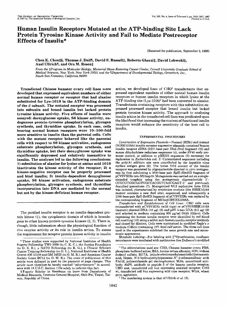
FIG. 8. Stimulation of thymidine incorporation. Cells were incubated with the indicated concentrations of insulin and with methyl t3H]thymidine (0.5 (rCi/well) as described under ”Experimen- tal Procedures.” After exposure to thymidine for 2 h, trichloroacetic acid-precipi~ble radioactivity was measured. All of the assays were performed in quadruplicate. Five independent experiments that did not differ from each other by more than 15% were averaged to yield each curve. The results are expressed as percentage of maximal stimulation by insulin. The average stimulation by insulin was 52.7% for parental CHO (10,750-16,415 cpm), 69.7% for CHO wt (11,180- 18,972 cpm), and 51.6% for CHO mut (9,950-15,084 cpm). 0, parental C H O 0, CHO wt; A, CHO mut.
the isolated insulin receptor in vitro can occur without affect- ing its insulin-binding properties. The inherent protein kinase activity of the receptor is also apparently not required for the cleavage of the receptor into subunits or for its insertion into the plasma membrane in an orientation that permits insulin binding by the intact cell. To examine the role of the protein kinase activity of the receptor in insulin action, five cellular functions were evaluated. The prediction was that a functional human insulin receptor would render the transfected CHO cells more sensitive to insulin, whereas a receptor incapable of coupling to the effector system would not. For each situa- tion examined, the increment of normal human receptors increased the sensitivity of the cell to insulin. For deoxyglu- cose uptake, sensitivity was increased 100-fold; for the other four effects, sensitivity was enhanced approximately 10-fold. The mutated receptor was unable to transduce any of these effects. In the case of phosphorylation of p150 and p35, this is perhaps not surprising since these proteins may be direct substrates for the insulin receptor kinase. The result with the S6 kinase in CHO wt directly demonstrates its requirement for the protein-tyrosine kinase activity of a growth factor receptor for activation. Ellis et al. (36) have shown that transfected human insulin receptors with either phenylala- nine substitutions at an autophosphorylation site in the p subunit (Tyr-1150, Tyr-1151) (37) or a deletion in the 8 subunit, that leads to loss of nearly the entire cytoplasmic domain of the receptor, have diminished kinase activity and respond less well than parental ceIls to insulin-promoted deoxyglucose uptake. The present experiments examined this question using a mutant receptor with intact subunits that is completely rather than partially deficient in kinase activity. Interestingly, unlike the situation with S6 kinase activation, endogenous substrate phospho~lation, glycogen synthesis, and thymidine incorporation, stimulation of deoxyglucose uptake in CHO wt occurred at concentrations of insulin that are 100-fold lower than those required by the parental cell line. In contrast, cells bearing the mutant receptor exhibited depressed uptake that was relatively insulin insensitive. This suggests that there is something different about either the coupling of the receptor to the glucose transport system compared to the other effector systems analyzed or the posi- tion of the glucose transport system in the cascade of reactions initiated by insulin. Certainly the expression of the mutant
receptors did not interfere subs~ntially with the ability of endogenous CHO receptors to transmit the signals for the other actions of insulin that were analyzed. It is possible that the mutant receptors competitively bind a rate-limiting com- ponent of the transport system, that clustering of normal receptors is more critical for this function than for the others and is interfered with by the mutant molecules, or that hybrid receptors composed of CHO and mutant human receptor subunits may transduce some but not all actions of insulin. It should also be pointed out that since some of the actions of insulin can also be elicited by interaction of this ligand with insulin-like growth factor receptors, the effect of the trans- fected human insulin receptors on other host cell growth factor receptors merits investigation.
In conclusion, although it is possible that the mutation induced in the receptor modifies properties in addition to receptor kinase activity such as the ability to internalize, cluster, or interact with other proteins, the simplest interpre- tation of our results is that the protein tyrosine kinase activity of the receptor or the immediate product of that reaction, the autophosphorylated receptor, is essential for mediating the effects of insulin on a protein serine kinase IS6 kinase), deoxyglucose transport, nonreceptor protein tyrosine phos- phorylation, glycogen formation, and thymidine incorporation into DNA.
A c ~ ~ ~ ~ ~ g ~ n ~ s - W e are grateful to Dr. Stuart Decker (Rocke- feller University) for the rabbit antiphosphotyrosine antisera elicited as described in Ref. 38 and to Dr. Pamela Stanley (AIbert Einstein College of Medicine) for the CHO cells used for the transfections.
REFERENCES 1. Petruzzelli, L., Herrera, R., and Rosen, 0. M. (1984) Proc. Natt.
2. Ullrich, A., Bell, J. R., Chen, E. Y., Herrera, R., Petruzzelli, L. M., Dull, T. J. , Gray, A., Coussens, L., Liao, Y-C., Tsubokawa, M., Mason, A., Seeburg, P. H., Grunfeld, C., Rosen, 0. M., and Ramachandran, J. (1985) Nature 313,756-761
3. Ehina, Y., Ellis, L., Jarnagin, K., Edery, M., Graf, L., Clauser, E., Ou, J.-H., Masiarz, F., Kan, Y. W . , Goldfine, I. D., Roth, R., and Rutter, W . (1985) Cell 46, 747-758
4. Simonsen, C. C., and Levinson, A. D. (1983) Proc. Natl. Acad. Sei. U. S. A. 80, 2495-2499
5. Lusky, M., and Botchan, M. (1981) Nature 293,79-81 6. Crowley, C. W., Liu, C-C., and Levinson, A. D. (1983) Mol. Celt.
7. Adelman, J. P., Hayflick, J. S., Vasser, M., and Seeburg, P. H.
8. Wigler, M., Pellicer, A., Silverstein, S., Axel, R., Urland, G., and Chasin, L. (1979) Proc. Natl. Acad. Sci. U. S. A. 76, 1373-1376
9. Southern, P. J., and Berg, P. (1982) J. Mol. Appl. Genet. 1,327- 341
Acad. Sci. U. S. A. 81,3327-3331
Biol. 3, 44-55
(1983) DNA 2,183-193
10. Goding, J. W. (1976) J. Immunol. Methods 10, 61-66 11. Petruzzelli, L. M., Ganguly, S., Smith, C. J., Cobb, M. H., Rubin,
C. S., and Rosen, 0. M. (1982) Proc. Nad Acad. Sei. U. S. A.
12. Ganguly, S., Petruzzelli, L. M., Herrera, R., Stadtmauer, L., and Rosen, 0. M. (1985) Cam. Top. Cell. Reg&. 27‘83-94
13. Herrera, R., Petruzzelli, L., Thomas, N., Bramson, H. N., Kaiser, E. T.. and Rosen. 0. M. (1985) Proc. NatL Acad. Sei. U. S. A.
79,6792-6796
82,7899-7903 ’
14. Herrera, R., Petruzzelli, L. M., and Rosen, 0. M. (1986) J. Biol. Chem. 261, 2489-2491
BWL Chem. 253,7570-7578 15. Rubin, C. S., Hirsch, A., Fung, C., and Rosen, 0. M. (1978) J.
16. Tabarini, D., Heinricb, J., and Rosen, 0. M. (1985) Proc. Natl.
17. Hofman, C., Marsh, J. W . , Miller, B., and Steiner, D. (1980)
18. Zapf, J., Schoenle, E., and Froesch, E. R. (1978) Eur. J. Biochem.
19. Laemmli, U. K. (1970) Nature 227,680-685
A ~ a d . Sei. U. S. A. 82,4369-4373
Diabetes 29,865-874
87,285-296

Kinase-deficient Insulin Receptors Fait to Mediate Insulin Actions 1847
20. Towbin, H., Staehelin, T., and Gorden, J. (1978) Prm. Natl. Acad.
21. Burnette, W. N. (1981) Anal. Biochern. 112,195-203 22. Simpson, I. A., and Cushman, S. W. (1986) Annu. Reu. Biochem.
23. Haselbacher, G. K., Humbel, R. E., and Thomas, G. (1978) FEBS
24. Smith, C. J., Wejksnora, P. J., Warner, J. B., Rubin, C. S., and Rosen, 0. M. (1979) Proc. Natl. Acad. Sci. U. S. A. 76, 2725- 2729
25. Perisic, O., and Traugh, J. A. (1983) J. Bwl. Chern. 258, 9589- 9592
26. Novak-Hofer, I., and Thomas, G. (1984) J. Biol. Chem. 259,
27. Maller, J. L., Pike, L. J., Freidenberg, G. R., Cordera, R., Stith, B. J., Olefsky, J. Me, and Krebs, E. G. (1986) Nature 320,459- 461
28. Blenis, J., and Erikson, R. L. (1985) Proc. Natl. Acad. Sci. U. S.
Sci. U. S. A. 76,3116-3121
55,1059-1090
Lett. 100,185-190
5995-6000
A. 82,7621-7625
29. White, M. F., Maron, R., and Kahn, C. R. (1985) Nature 318,
30. Hunter, T., and Cooper, J. A. (1985) Annu. Reu. Biochern. 54,
31. Fava, R., and Cohen, S. (1984) J. Biol. Chem. 259, 2636-2645 32. Larner, J., Lawrence, J. C., Walkenbach, R. J., Roach, P. J.,
Hazen, R. J., and Huang, L. C. (1978) Adu. Cyclic Nucleotide Res. 9,425-439
33. Kamps, M. P., Taylor, S. S., and Sefton, B. M. (1984) Nature
34. Weinmaster, G., Zoller, M. J., and Pawson, T. (1985) EMBO J.
35. Rosen, 0. M., Herrera, R., Olowe, Y., Petruzzelli, L. M., and Cobb, M. H. (1983) Prm. Natl. Acad. Sci. U. S. A. 80, 3237- 3240
36. Ellis, L., Clauser, E., Morgan, D., Edery, M., Roth, R., and Rutter, W. (1986) Cell 45, 721-732
37. Herrera, R., and Rosen, 0. M. (1986) J. Bwl. Chern. 261,
183-186
897-930
310,589-592
5, 69-76
11980-11985 38. Ross, A. H., Baltimore, D., and Eisen, H. N. (1981) Nature 294,
654-656