human male unexplained infertility
TRANSCRIPT
THE YALE JOURNAL OF BIOLOGY AND MEDICINE 65 (1992), 29-38
Use ofXenopus laevis Frog Egg Extract in DiagnosingHuman Male Unexplained Infertility
DAVID B. BROWN, Ph.D.,a AND MANUBAI NAGAMANI, M.D.b
aDepartment ofHuman Biological Chemistry and Genetics, bDepartment of Obstetricsand Gynecology, University of Texas Medical Branch, Galveston, Te-xas
Received July 22, 1991
Approximately one in six married couples find themselves involuntarily infertile. This ratiotranslates to between two and four million U.S. couples. Although numerous tests are availablefor diagnosing infertility problems, 5-10 percent of all couples who seek medical treatment arediagnosed with unexplained infertility. Several tests are presently available for diagnosing maleinfertility; however, none of the present procedures test for activation of the sperm nucleusfollowing entry into the fertilized egg, a series of events critical for the entry of the zygote intothe developmental program. We have developed an in vitro human sperm activation assay, usingXenopus laevis frog egg extract. When normal human sperm is permeabilized and then mixedwith frog egg extract, the sperm nuclei decondense, synthesize DNA, and recondense during athree-hour time course. We have tested this assay's utility in diagnosing previously unexplainedinfertility. We found that 20 percent of the male infertility patients produced sperm thatresponded abnormally in the assay (95 percent confidence interval, 4-48 percent; n = 15), whilesperm samples from 15 fertile males showed no abnormal responses (p = 0.112). Thesepreliminary results indicate that the human sperm activation assay may be a useful tool fordiagnosing some cases of human infertility.
INTRODUCTION
During fertilization, the sperm nucleus is activated by the egg cytoplasm. Theactivated sperm undergoes nuclear envelope breakdown and chromatin dispersion;the male pronucleus is formed and surrounded by a new nuclear envelope; DNA issynthesized in both the male and female pronuclei; and mitosis ensues, with nuclearenvelope breakdown and chromosome formation [1,2]. In order to identify thefactor(s) required for sperm activation, Lohka and Masui developed an in vitro frogsperm activation assay [3]. They demonstrated that incubating demembranatedXenopus laevis frog sperm in Rana pipiens frog egg extract induced sperm activation,including chromatin decondensation, pronuclei formation, DNA synthesis, andchromatin recondensation. These results were extended to the human system whenGorden et al. [4] and Brown et al. [5] demonstrated that mixing permeabilizedhuman sperm with Xenopus laevis frog egg extract promoted the early events ofhuman sperm activation, including chromatin decondensation, pronuclei formation,DNA synthesis, and chromatin recondensation. We now present results from thefirst clinical application of the human sperm activation assay (HSAA), analyzing the
29Abbreviation: GIFT: gamete intrafallopian transfer HSAA: human sperm activation assay IVF-
ET: in vitro fertilization-embryo transfer NIM: nuclear isolation medium SPA: sperm penetrationassay XEIM: Xenopus extract isolation mediumAddress reprint requests to: David B. Brown, Ph.D., University of Texas Medical Branch, Dept. HBC &
G, BSB-506H, F45, Galveston, TX 77550
Copyright © 1992 by The Yale Journal of Biology and Medicine, Inc.All rights of reproduction in any form reserved.

BROWN AND NAGAMANI
sperm from 15 fertile males and 15 male unexplained infertility patients. An abstractof this study has appeared elsewhere [6].
MATERIALS AND METHODS
Unexplained Infertility Patients
At the University of Texas Medical Branch Infertility Clinic, a couple is diagnosedas having unexplained infertility when the following conventional tests have beencompleted without finding a cause for the couple's infertility: the female partner willhave undergone complete infertility evaluations, including post-coital tests (to ruleout sperm motility loss in cervical mucus), a timed endometrial biopsy (to excludeluteal phase defect), a hysterosalpingogram (to establish tubal patency), and laparos-copy (to rule out pelvic pathology); in the male partner, semen analysis should havebeen normal on two occasions with sperm concentrations greater than 20 million/ml,total sperm numbers of 40 million or more, sperm motility greater than 60 percent,and normal morphology in more than 60 percent of the sperm [7]. The inexplicablyinfertile couples must also have been involuntarily infertile for over two years. In thisstudy, sperm was obtained from 15 unexplained infertility patients who have beeninvoluntarily infertile for more than five years (average of 7.3 years, range of five toten years). All of the semen samples from the unexplained infertility patients thatwere analyzed in this study had normal sperm concentrations, total sperm counts,motility, and morphology.
Human Sperm Preparation
All semen donors abstained from ejaculation for at least two days prior to thecollection of the semen samples that were obtained by masturbation.Semen samples from 15 unexplained infertility patients were obtained at the time
of the regularly scheduled procedures at the infertility clinic (e.g., sperm penetrationassay, SPA; in vitro fertilization-embryo transfer, IVF-ET; and gamete intrafallopiantransfer, GIFT). The portion of the sample not used for these procedures (usuallydiscarded) was used for the HSAA. These samples were kept in a 4°C refrigerator forup to seven days for parallel weekly assays on samples collected throughout the week.Semen samples from 15 fertile males were stored from one to seven days, mimickingthe storage conditions of the semen samples from the unexplained infertility pa-tients. Fresh samples are not needed for the HSAA, as stored semen samples from afertile male were found to respond the same in the human sperm activation assays asdid sperm collected from the same fertile male on the day the assay was performed,even after 4°C storage of up to a month (data not shown).On the day of the experiment, semen samples were incubated for 30 minutes at
37°C; each sample was then suspended in 10 ml of nuclear isolation medium (NIM,200 mM sucrose, 2.4 mM MgCl2, 10 mM Tris-HCl, and 5 mM maleic acid, pH 7.4). Asperm count was taken and an aliquot (containing 20 million sperm) was pelleted in acentrifuge for ten minutes at 400 g, then resuspended in 10 ml of NIM containing0.05 percent lysolecithin and 1 ,ug/ml soybean trypsin inhibitor. The mixture waskept at room temperature for five minutes and the sperm repelleted as before, thenwashed, first in NIM with 3 percent BSA and then with NIM containing 0.4 percentBSA, as described by Lohka and Masui [3]. Sperm were finally suspended inXenopusextract isolation medium (XEIM, 10 mM Tris-HCl, pH 7.5, 1.5 mM MgCl2, 100 mMKCI, and 50 mM DTT) at a concentration of 25,000 sperm/,ul and kept on ice for 45
30

HUMAN SPERM ACTIVATION ASSAY
minutes before mixing with the frog egg extract. This XEIM (DTT) pre-treatmentwas found to enhance the decondensation and DNA synthesis events (unpublishedobservations), presumably by reducing the protamines that in mammalian sperm arecross-linked by intermolecular disulfide bonds [8]. Xenopus laevis sperm contain"intermediate" protamines [9] that are not cysteine-rich [10], so the frog egg extractlacks the reducing factor(s) found in mammalian eggs that are required for reducingthe disulfide bridges of the human sperm; we must thus reduce the disulfide bondsexperimentally.
Xenopus laevis Egg Extract Isolation
To promote oocyte maturation, ovulation, and egg laying, adult female Xenopuslaevis frogs were injected with 500 units of human chorionic gonadotropin in themorning and evening of the day preceding the experiment. Mature eggs werecollected and dejellied with 2 percent cysteine-HCl, pH 7.6. The eggs were thenrinsed and incubated for one hour at room temperature in Barth's medium com-posed of 88 mM NaCl, 1.0 mM KCl, 0.83 mM MgSO4, 0.34 mM Ca (NO3)2, 0.41 mMCaCl2, 7.5 mM Tris-HCl, pH 7.6, 10 ,ug/ml penicillin, 10 Mg/ml streptomycin, and2.4 mM Na(CO3)2. The eggs were washed three times in XEIM. Excess buffer wasremoved and the eggs lysed by centrifugation at 10,000 g, 4°C, for 15 minutes in aswinging bucket rotor. The resulting middle layer between the lipids and the eggpellet was removed from the centrifuge tube, re-centrifuged, and the resultingmiddle layer used as the stock extract solution. The extract was stored on ice untilused for the HSAA.
Human Sperm Activation Assay (HSAA)
Permeabilized sperm from the normal controls and the infertility patients weremixed separately with frog egg extract at a concentration of 150,000 sperm/ 150 ,ul ofextract. The permeabilized sperm were incubated at 19°C in extracts containing3H-YTP at a concentration of 80 p,Ci/ml. Chromatin decondensation, DNA synthe-sis, and chromatin recondensation were evaluated. Phase-contrast microscopy wasused to assess chromatin decondensation and recondensation. Aliquots (5 [lI) weretaken, placed on glass slides, and covered with glass cover slips at five and tenminutes and at three hours following the addition of the sperm to the frog eggextract. At the five- and ten-minute time points, the percentage of sperm that haddecondensed (Fig. 1B) was determined. Between 95-100 percent of the controlsperm were normally decondensed within five minutes. At the ten-minute time point,between 95-100 percent normally have the appearance of the nucleus shown in Fig.1B. At the three-hour time point, it was determined if the sperm nuclei hadrecondensed their chromatin as may be seen by the smaller nucleus shown in Fig. 1Dwhen compared to the nucleus shown in Fig. 1B. Recondensation was typicallyobserved in 95-100 percent of the control sperm. DNA synthesis in the sperm nucleiwas assessed, using a 50 ,lI aliquot taken at the two-hour time point and diluted with50 pl of phosphate-buffered saline. The sperm were then affixed to glass slides bycentrifugation, dried, and fixed for five minutes in Carnoy's fixative (three partsice-cold methanol to one part glacial acetic acid). These fixed cytopreps were thendipped in 42°C Kodak nuclear track emulsion (NTB-2), stored at 4°C for two weeks,and then, following development, were Giemsa-stained through the emulsion. Wealso made cytopreps at the 15-minute time point to observe chromatin smearing andat the three-hour time point to see if the chromatin had recondensed to the level
31
BROWN AND NAGAMANI
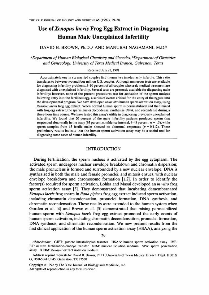
FIG. 1. Pictorial representation of permeabilized human sperm incubated in frog egg extract. Fig-ures lA-lD are phase-contrast micrographs. Figures la-Id are bright-field micrographs of Giemsa-stained nuclei. Figure lc is a Giemsa-stained autoradiograph. A,a: Time zero. B,b: 15-minuteincubation. C,c: Two-hour incubation. D,d: Three-hour incubation. Bar, 10 ,um.
shown in Fig. ld. The cytopreps were prepared and fixed as previously described,followed by Giemsa staining without autoradiography. All microscopy was doneusing a Leitz Orthoplan microscope. The micrographs in Figs. 1-4 were photo-graphed at 800 X magnification, using a Zeiss Photomicroscope III. Two hundrednuclei were scored for labeling and the results with control sperm (percentage of thenuclei showing label above background) compared to those with the patients' sperm.
RESULTS
Normal Sperm (Control) Response in the HSAA
A pictorial representation of a normal response of permeabilized human sperm tothe frog egg extract is shown in Fig. 1. The sperm nuclei shown in Figs. 1A-D andla-d were photographed (800 x magnification) and printed at the same magnifica-tion, with the bar representing 10 p,m. Phase-contrast microscopy was used whenphotographing the nuclei shown in Figs. 1A-D, while Figs. la-d are bright-fieldphotographs of Giemsa-stained cytopreps, with the nuclei shown in Fig. lc beingGiemsa-stained autoradiographs. Permeabilized sperm without extract treatmentare shown in Figs. 1A and la. Following a five-minute incubation in the frog eggextract, greater than 90 percent of the sperm are decondensing. After a 10- to15-minute incubation in the extract, greater than 95 percent of the sperm havecompletely decondensed nuclei. A typical completely decondensed sperm nucleus isshown in Fig. 1B. We have observed that at the 15-minute time point, the spermnuclei do not remain intact during the cytocentrifugation procedure used when
32
HUMAN SPERM ACTIVATION ASSAY
TABLE 1Human Sperm Activation Assay Rating Scheme
% of Control
<40 40-60 60-80 80-100
% Decondensing at five minutes - + + + +++% Decondensing at ten minutes - + + + +++% Synthesizing DNA at two hours - + + + + + +% Recondensing at three hours - + + + +++
making cytopreps; the nuclei become smeared on to the glass slide (Fig. lb).Following a two-hour incubation in the frog egg extract, the sperm nuclei have begunto recondense their chromatin. Notice the decrease in size of the sperm nucleusshown in Fig. 1C as compared to the nucleus shown in Fig. 1B. The recondensedsperm nuclei also withstand the cytocentrifugation procedure (note intact nuclei inFig. lc). The labeling of these nuclei indicates that they have undergone DNAsynthesis. Typically, >95 percent of the control nuclei are labeled following atwo-hour incubation in frog egg extract containing tritiated thymidine, while > 95percent are fully recondensed following a three-hour incubation. Examples ofrecondensed nuclei are shown in Figs. 1D and ld.
Use ofthe HSAA in the Analysis ofthe Sperm Obtainedfrom Fertile Males andUnexplained Infertility Patients
In this study, sperm samples were obtained from 15 unexplained infertility patientsand 15 fertile males and prepared for use in the HSAA as described in Methods. Ineach HSAA, a sperm sample from a fertile male (previously shown to produce spermthat have a normal response in the HSAA) was also assayed as a parallel control. Wehave developed a rating system (Table 1) comparing the HSAA responses of thecontrol sperm sample to the sperm samples obtained from 15 unexplained infertilitypatients and 15 fertile males. The results of the pilot study are shown in Table 2. Ascan quickly be ascertained by looking at the data summarized in Table 2, three of thesperm samples from the infertility patients responded abnormally in the HSAA,whereas all sperm from fertile men responded normally in the assay.
Case Reports ofPatients Producing Sperm That Responded Abnornally in the HSAA
Patient 4: Sperm from patient 4 sperm sample, although scored normal (60percent) in the sperm penetration assay (SPA; [11-13]), had a diminished deconden-sation response (50 percent decondensed after five minutes of incubation in extract),
TABLE 2Human Sperm Activation Assay Pilot Study Results
Decondensation Decondensation DNA Synthesis Recondensation(5 minutes) (10 minutes) (2 hours) (3 hours)
Fertile males 1-15 +++ +++ +++ +++Infertility patients 1-3, 5, 7-14 +++ +++ +++ +++Infertility patient 4 + + + +++ +++Infertility patient 6 +++ +++ -+++Infertility patient 15 + + + ?
33
BROWN AND NAGAMANI
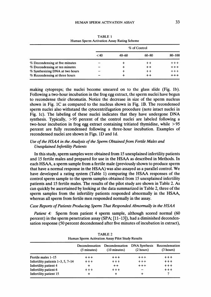
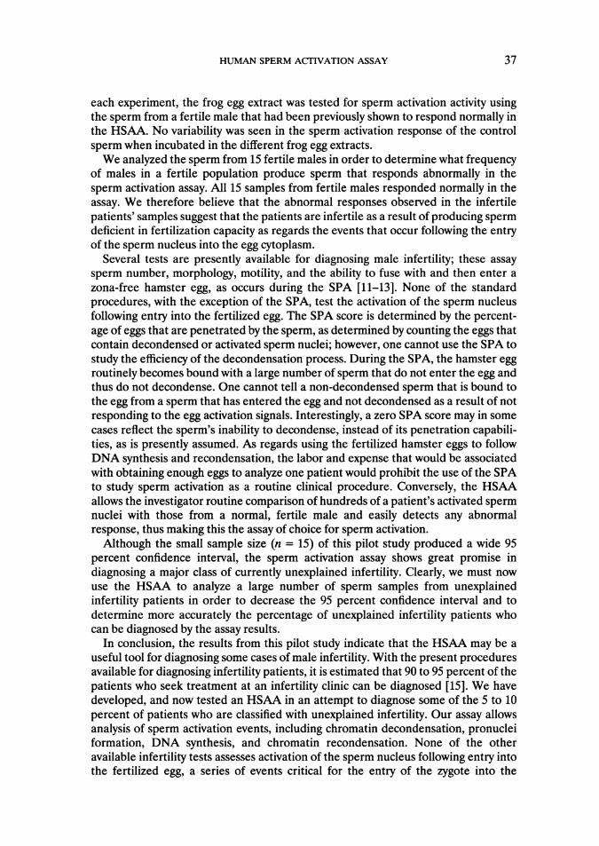
I IFIG. 2. Bright-field photographs of
Giemsa-stained autoradiographs ofspermnuclei following two hours of incubation infrog egg extract. A. Fertile control
A labeled nuclei. B. Patient 6 unlabelednuclei. Bar, 10 pWm.
by which time normal human sperm were 95 percent decondensed. Even after aten-minute incubation, only 80 percent of the patient's sperm had decondensed. Wehave repeated the HSAA for patient 4 using the excess sperm from an unsuccessfulGIFT attempt (four eggs transferred), finding 60 percent of the sperm decondensedafter five minutes of incubation in the extract, with 98 percent of the normal spermdecondensing during the same incubation period. Following a ten-minute incubationin the egg extract, 90 percent of the patient's sperm had decondensed, with 98percent of the control sperm being decondensed. When new samples of this patient'ssperm were obtained for use in the HSAA at 4, 11, and 13 months following theinitial analyses, this patient's sperm responded normally in the HSAA when com-pared to the control sperm. During the time period where the patient's sperm wasresponding normally in the HSAA, an IVF-ET attempt resulted in a successfulpregnancy; however, the pregnancy ended with a spontaneous miscarriage at sixweeks of pregnancy. The sperm is from the male member of a couple who have beentrying to conceive for ten years and at present, with the exception of our assay, thecouple has been found to be normal in every test performed.
Patient 6: Patient 6 has sperm that was seen to decondense and recondense in anormal fashion. Only 27 percent of the sperm nuclei were found to be labeledfollowing autoradiography, however, while 100 percent of the control sperm nucleiwere found to be labeled. Autoradiographs of typical labeled control nuclei andunlabeled patient nuclei are shown in Figs. 2A and 2B, respectively. This couple hasbeen trying to conceive for eight years. The sperm penetration assay has not beenperformed for this patient.
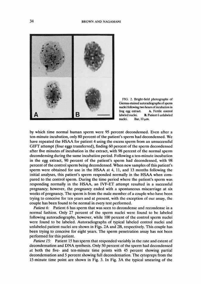
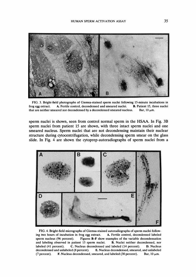
Patient 15: Patient 15 has sperm that responded variably in the rate and extent ofdecondensation and DNA synthesis. Only 50 percent of the sperm had decondensedat both the five- and ten-minute time points with 45 percent showing partialdecondensation and 5 percent showing full decondensation. The cytopreps from the15-minute time point are shown in Fig. 3. In Fig. 3A the typical smearing of the
34
HUMAN SPERM ACTIVATION ASSAY
FIG. 3. Bright-field photographs of Giemsa-stained sperm nuclei following 15-minute incubations infrog egg extract. A. Fertile control, decondensed and smeared nuclei. B. Patient 15, three nucleithat are neither smeared nor decondensed by a decondensed smeared nucleus. Bar, 10 pLm.
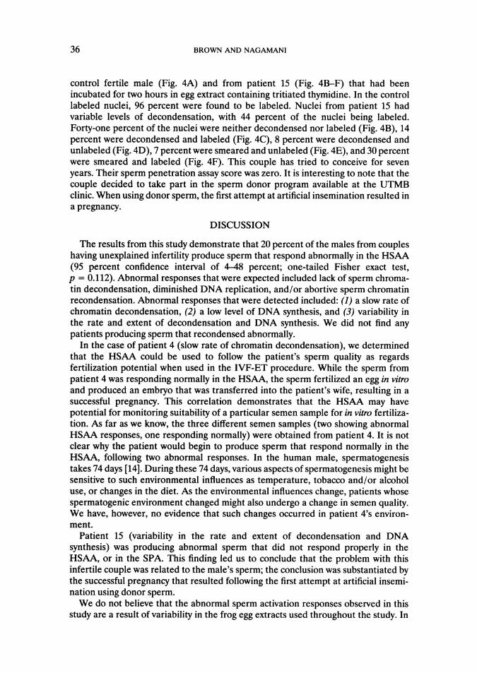
sperm nuclei is shown, seen from control normal sperm in the HSAA. In Fig. 3Bsperm nuclei from patient 15 are shown, with three intact sperm nuclei and onesmeared nucleus. Sperm nuclei that are not decondensing maintain their nuclearstructure during cytocentrifugation, while decondensing sperm smear on the glassslide. In Fig. 4 are shown the cytoprep-autoradiographs of sperm nuclei from a
FIG. 4. Bright-field micrographs of Giemsa-stained autoradiographs of sperm nuclei follow-ing two hours of incubation in frog egg extract. A. Fertile control, decondensed labeledsperm nucleus (96 percent). Figures B-F show examples of the variable decondensationand labeling observed in patient 15 sperm nuclei. B. Nuclei neither decondensed, norlabeled (41 percent). C. Nucleus decondensed and labeled (14 percent). D. Nucleusdecondensed and unlabeled (8 percent). E. Nucleus decondensed, smeared, and unlabeled(7 percent). F. Nucleus decondensed, smeared, and labeled (30 percent). Bar, 10 pL.m
t..
-..M-
35
BROWN AND NAGAMANI
control fertile male (Fig. 4A) and from patient 15 (Fig. 4B-F) that had beenincubated for two hours in egg extract containing tritiated thymidine. In the controllabeled nuclei, 96 percent were found to be labeled. Nuclei from patient 15 hadvariable levels of decondensation, with 44 percent of the nuclei being labeled.Forty-one percent of the nuclei were neither decondensed nor labeled (Fig. 4B), 14percent were decondensed and labeled (Fig. 4C), 8 percent were decondensed andunlabeled (Fig. 4D), 7 percent were smeared and unlabeled (Fig. 4E), and 30 percentwere smeared and labeled (Fig. 4F). This couple has tried to conceive for sevenyears. Their sperm penetration assay score was zero. It is interesting to note that thecouple decided to take part in the sperm donor program available at the UTMBclinic. When using donor sperm, the first attempt at artificial insemination resulted ina pregnancy.
DISCUSSION
The results from this study demonstrate that 20 percent of the males from coupleshaving unexplained infertility produce sperm that respond abnormally in the HSAA(95 percent confidence interval of 4-48 percent; one-tailed Fisher exact test,p = 0.112). Abnormal responses that were expected included lack of sperm chroma-tin decondensation, diminished DNA replication, and/or abortive sperm chromatinrecondensation. Abnormal responses that were detected included: (1) a slow rate ofchromatin decondensation, (2) a low level of DNA synthesis, and (3) variability inthe rate and extent of decondensation and DNA synthesis. We did not find anypatients producing sperm that recondensed abnormally.
In the case of patient 4 (slow rate of chromatin decondensation), we determinedthat the HSAA could be used to follow the patient's sperm quality as regardsfertilization potential when used in the IVF-ET procedure. While the sperm frompatient 4 was responding normally in the HSAA, the sperm fertilized an egg in vitroand produced an embryo that was transferred into the patient's wife, resulting in asuccessful pregnancy. This correlation demonstrates that the HSAA may havepotential for monitoring suitability of a particular semen sample for in vitro fertiliza-tion. As far as we know, the three different semen samples (two showing abnormalHSAA responses, one responding normally) were obtained from patient 4. It is notclear why the patient would begin to produce sperm that respond normally in theHSAA, following two abnormal responses. In the human male, spermatogenesistakes 74 days [14]. During these 74 days, various aspects of spermatogenesis might besensitive to such environmental influences as temperature, tobacco and/or alcoholuse, or changes in the diet. As the environmental influences change, patients whosespermatogenic environment changed might also undergo a change in semen quality.We have, however, no evidence that such changes occurred in patient 4's environ-ment.
Patient 15 (variability in the rate and extent of decondensation and DNAsynthesis) was producing abnormal sperm that did not respond properly in theHSAA, or in the SPA. This finding led us to conclude that the problem with thisinfertile couple was related to the male's sperm; the conclusion was substantiated bythe successful pregnancy that resulted following the first attempt at artificial insemi-nation using donor sperm.We do not believe that the abnormal sperm activation responses observed in this
study are a result of variability in the frog egg extracts used throughout the study. In
36
HUMAN SPERM ACTIVATION ASSAY
each experiment, the frog egg extract was tested for sperm activation activity usingthe sperm from a fertile male that had been previously shown to respond normally inthe HSAA. No variability was seen in the sperm activation response of the controlsperm when incubated in the different frog egg extracts.We analyzed the sperm from 15 fertile males in order to determine what frequency
of males in a fertile population produce sperm that responds abnormally in thesperm activation assay. All 15 samples from fertile males responded normally in theassay. We therefore believe that the abnormal responses observed in the infertilepatients' samples suggest that the patients are infertile as a result of producing spermdeficient in fertilization capacity as regards the events that occur following the entryof the sperm nucleus into the egg cytoplasm.
Several tests are presently available for diagnosing male infertility; these assaysperm number, morphology, motility, and the ability to fuse with and then enter azona-free hamster egg, as occurs during the SPA [11-13]. None of the standardprocedures, with the exception of the SPA, test the activation of the sperm nucleusfollowing entry into the fertilized egg. The SPA score is determined by the percent-age of eggs that are penetrated by the sperm, as determined by counting the eggs thatcontain decondensed or activated sperm nuclei; however, one cannot use the SPA tostudy the efficiency of the decondensation process. During the SPA, the hamster eggroutinely becomes bound with a large number of sperm that do not enter the egg andthus do not decondense. One cannot tell a non-decondensed sperm that is bound tothe egg from a sperm that has entered the egg and not decondensed as a result of notresponding to the egg activation signals. Interestingly, a zero SPA score may in somecases reflect the sperm's inability to decondense, instead of its penetration capabili-ties, as is presently assumed. As regards using the fertilized hamster eggs to followDNA synthesis and recondensation, the labor and expense that would be associatedwith obtaining enough eggs to analyze one patient would prohibit the use of the SPAto study sperm activation as a routine clinical procedure. Conversely, the HSAAallows the investigator routine comparison of hundreds of a patient's activated spermnuclei with those from a normal, fertile male and easily detects any abnormalresponse, thus making this the assay of choice for sperm activation.Although the small sample size (n = 15) of this pilot study produced a wide 95
percent confidence interval, the sperm activation assay shows great promise indiagnosing a major class of currently unexplained infertility. Clearly, we must nowuse the HSAA to analyze a large number of sperm samples from unexplainedinfertility patients in order to decrease the 95 percent confidence interval and todetermine more accurately the percentage of unexplained infertility patients whocan be diagnosed by the assay results.
In conclusion, the results from this pilot study indicate that the HSAA may be auseful tool for diagnosing some cases of male infertility. With the present proceduresavailable for diagnosing infertility patients, it is estimated that 90 to 95 percent of thepatients who seek treatment at an infertility clinic can be diagnosed [15]. We havedeveloped, and now tested an HSAA in an attempt to diagnose some of the 5 to 10percent of patients who are classified with unexplained infertility. Our assay allowsanalysis of sperm activation events, including chromatin decondensation, pronucleiformation, DNA synthesis, and chromatin recondensation. None of the otheravailable infertility tests assesses activation of the sperm nucleus following entry intothe fertilized egg, a series of events critical for the entry of the zygote into the
37
38 BROWN AND NAGAMANI
developmental program. The HSAA will allow diagnosis of otherwise unexplainedinfertility patients by determining that their infertility is a result of their sperm nucleinot responding properly to the egg cytoplasm following fertilization. Couples havingunexplained infertility spend a large amount of money on current treatments. It isestimated that as much as a billion dollars a year is spent by from 300,000 to onemillion couples in pursuit of pregnancy [16]. As with patient 15, the HSAA can beused to identify patients who cannot benefit from the available treatments ofinfertility, thereby saving the associated expense and discomfort. Eventually, wehope to develop methods to identify and perhaps ultimately to replace the defectiveor missing components of the sperm that prevent normal sperm activation.
ACKNOWLEDGEMENTS
The authors wish to extend sincere appreciation to Dr. David Konkel for critical reading of themanuscript and to Todd Pappas and Tatsuo Uchida for statistical analysis of the data. We also express ourthanks for technical assistance from Louise Henson.
REFERENCES
1. Longo FJ: Fertilization: A comparative ultrastructural review. Biol Reprod 9:149-215, 19732. Longo FJ, Kunkle M: Transformation of sperm nuclei upon insemination. Curr Top Dev Biol
12:149-184, 19783. Lohka MJ, Masui Y: Formation in vitro of sperm pronuclei and mitotic chromosomes induced by
amphibian ooplasmic components. Science 220:719-721, 19834. Gordon K, Brown DB, Ruddle FH: In vitro activation of human sperm induced by amphibian egg
extract. Exp Cell Res 157:409-418, 19855. Brown DB, Blake EJ, Wolgemuth DJ, Gordon K, Ruddle FH: Chromatin decondensation and DNA
synthesis in human sperm activated in vitro by using Xenopus laevis egg extracts. J Exp Zool242:215-231, 1987
6. Brown DB, Nagamani M: Use of frog egg extract in diagnosing human infertility. J Cell Biol 111:115a,1990
7. World Health Organization: Appendix LA, Normal values of semen variables. In WHO LaboratoryManual for the Examination of Human Semen and Semen-Cervical Mucus Interaction. New York,Cambridge University Press, 1987, p 27
8. Perreault SD, Barbee RR, Slott VL: The role of disulfide bond reduction during mammalian spermnuclear decondensation in vivo. Dev Biol 101:160-167, 1984
9. Kasinsky HE, Huang SY, Mann M, Roca J, Subirana JA: On the diversity of sperm histones in thevertebrates. IV. Cytochemical and amino acid analysis inAnura. J Exp Zool 234:33-45, 1985
10. Kasinsky HE: Specificity and distribution of sperm basic proteins. In Histones and Other Basic NuclearProteins. Edited by LS Hnilica, GS Stein, JL Stein. Boca Raton, FL, CRC Press Inc, 1989, pp 73-163
11. Yanagimachi R, Yanagimachi H, Rogers BJ: The use of zona-free animal ova as a test-system for theassessment of the fertilizing capacity of human spermatozoa. Biol Reprod 15:471-476, 1976
12. Overstreet JW, Yanagimachi R, Katz DF, Hayashi K, Hanson FW: Penetration of human spermatozoainto the human zona pellucida and the zona-free hamster egg: A study of fertile donors and infertilepatients. Fertil Steril 33:534-542, 1980
13. Wolf DP, Sokoloski JE: Characterization of the sperm penetration bioassay. J Androl 3:445-451, 198214. Johnson L: Spermatogenesis (Animal Species and Humans). In Gamete Physiology. Edited by RH
Asch, JP Balmaceda, I Johnston. Norwell, MA, Serono Symposia, USA, 1990, pp 3-1815. Webster BW, Cook AS, Garner CH: The infertility evaluation. In Handbook of the Laboratory
Diagnosis and Treatment of Infertility. Edited by BA Keel, BW Webster. Boca Raton, FL, CRC PressInc, 1990, pp 1-9
16. U.S. Congress, Office of Technology Assessment: Infertility: Medical and Social Choices. OTA-BA358. Washington DC, U.S. Government Printing Office, 1988