human oxidative phosphorylation (oxphos) …...mitochondrial oxidative phosphorylation (oxphos)...
TRANSCRIPT

Human Oxidative Phosphorylation (OXPHOS) Magnetic Bead Panel 96-Well Plate Assay Cat. # H0XPSMAG-16K

MILLIPLEX® MAP
HUMAN OXIDATIVE PHOSPHORYLATION (OXPHOS) MAGNETIC BEAD PANEL
96-Well Plate Assay
# H0XPSMAG-16K
TABLE OF CONTENTS PAGE Introduction 2 Principle 4 Storage Conditions Upon Receipt 4 Reagents Supplied 5 Materials Required But Not Provided 5 Safety Precautions 6 Technical Guidelines 6 Sample Collection And Storage 7 Preparation of Reagents for Immunoassay 11 Immunoassay Procedure 12 Plate Washing 14 Equipment Settings 14 Assay Characteristics 17 Troubleshooting Guide 20 Replacement Reagents 22 Ordering Information 23 Well Map 24
FOR RESEARCH USE ONLY. NOT FOR USE IN DIAGNOSTIC PROCEDURES
By purchasing this product, which contains fluorescently labeled microsphere beads authorized by Luminex Corporation (“Luminex”), you, the customer, acquire the right under Luminex’s patent rights, if
any, to use this product or any portion of this product, including without limitation the microsphere beads contained herein, only with Luminex’s laser based fluorescent analytical test instrumentation marketed
under the name of Luminex 100TM IS, 200TM, HTS, FLEXMAP 3D®, or MAGPIX®.
Cat# H0XPSMAG-15K Rev. 21-AUG-2013 PAGE 2 EMD MILLIPORE
Human Oxidative Phosphorylation (OXPHOS) Magnetic Bead Panel INTRODUCTION Mitochondrial Oxidative Phosphorylation (OXPHOS) produces more than 95% of the conserved cellular energy in the form of ATP under normal conditions. This process involves five different protein complexes, NADH–ubiquinone oxidoreductase (Complex I), succinate ubiquinone oxidoreductase (Complex II), ubiquinone cytochrome c oxidoreductase (Complex III), cytochrome c oxidase (Complex IV) and ATP synthase (Complex V). Nicotinamide nucleotide transhydrogenase (NNT) is an integral protein of the inner mitochondrial membrane whose structure and function are closely related to OXPHOS. Under most physiological conditions, the enzyme uses energy from the mitochondrial proton gradient to produce high concentrations of NADPH. The resulting NADPH is used for biosynthesis and in free radical detoxification. Since NNT is encoded solely in the nucleus, it serves as a good internal reference for mitochondrial proteins. The overall process of oxidative phosphorylation is tightly controlled by transcriptional regulation at the level of DNA and RNA, by substrate feedback inhibition and by post-translational modifications including phosphorylation and acetylation. Inefficient electron transfer through complexes I-IV leads to various human abnormalities, which is due in part to a loss of energy metabolism and a deregulation of critical enzymes, such as complexes I, II and III. Without proper regulation, the production of toxic reactive oxygen species is known to increase. Some diseases associated with impaired OXPHOS include diabetes, Parkinson’s disease, Alzheimer’s disease, cancer and the aging process itself. Many diverse classes of drugs inhibit OXPHOS and induce mitochondrial toxicity. Not surprisingly, the ability to monitor the expression levels of the OXPHOS complexes is a key element to the studies of many diseases and to the process of drug safety evaluation. The MILLIPLEX® MAP Human Oxidative Phosphorylation (OXPHOS) Magnetic Bead Panel includes Complex I, II, III, IV, V, and NNT, and allows the study of these multiple targets simultaneously in one reaction well. To study cellular metabolism, it might be necessary to screen different panels of specific pathway proteins, which often require some level of automation and/or high throughput assays. Magnetic beads can make the process of automation and high throughput screening easier with features such as walk-away washing. Advantages include:
• More flexible plate and plate washer options • Improved performance with turbid samples • Automated washing eliminating technical obstacles which may result during vacuum
manifold/manual washing (i.e. clogging of wells that contain viscous samples) The MILLIPLEX® MAP Human Oxidative Phosphorylation (OXPHOS) Magnetic Bead Panel enables users to focus on the OXPHOS pathway. Coupled with the Luminex xMAP® platform in a magnetic bead format, users receive the advantage of ideal speed and sensitivity by allowing the quantitative multiplex detection of multiple analytes simultaneously, resulting in dramatically improved productivity. This kit uses the most versatile system available for cellular metabolism research. A convenient “all-in-one” box format gives assurance that all the necessary reagents are provided to run an assay.

Cat# H0XPSMAG-15K Rev. 21-AUG-2013 PAGE 3 EMD MILLIPORE
EMD Millipore’s MILLIPLEX® MAP Human Oxidative Phosphorylation (OXPHOS)
Magnetic Bead Panel is available as a premixed panel and is to be used for the simultaneous detection of Complex I, II, III, IV, V, and NNT in human cell lysate or tissue extract. Full names and alternative names of analytes: Complex I: NADH–ubiquinone oxidoreductase Complex II: succinate ubiquinone oxidoreductase Complex III: ubiquinone cytochrome c oxidoreductase Complex IV: cytochrome c oxidase Complex V: ATP synthase NNT: nicotinamide nucleotide transhydrogenase For Research Use Only. Not for Use in Diagnostic Procedures. Please read entire protocol before use. It is important to use same assay incubation conditions throughout your study.

Cat# H0XPSMAG-15K Rev. 21-AUG-2013 PAGE 4 EMD MILLIPORE
PRINCIPLE MILLIPLEX® MAP is based on the Luminex xMAP® technology — one of the most respected multiplex technologies available. This technology finds applications throughout the life sciences and enables a variety of bioassays, including immunoassays, on the surface of fluorescent-coded beads known as MagPlex®-C microspheres.
• Luminex uses proprietary techniques to internally color-code microspheres with two fluorescent dyes. Through precise concentrations of these dyes, 100 distinctly colored bead sets can be created, each of which is coated with a specific capture antibody.
• After an analyte from a test sample is captured by the bead, a biotinylated detection antibody is introduced.
• The reaction mixture is then incubated with Streptavidin-PE conjugate, the reporter molecule, to complete the reaction on the surface of each microsphere.
• The microspheres are illuminated, and the internal dyes fluoresce, marking the microsphere set(s) used in a particular assay. A second illumination source excites PE, the fluorescent dye on the reporter molecule.
• Finally, high-speed digital-signal processors identify each individual microsphere and quantify the result of its bioassay based on fluorescent reporter signals.
The capability of adding multiple conjugated beads to each sample results in the ability to obtain multiple results from each sample. Open-architecture xMAP® technology enables multiplexing of many types of bioassays reducing time, labor and costs over traditional methods. STORAGE CONDITIONS UPON RECEIPT
• Recommended storage for kit components is 2 - 8°C.
• Once the lysate controls have been reconstituted, immediately transfer contents into polypropylene vials. DO NOT STORE RECONSITUTED CONTROLS IN GLASS VIALS. For long-term storage, freeze reconstituted controls at ≤ -70°C. Avoid multiple (>2) freeze/thaw cycles.
• DO NOT FREEZE Antibody-Immobilized Beads, Detection Antibody, and Streptavidin-Phycoerythrin.
Cat# H0XPSMAG-15K Rev. 21-AUG-2013 PAGE 5 EMD MILLIPORE
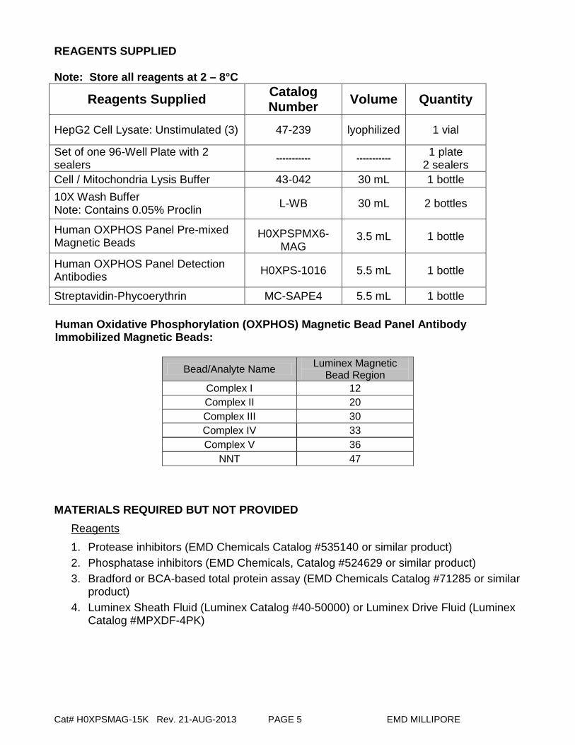
REAGENTS SUPPLIED Note: Store all reagents at 2 – 8°C
Reagents Supplied Catalog Number Volume Quantity
HepG2 Cell Lysate: Unstimulated (3) 47-239 lyophilized 1 vial
Set of one 96-Well Plate with 2 sealers ----------- ----------- 1 plate
2 sealers Cell / Mitochondria Lysis Buffer 43-042 30 mL 1 bottle 10X Wash Buffer Note: Contains 0.05% Proclin L-WB 30 mL 2 bottles
Human OXPHOS Panel Pre-mixed Magnetic Beads
H0XPSPMX6-MAG
3.5 mL 1 bottle
Human OXPHOS Panel Detection Antibodies H0XPS-1016 5.5 mL 1 bottle
Streptavidin-Phycoerythrin MC-SAPE4 5.5 mL 1 bottle Human Oxidative Phosphorylation (OXPHOS) Magnetic Bead Panel Antibody Immobilized Magnetic Beads:
Bead/Analyte Name Luminex Magnetic Bead Region
Complex I 12 Complex II 20 Complex III 30 Complex IV 33 Complex V 36
NNT 47 MATERIALS REQUIRED BUT NOT PROVIDED
Reagents 1. Protease inhibitors (EMD Chemicals Catalog #535140 or similar product) 2. Phosphatase inhibitors (EMD Chemicals, Catalog #524629 or similar product) 3. Bradford or BCA-based total protein assay (EMD Chemicals Catalog #71285 or similar
product) 4. Luminex Sheath Fluid (Luminex Catalog #40-50000) or Luminex Drive Fluid (Luminex
Catalog #MPXDF-4PK)

Cat# H0XPSMAG-15K Rev. 21-AUG-2013 PAGE 6 EMD MILLIPORE
MATERIALS REQUIRED BUT NOT PROVIDED (continued) Instrumentation / Materials 1. Adjustable Pipettes with Tips capable of delivering 25 µL to 1000 µL 2. Multichannel Pipettes capable of delivering 5 µL to 50 µL or 25 µL to 200 µL 3. Reagent Reservoirs 4. Polypropylene Microfuge Tubes 5. Rubber Bands 6. Aluminum Foil 7. Absorbent Pads 8. Laboratory Vortex Mixer 9. Sonicator (Branson Ultrasonic Cleaner Model # B200 or equivalent) 10. Titer Plate Shaker (Lab-Line Instruments Model #4625 or equivalent) 11. Luminex 200™, HTS, FLEXMAP 3D®, or MAGPIX® with xPONENT® software by
Luminex Corporation 12. Automatic Plate washer for magnetic beads (Bio-Tek ELx405, EMD Millipore catalog
#40-015 or equivalent) or handheld Magnetic Separation Block (EMD Millipore catalog #40-285 or equivalent)
Note: If a plate washer or handheld magnetic separation block for magnetic beads is not available, one can use a microtiter filter plate (EMD Millipore Catalog #MX-PLATE) to run the assay using a Vacuum Filtration Unit (EMD Millipore Vacuum Manifold Catalog #MSVMHTS00 or equivalent with EMD Millipore Vacuum Pump Catalog #WP6111560 or equivalent).
SAFETY PRECAUTIONS • All tissue or blood components and biological materials should be handled as
potentially hazardous. Follow universal precautions as established by the Centers for Disease Control and Prevention and by the Occupational Safety and Health Administration when handling and disposing of infectious agents.
• Sodium Azide or Proclin has been added to some reagents as a preservative. Although the concentrations are low, Sodium Azide and Proclin may react with lead and copper plumbing to form highly explosive metal azides. Dispose of unused contents and waste in accordance with international, federal, state, and local regulations.
TECHNICAL GUIDELINES To obtain reliable and reproducible results, the operator should carefully read this entire manual and fully understand all aspects of each assay step before running the assay. The following notes should be reviewed and understood before the assay is set up.
• FOR RESEARCH USE ONLY. NOT FOR USE IN DIAGNOSTIC PROCEDURES.
• Do not use beyond the expiration date on the label.
• Do not mix or substitute reagents with those from other lots or sources.

Cat# H0XPSMAG-15K Rev. 21-AUG-2013 PAGE 7 EMD MILLIPORE
TECHNICAL GUIDELINES (continued) • The Antibody-Immobilized Beads are light-sensitive and must be protected from light at
all times. Cover the assay plate containing beads with opaque plate lid or aluminum foil during all incubation steps.
• It is important to allow all reagents to equilibrate to room temperature (20-25°C) before use in the assay.
• Incomplete washing can adversely affect the assay outcome. All washing must be performed using the provided Wash Buffer.
• After hydration, the control must be transferred to polypropylene tubes.
• The reconstituted lysate control must be used within 1 hour of preparation. Discard any unused lysate control except the stock which may be stored at ≤ -20°C for 1 month and at ≤ -70°C for greater than one month.
• If samples fall outside the dynamic range of the assay, further dilute the samples with the appropriate diluent and repeat the assay.
• Any unused Premixed Antibody-Immobilized Beads may be stored at 2-8°C for up to one month.
• The plate should be read immediately after the assay is finished. If the plate cannot be read immediately, seal the plate and cover with aluminum foil or an opaque lid, and store the plate at 2-8°C for up to 24 hours. Prior to reading, agitate the plate on the plate shaker at room temperature for 10 minutes. Delay in reading a plate may result in decreased sensitivity for some analytes.
• The titer plate shaker should be set at a speed to provide maximum orbital mixing without splashing of liquid outside the wells. For the recommended plate shaker, this would be a setting of 5-7 which is approximately 500-800 rpm.
• Ensure that the needle probe is clean. This may be achieved by sonication and/or alcohol flushes.
• When reading the assay on Luminex 200™, adjust probe height according to the protocols recommended by Luminex to the kit filter plate using 3 alignment discs. When reading the assay on FLEXMAP 3D®, adjust probe height according to the protocols recommended by Luminex to the kit filter plate using 1 alignment disc. When reading the assay on MAGPIX®, adjust probe height according to the protocols recommended by Luminex to the kit filter plate using 2 alignment discs.
• For FLEXMAP 3D® when using the solid plate in the kit, the final suspension should be in 150 μL and 75 μL should be aspirated.
• Vortex all reagents well before adding to plate.
Cat# H0XPSMAG-15K Rev. 21-AUG-2013 PAGE 8 EMD MILLIPORE
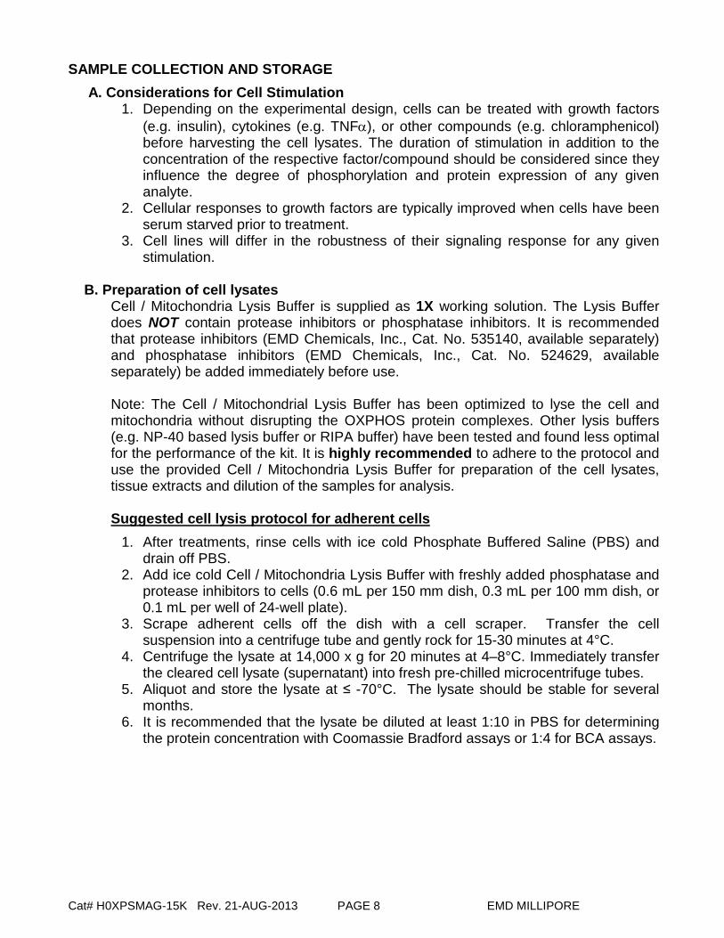
SAMPLE COLLECTION AND STORAGE A. Considerations for Cell Stimulation
1. Depending on the experimental design, cells can be treated with growth factors (e.g. insulin), cytokines (e.g. TNFα), or other compounds (e.g. chloramphenicol) before harvesting the cell lysates. The duration of stimulation in addition to the concentration of the respective factor/compound should be considered since they influence the degree of phosphorylation and protein expression of any given analyte.
2. Cellular responses to growth factors are typically improved when cells have been serum starved prior to treatment.
3. Cell lines will differ in the robustness of their signaling response for any given stimulation.
B. Preparation of cell lysates
Cell / Mitochondria Lysis Buffer is supplied as 1X working solution. The Lysis Buffer does NOT contain protease inhibitors or phosphatase inhibitors. It is recommended that protease inhibitors (EMD Chemicals, Inc., Cat. No. 535140, available separately) and phosphatase inhibitors (EMD Chemicals, Inc., Cat. No. 524629, available separately) be added immediately before use. Note: The Cell / Mitochondrial Lysis Buffer has been optimized to lyse the cell and mitochondria without disrupting the OXPHOS protein complexes. Other lysis buffers (e.g. NP-40 based lysis buffer or RIPA buffer) have been tested and found less optimal for the performance of the kit. It is highly recommended to adhere to the protocol and use the provided Cell / Mitochondria Lysis Buffer for preparation of the cell lysates, tissue extracts and dilution of the samples for analysis. Suggested cell lysis protocol for adherent cells
1. After treatments, rinse cells with ice cold Phosphate Buffered Saline (PBS) and drain off PBS.
2. Add ice cold Cell / Mitochondria Lysis Buffer with freshly added phosphatase and protease inhibitors to cells (0.6 mL per 150 mm dish, 0.3 mL per 100 mm dish, or 0.1 mL per well of 24-well plate).
3. Scrape adherent cells off the dish with a cell scraper. Transfer the cell suspension into a centrifuge tube and gently rock for 15-30 minutes at 4°C.
4. Centrifuge the lysate at 14,000 x g for 20 minutes at 4–8°C. Immediately transfer the cleared cell lysate (supernatant) into fresh pre-chilled microcentrifuge tubes.
5. Aliquot and store the lysate at ≤ -70°C. The lysate should be stable for several months.
6. It is recommended that the lysate be diluted at least 1:10 in PBS for determining the protein concentration with Coomassie Bradford assays or 1:4 for BCA assays.

Cat# H0XPSMAG-15K Rev. 21-AUG-2013 PAGE 9 EMD MILLIPORE
Suggested cell lysis protocol for non-adherent cells 1. Pellet the cells by centrifugation (500 – 1000 x g) in a tabletop centrifuge for 5
minutes. 2. Wash the cells in ice cold PBS. 3. Add ice cold Cell / Mitochondria Lysis Buffer with freshly added phosphatase and
protease inhibitors to cells (1 mL per 1 x 10⁷ cells). 4. Gently rock the lysate for 15-30 minutes at 4°C. 5. Centrifuge the lysate at 14,000 x g for 20 minutes at 4–8°C. Immediately transfer
the cleared cell lysate (supernatant) into fresh pre-chilled microcentrifuge tubes. 6. Aliquot and store the lysate at ≤ -70°C. The lysate should be stable for several
months. 7. It is recommended that the lysate be diluted at least 1:10 in PBS for determining
the protein concentration with Coomassie Bradford assays or 1:4 for BCA assays.
Cell lysis protocol for cells in sterile 96-well tissue culture plates Adherent or non-adherent cells seeded or grown in sterile 96-well tissue culture grade plates (see supplemental protocols) can be washed, treated, and lysed in the same plate, but need to be filtered in a separate 96-well filter plate. Wash the cells by centrifugation in a micro plate carrier for 2 minutes at 500 x g.
1. Remove the supernatant via aspiration and add 100 µL of ice-cold PBS. 2. Centrifuge and remove supernatant via aspiration. 3. Add 30-50 µL/well of ice cold Cell / Mitochondria Lysis Buffer with freshly added
phosphatase and protease inhibitors. 4. Place the plate on an orbital shaker (600 – 800 rpm) for 10-15 minutes at 4°C. 5. Transfer the lysate to a 96-well filter plate that has been pre-wetted with Cell /
Mitochondria Lysis Buffer. 6. Place a low protein binding, 96-well round bottom or V-bottom plate underneath
the filter plate. 7. Centrifuge the plates in a micro plate carrier for 5 minutes at 500 x g. 8. Store the filtered lysate at ≤ -70°C until ready for use. 9. It is recommended that the lysate be diluted at least 1:10 in PBS for determining
the protein concentration with Coomassie Bradford assays or 1:4 for BCA assays.
C. Preparation of tissue extracts Cell / Mitochondria Lysis Buffer is supplied as 1X working solution. The Lysis Buffer does NOT contain protease inhibitors or phosphatase inhibitors. It is recommended that protease inhibitors (EMD Chemicals, Inc., Cat. No. 535140, available separately) and phosphatase inhibitors (EMD Chemicals, Inc., Cat. No. 524629, available separately) be added immediately before use. Note: The Cell / Mitochondrial Lysis Buffer has been optimized to lyse the cell and mitochondria without disrupting the OXPHOS protein complexes. Other lysis buffers (e.g. NP-40 based lysis buffer or RIPA buffer) have been tested and found less optimal for the performance of the kit. It is highly recommended to adhere to the protocol and use the provided Cell / Mitochondria Lysis Buffer for preparation of the cell lysates, tissue extracts and dilution of the samples for analysis.

Cat# H0XPSMAG-15K Rev. 21-AUG-2013 PAGE 10 EMD MILLIPORE
SAMPLE COLLECTION AND STORAGE (continued) Suggested protocol for tissue extracts preparation
NOTE: This kit provides enough Cell / Mitochondria Lysis buffer to process ~ 25 tissue samples at 100mg following this protocol. Additional Cell / Mitochondria Lysis Buffer (Cat# 43-042) may be purchased from EMD Millipore.
1. Weigh out the appropriate amount of tissue. Expect protein recoveries at 5-10%
of total weight. Calculate total volume of lysis buffer needed for all samples. 2. Wash the sample tissue twice with ice-cold Phosphate Buffered Saline (PBS). 3. Mince the tissue with a scalpel and place in pre-chilled Dounce homogenizer. 4. Add 1.0 mL Cell / Mitochondria Lysis Buffer with freshly added phosphatase and
protease inhibitors to every 100mg of the tissue in the homogenizer. To rupture the cells, perform 20–50 Dounce strokes, use pestle A (large clearance) for the initial strokes, then use pestle B (small clearance) for the remaining strokes. Tissue homogenate can be stored at ≤-70°C in aliquots at this point.
5. Incubate on ice for 30 minutes with occasional vortexing. 6. Centrifuge the tissue extracts at 14,000 x g for 20 minutes at 4–8°C. Immediately
aliquot the cleared supernatant into fresh microcentrifuge tubes and discard the pellet.
7. It is recommended that the tissue extract be diluted at least 1:10 with PBS for determining the protein concentration with Coomassie Bradford assays or BCA assays.
8. Remove 100µL of tissue extract for further dilution. Store the concentrated tissue extract aliquots at ≤ -70°C. The tissue extract should be stable for several months.
9. Proceed with Immunoassay Procedure below.

Cat# H0XPSMAG-15K Rev. 21-AUG-2013 PAGE 11 EMD MILLIPORE
PREPARATION OF REAGENTS FOR IMMUNOASSAY
A. Preparation of Antibody-Immobilized Beads Sonicate the Human OXPHOS Panel Pre-mixed Magnetic Beads (1X) for 30 seconds and vortex for one minute just prior to use. The unused portion may be stored at 2-8°C for up to one month. (Note: Due to the composition of magnetic beads, you may notice a slight color in the bead solution. This does not affect the performance of the beads or the kit.)
B. Preparation of Wash Buffer
Bring the 10X Wash Buffer to room temperature and mix to bring all salts into solution. Dilute 30 mL of 10X Wash Buffer with 270 mL deionized water. Store the unused portion at 2-8°C for up to one month.
C. Preparation of lyophilized HepG2 Cell Lysate: Unstimulated (3) (Catalog #47-239)
HepG2 Cell Lysate: Unstimulated (3) (#47-239) is provided as a lyophilized stock of cell lysate prepared from unstimulated HepG2 cells and is used as a positive control. 1. Reconstitute the lyophilized cell lysate in 100 µL of Ultrapure water, this will
yield 100 µL of lysate at a total protein concentration of 2 mg/mL. 2. Gently vortex and incubate the reconstituted lysate for 5 min at RT (store on ice). 3. Add 300 µL of Cell / Mitochondria Lysis Buffer to 100µL of 2mg/mL lysate control.
The prepared cell lysate is now ready for use. If desired, unused lysate may be stored in polypropylene vials at ≤ -70°C for up to one month. Note: Please use Cell / Mitochondria Lysis Buffer to dilute cell lysate samples and controls for analysis. The Cell / Mitochondrial Lysis Buffer has been optimized to maintain the integrity of the OXPHOS complexes.
Cat# H0XPSMAG-15K Rev. 21-AUG-2013 PAGE 12 EMD MILLIPORE
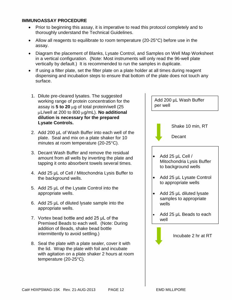
IMMUNOASSAY PROCEDURE • Prior to beginning this assay, it is imperative to read this protocol completely and to
thoroughly understand the Technical Guidelines.
• Allow all reagents to equilibrate to room temperature (20-25°C) before use in the assay.
• Diagram the placement of Blanks, Lysate Control, and Samples on Well Map Worksheet in a vertical configuration. (Note: Most instruments will only read the 96-well plate vertically by default.) It is recommended to run the samples in duplicate.
• If using a filter plate, set the filter plate on a plate holder at all times during reagent dispensing and incubation steps to ensure that bottom of the plate does not touch any surface.
1. Dilute pre-cleared lysates. The suggested
working range of protein concentration for the assay is 5 to 20 µg of total protein/well (25 µL/well at 200 to 800 µg/mL). No additional dilution is necessary for the prepared Lysate Controls.
2. Add 200 µL of Wash Buffer into each well of the plate. Seal and mix on a plate shaker for 10 minutes at room temperature (20-25°C).
3. Decant Wash Buffer and remove the residual amount from all wells by inverting the plate and tapping it onto absorbent towels several times.
4. Add 25 µL of Cell / Mitochondria Lysis Buffer to the background wells.
5. Add 25 µL of the Lysate Control into the appropriate wells.
6. Add 25 µL of diluted lysate sample into the appropriate wells.
7. Vortex bead bottle and add 25 μL of the Premixed Beads to each well. (Note: During addition of Beads, shake bead bottle intermittently to avoid settling.)
8. Seal the plate with a plate sealer, cover it with the lid. Wrap the plate with foil and incubate with agitation on a plate shaker 2 hours at room temperature (20-25°C).
Add 200 µL Wash Buffer per well
• Add 25 µL Cell /
Mitochondria Lysis Buffer to background wells
• Add 25 µL Lysate Control
to appropriate wells • Add 25 µL diluted lysate
samples to appropriate wells
• Add 25 µL Beads to each
well
Incubate 2 hr at RT
Shake 10 min, RT Decant

Cat# H0XPSMAG-15K Rev. 21-AUG-2013 PAGE 13 EMD MILLIPORE
9. Gently remove well contents and wash plate 3 times following instructions listed in the PLATE WASHING section.
10. Add 50 µL of Detection Antibodies into each well. (Note: Allow the Detection Antibodies to warm to room temperature prior to addition.)
11. Seal, cover with lid and incubate with agitation on a plate shaker for 1 hour at room temperature (20-25°C).
12. Gently remove well contents and wash plate 3 times following instructions listed in the PLATE WASHING section.
13. Add 50 µL Streptavidin-Phycoerythrin to each well.
14. Seal, cover with lid and incubate with agitation on a plate shaker for 30 minutes at room temperature (20-25°C).
15. Gently remove well contents and wash plate 3 times following instructions listed in the PLATE WASHING section.
16. Add 100 µL of Sheath Fluid (or Drive Fluid if using MAGPIX®) to all wells. Resuspend the beads on a plate shaker for 5 minutes.
17. Run plate on Luminex 200TM, HTS, FLEXMAP 3D® or MAGPIX® with xPONENT® software.
18. Save and analyze the Median Fluorescence Intensity (MFI) data. (Note: make sure all lysate samples in comparison are diluted by the same factor.)
Add 50 µL Detection Antibodies per well
Incubate 1 hr at RT
Add 50 µL Streptavidin-Phycoerythrin per well
Add 100 µL Sheath Fluid or Drive Fluid per well
Read on Luminex: 50 µL, 50 beads per bead set
Remove well contents and wash 3X with 200 µL Wash Buffer
Remove well contents and wash 3X with 200 µL Wash Buffer
Incubate 30 minutes at RT
Remove well contents and wash 3X with 200 µL Wash Buffer

Cat# H0XPSMAG-15K Rev. 21-AUG-2013 PAGE 14 EMD MILLIPORE
PLATE WASHING
1.) Solid Plate If using a solid plate, use either a handheld magnet or magnetic plate washer. A.) For handheld magnet, rest plate on magnet for 60 seconds to allow complete
settling of magnetic beads. Remove well contents by gently decanting the plate in an appropriate waste receptacle and gently tapping on absorbent pads to remove residual liquid. Wash plate with 200 µL of Wash Buffer by removing plate from magnet, adding Wash Buffer, shaking for 30 seconds, reattaching to magnet, letting beads settle for 60 seconds and removing well contents as previously described after each wash. Repeat wash steps as recommended in Assay Procedure.
B.) For magnetic plate washer, let plate “soak” on magnet for 60 seconds to allow complete settling of the magnetic beads. Remove well contents by aspiration. Wash plate with 200 µL/well of Wash Buffer, letting beads “soak” for 60 seconds and removing Wash Buffer by aspiration after each wash. Repeat wash steps as recommended in Assay Procedure.
2.) Filter Plate (EMD Millipore Cat. #MX-PLATE) If using a filter plate, use a vacuum filtration manifold to remove well contents. Wash plate with 200 µL/well of Wash Buffer, removing Wash Buffer by vacuum filtration after each wash. Repeat wash steps as recommended in the Assay Procedure.
INSTRUMENT SETTINGS Luminex 200™, HTS, FLEXMAP 3D® and MAGPIX® with xPONENT® software: These specifications are for the Luminex 200™, Luminex HTS, Luminex FLEXMAP 3D® and Luminex MAGPIX® with xPONENT® software. Luminex instruments with other software (e.g. MasterPlex®, STarStation, LiquiChip, Bio-Plex Manager™, LABScan™ 100) would need to follow instrument instructions for gate settings and additional specifications from the vendors for reading Luminex Magnetic Beads. For magnetic bead assays, the Luminex 200™ and HTS instruments must be calibrated with the xPONENT® 3.1 compatible Calibration Kit (EMD Millipore Catalog #40-275) and performance verified with the Performance Verification Kit (EMD Millipore Catalog #40-276). The Luminex FLEXMAP 3D® instrument must be calibrated with the FLEXMAP 3D® Calibrator Kit (EMD Millipore Catalog #40-028) and performance verified with the FLEXMAP 3D® Performance Verification Kit (EMD Millipore Catalog #40-029). The Luminex MAGPIX® instrument must be calibrated with the MAGPIX® Calibration Kit (EMD Millipore Catalog #40-049) and performance verified with the MAGPIX® Performance Verification Kit (EMD Millipore Catalog #40-050). NOTE: These assays cannot be performed on any instruments running Luminex IS 2.3
or Luminex 1.7 software. NOTE: When setting up for a magnetic bead assay using the xPONENT® software, you
must select [MagPlex] as the Bead Type in the Acquisition settings. The default setting is [MicroPlex] for polystyrene beads.
Cat# H0XPSMAG-15K Rev. 21-AUG-2013 PAGE 15 EMD MILLIPORE
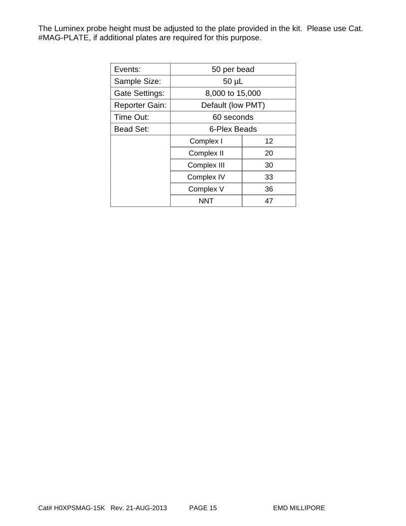
The Luminex probe height must be adjusted to the plate provided in the kit. Please use Cat. #MAG-PLATE, if additional plates are required for this purpose.
Events: 50 per bead Sample Size: 50 µL Gate Settings: 8,000 to 15,000 Reporter Gain: Default (low PMT) Time Out: 60 seconds Bead Set: 6-Plex Beads
Complex I 12 Complex II 20 Complex III 30 Complex IV 33
Complex V 36 NNT 47

Cat# H0XPSMAG-15K Rev. 21-AUG-2013 PAGE 16 EMD MILLIPORE
ASSAY CHARACTERISTICS Representative Data
Human Oxidative Phosphorylation (OXPHOS) Magnetic Bead Panel serial dilution curve. The human HepG2 hepatocellular carcinoma cell lysate was prepared according to the procedures described in the protocol and serially diluted (2-fold) with Cell / Mitochondria Lysis Buffer and analyzed with the Human Oxidative Phosphorylation (OXPHOS) Magnetic Bead Panel according to the assay protocol. The Median Fluorescence Intensity (MFI) was measured with the Luminex system.
Multiplexed analysis of different human cancer cell lines and heart tissue extract with Human Oxidative Phosphorylation (OXPHOS) Magnetic Bead Panel. Lysates from different human cancer cell lines (HepG2, HL-60, A431, HeLa, HEK293) and heart tissue were prepared according to the procedures described in the protocol. 6000 ng of cell line lysates, 300 ng of heart tissue extract and the Lysate Control (#47-239) were analyzed with the Human Oxidative Phosphorylation (OXPHOS) Magnetic Bead Panel according to the assay protocol. The Median Fluorescence Intensity (MFI) was measured with the Luminex system.

Cat# H0XPSMAG-15K Rev. 21-AUG-2013 PAGE 17 EMD MILLIPORE
ASSAY CHARACTERISTICS (continued)
Human OXPHOS changes upon drug induced mitochondrial toxicity. Human HepG2 hepatocellular carcinoma cells were cultured to 95% confluency then treated with either an antibiotic (20 µM of chloramphenicol) or an antiviral drug (20 µM of ddC, 2'-3'-dideoxycytidine) for 6 days to induce mitochondrial toxicity. DMSO mock-treated cells were used as the control. During the treatments, the cells were split in conditioned media if necessary. At the end of the treatments, the cell lysates were prepared according to the procedures described in the protocol. 10 µg of the cell lysates were analyzed with the Human Oxidative Phosphorylation (OXPHOS) Magnetic Bead Panel according to the assay protocol. Drug-induced mitochondrial toxicity was evaluated by normalizing the signal from each analyte to the mock-treated cells as a percentage. The results indicate that chloramphenicol and ddC clearly induced mitochondrial toxicity as demonstrated by the significant reduction of Complex I and IV while Complex II, V and NNT remained relatively unchanged.

Cat# H0XPSMAG-15K Rev. 21-AUG-2013 PAGE 18 EMD MILLIPORE
Specificity/Cross-Reactivity Each capture bead within the panel was tested for cross-reactivity to the other analytes with mass spectrometry. There was no or negligible cross-reactivity between the antibodies for an analyte and any of the other analytes within this panel. Precision Intra-assay precision is generated from the mean of the %CVs from 8 reportable results across two different concentrations of analytes in a single assay. Inter-assay precision is generated from the mean of the %CVs from 48 reportable results across two different concentrations of analytes from 6 different assays.
Analyte Intra-Assay CV Inter-Assay CV Complex I < 10.0% < 15.0% Complex II < 10.0% < 15.0%
Complex III < 10.0% < 15.0%
Complex IV < 10.0% < 15.0%
Complex V < 10.0% < 15.0%
NNT < 10.0% < 15.0%

Cat# H0XPSMAG-15K Rev. 21-AUG-2013 PAGE 19 EMD MILLIPORE
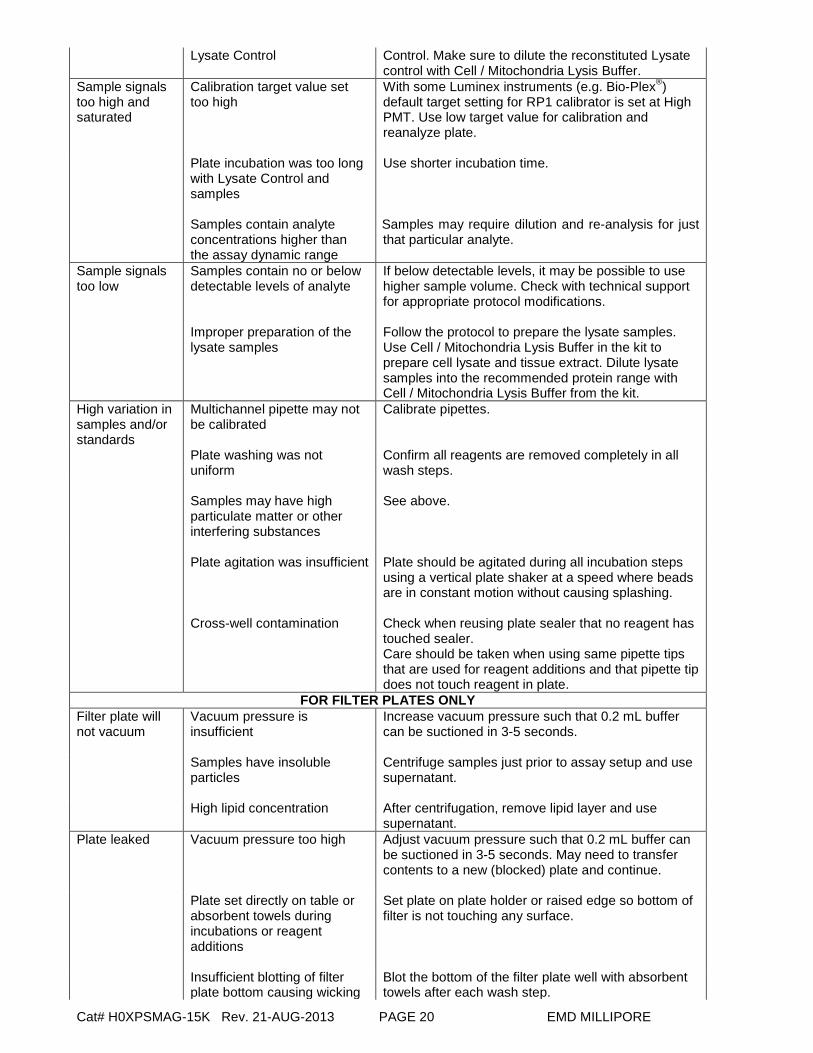
TROUBLESHOOTING GUIDE Problem Probable Cause Solution Insufficient bead count
Plate washer aspiration height set too low
Adjust aspiration height according to manufacturers’ instructions.
Bead mix prepared inappropriately
Sonicate bead vials and vortex just prior to adding to bead mix bottle according to protocol. Agitate bead mix intermittently in reservoir while pipetting this into the plate.
Samples cause interference due to particulate matter or viscosity
See above. Also sample probe may need to be cleaned with alcohol flush, back flush and washes; or if needed probe should be removed and sonicated.
Probe height not adjusted correctly
When reading the assay on Luminex 200™, adjust probe height to the kit solid plate or to the recommended Millipore filter plates using 3 alignment discs. When reading the assay on MAGPIX®, adjust probe height to the kit solid plate or to the recommended Millipore filter plates using 2 alignment discs. When reading the assay on FLEXMAP 3D®, adjust probe height to the kit solid plate using 1 alignment disc.
For FLEXMAP 3D® when using the solid plate in the kit, the final suspension should be in 150ul and 75ul should be aspirated.
Background is too high
Background wells were contaminated
Avoid cross-well contamination by using sealer appropriately and pipetting with multichannel pipettes without touching reagent in plate.
Insufficient washes Increase number of washes. Beads not in region or gate
Luminex instrument not calibrated correctly or recently
Calibrate Luminex instrument based on manufacturer’s instructions, at least once a week or if temperature has changed by >3oC.
Gate settings not adjusted correctly
Some Luminex instruments (e.g. Bio-Plex®) require different gate settings than those described in the kKit protocol. Use instrument default settings.
Wrong bead regions in protocol template
Check kit protocol for correct bead regions or analyte selection.
Incorrect sample type used Samples containing organic solvents or if highly viscous should be diluted or dialyzed as required.
Instrument not washed or primed
Prime the Luminex instrument 4 times to rid of air bubbles, wash 4 times with sheath fluid or water if there is any remnant alcohol or sanitizing liquid.
Beads were exposed to light Keep plate and bead mix covered with dark lid or aluminum foil during all incubation steps.
Signal for whole plate is same as background
Incorrect or no Detection Antibody was added
Add appropriate Detection Antibody and continue.
Streptavidin-Phycoerythrin was not added
Add Streptavidin-Phycoerythrin according to protocol.
Low signal for positive Lysate Control
Incubations done at inappropriate temperatures, timings or agitation Improper preparation of the
Assay conditions need to be checked. Follow the protocol to prepare the positive Lysate
Cat# H0XPSMAG-15K Rev. 21-AUG-2013 PAGE 20 EMD MILLIPORE
Lysate Control Control. Make sure to dilute the reconstituted Lysate control with Cell / Mitochondria Lysis Buffer.
Sample signals too high and saturated
Calibration target value set too high
With some Luminex instruments (e.g. Bio-Plex®) default target setting for RP1 calibrator is set at High PMT. Use low target value for calibration and reanalyze plate.
Plate incubation was too long with Lysate Control and samples Samples contain analyte concentrations higher than the assay dynamic range
Use shorter incubation time.
Samples may require dilution and re-analysis for just that particular analyte.
Sample signals too low
Samples contain no or below detectable levels of analyte Improper preparation of the lysate samples
If below detectable levels, it may be possible to use higher sample volume. Check with technical support for appropriate protocol modifications. Follow the protocol to prepare the lysate samples. Use Cell / Mitochondria Lysis Buffer in the kit to prepare cell lysate and tissue extract. Dilute lysate samples into the recommended protein range with Cell / Mitochondria Lysis Buffer from the kit.
High variation in samples and/or standards
Multichannel pipette may not be calibrated
Calibrate pipettes.
Plate washing was not uniform
Confirm all reagents are removed completely in all wash steps.
Samples may have high particulate matter or other interfering substances
See above.
Plate agitation was insufficient
Plate should be agitated during all incubation steps using a vertical plate shaker at a speed where beads are in constant motion without causing splashing.
Cross-well contamination
Check when reusing plate sealer that no reagent has touched sealer.
Care should be taken when using same pipette tips that are used for reagent additions and that pipette tip does not touch reagent in plate.
FOR FILTER PLATES ONLY Filter plate will not vacuum
Vacuum pressure is insufficient
Increase vacuum pressure such that 0.2 mL buffer can be suctioned in 3-5 seconds.
Samples have insoluble particles
Centrifuge samples just prior to assay setup and use supernatant.
High lipid concentration After centrifugation, remove lipid layer and use supernatant.
Plate leaked Vacuum pressure too high Adjust vacuum pressure such that 0.2 mL buffer can be suctioned in 3-5 seconds. May need to transfer contents to a new (blocked) plate and continue.
Plate set directly on table or absorbent towels during incubations or reagent additions
Set plate on plate holder or raised edge so bottom of filter is not touching any surface.
Insufficient blotting of filter plate bottom causing wicking
Blot the bottom of the filter plate well with absorbent towels after each wash step.

Cat# H0XPSMAG-15K Rev. 21-AUG-2013 PAGE 21 EMD MILLIPORE
Pipette touching plate filter
during additions
Pipette to the side of plate.
Probe height not adjusted correctly
Adjust probe to 3 alignment discs in well H6.
Sample too viscous May need to dilute sample.
REPLACEMENT REAGENTS Catalog # HepG2 Cell Lysate: Unstimulated (3) 47-239 Human OXPHOS Panel Pre-mixed Magnetic Beads H0XPSPMX6-MAG Human OXPHOS Panel Detection Antibodies H0XPS-1016 Cell / Mitochondria Lysis Buffer 43-042 Streptavidin-Phycoerythrin MC-SAPE4 Set of two 96-Well plates with Sealers MAG-PLATE 10X Wash Buffer L-WB

Cat# H0XPSMAG-15K Rev. 21-AUG-2013 PAGE 22 EMD MILLIPORE
ORDERING INFORMATION To place an order:
To assure the clarity of your custom kit order, please FAX the following information to our customer service department:
Include: • Your name, telephone and/or fax number • Customer account number • Shipping and billing address • Purchase order number • Catalog number and description of product • Quantity of kits
FAX: (636) 441-8050
Toll-Free US: (866) 441-8400 (636) 441-8400
Mail Orders: EMD Millipore Corporation 6 Research Park Drive St. Charles, Missouri 63304 U.S.A.
For International Customers:
To best serve our international customers in placing an order or obtaining additional information about MILLIPLEX® MAP products, please contact your multiplex specialist or sales representative or email our European Customer Service at [email protected].
Conditions of Sale
For Research Use Only. Not for Use in Diagnostic Procedures.
Material Safety Data Sheets (MSDS) Material Safety Data Sheets for EMD Millipore products may be ordered by fax or phone or through our website at www.emdmillipore.com/techlibrary/index.do.
Technical Services
http://www.emdmillipore.com/techservices To contact by phone For North America: Toll-Free US: 1-(800) 221-1975 or 1-(781) 533-8045 Outside North America, contact your local office http://www.emdmillipore.com/offices

Cat# H0XPSMAG-15K Rev. 21-AUG-2013 EMD MILLIPORE 23
WELL MAP
1 2 3 4 5 6 7 8 9 10 11 12
A Background Sample
3
B Background Sample
3
C Lysate
Control Etc.
D Lysate
Control
E Sample
1
F Sample
1
G Sample
2
H Sample
2