human tyrosine kinasep72syk associates src-family p53 ... · pdf fileptk, protein-tyrosine...
TRANSCRIPT

Proc. Natl. Acad. Sci. USAVol. 92, pp. 359-363, January 1995Immunology
Human spleen tyrosine kinase p72Syk associates with theSrc-family kinase p53/56Lyn and a 120-kDa phosphoprotein
(B-cell activation/surface IgM/protein-tyrosine kinase/phospholipase C pathway)
SVETLANA P. SIDORENKO*, CHE-LEUNG LAW, KAREN A. CHANDRAN, AND EDWARD A. CLARKtDepartment of Microbiology, University of Washington Medical Center, SC-42, Seattle, WA 98195
Communicated by Edwin G. Krebs, University of Washington, Seattle, WA, September 15, 1994
ABSTRACT The 72-kDa spleen tyrosine kinase (Syk) andSrc-family kinase p53/56Lyn (Lyn) contribute to signalingvia the B-cell antigen receptor complex. Here we show that Sykand Lyn from human B lymphocytes can interact directly. Sykand Lyn coimmunoprecipitated from mature and activatedB-cell lines, and gel-purified Syk and Lyn reassociated in vitro,demonstrating their direct interaction. This Syk-Lyn inter-action may be dependent on the stage of B-cell differentiation,since Syk-Lyn associations were not detected in pre-B andmyeloma cell lines and Syk from an immature B-cell line didnot reassociate with Lyn in vitro. Serine/threonine kinaseactivity was also associated with Syk Crosslinking of cellsurface IgM led to rapid activation of both tyrosine andserine/threonine protein kinase activities that resulted inphosphorylation in vitro of proteins coprecipitating withSyk-in particular, a serine/threonine phosphorylated pro-tein 120 kDa in size (ppl20). Several phosphoproteins, in-cluding one of 72 kDa and one of 120 kDa, coprecipitated withphospholipase C-,yl (PLCy1). Sequential immunoprecipita-tion identified the 72-kDa protein associated with PLCyl asSyk. The 120-kDa serine/threonine phosphorylated proteinthat coprecipitated with PLCy1 resembled the Syk-associatedppl20 by several criteria. Thus, pp120 may serve as a linkbetween Syk and PLC'yl, coupling the B-cell antigen receptorto the phosphatidylinositol pathway.
Two families of protein-tyrosine kinases (PTKs) interact withthe B-cell antigen receptor (BCR) and contribute to signaling(1). Src-family PTKs (Blk, Lyn, Fyn) may physically interactwith the components of the BCR complex (2) and the activitiesof these enzymes increase following receptor activation (3).The 72-kDa spleen tyrosine kinase (Syk) also associates withthe BCR (4-6). Unlike the Src-family PTKs, Syk has noN-terminal myristoylation site, but it possesses two Src-homology 2 (SH2) domains potentially capable of interactingwith tyrosine phosphorylated proteins (6, 7). Crosslinking theBCR results in both tyrosine phosphorylation of Syk andactivation of its enzyme activity (6, 8, 9). How Syk interactswith the BCR complex and the sequence of events aftersignaling are unknown. Nor is it clear which proteins canassociate and interact directly with Syk in B cells. One possi-bility is that Syk interacts directly with members of the Srcfamily of PTKs. In a mast cell line both Syk and Lyn associatewith the ,B subunit of the type I receptor for FcE (FcERI) (10),and after crosslinking of the BCR on B-cell lines, phosphor-ylated proteins 66-72 kDa in size coprecipitate with p53/56Lyn (Lyn) (11). However, it is unclear whether these kinasesinteract directly or whether the association is mediated viacomponents of Fc_RI or the BCR.A number of important effector molecules are tyrosine
phosphorylated and/or activated after BCR ligation, includingphosphatidylinositol 3-kinase (12, 13), Ras-GTPase-activating
protein (Ras-GAP) (14), and phospholipase C--y (PLCy) (15).An essential role for Syk in the activation of the PLCy pathwayin B cells has recently been demonstrated. Clustering ofchimeric molecules consisting of a CD16 extracellular domainand Syk results in tyrosine phosphorylation of PLCyl andtriggers mobilization of intracellular Ca2+ (16). Furthermore,crosslinking the BCR on a Syk-negative cell line does notactivate the PLC-y-dependent release of intracellular Ca2+(17). On the other hand, Src-family PTKs associated with theBCR, particularly Lyn, apparently can modulate enzymaticactivity of Syk and Syk-dependent activation of PLC-y (18).
In this study, we show that Syk from human B lymphocytesinteracts directly with Lyn. We also found that Syk associateswith a 120-kDa serine/threonine phosphorylated protein,ppl20, and coprecipitates with PLCyl. This Syk-associatedppl20 molecule may be a downstream component of Syk-dependent signal transduction pathways.
MATERIALS AND METHODSCells. The human cell lines B104 (provided by Mitsufumi
Mayumi, Kyoto University Medical School, Japan) (19), Reh,Nalm-6, Daudi, T5-1, DB, CESS, and RPMI-8226 and densetonsillar B cells were maintained and prepared as described(20).
Reagents. Rabbit anti-Syk serum was generated to a peptide,CRADENYYKAQTHG, derived from the Syk kinase domain(6). Rabbit antisera against Lyn, Fyn, or Lck were gifts fromJ. Bolen (Bristol-Myers Squibb Research Institute, Princeton,NJ), anti-p125FAK was provided by S. Kanner (Bristol-MeyersSquibb Research Institute, Seattle, WA), and rabbit antisera toPLC,yl was a gift from G. Carpenter (Vanderbilt University,Nashville, TN). Antibodies directed against Lyn, Blk, Csk,Ras-GAP (Santa Cruz Biotechnology, Santa Cruz, CA), Shc,Jakl, Jak2, p120, and phosphotyrosine (4G10) (Upstate Bio-technology) were used for immunoprecipitation and Westernblotting. F(ab')2 fragments of goat anti-human IgM and con-trol anti-mouse IgM antibodies (Jackson ImmunoResearch)were used for B-cell stimulation.
Biochemical Methods. Immunoprecipitation, SDS/PAGE,in vitro kinase assay, and phospho amino acid analysis wereperformed as described (6, 21). Sequential immunoprecipita-tion was performed after elution of phosphoproteins fromexcised bands in 50 mM ammonium bicarbonate/0.1% SDS(22). Western blotting with anti-Syk and anti-phosphotyrosineantibodies was performed as described (6).
Abbreviations: BCR, B-cell antigen receptor; GAP, GTPase-activating protein; NRS, normal rabbit serum; PLC, phospholipase C;PTK, protein-tyrosine kinase; SH2, Src homology 2; sIg, surfaceimmunoglobulin.*On leave from the Kavetsky Institute of Experimental Pathology,Oncology and Radiobiology, Ukrainian Academy of Sciences, Kiev,Ukraine.tTo whom reprint requests should be addressed.
359
The publication costs of this article were defrayed in part by page chargepayment. This artic!e must therefore be hereby marked "advertisement" inaccordance with 18 U.S.C. §1734 solely to indicate this fact.

360 Immunology: Sidorenko et at
RESULTSHuman Syk Is Associated with Phosphorylated Proteins
53/56 kDa and 120 kDa in Size. B-cell lines that representdifferent stages of maturation and tonsillar B cells were usedto study the phosphorylation pattern of Syk-associated pro-teins (Table 1; Fig. 1). Three main sets of phosphoproteins of53/56, 72, and 120 kDa were labeled in Syk immunoprecipi-tates after in vitro kinase assays. Sequential immunoprecipi-tation and Western blotting with anti-Syk serum revealed thatp72 corresponded to Syk (data not shown). In the pre-B-celllines Nalm-6 and Reh, only p72Syk could be phosphorylatedin vitro (Fig. 1A, lane 2). Phosphorylation of both p53/56 andppl20 was detected in Syk immunoprecipitates from Burkittlymphoma, Epstein-Barr virus-transformed B-lymphoblastoidcell lines, and tonsillar B cells. In the myeloma cell lineRPMI-8226, only ppi20 was coprecipitated with Syk (Fig. 1A,lane 11). Longer exposure of gels did not reveal phosphory-lation of the p53/56 doublet (data not shown). These dataimply that phosphorylation or association of these proteinswith Syk depends on the stage of B-cell differentiation.
In the CESS, Daudi, and B104 cell lines, Syk-transfectedCOS cells, and also tonsillar B cells, Syk was phosphorylatedon tyrosine, serine, and threonine residues when coprecipi-tated with . sociated proteins (Fig. 1C and data not shown).However, the Syk immunoprecipitated from the CESS cell lineand washed prior to kinase assay with high-salt buffers and0.1% SDS was phosphorylated only on tyrosine (6). p53/56 inSyk immunoprecipitates was also phosphorylated on all threeamino acids, but ppl20 showed phosphorylation only on serineand threonine (Fig. 1C). Although p53/56 in Syk immuno-precipitates was phosphorylated on serine, threonine, andtyrosine, the same band in Lyn immunoprecipitates was phos-phorylated exclusively on tyrosine (Fig. 1C). These datasuggest that Syk PTK can be a substrate for serine/threoninekinase(s) coprecipitated with it.Syk Can Associate Directly with Lyn. The p53/56 associated
with Syk is in the size range of Src-family PTKs (Fig. 2A). Theheavily labeled p53/56 doublet coprecipitated with Syk inCESS cells was eluted from gels after SDS/PAGE and sub-jected to reprecipitation with antibodies to various Src-familyPTKs (Fyn, Lyn, Blk and Lck), the Csk PTK, or the Shcprotein. Only anti-Lyn specifically reprecipitated the p53/56doublet (Fig. 2B). Anti-Lyn immunoprecipitate from theCESS cell line had a faintly labeled band of 72 kDa in additionto the heavily phosphorylated doublet p53/56Lyn (Fig. 2A,lane 3). Sequential immunoprecipitation and Western blotanalysis with anti-Syk serum confirmed that this 72-kDaphosphoprotein was in fact Syk (Fig. 2 B and C). Treatment ofexcised bands with Staphylococcus aureus V8 protease showedthat Syk-associated p53/56 in cell lines CESS, B104, DB, andT5-1 were identical (data not shown).
Table 1. Phosphorylation of proteins coprecipitated with Syk inlysates of human B-cell lines
Relative phosphorylation
Cell line Cell origin p53/56 p72 pp120Reh Pre-B ALL - +Nalm-6 Pre-B ALL - +Daudi Burkitt lymphoma + ++ +B104 B-cell lymphoma + + + +++T5-1 B lymphoblastoid + + + + +DB B lymphoblastoid + + + + +CESS B lymphoblastoid + + + + + + +RPMI-8226 Myeloma - + + +
Syk-associated proteins were immunoprecipitated with anti-Sykserum in digitonin or Nonidet P-40 lysis buffers, labeled by in vitrokinase assay, and resolved by SDS/PAGE under reducing conditions.ALL, acute lymphoblastic leukemia.
Proc. Nati Acad Sci USA 92 (1995)
A
ppl20-.
p72 -.
p53/56-
c
B TonsillarReh B104 CESS RPMI-8226 B cells
Cl)o* Cl) JCO C U)J CO
Co.Zto_enn Z) nZ mev coea Z co Z eM co Z X
00
..... -20sss 9 pl2971
- p53/572.i!
,-,',,s..'.'.'-''''.' 't' p53'56-43 -
13 14
Immunoprecipitatesanti-Syk anti-Lyn
+ T s
ppl 20 b
p7 T -S- T
p72
I Y YT
p53/56 p53/56
V11 VII k
- pH 1.9 +
FIG. 1. (A and B) Phosphoproteins 53/56, 72, and 120 kDa in sizewere coprecipitated from B-lineage cells with anti-Syk serum. Syk andLynwere immunoprecipitated from digitonin lysates of B-cell lines (A)and tonsillar B cells (B). Normal rabbit serum (NRS) was used asnegative control. Equivalent amounts of protein were used for allimmunoprecipitations. After in vitro kinase assays, immunoprecipi-tated phosphoproteins were resolved by SDS/PAGE under reducingconditions. (C) 32P-labeled p53/56, p72, and ppl20 proteins, copre-cipitated from the CESS cell line with anti-Syk or anti-Lyn sera, wereanalyzed for phospho amino acid content. Positions of standard aminoacids are shown by dotted lines (S, phosphoserine; T, phosphothreo-nine; Y, phosphotyrosine).
Coprecipitation of Syk and Lyn PTKs suggested that thesetwo kinases interact directly. It was unlikely that the ppl20which coprecipitated with Syk mediates this interaction, be-cause pp120 did not coprecipitate with Lyn (Figs. 1A and 2A);but we could not rule out the presence of other intermediatemolecules. To test the hypothesis that Syk and Lyn interactdirectly, we examined whether gel-purified Lyn and Syk couldreassociate in vitro. After in vitro kinase assays, Syk and Lynphosphoproteins were resolved by SDS/PAGE, eluted fromexcised bands, mixed back together, and subjected to second-ary immunoprecipitation with anti-Lyn serum. Fig. 3 (lane 3)demonstrates that phosphorylated Syk and Lyn proteins, iso-lated from -the CESS cell line, are able to reassociate inNonidet P-40 buffer and be reprecipitated with anti-Lynantibodies.
Since p53/56Lyn was not detected in Syk immunoprecipi-tates from the pre-B-cell line Nalm-6, we tested whether Sykand Lyn phosphoproteins could reassociate in vitro. Syk andLyn isolated from Nalm-6 and mixed in vitro did not copre-cipitate (Fig. 3, lane 13). However, Lyn from Nalm-6 didreassociate with Syk from the CESS cell line (Fig. 3, lane 6),but Syk from Nalm-6 did not reassociate with Lyn from CESS(lane 9). Longer exposure of the gels did not reveal Syk in lanes9 and 13 (see lane 10). These data suggest that the Syk-Lyn

Proc. Natl. Acad Sci USA 92 (1995) 361
200-
-be L .a
CD .j LL.
co .2. ._. ._ ._
z 0 X 0
1° ant-Syk anti-Lyn0
c
1-0 C/) CoUn .2.
az a! E
I- c
u) .2 .
ac cZ ci co3
- 200
97- -97
Sp5356**4p72_j4 p72_ .
43-"<S/6* i
1 2 3 4 5
A6 7 8 9 10 11
B
- 43
12 1314
C
FIG. 2. Association of Syk with Lyn in human B cells. (A) CESScells were lysed in digitonin, and immunoprecipitations from equiv-alent amounts of protein were performed with NRS or anti-Syk,anti-Lyn, anti-Fyn, or anti-Lck serum followed by protein A-Sepharosebeads. Immunoprecipitates were submitted to an in vitro kinase assayin the presence of [a-32P]ATP and resolved by SDS/PAGE underreducing conditions. p53/56 coprecipitated with anti-Syk serum washeavily phosphorylated, and p72 was phosphorylated in the anti-Lynimmunoprecipitate. (B) After primary immunoprecipitations withanti-Syk and anti-Lyn sera (10) and in vitro kinase assay as describedin A, bands containing p53/56 and p72 were excised from dried gelsand proteins were eluted in ammonium bicarbonate buffer with 0.1%SDS, concentrated with Microcon filters (Amicon), and diluted withlysis buffer prior to secondary immunoprecipitations (20). p53/56from the primary anti-Syk immunoprecipitate was specifically repre-cipitated by anti-Lyn serum, and p72 from the primary anti-Lynimmunoprecipitate was specifically reprecipitated by anti-Syk serum.(C) Syk and Lyn immunoprecipitates from CESS cells were resolvedby SDS/PAGE and Western blotted with anti-Syk serum.
in these cell lines (Fig. 1A, lanes 3 and 12). Our data do notformally rule out the possibility that in these cell lines someLyn can coprecipitate with Syk but not be phosphorylated invitro. However, Lyn immunoprecipitates from Nalm-6 and Rehdid not contain Syk detectable by Western blotting (data notshown). The reciprocal experiment of Western blotting of Sykimmunoprecipitates with anti-Lyn serum was not informative,because Lyn and heavy chains of immunoglobulin from theanti-Syk serum had the same electrophoretic mobility inSDS/PAGE.
Ligation of Cell Surface IgM (sIgM) Augments KinaseActivities Associated with Syk A substantial increase in in vitrophosphorylation of both ppl20 and p72 in Syk immunopre-cipitates was detected within 15 sec following crosslinking ofsIgM on Daudi cells or tonsillar B cells (Fig. 4A). Phospho-amino acid analysis revealed that after BCR ligation, the invitro phosphorylation patterns of ppl20 and Syk remained thesame as before ligation; i.e., ppl20 was phosphorylated only onserine and threonine, and Syk phosphorylation was enhancedon tyrosine as well as serine and threonine residues (data notshown). Western blotting with anti-phosphotyrosine antibod-
A -_
Stimulation o c o C
>.c 4e. >_U) U)C' U) U)
m'
U) .. IUC ..L U) .2 U)C, .2.Precipitation cc cc cc cc
z cu z ct z Ca z CZ
200-
116 - -
97-
66i
6-
:?: _ppl 20
40 __- p72
association is direct and specific and might depend on the stageof B-cell differentiation. The element dictating this associationappears to be Syk.
After in vitro kinase assays, we did not detect phosphory-lated Lyn in Syk immunoprecipitates from cell lines Reh,Nalm-6, and RPMI-8226. At the same time, Lyn was expressed
45-
NP-40 Digitonin
B
10 Syk/CESS Syk/CESS Syk/Nalm-6 Syk/Nalm-6Lyn/CESS Lyn/Nalm-6 Lyn/CESS Lyn/Nalm-6
2° 't coX
C v C
+ ) + ) + cn*~~~C a:^ n
cOZo ZcOZw z co CM u)z co
-97
pp72 Syk -a
pp53/56 Lyn -. i 0w:.....~
-68
1 2 3 4 5 6 7 8 9 10 11 1213
FIG. 3. Reassociation of Syk and Lyn in vitro. Syk and Lynphosphoproteins were immunoprecipitated from CESS and Nalm-6cell lines (1°), labeled and excised from the gels as described in Fig. 2,mixed, and reprecipitated with either NRS or anti-Lyn serum (20). Sykfrom the CESS cell line was coprecipitated with Lyn from either CESSor Nalm-6 cells; however, Syk from Nalm-6 was not. Longer exposureof the gel did not reveal p72Syk in lanes 9 (lane 10) and 13 (data notshown).
10 anti-Syk anti-p120 anti-GAP
20~
D2~C ° CL CU °C L .2 - a EL°5 cm CR a-NO C\C CD C% CM2cm)- cmJ<0 Cm1<
Q°C L -Q° C _° C2 co co OL 2 co 0 cL2 mcu
-200
pp120 _--97
-68
FIG. 4. (A) Crosslinking of the IgM BCR complex augments kinaseactivities associated with Syk. Daudi cells were stimulated by F(ab')2fragments of goat anti-human IgM antibodies (anti-,u) for 1 min. Goatanti-mouse IgM F(ab')2 was used as a control serum. Anti-Sykimmunoprecipitates obtained in the presence of Nonidet P-40 (NP-40)or digitonin were labeled and resolved as in Fig. 2A. (B) Sequentialimmunoprecipitation of ppl20 with anti-GAP and anti-p120-Src-family-substrate antibodies. After primary immunoprecipitation (10)with anti-Syk, anti-GAP, or anti-pl20-Src antibodies and in vitrokinase assays, the 120-kDa phosphoprotein was extracted as describedin Fig. 2A and subjected to secondary immunoprecipitation (20). IgGlpurified from MOPC21 ascites served as negative control. Syk-associated ppl20 did not reprecipitate with anti-GAP or anti-p120-Srcantibodies.
29-
Immunology: Sidorenko et at
362 Immunology: Sidorenko et at
ies also did not detect tyrosine phosphorylation of ppl20before or after IgM crosslinking (data not shown). These datasuggest that both PTK and Ser/Thr kinase activities copre-cipitated with Syk were augmented upon anti-IgM crosslink-ing.To identify the ppl20 associated with Syk, we performed
sequential immunoprecipitation with antibodies to knownphosphoproteins of similar size. This Syk-associated proteincould not be reprecipitated with antibodies to Ras-GAP(p120), the p120 substrate for Src-family kinases, pl25Fak, orJakl kinase (Fig. 4B and data not shown). Apparently, Syk-associated pp120 is a novel protein that potentially may beinvolved in signal transduction via the BCR.Syk Is Coprecipitated with PLCy1. Since Syk is involved in
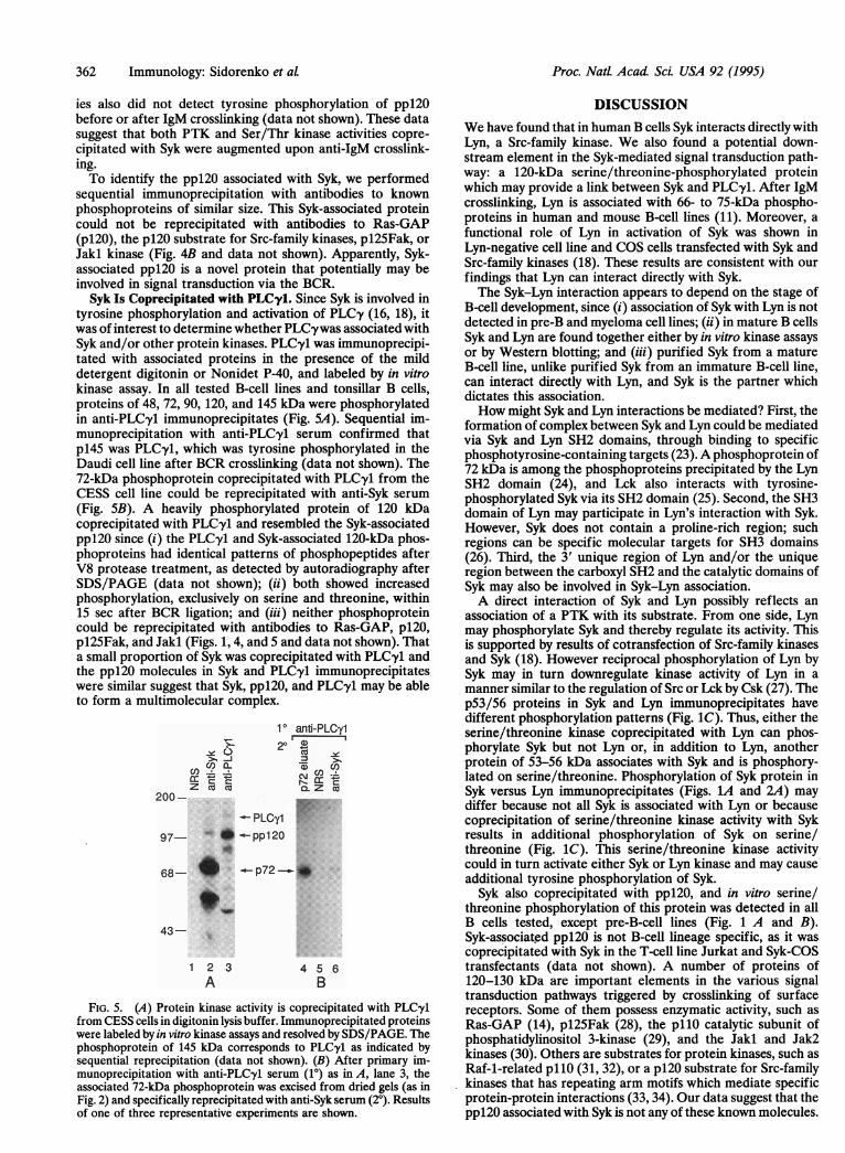
tyrosine phosphorylation and activation of PLCy (16, 18), itwas of interest to determine whether PLC'ywas associated withSyk and/or other protein kinases. PLCy1 was immunoprecipi-tated with associated proteins in the presence of the milddetergent digitonin or Nonidet P-40, and labeled by in vitrokinase assay. In all tested B-cell lines and tonsillar B cells,proteins of 48, 72, 90, 120, and 145 kDa were phosphorylatedin anti-PLCyl immunoprecipitates (Fig. 5A). Sequential im-munoprecipitation with anti-PLCyl serum confirmed thatp145 was PLCyl, which was tyrosine phosphorylated in theDaudi cell line after BCR crosslinking (data not shown). The72-kDa phosphoprotein coprecipitated with PLC-y1 from theCESS cell line could be reprecipitated with anti-Syk serum(Fig. SB). A heavily phosphorylated protein of 120 kDacoprecipitated with PLCy1 and resembled the Syk-associatedppl20 since (i) the PLC-yl and Syk-associated 120-kDa phos-phoproteins had identical patterns of phosphopeptides afterV8 protease treatment, as detected by autoradiography afterSDS/PAGE (data not shown); (ii) both showed increasedphosphorylation, exclusively on serine and threonine, within15 sec after BCR ligation; and (iii) neither phosphoproteincould be reprecipitated with antibodies to Ras-GAP, p120,pl25Fak, and Jakl (Figs. 1, 4, and 5 and data not shown). Thata small proportion of Syk was coprecipitated with PLCy1 andthe ppl20 molecules in Syk and PLCy1 immunoprecipitateswere similar suggest that Syk, ppl20, and PLCy1 may be ableto form a multimolecular complex.
1 anti-PLOyl
200- P F
97 *.-ppl20
68- * .-p72-_ *
_> _j
43-
12 3A
4 5 6B
FIG. 5. (A) Protein kinase activity is coprecipitated with PLCy1from CESS cells in digitonin lysis buffer. Immunoprecipitated proteinswere labeled by in vitro kinase assays and resolved by SDS/PAGE. Thephosphoprotein of 145 kDa corresponds to PLCyl as indicated bysequential reprecipitation (data not shown). (B) After primary im-munoprecipitation with anti-PLC-y1 serum (10) as in A, lane 3, theassociated 72-kDa phosphoprotein was excised from dried gels (as inFig. 2) and specifically reprecipitated with anti-Syk serum (20). Resultsof one of three representative experiments are shown.
DISCUSSIONWe have found that in human B cells Syk interacts directly withLyn, a Src-family kinase. We also found a potential down-stream element in the Syk-mediated signal transduction path-way: a 120-kDa serine/threonine-phosphorylated proteinwhich may provide a link between Syk and PLCyl. After IgMcrosslinking, Lyn is associated with 66- to 75-kDa phospho-proteins in human and mouse B-cell lines (11). Moreover, afunctional role of Lyn in activation of Syk was shown inLyn-negative cell line and COS cells transfected with Syk andSrc-family kinases (18). These results are consistent with ourfindings that Lyn can interact directly with Syk.The Syk-Lyn interaction appears to depend on the stage of
B-cell development, since (i) association of Syk with Lyn is notdetected in pre-B and myeloma cell lines; (ii) in mature B cellsSyk and Lyn are found together either by in vitro kinase assaysor by Western blotting; and (iii) purified Syk from a matureB-cell line, unlike purified Syk from an immature B-cell line,can interact directly with Lyn, and Syk is the partner whichdictates this association.How might Syk and Lyn interactions be mediated? First, the
formation of complex between Syk and Lyn could be mediatedvia Syk and Lyn SH2 domains, through binding to specificphosphotyrosine-containing targets (23).A phosphoprotein of72 kDa is among the phosphoproteins precipitated by the LynSH2 domain (24), and Lck also interacts with tyrosine-phosphorylated Syk via its SH2 domain (25). Second, the SH3domain of Lyn may participate in Lyn's interaction with Syk.However, Syk does not contain a proline-rich region; suchregions can be specific molecular targets for SH3 domains(26). Third, the 3' unique region of Lyn and/or the uniqueregion between the carboxyl SH2 and the catalytic domains ofSyk may also be involved in Syk-Lyn association.A direct interaction of Syk and Lyn possibly reflects an
association of a PTK with its substrate. From one side, Lynmay phosphorylate Syk and thereby regulate its activity. Thisis supported by results of cotransfection of Src-family kinasesand Syk (18). However reciprocal phosphorylation of Lyn bySyk may in turn downregulate kinase activity of Lyn in amanner similar to the regulation of Src or Lck by Csk (27). Thep53/56 proteins in Syk and Lyn immunoprecipitates havedifferent phosphorylation patterns (Fig. 1C). Thus, either theserine/threonine kinase coprecipitated with Lyn can phos-phorylate Syk but not Lyn or, in addition to Lyn, anotherprotein of 53-56 kDa associates with Syk and is phosphory-lated on serine/threonine. Phosphorylation of Syk protein inSyk versus Lyn immunoprecipitates (Figs. 1A and 2A) maydiffer because not all Syk is associated with Lyn or becausecoprecipitation of serine/threonine kinase activity with Sykresults in additional phosphorylation of Syk on serine/threonine (Fig. 1C). This serine/threonine kinase activitycould in turn activate either Syk or Lyn kinase and may causeadditional tyrosine phosphorylation of Syk.
Syk also coprecipitated with ppl20, and in vitro serine/threonine phosphorylation of this protein was detected in allB cells tested, except pre-B-cell lines (Fig. 1 A and B).Syk-associated ppl20 is not B-cell lineage specific, as it wascoprecipitated with Syk in the T-cell line Jurkat and Syk-COStransfectants (data not shown). A number of proteins of120-130 kDa are important elements in the various signaltransduction pathways triggered by crosslinking of surfacereceptors. Some of them possess enzymatic activity, such asRas-GAP (14), pl25Fak (28), the pllO catalytic subunit ofphosphatidylinositol 3-kinase (29), and the Jakl and Jak2kinases (30). Others are substrates for protein kinases, such asRaf-1-related pllO (31, 32), or a p120 substrate for Src-familykinases that has repeating arm motifs which mediate specificprotein-protein interactions (33, 34). Our data suggest that theppl20 associated with Syk is not any of these known molecules.
Proc. Natl. Acad Sci. USA 92 (1995)

Proc. Natl. Acad Sci USA 92 (1995) 363
Crosslinking the BCR results in rapid augmentation of PTKactivity associated with Syk (9) and also induces a serine/threonine kinase activity coprecipitated with Syk which leadsto increased serine/threonine phosphorylation ofboth Syk andppl20. Syk-associated ppl20 does not appear to be a substratefor Syk but might be a substrate for a coprecipitated serine/threonine kinase(s). Another possibility is that ppl20 itself isa serine/threonine kinase which can phosphorylate and reg-ulate Syk.Although a number of Src-family kinases, including Lyn, Fyn
and Lck, can phosphorylate purified PLCy in vitro (35) anddirectly interact with PLCy via SH2 domains (36), there is nodirect evidence that PLC'y isoforms are natural substrates forthese kinases in B lymphocytes. The fact that Syk can becoprecipitated with PLCyl (Fig. 5) is consistent with a modelthat Syk couples the BCR to the phosphatidylinositol pathwayand that PLCy may be a natural substrate for Syk. Thesimilarities between the ppi20 species that are coprecipitatedwith Syk and PLCOyl suggest that ppl20 may serve as a link tomediate interactions between Syk and PLCy. Serine phosphor-ylation ofPLCy has been shown to mediate negative regulationof its functional activity (37). One testable possibility is thatppl20 functions to influence the phosphorylation and activ-ity of PLC-y.The ppi20 protein is coprecipitated with Syk but not with
Lyn or IgM, and the p53/56 protein in Syk and Lyn immuno-precipitates has a different pattern of phosphorylation. Thissuggests there are at least two separate pools of Syk in humanB cells. The first pool is located near the cell membrane andthus could potentially interact with the components of BCRsuch as Iga, Ig,B, CD22, and Lyn and would be rapidlyphosphorylated and activated upon stimulation of BCR. Asecond cytoplasmic pool of Syk may be associated with ppl20and PLCy and might be activated and relocated to the cellmembrane after BCR engagement.
We thank Kate Elias for editorial assistance and Marge Dom-enowske for assistance in preparing figures. We thank Drs. JosephBolen, Graham Carpenter, and Steven Kanner for generous gifts ofantibodies. This work was supported by National Institutes of HealthGrants GM42508 and GM37905 (E.A.C.), National Institutes ofHealth Center Grant RR00166, and a special fellowship from theLeukemia Society of America (C.-L.L.).
1. Cambier, J. C., Pleiman, C. M. & Clark, M. R. (1994) Annu. Rev.Immunol. 12, 457-486.
2. Clark, M. R., Campbell, K. S., Kazlauskas, A., Johnson, S. A.,Hertz, M., Potter, T. A., Pleiman, C. & Cambier, J. C. (1992)Science 258, 123-126.
3. Burkhardt, A. L., Brunswick, M., Bolen, J. B. & Mond, J. J.(1991) Proc. Natl. Acad. Sci. USA 88, 7410-7414.
4. Hutchcroft, J. E., Harrison, M. L. & Geahlen, R. L. (1992) J.Bio. Chem. 267, 8613-8619.
5. Leprince, C., Draves, K. E., Geahlen, R. L., Ledbetter, J. A. &Clark, E. A. (1993) Proc. Natl. Acad. Sci. USA 90, 3236-3240.
6. Law, C.-L., Sidorenko, S. P., Chandran, K. A., Draves, K. E.,Chan, A. C., Weiss, A., Edelhoff, S., Disteche, C. M. & Clark, E.A. (1994) J. Bio. Chem. 269, 12310-12319.
7. Taniguchi, T., Kobayashi, T., Kondo, J., Takahashi, K., Naka-mura, H., Suzuki, J., Nagai, K., Yamada, T., Nakamura, S. &Yamamura, H. (1991) J. Bio. Chem. 266, 15790-15796.
8. Hutchcroft, J. E., Harrison, M. L. & Geahlen, R. L. (1991) J.Bio. Chem. 266, 14846-14849.
9. Yamada, T., Taniguchi, T., Yang, C., Yasue, S., Saito, H. &Yamamura, H. (1993) Eur. J. Biochem. 213, 455-459.
10. Hutchcroft, J. E., Geahlen, R. L., Deanin, G. G. & Oliver, J. M.(1992) Proc. Natl. Acad. Sci. USA 89, 9107-9111.
11. Yamamoto, T., Yamanashi, Y. & Toyoshima, K. (1993) Immunol.Rev. 132, 187-206.
12. Gold, M. R., Chan, V. W., Turck, C. W. & DeFranco, A. L.(1992) J. Immunol. 148, 2012-2022.
13. Pleiman, C. M., Hertz, W. M. & Cambier, J. C. (1994) Science263, 1609-1612.
14. Gold, M. R., Crowley, M. T., Martin, G. A., McCormick, F. &DeFranco, A. L. (1993) J. Immunol. 150, 377-386.
15. Carter, R. H., Park, D. J., Rhee, S. G. & Fearon, D. T. (1991)Proc. Natl. Acad. Sci. USA 88, 2745-2749.
16. Kolanus, W., Romeo, C. & Seed, B. (1993) Cell 74, 171-183.17. Takata, M., Sabe, H., Hata, A., Inazu, T., Homma, Y., Nukada,
T., Yamamura, H. & Kurosaki, T. (1994)EMBO J. 13,1341-1349.18. Kurosaki, T., Takata, M., Yamanashi, Y., Inazu, T., Tanigushi,
T., Yamamoto, T. & Yamamura, H. (1994) J. Exp. Med. 179,1725-1729.
19. Kim, K. M., Yoshimura, T., Watanabe, H., Ishigami, T., Nambu,M., Hata, D., Higaki, Y., Sasaki, M., Tsutsui, T., Mayumi, M. &Mikawaw, H. (1991) J. Immunol. 146, 819-825.
20. Clark, E. A. & Shu, G. (1987) J. Immunol. 138, 720-726.21. Leprince, C., Draves, K. E., Ledbetter, J. A., Torres, R. M. &
Clark, E. A. (1992) Eur. J. Immunol. 22, 2093-2099.22. Silva, d. A. J., Janssen, 0. & Rudd, C. E. (1993) J. Exp. Med. 178,
2107-2113.23. Zhou, S., Shoelson, S. E., Chaudhuri, M., Gish, G., Pawson, T.,
Haser, W. G., King, F., Roberts, T., Ratnofsky, S., Lechleider,R. J., Neel, B. G., Birge, R. B., Fajardo, J. E., Chou, M. M.,Hanafusa, H., Schaffhausen, B. & Cantley, L. C. (1993) Cell 72,767-778.
24. Malek, S. N. & Desiderio, S. (1993) J. Biol. Chem. 268, 22557-22565.
25. Couture, C., Baier, G., Altman, A. & Mustelin, T. (1994) Proc.Natl. Acad. Sci. USA 91, 5301-5305.
26. Yu, H., Chen, J. K., Feng, S., Dalgarno, D. C., Brauer, A. W. &Schreiber, S. L. (1994) Cell 76, 933-945.
27. Okada, M., Nada, S., Yamanashi, Y., Yamamoto, T. & Naka-gawa, H. (1991) J. Biol. Chem. 266, 24249-24252.
28. Schaller, M. D., Borgman, C. A., Cobb, B. S., Vines, R. R.,Reynolds, A. B. & Parsons, J. T. (1992) Proc. Natl. Acad. Sci.USA 89, 5192-5196.
29. Hiles, I. D., Otsu, M., Volinia, S., Fry, M. J., Gout, I., Dhand, R.,Panayotou, G., Ruiz, L. F., Thompson, A., Totty, N. F., Hsuan,J. J., Courtneidge, S. A., Parker, P. J. & Waterfield, M. D. (1992)Cell 70, 419-429.
30. Wilks, A. F., Harpur, A. G., Kurban, R. R., Ralph, S. J., Zurcher,G. & Ziemiecki, A. (1991) Mol. Cell. Biol. 11, 2057-2065.
31. Prasad, K. V. & Rudd, C. E. (1992) Mol. Cell. Biol. 12, 5260-5267.
32. Siegel, J. N., June, C. H., Yamada, H., Rapp, U. R. & Samelson,L. E. (1993) J. Immunol. 151, 4116-4127.
33. Reynolds, A. B., Herbert, L., Cleveland, J. L., Berg, S. T. & Gaut,J. R. (1992) Oncogene 7, 2439-2445.
34. Peifer, M., Berg, S. & Reynolds, A. B. (1994) Cell 76, 789-791.35. Liao, F., Shin, H. S. & Rhee, S. G. (1993) Biochem. Biophys. Res.
Commun. 191, 1028-1033.36. Pleiman, C. M., Clark, M. R., Gauen, L. K., Winitz, S., Cogge-
shall, K. M., Johnson, G. L., Shaw, A. S. & Cambier, J. C. (1993)Mol. Cell. Biol. 13, 5877-5887.
37. Berridge, M. J. (1993) Nature (London) 361, 315-325.
Immunology: Sidorenko et aL