hydrocephalus - neurosurgery · pdf filehydrocephalus is an abnormal accumulation of ......
TRANSCRIPT


Hydrocephalus is an abnormal accumulation of cerebrospinal fluid (CSF) within cavities called the ventricles inside the brain. The CSF is produced by the choroid plexus inside the ventricular system and its function is to cushion the brain and spinal cord tissue from injury and maintain a natural balance in the central nervous system. There are four ventricles inside the brain: two lateral ventricles, the third ventricle and the fourth ventricle. These ventricles are interconnected by narrow passageways. Normally, the CSF produced in the ventricles is reabsorbed in the bloodstream, however, when there is a blockage in flow, inadequate re absorption of CSF, or an over-production of CSF, there may be increased intra-cranial pressure (ICP) in the head, resulting in a life-long condition called hydrocephalus.
Intracranial pressure naturally varies throughout the day. Your ICP is highest in the morning before you get out of bed. This often results in high pressure headaches in the morning for untreated hydrocephalus or if there is a shunt malfunction. High pressure headache symptoms include a severe headache first thing in the morning (often slightly resolving throughout the day), vomiting, along with light and noise sensitivity.
If a shunt is draining too much CSF, it will cause low pressure headaches. Low pressure headaches most commonly occur closer to the end of the day. These headaches can be severe and are often described as pain behind the eyes. Some children will ask to lay down in an effort to naturally stop the over drainage of CSF from their ventricles.
hydrocephalus
diagram of the brain and the csF pathways
diagram of the ventricles and the passageways
What is a shunt?
Shunts help to stabilize intracranial pressure and reduce the risk of damage to the central nervous system (CNS). Shunts drain cerebrospinal fluid (CSF) away from the brain or spinal cord into another part of the body, where the CSF can be absorbed by the bloodstream.
A shunt system consists of three parts: Two silicone catheters and a one way valve. The catheter inside the ventricles is called the proximal catheter because it is closest to the ventricles. The other catheter which is placed in the peritoneal cavity or in the right atrium of the heart is called the distal catheter because it is farthest away from the ventricles. The proximal and the distal catheters are connected via a one-way valve. This valve allows CSF flow in a single direction only (away from the brain). It opens automatically when the pressure exceeds a certain limit and allows CSF to drain. The valve closes again when the intracranial pressure returns to the permitted level. Most shunt systems have a reservoir that allows for shunt taps to be performed in order to obtain CSF samples.
SHUNTS
What is a ventriculo-peritoneal (VP) shunt?
A VP shunt diverts CSF from the ventricles in the brain to the peritoneal cavity. The tip of the distal catheter rests around the digestive organs and not within them. The CSF that is drained to this area is reabsorbed by the bloodstream. In infants and children, the neurosurgeon will often place up to 4 ft of shunt tubing in this space to allow for the system to lengthen as the child grows.
What is a ventriculo-atrial (VA) shunt?
VA shunts divert CSF away from the ventricles in the brain via the internal jugular vein to the right atrium of the heart. The CSF enters the bloodstream directly. VA shunts are most often performed when the peritoneum no longer absorbs the CSF. Infants and children will need revisions because the neurosurgeon cannot place additional tubing to accommodate for the child’s growth.

ULTRASOUND
Ultrasound uses high frequency sound waves to outline the structures within the head. It can be used to diagnose congenital hydrocephalus while the baby is still in the mother’s womb. Ultrasound is most often used on infants as their soft spot is still open. Although the ultrasound allows the ventricle size to be measured accurately, it cannot image the surface of the brain.
SHUNT SERIES
Shunt series are 3 X-rays taken along the shunt tract. The purpose of running a shunt series is to check for shunt obstruction or disconnection. It is often done in conjunction with a Fast Head MRI or CT scan. The procedure is quick, painless and does not require sedation.
MRI OR MR SCANS
MRI uses radio signals and a very powerful magnet to scan inside the brain. It is a painless procedure and has no known side effects. There are two types of MRI scans: (1) Fast Head MRI – scan takes about 30 seconds and does not require sedation. (2) Full MRI – scan can take 30 – 60 minutes however gives more minute details than a Fast head MRI can. Most infants and children will require sedation to minimize movement that would cause blurring of the images.
CT OR CAT SCANS
CT scanning is a safe, reliable and painless procedure for diagnosing and assisting in the management of hydrocephalus. Patients must remain motionless for the duration of the test which takes only two to five minutes and therefore usually can be performed with no sedation. One advantage of a CT scan is that it shows catheter placement better than an MRI scan.
SHUNT TAP
Shunt taps are performed to withdraw small samples of CSF to look for any signs of infection. Shunts taps are performed by taking a very small needle and inserting it in the bulb reservoir of the shunt valve. The procedure usually takes about 5-10 minutes.
OTHER
Blood and urine tests may be performed as needed.
TesTs & scans
Measuring Head Circumference Accurately
Head circumference (HC) is a measurement of the circumference of the child’s head at its largest area. The tape measure should sit above the eyebrows and ears and around the back of the head. You should take the measurement twice to ensure
accuracy. Measurements should be taken in centimeters.The HC should be written on the back of the pink or blue “Head Circumference Chart” and then can be plotted on the graph on the same line as your child’s age. Babies that are more than 4 weeks premature should adjust their age up to the age of 2 years. Each child is unique and so are their measurements. The importance of the HC is that the child’s head growth is following along a natural and consistent growth curve.
It is very important to note that after the initial shunt insertion, the HC may stabilize or even decrease for the first 2-3 weeks after surgery.
The HC should be measured every two weeks for the first 3 monthsafter a shunt insertion or revision to pick up early signs of a malfunctioning shunt. Otherwise perform HC measurements every two weeks for the first year of life, and then monthly until they are 2 years old.
Tracking Patterns & Symptoms of Shunt Malfunction: Keeping a ‘Symptom Diary’.
Tracking symptoms of your child’s condition or illnesses is a very valuable tool in helping determine if your child’s shunt is functioning optimally. Many parents have found it useful to keep a ‘Symptom Diary’ where dates, days, times, and symptoms are recorded. Often, they are then able to see a pattern emerge and help make it easier for diagnosis of any problems.
Important symptoms to keep track of include, but are not limited to; • Headaches – Time of day they start, vomiting with headaches, what headaches are relieved by, how long they last • Head Circumference-See Head Circumference for details • Shunt Tract Pain-Where the pain is, any swelling associated with pain, relieved by anything • Other Symptoms-fever, urinary problems (painful urination, having to pee more often than normal), constipation (frequency)
Remember that tests and scans are not always perfect and sometimes they do not reflect what you are observing at home. We always value and believe what you are reporting and it may take a little time for a shunt malfunction to fully declare itself.
Head CirCumferenCe Symptom diary

Signs & Symptoms of Shunt Malfunction or Infection in Infants
♥ Enlargement of baby’s head ♥ Sleepiness ♥ Fontanelle full and tense when infant is upright and quiet ♥ Downward deviation of the eyes ♥ Prominent scalp veins ♥ Poor feeding ♥ Swelling or bogginess around shunt tract or valve ♥ Seizures ♥ Vomiting ♥ Fever * ♥ Irritability ♥ Redness along shunt tract *
*Fever and redness along shunt tract both indicate infection.
After a Shunt Insertion or Revision
What should I watch for at home?
After a shunt operation, you should watch for signs of INFECTION and MALFUNCTION (shunt not working well). Contact your neurosurgeon if you notice any of the following:
♥ Fever (greater than 38.5 °C) ♥ Soft spot (fontanelle) full and bulging when baby is quiet ♥ Redness, swelling or pustules anywhere along the shunt ♥ Fluid collecting anywhere along shunt or leaking from shunt ♥ Intense abdominal pain (tummy) pain ♥ Bulging at the shunt valve (on the head) ♥ Vomiting ♥ Head size that increases beyond the usual growth pattern ♥ Unusually tired and/or irritable ♥ Seizures
Infants
Does my baby have any stitches?
The stitches are on the inside and are absorbable. Occasionally you will notice little white thread coming out of a tiny pin hole along the incision line. This is just a dissolvable stitch that didn’t dissolve and is perfectly normal. Simply leave the thread alone and it will fall off on its own. Do not try to trim, cut or pull the thread. Your baby will have steri-strip tapes on all of the incisions. The steri-strips fall off in about 2 weeks. If they have not peeled off in 14 days, you may gently peel them off. This is easier after having soaked the steri-strips in the bath.
When can I bathe my baby?
Your baby may be bathed 24 hours after the surgery. Ensure that the water level is below the level of the steri-strips on the head and abdomen/neck. Be sure to rinse all the incisions with a cup of clean water, not the bath water.

Signs & Symptoms of Shunt Malfunction or Infection in Toddlers
Head enlargement Loss of previous ability (sensory or motor function) Vomiting Swelling/bulging along the shunt tract or valve Headache Seizures Irritability Fever* Sleepiness Redness along shunt tract*
*Fever and redness along shunt tract both indicate infection.
After a Shunt Insertion or Revision
What should I watch for at home?
After a shunt operation, you should watch for signs of INFECTION and MALFUNCTION (not working well). Contact your neurosurgeon if you notice any of the following:
Fever (greater than 38.5°C) Unusually tired and/or irritable Redness, swelling or pustules anywhere along the shunt Problems with vision Intense abdominal (tummy) pain Fluid collecting anywhere along the shunt or leaking from the shunt Headache after the first 7-10 days Bulging/fluid collection at the shunt valve (on the head) Vomiting Seizures
Does my toddler have any stitches?
The stitches are on the inside and are absorbable. Occasionally you will notice little white thread coming out of a tiny pin hole along the incision line. This is just a dissolvable stitch that didn’t dissolve and is perfectly normal. Simply leave the thread alone and it will fall off on its own. Do not try to trim, cut or pull the thread. Your toddler will have steri-strip tapes on all of the incisions. The steri-strips fall off in about 2 weeks. If they have not peeled off in 14 days, you may gently peel them off. This is easier after having soaked the steri-strips in the bath.
Toddlers
When can my toddler have a bath?
Your toddler can have a bath 24 hours after the surgery if they are up to it. When possible, it is better if you can shower your toddler for the first 2 weeks after the surgery, as soaking in the tub can loosen the steri-strips early and risk infection. If you must bathe your toddler, ensure that the water level is below the steri-strips on the head and abdomen/neck. Be sure to rinse all the incisions with a cup of clean water, not the bath water.
When can my toddler go back to daycare?
Your toddler can go back to daycare when you feel they are ready and provided they is no open area, redness or swelling at any of their incision sites. Some toddlers begin with half days so that they do not get too tired. Be sure to keep your toddler’s immunizations up to date.
When can my toddler go swimming?
Your toddler can return to swimming two weeks after surgery provided there is no open area, redness or swelling at any of their incision sites.
What about physical activity?
Your toddler can return to their physical activities at their own pace. Some children take up to 3 months to readjust after shunt surgery. Ask your neurosurgeon about specific activities...and remember to always wear a helmet on your tricycle, etc.

Signs & Symptoms of Shunt Malfunction or Infection in Child & Adolescent
Vomiting Swelling along shunt tract Headache Difficulty in waking up or staying awake Vision problems Decline in academic performance Irritability and/or tiredness Seizures Personality changes Fever* Loss of coordination or balance Redness along shunt tract*
*Fever and redness along shunt tract both indicate infection.
After a Shunt Insertion or Revision
What should I watch for at home?
After a shunt operation, you should watch for signs of INFECTION and MALFUNCTION (not working well). Contact your neurosurgeon if you notice any of the following: Fever (greater than 38.5°C) Unusually tired and/or irritable Redness, swelling or pustules anywhere along the shunt Problems with vision Intense abdominal (tummy) pain Fluid collecting/leaking anywhere along the Headache after the first 7-10 days Bulging/fluid collection at the shunt valve (on the head) Vomiting Seizures
Do I have any stitches?
The stitches are on the inside and are absorbable. Occasionally you will notice little white thread coming out of a tiny pin hole along the incision line. This is just a dissolvable stitch that didn’t dissolve and is perfectly normal. Simply leave the thread alone and it will fall off on its own. Do not try to trim, cut or pull the thread. You will have steri-strip tapes on all of the incisions. The steri-strips fall off in about 2 weeks. If they have not peeled off in 14 days, you may gently peel them off. This is easier after having soaked the steri-strips in the bath.
When can I have a bath?
You may have a bath 24 hours after the surgery if you are up to it. When possible, it is better if you may shower for the first 2 weeks after the surgery, as soaking in the tub can loosen the steri-strips early and risk infection. If you really want to have a bath, ensure that the water level is below the steri-strips on the belly (if there are any). Be sure to rinse all the incisions with a cup of clean water, not the bath water.
When can I go back to school?
You can go back to school when you feel you are ready and provided you have no open areas, redness or swelling at any of their incision sites. You might need to begin with half days so you do not get too tired.
When can I go swimming?
Your toddler can return to swimming in a pool two weeks after surgery and in a lake one month after surgery provided there is no open area, redness or swelling at any of your incision sites.
What about physical activity?
You can return to their physical activities at your own pace. Some children take up to 3 months to readjust after shunt surgery. Ask your neurosurgeon about specific activities...and remember to always wear a helmet on your bike, etc.
Child / AdolesCent

Atrium: One of the two upper chambers of the heart.
Bulb reservoir: A small dome on most shunt valves. It is used by medical professionals to test the condition of the shunt system.
CSF: The cerebrospinal fluid – the fluid filling the ventricles of the brain and surrounding the brain and spinal cord.
CAT or CT scan: A special x-ray technique which outlines the ventricles and other structures of the brain in cross section.
Choroid plexus: Delicate structures in the ventricles of the brain that produce CSF.
Congenital: A condition present since birth.
Cyst: Benign sacs or closed cavities that are filled with fluid; cysts in the brain are typically filled with CSF.
Downward gaze: The ability to look down. A loss of downward gaze, or sun-setting of the eyes, is often a sign of increased intracranial pressure.
Endoscope: A small camera used during surgery that allows the surgeon to observe internal structures of the body on a television monitor.
External ventricular drainage (EVD): A drainage system often used when the ventricles of the brain have been compromised by infect ion. The entire shunts system is replaced by a single proximal catheter that is connected to a bag to collect CSF as it drains from the ventricles. An EVD also permits the health professionals to administer antibiotics directly into the ventricles through the catheter.
Fontanelle: An opening between the sutures of the skull in infants and young children. If hydrocephalus is present, the fontanelle may be enlarged or tense. Also known as the ‘soft spot’.
Hydrocephalus: Excessive build-up of CSF in the ventricles in the brain, causing head enlargement and brain compression.
Internal jugular vein: A vein in the neck that returns blood from the head to the right atrium of the heart.
Intracranial: Within the cranium: within the skull.
Intracranial pressure (ICP): The pressure of CSF within the brain.
Intraventricular hemorrhage (IVH): A bleed within the ventricular system.
MRI or MR: A computerized body imaging process that provides three-dimensional images of the body.
Papilledema: Swelling of the optic nerve caused as a result of increased ICP.
Peritoneal cavity: The area of the body between the chest and pelvis containing the liver, intestines, kidneys and other organs. Also known as the abdominal cavity.
Stenosis: A blockage.
Sutures: (1) Stitches used to close an incision; (2) the area of the skull formed when the bones of the skull are joined together.
Third ventriculostomy: A procedure where small holes are punctured through the floor of the third ventricle to permit the flow of CSF from the third to the fourth ventricle of the brain.
Ultrasound: High frequency sound waves to outline the structures within the brain.
Valve: A one way, pressure or flow resistance device used to control the drainage of excess fluid from the brain.
Ventricles: The four cavities (two lateral, one third, and one fourth) lying within the brain, which are connected to each other by way of narrow passageways.
GloSSARY
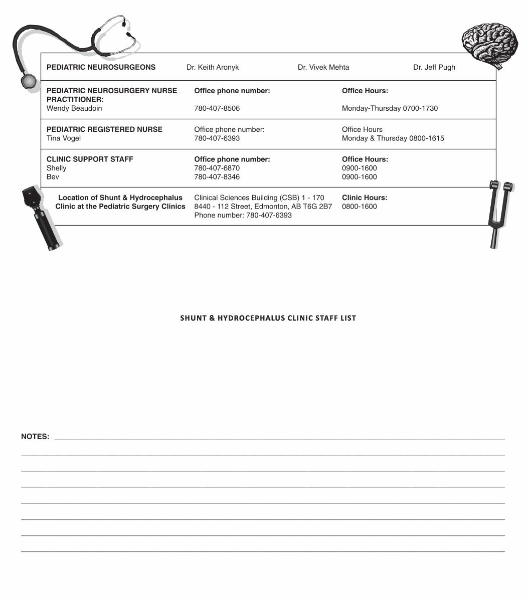
SHUNT & HYDROCEPHALUS CLINIC STAFF LIST
PEDIATRIC NEUROSURGEONS Dr. Keith Aronyk Dr. Vivek Mehta Dr. Jeff Pugh
PEDIATRIC NEUROSURGERY NURSE PRACTITIONER:Wendy Beaudoin
Office phone number:
780-407-8506
Office Hours:
Monday-Thursday 0700-1730
CLINIC SUPPORT STAFFShellyBev
Office phone number:780-407-6870780-407-8346
Office Hours:0900-16000900-1600
Location of Shunt & Hydrocephalus Clinic at the Pediatric Surgery Clinics
Clinical Sciences Building (CSB) 1 - 1708440 - 112 Street, Edmonton, AB T6G 2B7Phone number: 780-407-6393
Clinic Hours:0800-1600
PEDIATRIC REGISTERED NURSETina Vogel
Office phone number:780-407-6393
Office HoursMonday & Thursday 0800-1615
NOTES: ____________________________________________________________________________________________________________
____________________________________________________________________________________________________________________
____________________________________________________________________________________________________________________
____________________________________________________________________________________________________________________
____________________________________________________________________________________________________________________
____________________________________________________________________________________________________________________
____________________________________________________________________________________________________________________
____________________________________________________________________________________________________________________