hydrogel biophysical properties instruct coculture...
TRANSCRIPT
The FASEB Journal • Research Communication
Hydrogel biophysical properties instruct coculture-mediatedosteogenic potential
Kaitlin C. Murphy,* Roberta S. Stilhano,*,† Debika Mitra,* Dejie Zhou,* Samir Batarni,*Eduardo A. Silva,* and J. Kent Leach*,‡,1
*Department of Biomedical Engineering and ‡Department of Orthopaedic Surgery, School of Medicine,University of California, Davis, Davis, California, USA; †Department of Biophysics, Federal University ofSao Paulo, Sao Paulo, Brazil
ABSTRACT Cell-based approaches for bone formationrequire instructional cues from the surrounding environ-ment. As an alternative to pharmacological strategies ortransplanting single cell populations, one approach is tocoimplant populations that can establish a new vasculatureand differentiate to bone-forming osteoblasts. Mesenchy-mal stem/stromal cells (MSCs) possess osteogenic poten-tial and produce numerous angiogenic growth factors.Endothelial colony-forming cells (ECFCs) are a subpop-ulation of endothelial progenitor cells capable of vasculo-genesis in vivo and may provide endogenous cues tosupportMSC function.We investigated the contributionofthe carrier biophysical properties to instruct entrappedhumanMSCs and ECFCs to simultaneously promote theirosteogenic and proangiogenic potential. Compared withgels containing MSCs alone, fibrin gels engineered withincreased compressive stiffness simultaneously increasedthe osteogenic and proangiogenic potential of entrappedcocultured cells. ECFCs produced bone morphogeneticprotein-2 (BMP-2), a potent osteoinductive molecule, andincreases in BMP-2 secretion correlated with gel stiffness.Coculture of MSCs with ECFCs transduced to knockdownBMP-2 production abrogated the osteogenic response tolevels observed with MSCs alone. These results demon-strate that physical properties of engineered hydrogelsmodulate the function of cocultured cells in the absence ofinductive cues, thus increasing the translational potential ofcoimplantation to speed bone formation and repair.—Murphy,K.C.,Stilhano,R.S.,Mitra,D.,Zhou,D.,Batarni, S.,Silva, E. A., Leach, J. K. Hydrogel biophysical propertiesinstruct coculture-mediated osteogenic potential. FASEB J.30, 000–000 (2016). www.fasebj.org
Key Words: fibrin • mesenchymal stem cell • ECFC • BMP-2 •
osteogenesis
Ofthemore than6.2million fractures thatoccureachyear,approximately 5–10% will suffer from impaired healing,resulting in nonunions (1). Current treatments includeautograft and allograft bone, but their limitations have
prompted the search for alternative treatment strategiesincluding synthetic biomaterials and the use of recombi-nant osteoinductive proteins such as bonemorphogeneticprotein-2 (BMP-2).However, theseapproaches suffer frominherent delays in tissue repair that are dependent uponthe presence of the patient’s own responsive cells toachieve bone healing.
Cell-based therapies for bone regeneration aim toovercome limited availability of locally responsive endog-enous cells by implanting autologous or allogenic cellpopulations to directly or indirectly contribute to boneformation. Mesenchymal stem/stromal cells (MSCs) areunder investigation for restoring lost bone volume; theyhave the potential to undergo differentiation toward theosteoblastic lineage upon osteogenic induction (2, 3) andsecrete trophic factors that stimulate angiogenesis (4, 5).Endothelial cells have been implanted into bone defects topromote vascularization in situ and participate in the for-mation of new blood vessels, allowing the native repairprocesses to unfold (6). WhenMSCs and endothelial cellsare codelivered, synergistic paracrine signaling enhancesneovascularization and mineralization (7–9). However,the collection of autologous endothelial cells requirespainful dermal biopsies that fail to provide clinically rele-vant numbers of cells, thereby requiring prolonged cellculture and delaying treatment. Alternatively, endothelialcolony-forming cells (ECFCs) are a subpopulation of en-dothelial progenitor cells that can be obtained from pe-ripheral blood in high numbers and are easily expanded(10, 11), exhibit robust proliferative and vasculogenic po-tential (12), and represent a promising, clinically relevantcell population. Under hypoxic conditions in vitro, ECFCsproliferate and migrate better than human microvascularendothelial cells, a commonly used endothelial cell type(12). Despite their seemingly superior therapeutic poten-tial, the capacity of ECFCs to promote bone formationwhen deployed with MSCs is poorly understood.
The injection of cells to the target site seeks to localizecells at the defect and eliminate delays or off-target effectsassociatedwith cell homing strategies. Localdeploymentofcells using biomaterials further enhances efficacy of cell
Abbreviations: a-MEM, minimum essential medium a; ALP,alkaline phosphatase; BMP-2, bone morphogenetic protein-2;ECFC, endothelial colony-forming cell; EGM-2, endothelialcell growth medium-2; GF-Def, growth factor-deficient; MMP,matrix metalloproteinase; MSCs, mesenchymal stem/stromalcells; TBST, Tris-buffered saline supplemented with Tween 20
1 Correspondence: Department of Biomedical Engineering,University of California, Davis, 451 Health Sciences Dri.,Davis, CA 95616, USA. E-mail: [email protected]: 10.1096/fj.15-279984
0892-6638/16/0030-0001 © FASEB 1
The FASEB Journal article fj.15-279984. Published online October 6, 2015.
therapies by reducing the number of cells that migratefrom the defect while enhancing cell persistence andproviding instructional cues to direct cell function. In viewof deploying coculture populations for tissue repair, it isparticularly important to consider the needs of each celltype. Endothelial cell populations remodel their micro-environment to promote vasculogenesis, and thereforebenefit from more compliant materials (13, 14). Con-versely, MSCs differentiate into cells of the osteoblasticlineage more robustly in stiffer biomaterials (15–17).Therefore, many biomaterials used for cell-based thera-pies of bone formation are ill suited for endothelial celldelivery (18). Fibrin hydrogels are a promising platformfor cell delivery as fibrin naturally occurs in the body,serving as a scaffold for leukocytes and endothelial cellsduring tissue regeneration (19). We previously reportedthat supplementation of the pregel fibrinogen solutionwith 2.3% (w/v) NaCl modifies the fibrin fiber structureand increases compressive stiffness, thereby promotingthe osteogenic and proangiogenic potential of entrappedMSCs while avoiding osmolarity-related effects (16, 20).Entrapped cells are briefly exposed to salinity levelshigher than physiologic values, but the NaCl diffuses outof the gels within the first hour (20). Importantly, thesegels are formed from relatively low fibrinogen concen-trations and undergo gelation in a time course that ena-bles survival of entrapped cells. Thus, themanipulation ofthe physical properties of fibrin gels may provide a viableplatform to simultaneously instruct endothelial and MSCfunction for bone formation.
We hypothesized that tailoring the biophysical proper-ties of the cell carrier would instruct heterotypic cellfunction for enhancing bone formation. To explore thishypothesis, we entrapped humanMSCs and ECFCs withinfibrin gels engineered via supplementation with NaCl tomodulate biophysical properties while keeping composi-tion constant. We assessed gel material properties and theosteogenic and proangiogenic potential of entrappedcells. Furthermore, we explored the mechanism of howECFCs modulate osteogenic potential of the system. Theresults of these studies offer enhanced translational rele-vance for using cell-based therapies in tissue repair.
MATERIALS AND METHODS
Cell culture
Human bone marrow-derived MSCs (Lonza, Walkersville, MD,USA) were used without additional characterization. MSCs wereexpanded in standard culture conditions (37°C, 21% O2, 5%CO2) in a-MEM supplemented with 10% fetal bovine serum (JRScientific,Woodland, CA, USA) and 1% penicillin/streptomycin(Mediatech, Herndon, VA, USA) until use at passages 4–5. Hu-man umbilical cord blood ECFCs were generously provided byDr. Mervin Yoder (Indiana University, Indianapolis, IN, USA) andisolatedusing aprotocol approvedby the InstitutionalReviewBoardof the IndianaUniversity SchoolofMedicine aspreviously described(21).Adherent ECFCswere culturedon tissue cultureplastic coatedwith 5 mg/cm2 rat tail collagen I (BD Biosciences, San Jose, CA,USA) in endothelial cell growth medium-2 (EGM-2) mediumwithLonza’sSingleQuot supplements (hydrocortisone,gentamicin,human VEGF, human basic fibroblast growth factor, humanepidermal growth factor, human insulin-like growth factor
[IGF], and heparin) and further supplemented with 5% fetal bo-vine serumand1%penicillin/streptomycin.Growth factor-deficient(GF-Def) EGM-2 was prepared with serum-containing EGM-2but lacking VEGF, fibroblast growth factor, and IGF. Culture-expanded ECFCs (passages 12–13) were used for all studies.
Fibrin gel preparation
Fibrin gels were formed as we previously described (16, 20, 22).This fabrication process resulted in fibrin gels with a final fibrin-ogen concentration of 20 mg/mL (Calbiochem, Gibbstown, NJ,USA), 2.5 U/mL thrombin (Calbiochem), 20mMCaCl2 (Sigma-Aldrich, St. Louis, MO, USA), and 250 KIU/mL aprotinin (SantaCruz Biotechnology, Santa Cruz, CA,USA), all in PBS.GelsmadeinPBSwithout additionalNaCl supplementation contained 0.8%(w/v) NaCl (Sigma-Aldrich), whereas gels supplemented withNaCl had a final concentration of 2.3% (w/v) NaCl. A total vol-ume of 80mLwas added to each cylindrical polydimethylsiloxanemold (5 mm in diameter), and the contents were allowed to gelfor 1 h in standard culture conditions. The polydimethylsiloxanesheet was then carefully lifted from the culture dish, leaving be-hind the undisturbed fibrin gels, and the gels were transferred to24-well tissuecultureplates containingmedium.Themediumwasrefreshed after 1 h, ensuring that cellular responses were due tothe material properties of the hydrogels and not NaCl content.
To add clinical relevance, all cells were used directly from thecryovial. The cryopreservation solution was removed via centri-fugation, and cells were resuspended in the fibrinogen pre-gel solution. Fibrin gels contained 4 3 105 MSCs, 4 3 105
ECFCs, or a combination of 2 3 105 MSCs and 2 3 105 ECFCsfor a final concentration of 5 3 106 cells/mL in each gel. All gelswere cultured in a 1:1 mixture of a-MEM and GF-Def EGM-2without osteogenic supplements or growth factors to promotethe survival of both cell populations. The day of gel fabricationwas denoted as d 21. Gels were maintained in standard cellculture conditions with medium changes every 3 d.
Assessment of gel mechanical and morphologic properties
Rheological properties of fibrin gels, with or without cells, weremeasured on a Discovery HR-2 hybrid stress-controlled rheome-ter (Thermal Analysis Instruments, New Castle, DE, USA) equip-ped with an 8 mm parallel plate geometry on a stage heated to37°C. Gels were tested at a frequency of 0.5 Hz and a logarithmicsweep from 0.1 to 10 mN·mwith 10 points per decade. The shearstorage modulus was determined by averaging at least 10 pointsin the linear viscoelastic region.
The compressivemoduli offibrin gels weremeasured using anInstron 3345 Compressive Testing System (Norwood, MA, USA).Gelswere allowed to swell for 1 h inPBS, blotted, and then loadedbetween 2 flat platens and compressed at 1 mm/min. Compres-sive moduli were calculated from the linear portions of theforce–displacement graph for strain ranging from 0 to 5% (16).
The contraction of fibrin gels due to activity of entrapped cellswas measured at 0 and 21 d by visually following morphologicchanges in gel volume. Gels were imaged using a Nikon EclipseTE2000Umicroscope (Melville, NY,USA) and Andor Zyla digitalcamera (Oxford Instruments, Abingdon, United Kingdom) andgel area was calculated in NIS Elements (Nikon).
Cellular response to engineered fibrin gels
The osteogenic response of MSCs entrapped within engineeredfibrin gels was assessed from gels collected at 0, 7, 14, and 21 d.Gels were rinsed in PBS and sonicated in 400 mL passive lysis
2 Vol. 30 January 2016 MURPHY ET AL.The FASEB Journal x www.fasebj.org
buffer (Promega, Madison, WI, USA). Samples were centrifugedat 5000 rpm for 10 min to pellet the cell debris, and the super-natant was collected. The supernatant was analyzed for in-tracellular alkaline phosphatase (ALP) activity using ap-nitrophenyl phosphate colorimetric assay, and cell-secretedmineral within fibrin hydrogels was measured usingo-cresolphthalein complexone as previously described (16, 20,23). DNA content was quantified from the supernatant usingthe Quant-iT PicoGreen dsDNA Assay Kit (Thermo Fisher Sci-entific, Rochester, NY, USA). ECFC secretion of BMP-2 wasquantitatively determined from the conditioned medium after7 d in culture. The medium was refreshed 24 h before collec-tion, and the concentration of BMP-2 was determined usinga human-specific BMP-2 ELISA kit (R&D Systems,Minneapolis,MN, USA) according to the manufacturer’s instructions.
MSC secretion of VEGF was quantitatively determined fromthe conditioned medium of coculture-containing fibrin gels atdesignated time points. The medium was refreshed 24 h beforecollection, and the concentration of VEGFwas determined usinga human-specific VEGF ELISA kit (R&D Systems) according tothe manufacturer’s instructions.
To investigate the proangiogenic potential of entrapped cells,conditioned medium was collected from fibrin gels at 7 d andused to stimulate ECFC proliferation and tubule formation as wepreviously described (24). The mitogenic response of ECFCs toconditioned medium was determined by seeding ECFCs at7500 cells/cm2 in EGM-2 in a 12 well plate and allowing cells toattach overnight. The medium was then refreshed with a 1:4 volumeratio of coculture-conditioned medium to GF-Def EGM-2 andcultured for 72 h. Each well was then rinsed with PBS and cellswere lysed with passive lysis buffer. DNA content was quantifiedas described above. For tubule formation, 100 mL of GrowthFactor ReducedMatrigel (BDBiosciences) was pipetted into 48well plates and allowed to gel at 37°C for 1h. ECFCswere seededon Matrigel at 30,000 cells/cm2 in GF-Def EGM-2, and a mix-ture of conditioned medium and GF-Def EGM-2 was added ina 1:4 volume ratio. Cells were cultured for 16 h, after which theywere stainedwith calcein acetoxymethyl ester (3mg/mL in PBS;Invitrogen, Carlsbad, CA, USA) for 30 min. Average number ofbranch points per field of view and average tubule length werequantified usingNIS Elements (Nikon). For both assays, EGM-2served as the positive control, while GF-Def EGM-2 served as thenegative control.
Detection of BMP-2 production in ECFCs by Western blot
To quantify BMP-2 expression by ECFCs entrapped in fibrin gels,gels were first collected in sample buffer [0.1% Triton-X 100,0.01% Tris-EDTA, 0.1% protease inhibitor cocktail (Calbio-chem)]. Samples were sonicated and spun down, and total pro-tein was quantified using the BCA assay (Thermo FisherScientific).Protein(20mgper sample)wasadded to10mL43XTsample buffer (Bio-Rad, Hercules, CA, USA), boiled for 5 min toreduce protein, and loaded onto 12% Tris-HCl polyacrylamidegels (Bio-Rad). Gels were electrophoresed for 60 min at 180 V.Protein was then transferred to nitrocellulose membranes (Invi-trogen) for blotting, and protein was electrophoretically trans-ferred via the iBlot system (Invitrogen). After transfer, blots wereincubated in Tris-buffered saline supplemented with Tween 20(TBST; 10 mM Tris, 100 mM NaCl, 0.1% Tween-20, pH 7.5)containing 5% nonfat milk for 1 h to block nonspecific proteinbinding. After blocking, blots were incubated overnight at 4°Cwith a primary polyclonal anti-humanBMP-2 antibody (ab82511,Abcam, Cambridge, MA, USA; 1:100 in TBST) or anti-humanb-actin (Cell Signaling Technology, Danvers, MA, USA; 1:200 inTBST). After rinsingwithTBST, blots were incubated for 1 hwitha secondary goat anti-rabbit horseradish peroxidase-conjugatedantibody (1:1000; Cell Signaling Technology). Signal detection
was achieved with the ECL Western Blotting Substrate (ThermoFisher Scientific) and signals were recorded using theChemiDocMP System (Bio-Rad). Between antibodies, the membrane wasstripped at 55°C for 60min in stripping buffer (0.1M glycine, pH2.8). Protein bands were quantified using Image J software (Na-tional Institutes of Health, Bethesda, MD, USA) and normalizedto the levels of b-actin as the loading control.
Investigating cellular crosstalk when entrapped in fibrin gels
To probe the interaction between cells in coculture, we blockedcommon participants in matrix remodeling, angiogenesis, andbone formation.Matrixmetalloproteinases (MMPs) were blockedwith GM6001 (Calbiochem, 10 mM), a broad-spectrum MMP in-hibitor, by addition directly to the fibrin gels during fabrication(25). The bioactivity of secreted VEGF and BMP-2 was blockedby adding antibodies to VEGF165/121 (500 ng/mL) or BMP-2(2 mg/mL; R&D Systems) to the medium as described (24, 26).Medium was refreshed every 2 d, and gels were collected at7 and 21 d to assess mineral deposition.
To knock down BMP-2 expression, ECFCs were transduced(multiplicity of infection 10) using Mission shRNA lentivirustransduction particles (TRCN0000058193) and pLKO-puroNon-Target shRNA Control Transduction particles (Sigma-Aldrich) in growth medium containing 8 mg/mL polybrene(Sigma-Aldrich). Cells were placed under selective pressure 48 hafter infection using 2 mg/mL puromycin (Sigma-Aldrich). Fordetermination of transduction efficiency, all cell types weretransduced using a lentiviral-driven GFP construct (pLVTHM)(Addgene plasmid #12247). Knocked-down expression was con-firmed by Western blot for BMP-2.
Statistical analysis
Data are presented as means 6 SD. Statistical analysis was per-formed using 2-way ANOVA with Bonferroni correction formultiple comparisons or paired t tests when appropriate. All sta-tistical analysis was performed in Prism 6 software (GraphPad, LaJolla, CA, USA). Values of P , 0.05 were considered statisticallysignificant.
RESULTS
Gel contraction by entrapped cells is modulated byhydrogel physical properties
Changes in construct morphology were assessed by ex-amining gel area over time (Fig. 1A, B). At d 0 (24 h aftersynthesis), constructs formed without additional salt hadcontracted to half their original area and continued tocontract over the 21 d study. Conversely, fibrin gelsformed with 2.3% (w/v) NaCl did not contract signifi-cantly over time.
The initial rheological properties of fibrin hydrogelswere characterized with and without cells to elucidate thecontributions of NaCl and cells to the material properties.In the absence of cells, fibrin gels formed with 2.3% (w/v)NaClwerecomparable in storagemodulus to those formedwithout NaCl supplementation (Fig. 1C). When cells werepresent, the storage modulus decreased significantly ingels formedwith 2.3%(w/v)NaCl; however, therewere nosignificant differences in gels formed without NaCl sup-plementation. In contrast to the storage moduli, acellular
GEL PROPERTIES INSTRUCT COCULTURE OSTEOGENESIS 3
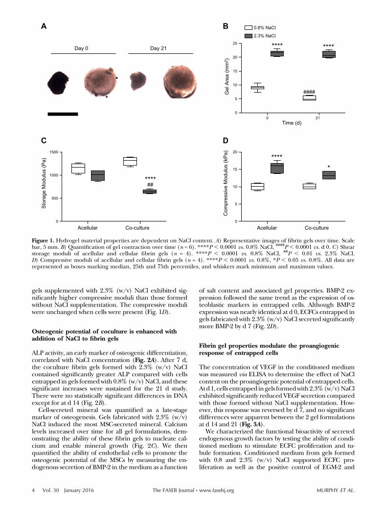
gels supplemented with 2.3% (w/v) NaCl exhibited sig-nificantly higher compressive moduli than those formedwithout NaCl supplementation. The compressive moduliwere unchanged when cells were present (Fig. 1D).
Osteogenic potential of coculture is enhanced withaddition of NaCl to fibrin gels
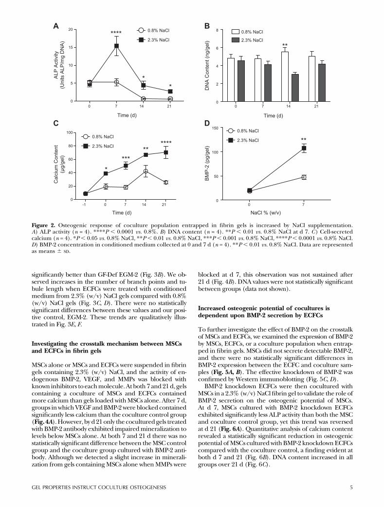
ALP activity, an early marker of osteogenic differentiation,correlated with NaCl concentration (Fig. 2A). After 7 d,the coculture fibrin gels formed with 2.3% (w/v) NaClcontained significantly greater ALP compared with cellsentrapped in gels formedwith 0.8% (w/v)NaCl, and thesesignificant increases were sustained for the 21 d study.There were no statistically significant differences in DNAexcept for at d 14 (Fig. 2B).
Cell-secreted mineral was quantified as a late-stagemarker of osteogenesis. Gels fabricated with 2.3% (w/v)NaCl induced the most MSC-secreted mineral. Calciumlevels increased over time for all gel formulations, dem-onstrating the ability of these fibrin gels to nucleate cal-cium and enable mineral growth (Fig. 2C). We thenquantified the ability of endothelial cells to promote theosteogenic potential of the MSCs by measuring the en-dogenous secretion of BMP-2 in themedium as a function
of salt content and associated gel properties. BMP-2 ex-pression followed the same trend as the expression of os-teoblastic markers in entrapped cells. Although BMP-2expression was nearly identical at d 0, ECFCs entrapped ingels fabricated with 2.3% (w/v) NaCl secreted significantlymore BMP-2 by d 7 (Fig. 2D).
Fibrin gel properties modulate the proangiogenicresponse of entrapped cells
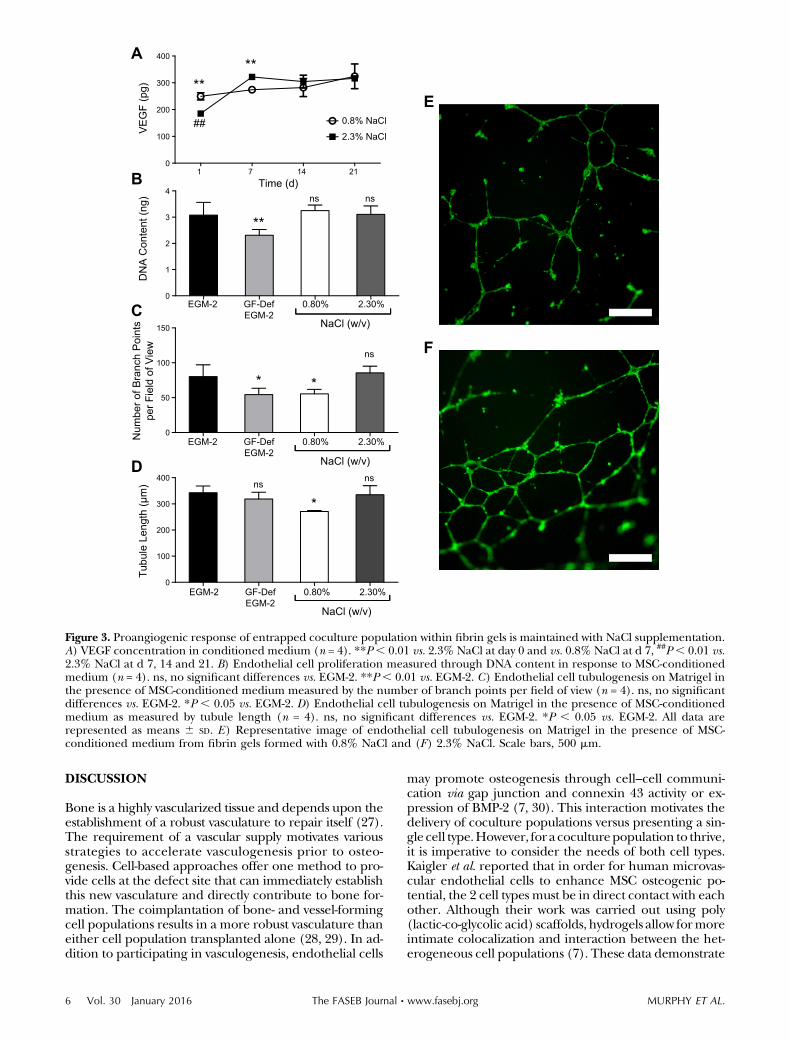
The concentration of VEGF in the conditioned mediumwas measured via ELISA to determine the effect of NaClcontent on theproangiogenic potential of entrapped cells.Atd1, cells entrapped ingels formedwith2.3%(w/v)NaClexhibited significantly reduced VEGF secretion comparedwith those formed without NaCl supplementation. How-ever, this response was reversed by d 7, and no significantdifferences were apparent between the 2 gel formulationsat d 14 and 21 (Fig. 3A).
We characterized the functional bioactivity of secretedendogenous growth factors by testing the ability of condi-tioned medium to stimulate ECFC proliferation and tu-bule formation. Conditioned medium from gels formedwith 0.8 and 2.3% (w/v) NaCl supported ECFC pro-liferation as well as the positive control of EGM-2 and
0 210
5
10
15
20
25
Time (d)
0.8% NaCl
2.3% NaCl
**** ****
####
Acellular Co-culture0
500
1000
1500
Sto
rage
Mod
ulus
(Pa)
****##
A B
C D
Day 0 Day 21
Acellular Co-culture0
5
10
15
20
Com
pres
sive
Mod
ulus
(kP
a) *****
Gel
Are
a (m
m2 )
Figure 1. Hydrogel material properties are dependent on NaCl content. A) Representative images of fibrin gels over time. Scalebar, 5 mm. B) Quantification of gel contraction over time (n = 6). ****P , 0.0001 vs. 0.8% NaCl, ####P , 0.0001 vs. d 0. C) Shearstorage moduli of acellular and cellular fibrin gels (n = 4). ****P , 0.0001 vs. 0.8% NaCl, ##P , 0.01 vs. 2.3% NaCl.D) Compressive moduli of acellular and cellular fibrin gels (n = 4). ****P , 0.0001 vs. 0.8%, *P , 0.05 vs. 0.8%. All data arerepresented as boxes marking median, 25th and 75th percentiles, and whiskers mark minimum and maximum values.
4 Vol. 30 January 2016 MURPHY ET AL.The FASEB Journal x www.fasebj.org
significantly better than GF-Def EGM-2 (Fig. 3B). We ob-served increases in the number of branch points and tu-bule length when ECFCs were treated with conditionedmedium from 2.3% (w/v) NaCl gels compared with 0.8%(w/v) NaCl gels (Fig. 3C, D). There were no statisticallysignificant differences between these values and our posi-tive control, EGM-2. These trends are qualitatively illus-trated in Fig. 3E, F.
Investigating the crosstalk mechanism between MSCsand ECFCs in fibrin gels
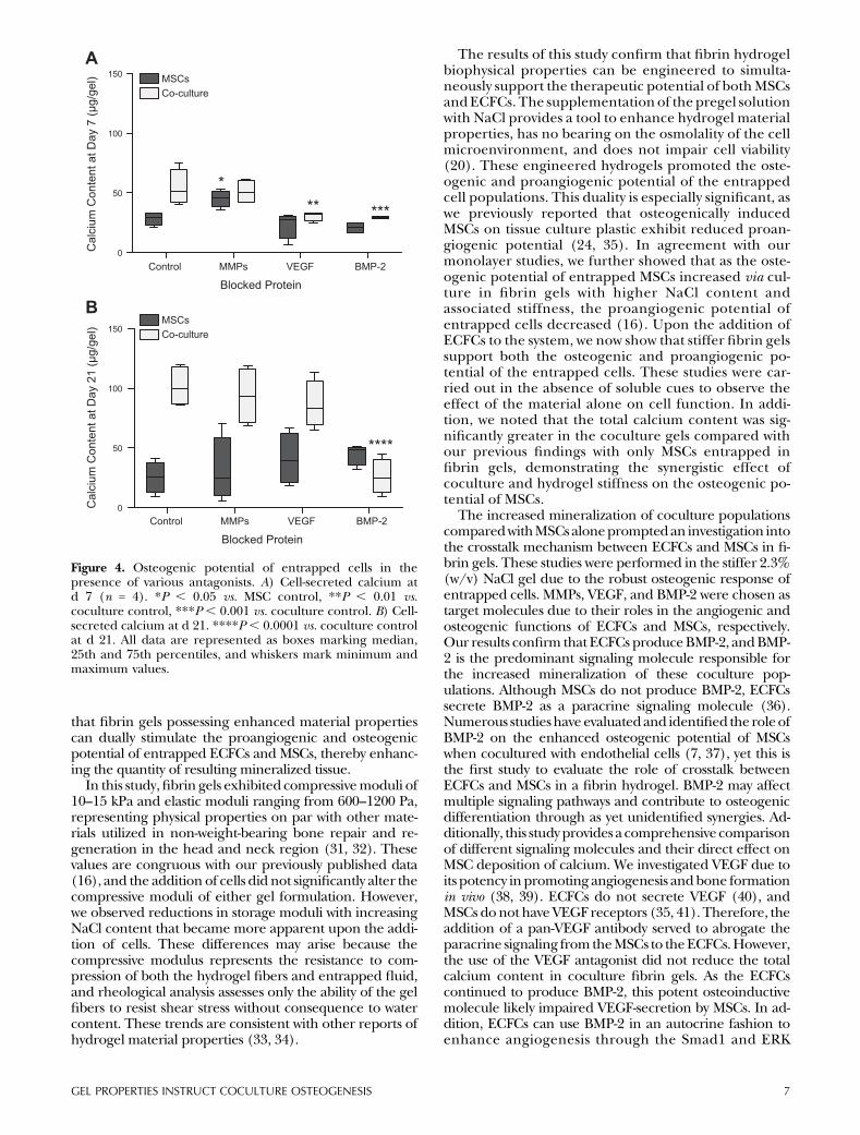
MSCs alone or MSCs and ECFCs were suspended in fibringels containing 2.3% (w/v) NaCl, and the activity of en-dogenous BMP-2, VEGF, and MMPs was blocked withknown inhibitors to eachmolecule.At both 7 and21d, gelscontaining a coculture of MSCs and ECFCs containedmore calcium than gels loaded withMSCs alone. After 7 d,groups inwhichVEGF andBMP-2 were blocked containedsignificantly less calcium than the coculture control group(Fig. 4A).However, by d21only the coculturedgels treatedwith BMP-2 antibody exhibited impairedmineralization tolevels below MSCs alone. At both 7 and 21 d there was nostatistically significant difference between theMSC controlgroup and the coculture group cultured with BMP-2 anti-body. Although we detected a slight increase in minerali-zation from gels containingMSCs alone whenMMPs were
blocked at d 7, this observation was not sustained after21 d (Fig. 4B). DNA values were not statistically significantbetween groups (data not shown).
Increased osteogenic potential of cocultures isdependent upon BMP-2 secretion by ECFCs
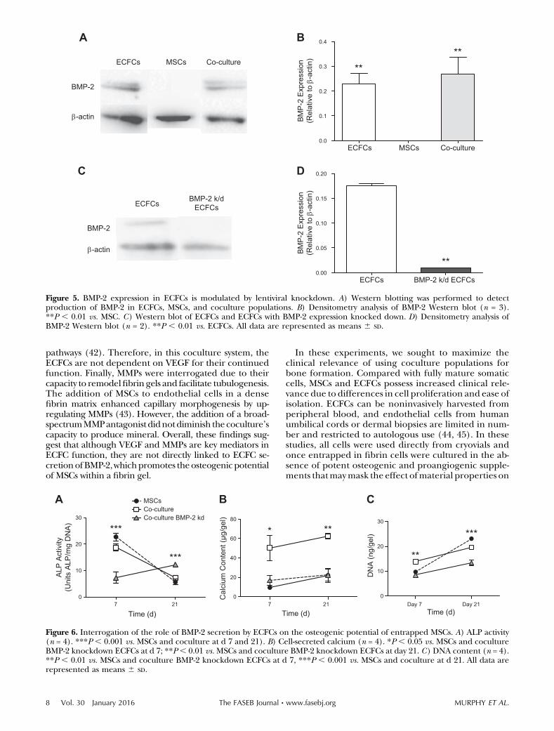
To further investigate the effect of BMP-2 on the crosstalkof MSCs and ECFCs, we examined the expression of BMP-2by MSCs, ECFCs, or a coculture population when entrap-ped in fibrin gels. MSCs did not secrete detectable BMP-2,and there were no statistically significant differences inBMP-2 expression between the ECFC and coculture sam-ples (Fig. 5A, B). The effective knockdown of BMP-2 wasconfirmed by Western immunoblotting (Fig. 5C, D).
BMP-2 knockdown ECFCs were then cocultured withMSCs in a 2.3% (w/v)NaClfibrin gel to validate the role ofBMP-2 secretion on the osteogenic potential of MSCs.At d 7, MSCs cultured with BMP-2 knockdown ECFCsexhibited significantly less ALP activity than both theMSCand coculture control group, yet this trend was reversedat d 21 (Fig. 6A). Quantitative analysis of calcium contentrevealed a statistically significant reduction in osteogenicpotential ofMSCs culturedwithBMP-2 knockdown ECFCscompared with the coculture control, a finding evident atboth d 7 and 21 (Fig. 6B). DNA content increased in allgroups over 21 d (Fig. 6C).
A
C
0 7 14 210
5
10
15
20
Time (d)
****
**
-1 0 7 14 210
20
40
60
80
100
Time (d)
*
******
***
B
D
0 7 14 210
2
4
6
8
Time (d)
DN
A C
onte
nt (n
g/ge
l)
0.8% NaCl
2.3% NaCl**
0 70
50
100
150
NaCl % (w/v)B
MP
-2 (p
g/ge
l)
**
0.8% NaCl
0.8% NaCl0.8% NaCl
2.3% NaCl2.3% NaCl
2.3% NaCl
ALP
Act
ivity
(Uni
ts A
LP/m
g D
NA
)C
alci
um C
onte
nt(μ
g/ge
l)
Figure 2. Osteogenic response of coculture population entrapped in fibrin gels is increased by NaCl supplementation.A) ALP activity (n = 4). ****P , 0.0001 vs. 0.8%. B) DNA content (n = 4). **P , 0.01 vs. 0.8% NaCl at d 7. C) Cell-secretedcalcium (n = 4). *P, 0.05 vs. 0.8% NaCl, **P, 0.01 vs. 0.8% NaCl, ***P , 0.001 vs. 0.8% NaCl, ****P, 0.0001 vs. 0.8% NaCl.D) BMP-2 concentration in conditioned medium collected at 0 and 7 d (n = 4). **P , 0.01 vs. 0.8% NaCl. Data are representedas means 6 SD.
GEL PROPERTIES INSTRUCT COCULTURE OSTEOGENESIS 5
DISCUSSION
Bone is a highly vascularized tissue and depends upon theestablishment of a robust vasculature to repair itself (27).The requirement of a vascular supply motivates variousstrategies to accelerate vasculogenesis prior to osteo-genesis. Cell-based approaches offer one method to pro-vide cells at the defect site that can immediately establishthis new vasculature and directly contribute to bone for-mation. The coimplantation of bone- and vessel-formingcell populations results in a more robust vasculature thaneither cell population transplanted alone (28, 29). In ad-dition to participating in vasculogenesis, endothelial cells
may promote osteogenesis through cell–cell communi-cation via gap junction and connexin 43 activity or ex-pression of BMP-2 (7, 30). This interaction motivates thedelivery of coculture populations versus presenting a sin-gle cell type.However, for a coculturepopulation to thrive,it is imperative to consider the needs of both cell types.Kaigler et al. reported that in order for human microvas-cular endothelial cells to enhance MSC osteogenic po-tential, the 2 cell types must be in direct contact with eachother. Although their work was carried out using poly(lactic-co-glycolic acid) scaffolds, hydrogels allow formoreintimate colocalization and interaction between the het-erogeneous cell populations (7). These data demonstrate
E
F
C
D
EGM-2 0.80% 2.30%0
1
2
3
4
DN
A C
onte
nt (n
g)
**
ns ns
NaCl (w/v)
0
50
100
150
* *
ns
0
100
200
300
400
*ns
ns
A
B 1 7 14 210
100
200
300
400
0.8% NaCl
2.3% NaCl
Time (d)
VE
GF
(pg) **
##
**
Num
ber o
f Bra
nch
Poi
nts
per F
ield
of V
iew
Tubu
le L
engt
h (µ
m)
GF-DefEGM-2
EGM-2 0.80% 2.30%
NaCl (w/v)
GF-DefEGM-2
EGM-2 0.80% 2.30%
NaCl (w/v)
GF-DefEGM-2
Figure 3. Proangiogenic response of entrapped coculture population within fibrin gels is maintained with NaCl supplementation.A) VEGF concentration in conditioned medium (n = 4). **P, 0.01 vs. 2.3% NaCl at day 0 and vs. 0.8% NaCl at d 7, ##P, 0.01 vs.2.3% NaCl at d 7, 14 and 21. B) Endothelial cell proliferation measured through DNA content in response to MSC-conditionedmedium (n = 4). ns, no significant differences vs. EGM-2. **P , 0.01 vs. EGM-2. C) Endothelial cell tubulogenesis on Matrigel inthe presence of MSC-conditioned medium measured by the number of branch points per field of view (n = 4). ns, no significantdifferences vs. EGM-2. *P , 0.05 vs. EGM-2. D) Endothelial cell tubulogenesis on Matrigel in the presence of MSC-conditionedmedium as measured by tubule length (n = 4). ns, no significant differences vs. EGM-2. *P , 0.05 vs. EGM-2. All data arerepresented as means 6 SD. E) Representative image of endothelial cell tubulogenesis on Matrigel in the presence of MSC-conditioned medium from fibrin gels formed with 0.8% NaCl and (F) 2.3% NaCl. Scale bars, 500 mm.
6 Vol. 30 January 2016 MURPHY ET AL.The FASEB Journal x www.fasebj.org
that fibrin gels possessing enhanced material propertiescan dually stimulate the proangiogenic and osteogenicpotential of entrapped ECFCs and MSCs, thereby enhanc-ing the quantity of resulting mineralized tissue.
In this study,fibrin gels exhibited compressivemoduli of10–15 kPa and elastic moduli ranging from 600–1200 Pa,representing physical properties on par with other mate-rials utilized in non-weight-bearing bone repair and re-generation in the head and neck region (31, 32). Thesevalues are congruous with our previously published data(16), and the addition of cells did not significantly alter thecompressive moduli of either gel formulation. However,we observed reductions in storage moduli with increasingNaCl content that became more apparent upon the addi-tion of cells. These differences may arise because thecompressive modulus represents the resistance to com-pression of both the hydrogel fibers and entrapped fluid,and rheological analysis assesses only the ability of the gelfibers to resist shear stress without consequence to watercontent. These trends are consistent with other reports ofhydrogel material properties (33, 34).
The results of this study confirm that fibrin hydrogelbiophysical properties can be engineered to simulta-neously support the therapeutic potential of bothMSCsandECFCs. The supplementation of the pregel solutionwith NaCl provides a tool to enhance hydrogel materialproperties, has no bearing on the osmolality of the cellmicroenvironment, and does not impair cell viability(20). These engineered hydrogels promoted the oste-ogenic and proangiogenic potential of the entrappedcell populations. This duality is especially significant, aswe previously reported that osteogenically inducedMSCs on tissue culture plastic exhibit reduced proan-giogenic potential (24, 35). In agreement with ourmonolayer studies, we further showed that as the oste-ogenic potential of entrapped MSCs increased via cul-ture in fibrin gels with higher NaCl content andassociated stiffness, the proangiogenic potential ofentrapped cells decreased (16). Upon the addition ofECFCs to the system, we now show that stiffer fibrin gelssupport both the osteogenic and proangiogenic po-tential of the entrapped cells. These studies were car-ried out in the absence of soluble cues to observe theeffect of the material alone on cell function. In addi-tion, we noted that the total calcium content was sig-nificantly greater in the coculture gels compared withour previous findings with only MSCs entrapped infibrin gels, demonstrating the synergistic effect ofcoculture and hydrogel stiffness on the osteogenic po-tential of MSCs.
The increased mineralization of coculture populationscomparedwithMSCsalonepromptedan investigation intothe crosstalk mechanism between ECFCs and MSCs in fi-brin gels. These studies were performed in the stiffer 2.3%(w/v) NaCl gel due to the robust osteogenic response ofentrapped cells. MMPs, VEGF, and BMP-2 were chosen astarget molecules due to their roles in the angiogenic andosteogenic functions of ECFCs and MSCs, respectively.Our results confirm that ECFCs produceBMP-2, andBMP-2 is the predominant signaling molecule responsible forthe increased mineralization of these coculture pop-ulations. Although MSCs do not produce BMP-2, ECFCssecrete BMP-2 as a paracrine signaling molecule (36).Numerous studieshave evaluatedand identified the roleofBMP-2 on the enhanced osteogenic potential of MSCswhen cocultured with endothelial cells (7, 37), yet this isthe first study to evaluate the role of crosstalk betweenECFCs and MSCs in a fibrin hydrogel. BMP-2 may affectmultiple signaling pathways and contribute to osteogenicdifferentiation through as yet unidentified synergies. Ad-ditionally, this studyprovides a comprehensive comparisonof different signaling molecules and their direct effect onMSC deposition of calcium. We investigated VEGF due toits potency in promoting angiogenesis andbone formationin vivo (38, 39). ECFCs do not secrete VEGF (40), andMSCs donot haveVEGF receptors (35, 41).Therefore, theaddition of a pan-VEGF antibody served to abrogate theparacrine signaling from theMSCs to theECFCs.However,the use of the VEGF antagonist did not reduce the totalcalcium content in coculture fibrin gels. As the ECFCscontinued to produce BMP-2, this potent osteoinductivemolecule likely impaired VEGF-secretion by MSCs. In ad-dition, ECFCs can use BMP-2 in an autocrine fashion toenhance angiogenesis through the Smad1 and ERK
A
0
50
100
150
*** ***
0
50
100
150
MSCsCo-culture
****
B
Control MMPs VEGF BMP-2
Blocked Protein
Cal
cium
Con
tent
at D
ay 7
(µg/
gel)
Cal
cium
Con
tent
at D
ay 2
1 (µ
g/ge
l)
Control MMPs VEGF BMP-2
Blocked Protein
MSCsCo-culture
Figure 4. Osteogenic potential of entrapped cells in thepresence of various antagonists. A) Cell-secreted calcium atd 7 (n = 4). *P , 0.05 vs. MSC control, **P , 0.01 vs.coculture control, ***P , 0.001 vs. coculture control. B) Cell-secreted calcium at d 21. ****P , 0.0001 vs. coculture controlat d 21. All data are represented as boxes marking median,25th and 75th percentiles, and whiskers mark minimum andmaximum values.
GEL PROPERTIES INSTRUCT COCULTURE OSTEOGENESIS 7
pathways (42). Therefore, in this coculture system, theECFCs are not dependent on VEGF for their continuedfunction. Finally, MMPs were interrogated due to theircapacity to remodelfibrin gels and facilitate tubulogenesis.The addition of MSCs to endothelial cells in a densefibrin matrix enhanced capillary morphogenesis by up-regulating MMPs (43). However, the addition of a broad-spectrumMMPantagonist didnotdiminish thecoculture’scapacity to produce mineral. Overall, these findings sug-gest that although VEGF and MMPs are key mediators inECFC function, they are not directly linked to ECFC se-cretionofBMP-2,whichpromotes theosteogenic potentialof MSCs within a fibrin gel.
In these experiments, we sought to maximize theclinical relevance of using coculture populations forbone formation. Compared with fully mature somaticcells, MSCs and ECFCs possess increased clinical rele-vance due to differences in cell proliferation and ease ofisolation. ECFCs can be noninvasively harvested fromperipheral blood, and endothelial cells from humanumbilical cords or dermal biopsies are limited in num-ber and restricted to autologous use (44, 45). In thesestudies, all cells were used directly from cryovials andonce entrapped in fibrin cells were cultured in the ab-sence of potent osteogenic and proangiogenic supple-ments thatmaymask the effect ofmaterial properties on
A B
D
BMP-2
-actin
ECFCs MSCs Co-culture
C
BMP-2
-actin
ECFCsBMP-2 k/d
ECFCs
ECFCs MSCs Co-culture0.0
0.1
0.2
0.3
0.4
****
BM
P-2
Exp
ress
ion
(Rel
ativ
e to
-a
ctin
)
ECFCs BMP-2 k/d ECFCs0.00
0.05
0.10
0.15
0.20
**
BM
P-2
Exp
ress
ion
(Rel
ativ
e to
-a
ctin
)Figure 5. BMP-2 expression in ECFCs is modulated by lentiviral knockdown. A) Western blotting was performed to detectproduction of BMP-2 in ECFCs, MSCs, and coculture populations. B) Densitometry analysis of BMP-2 Western blot (n = 3).**P , 0.01 vs. MSC. C) Western blot of ECFCs and ECFCs with BMP-2 expression knocked down. D) Densitometry analysis ofBMP-2 Western blot (n = 2). **P , 0.01 vs. ECFCs. All data are represented as means 6 SD.
A B C
7 210
10
20
30
Time (d)
MSCsCo-cultureCo-culture BMP-2 kd
***
***
7 210
20
40
60
80
Time (d)
* **
Day 7 Day 210
10
20
30
DN
A (n
g/ge
l)
Time (d)
**
***
ALP
Act
ivity
(Uni
ts A
LP/m
g D
NA
)
Cal
cium
Con
tent
(µg/
gel)
Figure 6. Interrogation of the role of BMP-2 secretion by ECFCs on the osteogenic potential of entrapped MSCs. A) ALP activity(n = 4). ***P , 0.001 vs. MSCs and coculture at d 7 and 21). B) Cell-secreted calcium (n = 4). *P , 0.05 vs. MSCs and cocultureBMP-2 knockdown ECFCs at d 7; **P, 0.01 vs.MSCs and coculture BMP-2 knockdown ECFCs at day 21. C) DNA content (n = 4).**P , 0.01 vs. MSCs and coculture BMP-2 knockdown ECFCs at d 7, ***P , 0.001 vs. MSCs and coculture at d 21. All data arerepresented as means 6 SD.
8 Vol. 30 January 2016 MURPHY ET AL.The FASEB Journal x www.fasebj.org
instructing cell function. Finally, fibrin is approved bytheU.S. Food andDrugAdministration as a biomaterial.The physiological concentrations of fibrinogen andthrombin necessary to fabricate these materials couldfeasibly be harvested from patients to use autologousproteins (46).
The resultsof this study confirm thatNaClcanbeused totailor fibrin gels to support both the proangiogenic andosteogenic potential ofMSCswhencoculturedwithECFCsin vitro. Furthermore, this coculture population exhibitsincreased osteogenic potential compared with a mono-culture of MSCs, and this enhanced function is due to theECFC secretion of BMP-2.
This work was supported by a U.S. National Institutes ofHealth, National Institute of Dental and Craniofacial Re-search Grant R03-DE021704, and the AO Foundation (Davos,Switzerland) (C10-39L to J.K.L.). K.M. was supported by theAmerican Heart Association Western States Affiliate Pre-doctoral Fellowship. The authors declare no conflicts ofinterest.
REFERENCES
1. American Academy of Orthopaedic Surgeons (2000)MusculoskeletalInjuries Report: Incidence, Risk Factors and Prevention, Rosemont, IL
2. Bajada, S., Mazakova, I., Richardson, J. B., and Ashammakhi, N.(2008) Updates on stem cells and their applications in regenerativemedicine. J. Tissue Eng. Regen. Med. 2, 169–183
3. Hoch, A. I., and Leach, J. K. (2014) Concise review: optimizingexpansion of bone marrow mesenchymal stem/stromal cells forclinical applications. Stem Cells Transl. Med. 3, 643–652
4. Leu, A., Stieger, S. M., Dayton, P., Ferrara, K. W., and Leach, J. K.(2009) Angiogenic response to bioactive glass promotes bonehealing in an irradiated calvarial defect. Tissue Eng. Part A 15,877–885
5. He, J., Decaris, M. L., and Leach, J. K. (2012) Bioceramic-mediatedtrophic factor secretion bymesenchymal stemcells enhances in vitroendothelial cell persistence and in vivo angiogenesis.Tissue Eng. PartA 18, 1520–1528
6. Kaigler, D., Krebsbach, P. H., Wang, Z., West, E. R., Horger, K., andMooney, D. J. (2006) Transplanted endothelial cells enhanceorthotopic bone regeneration. J. Dent. Res. 85, 633–637
7. Kaigler, D., Krebsbach, P. H., West, E. R., Horger, K., Huang, Y. C.,and Mooney, D. J. (2005) Endothelial cell modulation of bonemarrow stromal cell osteogenic potential. FASEB J. 19, 665–667
8. Grellier, M., Granja, P. L., Fricain, J. C., Bidarra, S. J., Renard, M.,Bareille, R., Bourget, C., Amedee, J., and Barbosa, M. A. (2009) Theeffect of the co-immobilization of human osteoprogenitors and en-dothelial cells within alginate microspheres on mineralization ina bone defect. Biomaterials 30, 3271–3278
9. Seebach, C., Henrich, D., Wilhelm, K., Barker, J. H., and Marzi, I.(2012) Endothelial progenitor cells improve directly and indirectlyearly vascularization of mesenchymal stem cell-driven bone re-generation in a critical bone defect in rats. Cell Transplant. 21,1667–1677
10. Melero-Martin, J. M., Khan, Z. A., Picard, A., Wu, X., Paruchuri, S.,and Bischoff, J. (2007) In vivo vasculogenic potential of humanblood-derived endothelial progenitor cells. Blood 109, 4761–4768
11. Ingram, D. A., Mead, L. E., Tanaka, H., Meade, V., Fenoglio, A.,Mortell, K., Pollok, K., Ferkowicz, M. J., Gilley, D., and Yoder, M. C.(2004) Identification of a novel hierarchy of endothelial progenitorcells using human peripheral and umbilical cord blood. Blood 104,2752–2760
12. Decaris,M. L., Lee,C. I., Yoder,M.C., Tarantal, A. F., andLeach, J. K.(2009) Influence of the oxygen microenvironment on theproangiogenic potential of human endothelial colony formingcells. Angiogenesis 12, 303–311
13. Fioretta, E. S., Fledderus, J. O., Baaijens, F. P., and Bouten, C. V.(2012) Influence of substrate stiffness on circulating progenitor cellfate. J. Biomech. 45, 736–744
14. Ghajar,C.M., Chen,X.,Harris, J.W., Suresh, V.,Hughes,C. C., Jeon,N. L., Putnam, A. J., and George, S. C. (2008) The effect of matrixdensity on the regulation of 3-D capillary morphogenesis. Biophys. J.94, 1930–1941
15. Engler, A. J., Sen, S., Sweeney,H.L., andDischer,D.E. (2006)Matrixelasticity directs stem cell lineage specification. Cell 126, 677–689
16. Murphy, K. C., Hughbanks, M. L., Binder, B. Y., Vissers, C. B., andLeach, J. K. (2015) Engineered fibrin gels for parallel stimulation ofmesenchymal stem cell proangiogenic and osteogenic potential.Ann. Biomed. Eng. 43, 2010–2021
17. Kim, J., Park, Y., Tae, G., Lee, K. B., Hwang, C. M., Hwang, S. J., Kim,I. S., Noh, I., and Sun, K. (2009) Characterization of low-molecular-weight hyaluronic acid-based hydrogel and differential stem cell re-sponses in the hydrogel microenvironments. J. Biomed. Mater. Res. A88, 967–975
18. Chen, Y. C., Lin, R. Z., Qi, H., Yang, Y., Bae, H., Melero-Martin, J. M.,andKhademhosseini, A. (2012) Functional human vascular networkgenerated in photocrosslinkable gelatin methacrylate hydrogels.Adv. Funct. Mater. 22, 2027–2039
19. Janmey, P. A., Winer, J. P., and Weisel, J. W. (2009) Fibrin gels andtheir clinical and bioengineering applications. J. R. Soc. Interface 6,1-10
20. Davis, H. E., Miller, S. L., Case, E. M., and Leach, J. K. (2011)Supplementation of fibrin gels with sodium chloride enhancesphysical properties and ensuing osteogenic response. Acta Biomater.7, 691–699
21. Yoder,M.C.,Mead,L.E., Prater,D.,Krier,T.R.,Mroueh,K.N.,Li, F.,Krasich, R., Temm, C. J., Prchal, J. T., and Ingram, D. A. (2007)Redefining endothelial progenitor cells via clonal analysis andhematopoietic stem/progenitor cell principals. Blood 109,1801–1809
22. Murphy, K. C., and Leach, J. K. (2012) A reproducible, highthroughput method for fabricating fibrin gels. BMC Res. Notes 5, 423
23. Decaris, M. L., and Leach, J. K. (2011) Design of experimentsapproach to engineer cell-secretedmatrices for directing osteogenicdifferentiation. Ann. Biomed. Eng. 39, 1174–1185
24. Hoch, A. I., Binder, B. Y., Genetos, D. C., and Leach, J. K. (2012)Differentiation-dependent secretion of proangiogenic factors bymesenchymal stem cells. PLoS One 7, e35579
25. Ghajar, C. M., Kachgal, S., Kniazeva, E., Mori, H., Costes, S. V.,George, S. C., and Putnam, A. J. (2010)Mesenchymal cells stimulatecapillary morphogenesis via distinct proteolytic mechanisms. Exp.Cell Res. 316, 813–825
26. Edgar, C. M., Chakravarthy, V., Barnes, G., Kakar, S., Gerstenfeld,L. C., and Einhorn, T. A. (2007) Autogenous regulation ofa network of bone morphogenetic proteins (BMPs) mediates theosteogenic differentiation in murine marrow stromal cells. Bone40, 1389–1398
27. Das, A., and Botchwey, E. (2011) Evaluation of angiogenesis andosteogenesis. Tissue Eng. Part B Rev. 17, 403–414
28. Yu,H., VandeVord, P. J.,Mao, L.,Matthew,H.W.,Wooley, P.H., andYang, S. Y. (2009) Improved tissue-engineered bone regenerationbyendothelial cell mediated vascularization. Biomaterials 30, 508–517
29. Koike,N., Fukumura,D., Gralla,O., Au, P., Schechner, J. S., and Jain,R. K. (2004) Tissue engineering: creation of long-lasting blood ves-sels. Nature 428, 138–139
30. Herzog,D.P.,Dohle,E.,Bischoff, I., andKirkpatrick,C. J. (2014)Cellcommunication in a coculture system consisting of outgrowthendothelial cells and primary osteoblasts. Biomed Res. Int. 2014, 1–15
31. Kim, J., Kim, I. S., Cho, T. H., Lee, K. B., Hwang, S. J., Tae, G.,Noh, I., Lee, S. H., Park, Y., and Sun, K. (2007) Boneregeneration using hyaluronic acid-based hydrogel with bonemorphogenic protein-2 and human mesenchymal stem cells.Biomaterials 28, 1830–1837
32. Chung, Y. I., Ahn, K.M., Jeon, S. H., Lee, S. Y., Lee, J.H., and Tae, G.(2007) Enhanced bone regeneration with BMP-2 loaded functionalnanoparticle-hydrogel complex. J. Control. Release 121, 91–99
33. Oelker, A. M., Berlin, J. A., Wathier, M., and Grinstaff, M. W. (2011)Synthesis and characterization of dendron cross-linked PEG hydro-gels as corneal adhesives. Biomacromolecules 12, 1658–1665
34. Wingate, K., Bonani, W., Tan, Y., Bryant, S. J., and Tan, W. (2012)Compressive elasticity of three-dimensional nanofiber matrixdirects mesenchymal stem cell differentiation to vascular cellswith endothelial or smooth muscle cell markers. Acta Biomater. 8,1440–1449
35. Binder, B. Y., Genetos, D. C., and Leach, J. K. (2014)Lysophosphatidic acid protects human mesenchymal stromal cells
GEL PROPERTIES INSTRUCT COCULTURE OSTEOGENESIS 9
from differentiation-dependent vulnerability to apoptosis. TissueEng. Part A 20, 1156–1164
36. Smadja, D. M., Bieche, I., Silvestre, J. S., Germain, S., Cornet, A.,Laurendeau, I., Duong-Van-Huyen, J. P., Emmerich, J., Vidaud, M.,Aiach, M., and Gaussem, P. (2008) Bone morphogenetic proteins 2and 4 are selectively expressed by late outgrowth endothelialprogenitor cells and promote neoangiogenesis. Arterioscler. Thromb.Vasc. Biol. 28, 2137–2143
37. Saleh, F. A., Whyte, M., Ashton, P., and Genever, P. G. (2011)Regulation of mesenchymal stem cell activity by endothelial cells.Stem Cells Dev. 20, 391–403
38. Young, S., Patel, Z. S., Kretlow, J. D., Murphy, M. B., Mountziaris,P.M., Baggett, L. S.,Ueda,H., Tabata, Y., Jansen, J. A.,Wong,M., andMikos, A. G. (2009) Dose effect of dual delivery of vascularendothelial growth factor and bone morphogenetic protein-2 onbone regeneration in a rat critical-size defect model. Tissue Eng. PartA 15, 2347–2362
39. Leach, J. K., Kaigler, D., Wang, Z., Krebsbach, P. H., andMooney,D. J. (2006) Coating of VEGF-releasing scaffolds with bioactiveglass for angiogenesis and bone regeneration. Biomaterials 27,3249–3255
40. Joo, H. J., Song, S., Seo, H. R., Shin, J. H., Choi, S. C., Park, J. H., Yu,C. W., Hong, S. J., and Lim, D. S. (2015) Human endothelial colonyforming cells from adult peripheral blood have enhanced sproutingangiogenicpotential throughup-regulatingVEGFR2 signaling. Int. J.Cardiol. 197, 33–43
41. Fierro, F. A., Kalomoiris, S., Sondergaard, C. S., and Nolta, J. A.(2011) Effects on proliferation and differentiation of multipotentbone marrow stromal cells engineered to express growth factors forcombined cell and gene therapy. Stem Cells 29, 1727–1737
42. Dyer, L. A., Pi, X., and Patterson, C. (2014) The role of BMPs inendothelial cell function and dysfunction. Trends Endocrinol. Metab.25, 472–480
43. Ghajar, C. M., Blevins, K. S., Hughes, C. C., George, S. C., andPutnam, A. J. (2006) Mesenchymal stem cells enhance angiogenesisin mechanically viable prevascularized tissues via early matrixmetalloproteinase upregulation. Tissue Eng. 12, 2875–2888
44. Lin, R. Z., Moreno-Luna, R., Li, D., Jaminet, S. C., Greene, A. K., andMelero-Martin, J. M. (2014) Human endothelial colony-formingcells serve as trophic mediators for mesenchymal stem cell engraft-ment via paracrine signaling. Proc. Natl. Acad. Sci. USA 111,10137–10142
45. Melero-Martin, J. M., De Obaldia, M. E., Kang, S. Y., Khan, Z. A.,Yuan, L.,Oettgen, P., andBischoff, J. (2008) Engineering robust andfunctional vascular networks in vivo with human adult and cordblood-derived progenitor cells. Circ. Res. 103, 194–202
46. Spotnitz, W. D. (2014) Fibrin sealant: the only approved hemostat,sealant, and adhesive-a laboratory and clinical perspective. ISRNSurg. 2014, 203943
Received for publication July 31, 2015.Accepted for publication September 21, 2015.
10 Vol. 30 January 2016 MURPHY ET AL.The FASEB Journal x www.fasebj.org