hydrolysis of gtp by the α-chain of gs and other gtp binding proteins
TRANSCRIPT
PROTEINS: Structure, Function, and Genetics 6:222-230 (1989)
Hydrolysis of GTP by the a-Chain of G, and Other GTP Binding Proteins Henry R. Bourne, Claudia A. Landis, and Susan B. Masters Department of Pharmacology, Medicine, and the Cardiovascular Research Institute, University of California, San Francisco, California 94143-0450
ABSTRACT The functions of G proteins- like those of bacterial elongation factor (EF) Tu and the 21 kDa rus proteins (p21Y-depend upon their abilities to bind and hydrolyze GTP and to assume different conformations in GTP- and GDP-bound states. Similarities in function and amino acid sequence indicate that EF-Tu, p21"", and G protein a-chains evolved from a primordial GTP-binding protein. Proteins in all three families appear to share common mechanisms for GTP-dependent conforma- tional change and hydrolysis of bound GTP. Biochemical and molecular genetic studies of the a-chain of G, (a,) point to key regions that are involved in GTP-dependent conformational change and in hydrolysis of GTP. Tumorigenic mutations of a, in human pituitary tumors in- hibit the protein's GTPase activity and cause constitutive elevation of adenylyl cyclase activ- ity. One such mutation replaces a Gln residue in a, that corresponds to Gln-61 of p21""; muta- tional replacements of this residue in both pro- teins inhibit their GTPase activities. A second class of GTPase inhibiting mutations in a, oc- curs in the codon for an Arg residue whose co- valent modification by cholera toxin also inhib- its GTP hydrolysis by a,. This Arg residue is located in a domain of a, not represented in EF- Tu or p21"". We propose that this domain con- stitutes an intrinsic activator of GTP hydroly- sis, and that it performs a function analogous to that performed for EF-Tu by the programmed ribosome and for p21"" by the recently discov- ered GTPase-activating protein. Owing to their inherited similarities of structure and function, what we learn about a,, p21m", or EF-Tu as in- dividual molecules helps us to understand cru- cial functions of other members of the super- family.
Key words: G proteins, p21", GTPase, cholera toxin, GTPase-activating protein, amino acid sequence, protein struc- ture, conformational change
INTRODUCTION Recent biochemical and genetic studies have re-
vealed shared structural and functional features of
0 1989 ALAN R. LISS, INC.
many proteins that bind and hydrolyze GTP. Three well-defined families of such proteins are under- stood in some detail: The initiation and elongation factors required for ribosomal protein synthesis, ex- emplified by bacterial elongation factor (EF) Tu, the 21 kDa products of the rus protooncogenes ( ~ 2 1 7 and the heterotrimeric G proteins that couple cell surface receptors for hormones and sensory stimuli to generation of intracellular second messengers. The structures and general functions of proteins in each family have been tightly conserved in evolu- tion, from unicellular organisms to mammals.
This review will focus on the enzymatic reaction catalyzed by all these proteins, hydrolysis of bound GTP. We will first consider the widely differing cel- lular functions served by binding and hydrolysis of GTP in the three families of GTP-binding proteins. Then the focus will shift to the structure and func- tion of the a-subunits of signaling G proteins. We will describe a structure-function model of these GTP-binding a-chains and summarize experiments designed to test it. Finally, we will focus on cognate mutations that inhibit GTP hydrolysis in p21"" and a G protein a-chain. In combination with biochemi- cal and crystallographic evidence, these mutations begin to elucidate the mechanism and regulation of GTP hydrolysis.
Irving Sigal directly influenced many of the ideas presented in this review. Like many others, we are grateful to Irving for the generous advice and help he provided on many occasions. Even more, we are thankful for the example he set, as an ideally imag- inative, creative, and thoughtful scientist. Irving's example continues to challenge and inspire us.
CELLULAR FUNCTIONS OF DIFFERENT GTPases
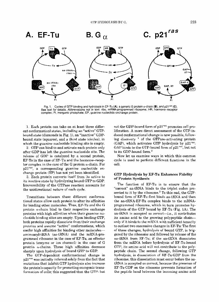
Figure 1 depicts the cycles of GTP binding and hydrolysis by EF-Tu, generic G protein a-chain, and p21"". The three cycles share the following general features:
Received May 12, 1989; accepted June 28, 1989. Address reprint requests to Henry R. Bourne, Box 0450, Uni-
versity of California Medical Center, San Francisco, CA 94143.
GTP HYDROLYSIS BY G, 223
A. EF-TU B. G a ras c. p21
PI $1 1 P I
Fig. 1. Cycles of GTP binding and hydrolysis in EF-TU (A), a generic G protein a-chain (B), and p2lraS (C). See text for details. Abbreviations not in text: ribo, mRNA-programmed ribosome; HR, hormone-receptor complex; Pi, inorganic phosphate; EP, guanine nucleotide exchange protein.
1. Each protein can take on at least three differ- ent conformation1 states, including an “active” GTP- bound state (diamonds in Fig. 11, an “inactive” GDP- bound state (squares), and a third state (circles), in which the guanine nucleotide binding site is empty.
2. GTP can bind to and activate each protein only after GDP has left the guanine nucleotide site. The release of GDP is catalyzed by a second protein, EF-Ts in the case of EF-Tu and the hormone-recep- tor complex in the case of the G protein a-chain. For p21“”, a corresponding guanine nucleotide ex- change protein (EP) has not yet been identified.
3. Each protein converts itself from its active to its inactive state by hydrolyzing bound GTP to GDP. Irreversibility of the GTPase reaction accounts for the unidirectional nature of each cycle.
Transitions between these different conforma- tional states allow each protein to alter its affinities for binding other molecules. Thus, EF-Tu and the G protein a-chain bind to their respective exchange proteins with high affinities when their guanine nu- cleotide binding sites are empty. Upon binding GTP, both proteins rapidly dissociate from their exchange proteins and assume “active” conformations, which confer high affinities for binding other molecules- aminoacyl-tRNA (aa-tRNA) and the mRNA-pro- grammed ribosome in the case of EF-Tu, an effector protein (enzyme or ion channel) in the case of G protein a-chains. These high affinities decrease sharply upon hydrolysis of bound GTP to GDP.
The GTP-dependent conformational change in p21““ was initially inferred solely from the fact that mutations that inhibit GTP hydrolysis also increase the protein’s capacity for promoting oncogenic trans- formation of cells; this suggested that the GTP- but
not the GDP-bound form of ~21‘““ promotes cell pro- liferation. A more direct assessment of the GTP-in- duced conformational change is now possible, follow- ing discovery ’ of the GTPase-activating protein (GAP), which activates GTP hydrolysis by p21““: GAP binds to the GTP-bound form of p21“”, but not to its GDP-bound form.2
Now let us examine ways in which this common cycle is used to perform different functions in the cell.
GTP Hydrolysis by EF-Tu Enhances Fidelity of Protein Synthesis
The function of EF-Tu is to ensure that the “correct” aa-tRNA binds to the triplet codon pre- sented to it by the r i b o ~ o m e . ~ To this end, the GTP- bound form of EF-Tu first binds aa-tRNA and then the aa-tRNA.EF-Tu complex binds to the mRNA- programmed ribosome, which in turn promotes hy- drolysis of the GTP bound by EF-Tu (Fig. 1A). The aa-tRNA is accepted as correct-i.e., it contributes its amino acid to the growing polypeptide chain- only if it binds to the mRNA with sufficient tenacity to outlast two successive changes in EF-Tu: The first of these changes, hydrolysis of bound GTP, is trig- gered by the ribosome and followed by release of the aa-tRNA from EF-Tu; if the aa-tRNA dissociates from the mRNA before hydrolysis of EF-Tu-bound GTP, its amino acid will not contribute to the poly- peptide chain. The second change, following GTP hydrolysis, is dissociation of EF-Tu.GDP from the ribosome; this dissociation must occur before the aa- tRNA is accepted as correct, because the presence of EF-Tu-GDP on the ribosome prevents formation of the peptide bond between the incoming amino acid
224 H.R. BOURNE ET AL.
and the polypeptide chain. Thus the ribosome uses EF-Tu to "proofread" each aa-tRNA twice, by se- quentially assessing the rate of dissociation of aa- tRNA from the ribosome against two temporal in- ternal standards, the rate of GTP hydrolysis and the rate of dissociation of EF-Tu.GDP from the ribosome.* The rate of ribosome-dependent GTP hy- drolysis by EF-Tu plays a major role in determining both the speed and the accuracy of translation.
Signal Amplification by G Protein a-Chains The GTP-dependent conformational change
serves a function in G protein a-chains different from that described for EF-Tu. Instead of ensuring a correct match between two other ligands, the GTP- bound a-chain amplifies the initial signal provided by the hormone-receptor In this way, the a-chain acts as a timer or memory device. For example, the binding of an average hormone ligand molecule to its receptor may persist for a few tens or hundreds of milliseconds. During this time a hor- mone-receptor complex may catalyze GDP release from one or more G protein a-chains. Upon binding GTP and dissociating from the hormone receptor, the active form of the a-chain can stimulate activity of an effector (E in Fig. 1B) for a time period much longer than the few milliseconds that were required to generate the GTP-bound a-chain. The bound GTP is hydrolyzed after a delay of 10-15 seconds. This slow hydrolysis of GTP amplifies the hormone signal by allowing many seconds of effector stimulation in return for a few milliseconds of occupancy of the receptor by hormone. In G protein a-chains and EF- Tu, the bond energy in the y phosphoryl of GTP is used, respectively, to purchase amplified signals and to purchase fidelity of translation.
A crucial difference between the two systems is that GTP hydrolysis is conditional in one but not in the other. Thus, GTP is hydrolyzed by EF-Tu only when the EF-Tu.aa-tRNA complex has bound to the mRNA-programmed ribosome. In contrast, G pro- tein a-chains hydrolyze GTP a t the same rate whether or not effector is present'; intrinsic proper- ties of the a-chain determine the time required for GTP hydrolysis.
Another difference between the two systems is that the GDP-bound inactive form of the G protein a-chain is associated in a heterotrimer with two other polypeptide chains, p and y, which form the by subunit complex. Binding of GTP to the a-chain causes dissociation of a-GTP from the Py subunit. Binding of by to the GDP-bound form serves to sta- bilize GDP binding by the a-chain and also to present the a-chain to the hormone-receptor com- plex. EF-Tu does not possess subunits analogous to p and y.
Finally, it should be noted that inhibition of GTP hydrolysis in the two systems-by mutation, by co- valent modification, or by binding of a hydrolysis
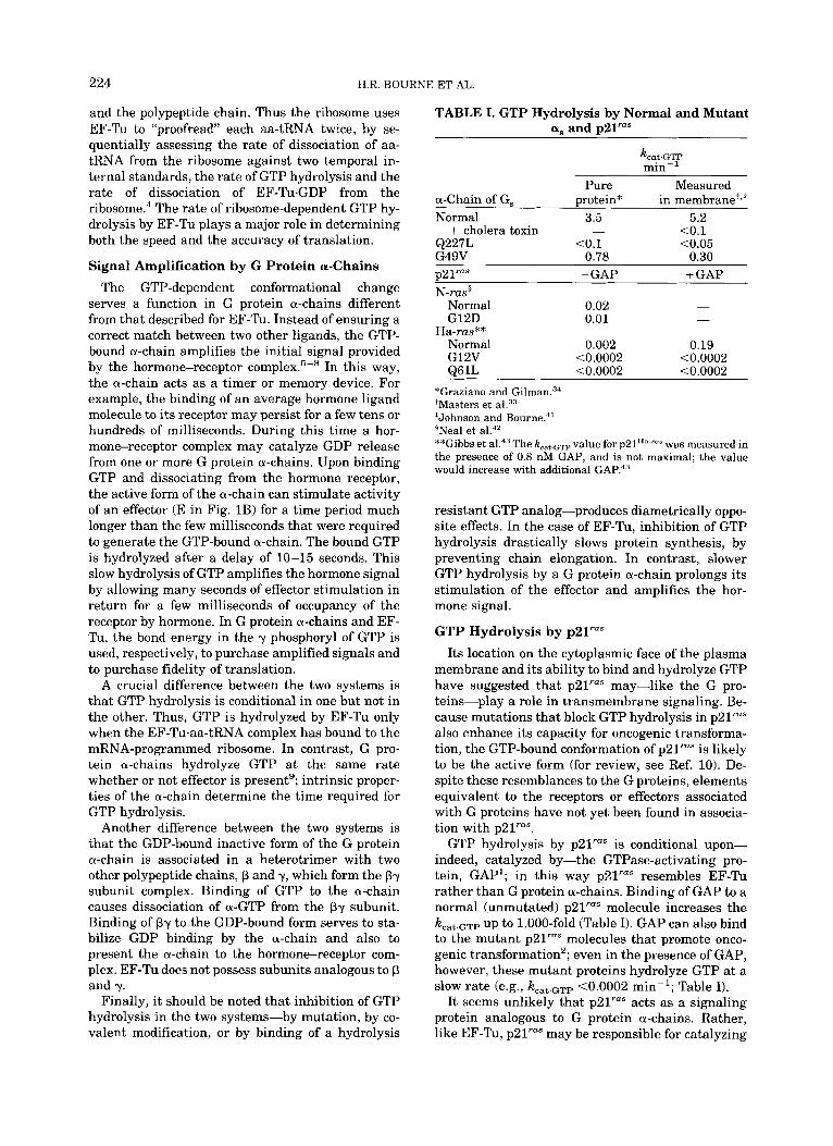
TABLE I. GTP Hydrolysis by Normal and Mutant a, and ~21""
Pure Measured a-Chain of G, protein* in membrane',? Normal 3.5 5.2 + cholera toxin - <0.1 Q227L <0.1 C0.05 G49V 0.78 0.30 p21r'"s -GAP + GAP N-ras$
Normal 0.02 - G12D 0.01 -
Normal 0.002 0.19 G12V <0.0002 <0.0002 861L <0.0002 <0.0002
Ha-rus**
*Graziano and Gilman.34 +Masters et *Johnson and Bourne?' 'Neal et al.42 **Gibbs et al.43 The k,,, GTP value for p21H"-ra" was measured in the presence of 0.8 nM GAP, and is not maximal; the value would increase with additional GAP.43
resistant GTP analog-produces diametrically oppo- site effects. In the case of EF-Tu, inhibition of GTP hydrolysis drastically slows protein synthesis, by preventing chain elongation. In contrast, slower GTP hydrolysis by a G protein a-chain prolongs its stimulation of the effector and amplifies the hor- mone signal.
GTP Hydrolysis by p21""
Its location on the cytoplasmic face of the plasma membrane and its ability to bind and hydrolyze GTP have suggested that p21"" may-like the G pro- teins-play a role in transmembrane signaling. Be- cause mutations that block GTP hydrolysis in p21"" also enhance its capacity for oncogenic transforma- tion, the GTP-bound conformation of p21"" is likely to be the active form (for review, see Ref. 10). De- spite these resemblances to the G proteins, elements equivalent to the receptors or effectors associated with G proteins have not yet been found in associa- tion with p21"".
GTP hydrolysis by p21"" is conditional upon- indeed, catalyzed by-the GTPase-activating pro- tein, GAP1; in this way p21"" resembles EF-Tu rather than G protein a-chains. Binding of GAP to a normal (unmutated) p21"" molecule increases the kcat.GTP up to 1,000-fold (Table I). GAP can also bind to the mutant p21"" molecules that promote onco- genic transformation2; even in the presence of GAP, however, these mutant proteins hydrolyze GTP at a slow rate (e.g., kcat.GTP <0.0002 min-'; Table I).
It seems unlikely that p21"" acts as a signaling protein analogous to G protein a-chains. Rather, like EF-Tu, p21"" may be responsible for catalyzing
GTP HYDROLYSIS BY G, 225
and regulating association between other mole- cules-e.g., GAP and "protein X" (see Fig. 1C and Ref. 11). In this view, the p21"".GAP.X complex is presumed to initiate the (not yet identified) chain of biochemical events responsible for promoting cell proliferation. This interpretation is consistent with several experimental observations: First, mutations that replace residues in the so-called "effector region"" of p2lrU"-i.e., between positions 33 and 40-produce proteins that cannot interact with GAP and that exhibit markedly diminished capacities for promoting oncogenic transformation."* l2 Pheno- types produced by these mutations argue against the possiblity that GAP acts only to stimulate GTP hydrolysis. Second, GAP stimulates the GTPase ac- tivity of a protein (p21R-"") that-unlike p21Ha-rus- does not promote oncogenic transformation of cells.13 This makes it unlikely that GAP is the only "effector" of p2IHa-"".
Other Small GTP-Binding Proteins Recent investigations have discovered a very
large number of GTP-binding proteins in the size range of 20-26 kDa. Stretches of amino acid se- quence in these proteins closely resemble sequences in the guanine nucleotide binding domain of p21"". Although the biochemical functions of these pro- teins are unknown, genetic and biochemical evi- dence suggests14 that many such proteins are in- volved in regulating vectorial transport of secretory and endocytic vesicles between specific membrane compartments within the cell. These GTP-binding proteins could serve-by analogy to EF-Tu-to guar- antee that a specific class of donor vesicles docks specifically and irreversibly on an appropriate ac- ceptor membrane.
STRUCTURE AND FUNCTION OF G PROTEIN &-CHAINS
Biochemical and molecular cloning techniques have led to identification of 11 distinct G protein a-chains in mammals and a multitude of a-chains in many other organisms. Much genetic and biochem- ical investigation has focused on the a-chains of transducin and G,, termed at and a,, respectively. G, mediates stimulation of adenylyl cyclase by hor- mone-receptor complexes, while transducin couples photorhodopsin to stimulation of cGMP phosphodi- esterase in retinal rod cells. Other G proteins couple receptors to other functions-e.g., G, mediates hor- monal inhibition of adenylyl cyclase. The diverse re- ceptoreffector coupling functions of these proteins are reviewed el~ewhere.~, 6, 7* Here we will focus on shared features of these polypeptides in an effort to elucidate the relations between molecular structure and GTP binding and hydrolysis that are presum- ably common to all G proteins.
G protein a-chains contain several stretches of highly conserved amino acid sequence. Small por-
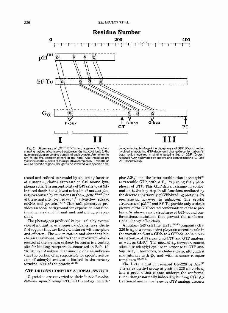
tions of these conserved sequences are similar to stretches of sequence that participate in the GDP- binding pockets of p2l"" and EF-Tu (Fig. 2, top). These portions presumably form the core of the pro- tein machine that harnesses binding and hydrolysis of GTP to amplification of hormonal signals. We used these similarities in amino acid sequence to construct a structure/function model of G protein a- chains.15 Holbrook and Kim" have refined and ex- tended the model, basing it primarily on the three- dimensional structure of ~ 2 l " " . ~ ~ Figure 2 (bottom) depicts the regions of primary structure that con- tribute to the presumptive guanine nucleotide bind- ing domain of G protein a-chains, located with re- spect to amino acid residues in mammalian as. Moving from amino to carboxy terminus, these in- clude:
1. A phosphoryl binding loop (the P-box in Fig. 2), located near the a and f3 phosphoryls of GDP in both EF-Tu"~~' and ~ 2 l ' " " . ~ ~ This glycine-rich region, corresponding to loop 1 (L1)in p21"", is thought to play a key role in catalyzing hydrolysis of bound GTP."
2. The so-called "switch-box" (S-box) region,16 which includes the conserved DXXGQ sequence mo- tif. In p21"" this region is composed of a p-strand and a loop (L4) that interact with the phosphoryl binding lo0pl7 and are postulated'' to play a role in switching the protein from its GDP- to its GTP- bound conformation.
3. The "G-box,"16 containing the NKXD sequence motif. In EF-Tu18,19 and in p2lrU"l7 this region is composed of a loop that interacts with the guanine ring of GDP.
The highly conserved regions that contribute to the guanine nucleotide binding domain divide the rest of the G protein a-chain into three other poten- tial domains15: Domain I, located a t the extreme amino terminus, is poorly conserved among differ- ent G protein a-chains and has no counterpart in p21"". Domain 11, located between the P-box and S-box regions (Fig. 21, is also poorly conserved among different a-chains. Domain I11 contains the carboxy terminal 100 residues of as. A short region (Cys-365 and Ala-366 of a,) in this domain is postulated16 to play a role similar to that of Ser-145 and Ala-146 of p21"", which participate in binding the guanine ring and are thus designated as addi- tional portions of a putative G box. The a-chain model of Holbrook and Kim" proposes that the three-dimensional structure of domain I11 resembles that of the carboxy terminal 60 residues of p21"".
A major goal of our analysis of G protein a-chains is to assign functions to these domains and to un- derstand how they participate in a-chain interac- tions with other proteins-the f3y complex, recep- tors, and effectors-under the control of the guanine nucleotide-binding domain. To this end, we have
226 H.R. BOURNE ET AL.
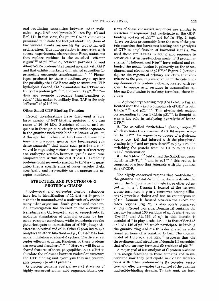
Residue Number
p2ias
EF-TU 1
CT PT u i 1 -
I I1 Fig. 2. Alignments of p21ra”, EF-Tu, and a generic G,-chain,
showing regions of conserved sequence (G) that contribute to the guanine nucleotide binding domain of each protein. Amino termini are at the left, carboxy termini at the right. Also indicated are locations on the a-chain of three putative domains (I, 11, and Ill), as well as specific regions thought to be involved with specific func-
tested and refined our model by analyzing function of mutant a, chains expressed in S49 mouse lym- phoma cells. The susceptibility of S49 cells to CAMP- induced death has allowed selection of mutant phe- notypes caused by mutations in the a, gene.21,22 One of these mutants, termed CYC-,’~ altogether lacks a, mRNA and p r ~ t e i n . ~ ~ , ’ ~ This null phenotype pro- vides an ideal background for expression and func- tional analysis of normal and mutant a, polypep- tides.
The phenotypes produced in cyc- cells by expres- sion of mutant a, or chimeric a-chains have identi- fied regions that are likely to interact with receptors and effectors. The unc mutation and abundant bio- chemical evidence indicate that a predicted a-helix located a t the a-chain carboxy terminus is a contact site for binding receptors (summarized in Refs. 15, 25, 26, 27). Analysis of chimeric a-chains indicates that the portion of a, responsible for specific activa- tion of adenylyl cyclase is located in the carboxy terminal 40% of the pr~tein.’~,~’
GTP-DRIVEN CONFORMATIONAL SWITCH G proteins are converted to their “active” confor-
mations upon binding GTP, GTP analogs, or GDP
I11 tions, including binding of the phosphoryls of GDP (P-box); region involved in mediating GTP-dependent change in conformation (S- box); region involved in binding guanine ring of GDP (G-box); residues ADP-ribosylated by cholera and pertussis toxins (CT and PT, respectively).
plus A1F4- ion; the latter combination is thoughtz9 to resemble GTP, with AlF,- replacing the y phos- phoryl of GTP. This GTP-driven change in confor- mation is the key step in all functions mediated by the diverse superfamily of GTP-binding proteins. Its mechanism, however, is unknown. The crystal structures of p21‘”” and EF-Tu provide only a static picture of the GDP-bound conformation of these pro- teins. While we await structures of GTP-bound con- formations, mutations that prevent the conforma- tional change offer clues.
A mutant S49 cell line, H21a,22,30 pinpoints Gly- 226 in a, as a residue that plays an essential role in the transition from a GDP- to a GTP-dependent con- formation. aS-H2la can bind GTP and GTP analogs, as well as GDP.31 The mutant as, however, cannot stimulate adenylyl cyclase in response to GTP ana- logs, AlF4-, hormones, or cholera toxin, although it can interact with Py and with hormone-receptor c o m p l e ~ e s . ’ ~ ~ ~ ~ ~ ~ ~
The H21a mutation replaced Gly-226 by Ala.31 The extra methyl group at position 226 converts a, into a protein that cannot undergo the conforma- tional change normally induced by binding GTP. Ac- tivation of normal a-chains by GTP analogs protects
GTP HYDROLYSIS BY G, 227
them from cleavage by trypsin a t a conserved site, probably corresponding to Arg-232 of a,. In contrast to the a, of wild-type S49 cells, the GTP-bound mu- tant a, of H21a is not protected from t r y p ~ i n . ~ ~
The Gly residue replaced in a,-H2la is conserved in all GTP-binding proteins, including p21"" and EF-Tu. In p21" it is located in the S-box, at the junction of a P-strand and a turn or loop (L4) that interacts with the phosphoryl-binding loop (Ll). The Gly+Ala substitution in a,-H2la presumably de- creases flexibility of this region and thereby pre- vents transmission to the rest of the protein of the conformational change that should be initiated by binding GTP. In keeping with this hypothesis, the same substitution (Gly-+Ala) at the cognate position (Gly-83) of EF-Tu produces a parallel phenotype: The mutant EF-Tu can bind GTP but is unable to bind aa-tRNA.32 Parallel effects of this subtle struc- tural change in two quite different proteins indicate that they share similar mechanisms of GTP-depen- dent activation, and that the so-called "switch box" (Fig. 2 and Refs. 16, 20) plays a crucial role in this mechanism.
as MUTATIONS DESIGNED TO INHIBIT GTP HYDROLYSIS
Many oncogenic mutations in ~21'"" decrease the protein's ability to hydrolyze GTP. Most such muta- tions are found in two p21"" codons, Gly-12 and Gln- 61." These residues and the amino acid sequences surrounding them are conserved in G protein a- chains. Site-directed mutations of these codons in a, provided an attractive way of confirming that the cognate regions of G protein a-chains participate in GTP binding and hydrolysis. Our laboratory33 ana- lyzed CY, chains carrying these mutations by express- ing them S49 cyc- cells. Graziano and Gilman34 studied the mutant proteins purified from E. coli. The substitutions are Leu for Gln-227 (Q227L) and Val for Gly-49 (G49V). The first, at a position just downstream from the Gly residue replaced by Ala in a,-H2la, corresponds to the Q61L substitution in p21'"", while the second corresponds to the G12V substitution in the phosphoryl-binding loop of p21"".
Broadly speaking, the results confirm the infer- ence, drawn from similarities in primary structure, that amino acid residues surrounding Gly-49 and Gln-227 of a, belong to the protein's guanine nucle- otide-binding domain. The inference may safely be extended to include cognate regions of other G pro- tein a-chains. The results also show, however, clear differences between the guanine nucleotide-binding domains of G protein a-chains and that of ~21"". For example, kcat.GTP values for pure as are nearly iden- tical to those measured with a, in its normal mem- brane environment (Table I). This confirms that GTP hydrolysis by a, does not depend upon the pres-
ence of accessory molecules, unlike the GTPase ac- tivities of EF-Tu and p21"".
Q227L Phenotype The Q227L mutation closely mimics the Q6lL
mutation in p21"". Both mutations cause a substan- tial reduction in kcat.GTp (Table I). The effect of the Q61L ras mutation is emphasized by comparing kcat.GTP values measured in the presence of GAP. a,-Q227L stimulates cAMP synthesis briskly in the presence of GTP alone33.34 and causes cAMP accu- mulation in cells not exposed to stimulating hor- m ~ n e , ~ ~ just as p21"" with the cognate mutation stimulates oncogenic transformation of fibroblasts in the absence of exogenous growth factors.
It is worth emphasizing that-despite its location just downstream of Gly-226 in the "switch-box" re- gion-the Q227L mutation does not affect the "switch" function of a,. a,-Q227L is unable to stim- ulate adenylyl cyclase when its guanine nucleotide- binding site is occupied by GDP. Moreoever, bound GDP fails to protect aS-Q227L from tryptic cleavage. These observation^^^.^^ indicate that the Q227L mu- tation leaves the switch mechanism itself i n t a c t i.e., it does not prevent the protein from taking on the inactive GDP-bound conformation. Instead, the mutation seems to slow a timing device that turns off the switch by triggering hydrolysis of bound GTP.
G49V Phenotype In parallel with substitution of Val for Gly-12 in
p21"", the G49V mutation did reduce GTPase activ- ity of a,. This inhibition of GTPase was not accom- panied, however, by the expected increase in maxi- mal GTP-dependent adenylyl cyclase activity. Instead, relative to normal a,, the G49V protein me- diated a somewhat reduced response to p-adreno- ceptor agonist and virtually no response to A1F4- or hydrolysis-resistant GTP analogs. The reduced ef- fectiveness of a,-G49V may have resulted from rel- ative inability of this protein to take on an "active" conformation, as suggested by the inability of hy- drolysis-resistant GTP analogs to protect the mu- tant protein from tryptic cleavage. In this respect, the as-G49V defect resembles that of a,-H21a.
The effect of the G49V mutation in a, differs pro- foundly from that of the cognate mutation in ~21'"". The small fold reduction in kcat.GTP, relative to nor- mal a,, superficially resembles the similarly small (2- to 10-fold) changes produced by cognate muta- tions in p21'"", when they are measured in the ab- sence of GAP (Table I). This resemblance pales in view of the fact that the kcat.GTP of as-G49V is quite close to that of GAP-stimulated normal p21"" (-0.30 vs. 0.19 min-l), but very much greater than the kcat.GTP of p21ra"-G12V, with or without GAP (0.0002 min-') (Table I). The G49V mutation re- duces kcat.GTP much less than does the Q227L mu-
228 H.R. BOURNE ET AL
tation. In summary, the data suggest that Gly-49 is probably located in the guanine nucleotide-binding domain of a,, although its linkage to the GTPase mechanism in a, differs from the relation of Gly-12 to the GTPase mechanism in p2lrU".
Taken together, the Q227L nad G49V mutations suggest that caution should be exercised in desig- nating structural features of guanine nucleotide binding domains by names that imply function. Al- though the "phosphoryl loop" of p21"" (Ll) may "straddle" the predicted location of the bond be- tween (3 and y phosphoryls of GTP,'6,20 the G49V a, mutation in this loop seems to affect the "switch" more than the GTPase. Conversely, the Q227L and Q6lL mutations profoundly diminish GTPase activ- ity of their respective proteins but-despite location in a region designatedl6Sz0 as a "switch-box"-do not seem to alter the switch at all.
ONCOGENIC MUTATIONS IN as The similar biochemical phenotypes of aS-Q227L
and both p21ra"-Q61L and p2lrU"-G12V suggested that GTPase-inhibiting mutations in G protein a- chains may also prove oncogenic. In order to convert an a, gene into an oncogene, the mutation theoret- ically should occur in a cell that is normally induced to proliferate by hormones that stimulate adenylyl ~ y c l a s e ~ ~ ; such cells include endocrine target cells in thyroid, adrenal cortex, gonads, and pituitary. We have recently identified mutations that inhibit the GTPase of a, in human pituitary tumors.
Growth hormone (GH)-secreting pituitary cells are stimulated to proliferate by GH-releasing hor- mone (GHRH), which acts via intracellular CAMP.^^ Vallar et al.37 reported that a subset of such tumors exhibits constitutively elevated GH secretion, cellu- lar CAMP, and adenylyl cyclase activity. Moreover, G, in membrane extracts from the same tumors is also constitutively active,37 making these tumors excellent candidates for harboring oncogenic a,. In collaboration with Vallar, our laboratory has begun to clone and sequence a, cDNAs from these tumors. An a, mutation that inhibits GTP hydrolysis has been found in each of four tumors so far tested.44
The a, mutation in one tumor is located in the Gln-227 codon, exactly as might have been pre- dicted. In this case Gln-227 is replaced by Arg rather than by Leu. The effect on a, is nonetheless identical to that seen with a,-Q227L, just as the Q61R sub- stitution functionally mimics Q61L in p21m".38 This mutation neatly confirms the hypothesis that a, mu- tations can be oncogenic, but does not materially extend our understanding of the mechanism that regulates GTP hydrolysis.
Mutations in the other three tumors are more in- structive. cDNAs obtained from each tumor exhibit mutations in the Arg-201 codon; two mutations re- place the Arg with Cys, while one substitutes His. A. G. Gilman's laboratory has analyzed functional
changes in a, caused by site-directed mutations that replace Arg-201 by Val, Ala, Lys, and Glu (A. G. Gilman, personal communication). Whether found in human tumors or studied in recombinant mole- cules purified from E. coli, each mutant protein ex- hibits the same phenotype: The R201X proteins re- semble aS-Q227L in their enhanced abilities to stimulate adenylyl cyclase in the presence of GTP. Moreover, R201X mutations dramatically reduce kcat.GTP (results not shown).
Arg-201 is the a, residue that is ADP-ribosylated by cholera toxin, as described above. Effects of the toxin-catalyzed covalent modification are virtually identical to those produced by mutational replace- ments of the same residue: enhanced GTP-depen- dent stimulation of adenylyl cyclase and reduced k,,t.GTp. This functional similarity indicates inti- mate involvement of the side chain of Arg-201 in regulating the GTPase timing device in a,. Duplica- tion of the functional change by mutational replace- ment of Arg-201 by many different amino acids, in- cluding Lys, makes it quite likely that the side chain of this residue interacts with or forms a part of the molecular timing device that regulates
All known G protein a-chains contain an Arg res- idue at the position corresponding to Arg-201.l' Of the other a-chains, only that of transducin is effi- ciently ADP-ribosylated by cholera toxin; this cova- lent modification reduces GTPase activity of just as it does that of a,. Mutational replacements of the conserved Arg residue will reveal whether i t plays a similar role in regulating GTPase in other a-chains, even though it may not be accessible to enzymatic modification by cholera toxin.
POSSIBLE ROLE OF DOMAIN I1 IN REGULATING GTP HYDROLYSIS
The location of Arg-201 and its cognate residues in other a-chains prompts an additional speculation, derived from the ability of GAP to "reset" the GTPase timing device of p21"" and the contrasting freedom of G protein a-chains from any requirement for a GAP-like protein.
Arg-201 is located in domain 11, between the pre- sumptive phosphoryl loop and switch box regions of a, (Fig. 2). The 162 residues of this presumptive do- main make it considerably longer than the topolog- ically equivalent sequence of p21"" (31 residues). In p21"" this sequence contains residues 33-40, termed the "effector region" by Sigal et a1.I' because mutational replacements of these residues abolish the ras protein's capacity for promoting oncogenic transformation. These mutations also prevent GAP from stimulating GTP hydrolysis and appear even to prevent GAP from binding to ~ 2 1 ~ " . ~ 2 ''* 40 The lo- cation of these residues in L2, which interacts with the phosphoryl binding loop of p21"", strongly suggests" that L2 is the part of the molecule to which GAP binds when it triggers GTP hydrolysis.
GTP HYDROLYSIS BY G, 229
In this context, we propose that domain I1 serves as an intrinsic or "built-in" GAP for G protein a- chains. This notion provides tentative explanations for 1) the large "excess" sequence in domain 11, rel- ative to p21"" and EF-Tu, proteins whose GTPase activities require stimulation by other molecules; 2) location of the excess a, sequence in a region topo- logically equivalent to L2 of p21""; 3) the fact that a, (and other G protein a-chains) do not require an exogenous GAP activity; indeed, the k,,t.GTP of a, is identical for the pure recombinant protein, the pure protein in the presence of adenylyl cyclase, and the protein expressed in cyc-membranes (Table I and Ref. 9). In this scenario, Arg-201 and cognate Arg residues in other G protein a-chains play key roles in the interaction between the GTP-binding do- mains of these proteins and their intrinsic GAP-like domains. This interaction, conserved in all G protein a-chains, is interrupted by oncogenic mutations that replace Arg-201 or by bacterial toxins that modify its side chain.
Although the scenario is highly speculative, some of its elements can be readily tested, by examining phenotypes produced by additional mutations and deletions in domain 11, the Arg-201 region, and the guanine nucleotide-binding domain.
PERSPECTIVE Early in the course of evolution, the GTP-binding
site of a primordial protein combined an ability to change the protein's conformation with an ability to hydrolyze bound GTP, thereby creating a switch- and-timer mechanism of immense power and versa- tility. Duplication and divergence of genes created many new versions of this machine, which we now study as members of the superfamily of GTP binding proteins. Owing to their apparently inherited simi- larities of structure and function, much of what we learn about a,, p21"", or EF-Tu as individual mol- ecules helps us to understand crucial functions of other members of the superfamily.
For understanding how the basic machine works, the signaling G proteins will eventually prove as useful as p21"" or EF-Tu-and perhaps more so- despite the availability of crystal structures for the latter two proteins. Knowledge of the functions of G proteins and of the proteins with which they interact provides multiple handles for their biochemical and genetic manipulation.
Aside from their intrinsic biological interest, we may find other reasons to explore the conserved mo- lecular machinery utilized by these proteins. Muta- tions or bacterial toxins cause disease by impairing the switch-and-timer mechanisms of rus proteins and the a-chain of G,. Other G proteins carry signals that regulate important cell functions, including proliferation, through many pathways in addition to CAMP. Malfunctions of these other G proteins are highly like to cause disease. Applying our knowl-
edge of p21"" and a, to these additional proteins may also turn out to have significant practical con- sequences.
REFERENCES 1. Trahey, M., McCormick, F. A cytoplasmic protein stimu-
lates normal N-ras p21 GTPase, but does not affect onco- genic mutants. Science 238:542-545, 1987.
2. Vogel, U.S., Dixon, R.A.F., Schaber, M.D., Diehl, R.E., Marshall, M.S., Scolnick, E.M., Sigal, IS., Gibbs, J.B. Cloning of bovine GAP and its interaction with oncogenic rus p21. Nature (London) 33590-93, 1988.
3. Kaziro, Y. The role of guanosine 5'-triphosphate in poly- peptide chain elongation. Biochim. Biophys. Acta 505:95- 127, 1978.
4. Thompson, R.C., Dix, D.B., Karim, A.M. The reaction of ribosomes with elongation factor Tu-GTP complexes. Ami- noacyl-tRNA-independent reactions in the elongation cy- cle determine the accuracy of protein synthesis. J . Biol. Chem. 261:4868-4874, 1986.
5. Stryer, L., Bourne, H.R. G proteins: A family of signal transducers. Annu. Rev. Cell Biol. 2:391-419, 1986.
6. Birnbaumer, L., Codina, J., Mattera, R., Yatani, A,, Scherer, N., Toro, M.J., Brown, A.M. Signal transduction by G proteins. Kidney Int (Suppl.) 32:S14S37, 1987.
7. Casey, P.J., Gilman, A.G. G protein involvement in recep- tor-effector coupling. J . Biol. Chem. 2632577-2580, 1988.
8. Neer, E.J., Clapham, D.E. Roles of G protein subunits in transmembrane signalling. Nature (London) 333:129- 134,1988.
9. Graziano, M.P., Freissmuth, M., Gilman, A.G. Expression of G,, in Escherichiu coli. Purification and properties of two forms of the protein. J. Biol. Chem. 264:409-418, 1989.
10. Barbacid, M. rus Genes. Annu. Rev. Biochem. 56:779-827, 1987.
11. McCormick, F. ras GTPase activating protein: Signal transmitter and signal terminator. Cell 565-8, 1989.
12. Sigal, IS,. Gibbs, J.B., DAlonzo, J.S., Scolnick, E.M. Iden- tification of effector residues and a neutralizing epitope of Ha rus p21. Proc. Natl. Acad. Sci. U.S.A. 83:4725-4729, 1986.
13. Garrett, M.D., Self, A.J., van Oers, C., Hall, A. Identifica- tion of distinct cytoplasmic targets for ras/R-ras and rho reeulatorv uroteins. J. Biol. Chem. 264:lO-13. 1989.
14. Bturne, H.R. Do GTPases direct membrane traffc in se-
15. Masters, S.B., Stroud, R.M., Bourne, H.R. Family of G pro- cretion? Cell 53:669-671, 1988.
tein 01 chains: Amphipathic analysis and predicted s&- ture of functional domains. Protein Engineer. 1:47-54, 1986.
16. Holbrook, S.R., Kim, S.H. Molecular model of the G pro- tein a subunit based on the crystal structure of the HRAS protein. Proc. Natl. Acad. Sci. U.S.A. 86:1751-1755, 1989.
17. de Vos, A.M., Tong, L., Milburn, M.V., Matias, P.M., Jan- carik, J., Noguchi, S., Nishimura, S., Miura, K., Ohtsuka, E., Kim, S.H. Three-dimensional structure of an oncogene protein: Catalytic domain of human c-H-rus p21. Science 239:888-893, 1988.
18. Jurnak, F. Structure of the GDP domain of EF-Tu and location of the amino acids homologous to ras oncogene proteins. Science 230:32-36, 1985.
19. La Cour, T.F.M., Nyborg, J., Thirup, S., Clark, B.F.C. Structural details of the binding of guanosine diphosphate to elongation factor Tu from E. coli as studied by X-ray crystallography. EMBO J. 4:2385-2388, 1985.
20. Kim, S.H., de Vos, A.M., Tong, L., Milburn, M.V., Matias, P.M., Jancarik, J., Nishimura, S. Ras oncogene proteins: Three-dimensional structures, functional implications, and a model for signal transducer. Cold Spring Harbor Symp. 53:273-281, 1988.
21. Bourne, H.R., Coffino, P., Tomkins, G.M. Selection of a variant lymphoma cell deficient in adenylate cyclase. Sci- ence 187:750-752, 1975.
22. Salomon, M.R., Bourne, H.R. Novel S49 lymphoma vari- ants with aberrant cyclic AMP metabolism. Mol. Pharma- col. 19:109-116, 1981.
23. Harris, B.A.. Robishaw. J.D., Mumbv, S.M., Gilman, A.G. Molecular cloning of complementary DNA' for the alpha
230 H.R. BOURNE ET AL.
subunit of the G protein that stimulates adenylate cyclase. Science 229:1274-1277, 1985.
24. Robishaw, J.D., Russell, D.W., Harris, B.A., Smigel, M.D., Gilman, A.G. Deduced primary structure of the OL subunit of the GTP-binding stimulatory protein of adenylate cy- clase. Proc. Natl. Acad. Sci. U.S.A. 83:1251-1255, 1986.
25. Sullivan, K.A., Miller, R.T., Masters, S.B., Beiderman, B., Heideman, W., Bourne, H.R. Identification of receptor con- tact site invovled in receptor-G protein coupling. Nature (London) 330:758-760, 1987.
26. Rall, T., Harris, B.A. Identification of the lesion in the stimulatory GTP-binding protein of the uncoupled S49 lymphoma. FEBS Lett. 224:365-371, 1987.
27. Bourne, H.R., Masters, S.B., Miller, R.T., Sullivan, K.A., Heideman, W. Mutations probe structure and function of G protein OL chains. Cold Spring Harbor Symp. Quant. Biol. 53221-228, 1988.
28. Masters, S.B., Sullivan, K.A., Miller, R.T., Beiderman, B., Lopez, N.G., Ramachandran, J., Bourne, H.R. Carboxy ter- minal domain of G,, specifies coupling of receptors to stim- ulation of adenylyl cyclase. Science 241:448-451, 1988.
29. Bigay, J., Deterre, P., Pfister, C., Chabre, M. Fluoroalumi- nates activate transducin-GDP by mimicking the y-phos- phate of GTP in its binding site. FEBS Lett. 191:181-185, 1985.
30. Bourne, H.R., Kaslow, D., Kaslow, H.R., Salomon, M.R., Licko, V. Hormone-sensitive adenylate cyclase: Mutant phenotype with normally regulated P-adrenergic receptors uncoupled from catalytic adenylate cyclase. Mol. Pharma- col. 20:435-441, 1981.
31. Miller, R.T., Masters, S.B., Sullivan, K.A., Beiderman, B., Bourne, H.R. A mutation that prevents GTP-dependent activation of the (I chain of G,. Nature (London) 334:712- 715, 1988.
32. Hwang, Y.W., Jurnak, F., Miller, D.L. A mutation that hinders the GTP induced aminoacyl-tRNA binding of elon- gation factor TU. In: “The Guanine-Nucleotide Binding Proteins: Common Structural and Functional Properties.” New York: Plenum, 1989: In press.
33. Masters, S.B., Miller, R.T., Chi, M.H., Chang, F.H., Bei-
derman, B., Lopez, N.G., Bourne, H.R. Mutations in the GTP-binding site of G,, alter stimulation of adenylyl cy- clase. J. Biol. Chem., 264:15467-15474, 1989.
34. Graziano, M., Gilman, A.G. Synthesis in Escherichia coli of GTPase-deficient mutants of Ge,. J. Biol. Chem., 264: 15475-15482, 1989.
35. Bourne, H.R. G proteins and CAMP. Discovery of a new oncogene in pituitary tumours? Nature (London) 330517- 518, 1987.
36. Billestrup, N., Swanson, L.W., Vale, W. Growth hormone- releasing factor stimulates proliferation of somatotrophs in uitro. Proc. Natl. Acad. Sci. U.S.A. 83:6854-6857, 1986.
37. Vallar, L., Spada, A,, Giannattasio, G. Altered G, and ade- nylate cyclase activity in human GH-secreting pituitary adenomas. Nature (London) 330:566-568, 1987.
38. Der, C.J., Finkel, T., Cooper, G.M. Biological and biochem- ical properties of human rusH genes mutated a t codon 61. Cell 44:167-176, 1986.
39. Abood, M.E., Hurley, J.B., Pappone, M.C., Bourne, H.R., Stryer, L. Functional homology between signal-coupling proteins: Cholera toxin inactivates the GTPase activity of transducin. J . Biol. Chem. 257:10540-10543, 1982.
40. Adari, H., Lowry, D.R., Willumsen, B.M., Der, C.J., Mc- Cormick, F. Guanosine triphosphatase activating protein (GAP) interacts with the p21““ effector binding domain. Science 240:518-521, 1988.
41. Johnson, G.L., Bourne, H.R. Influence of cholera toxin on the regulation of adenylate cyclase by GTP. Biochem. Bio- phys. Res. Commun. 78:792-798, 1977.
42. Neal, S.E., Eccleston, J.F., Hall, A,, Webb, M.R. Kinetic analysis of the hydrolysis of GTP by p21N-ras. The basal GTPase mechanism. J . Biol. Chem. 263:19718-19722, 1988.
43. Gibbs, J.B., Schaber, M.D., Allard, W.J., Sigal, I.S. Purifi- cation of ras GTPase activating protein from bovine brain. Proc. Natl. Acad. Sci. U.S.A. 855026-5030, 1988.
44. Landis, C.A., Masters, S.B., Spada, A,, Pace, A.M., Bourne, H.R., Vallar, L. GTPase inhibiting mutations activate the a-chain of G, and stimulate adenylyl cyclase in human pituitary tumors. Nature (London) 340:692-696, 1989.