hyperbaric oxygen therapy for the adjunctive treatment of … · the early period following tbi may...
TRANSCRIPT

Hyperbaric oxygen therapy for the adjunctive treatment of traumatic
brain injury Bennett MH, Trytko B, Jonker B
This review should be cited as: Bennett MH, Trytko B, Jonker B. Hyperbaric oxygen therapy for the adjunctive treatment of traumatic brain injury (Cochrane Review). In: The Cochrane Library,
Issue 1, 2006. Oxford: Update Software.
A substantive amendment to this systematic review was last made on 16 June 2004. Cochrane reviews are regularly checked and updated if necessary.
Abstract Background: Traumatic brain injury is common and presents a health problem with significant effect on quality of life. Hyperbaric oxygen therapy (HBOT) has been suggested to improve oxygen supply to the injured brain and, therefore, to reduce the volume of brain that will ultimately perish. It is postulated that the addition of HBOT to the standard intensive care regimen may result in a reduction in patient death and disability as a result of these additional brain-preserving effects.
Objective: To assess the benefits and harms of adjunctive HBOT for treating traumatic brain injury.
Search strategy: We searched CENTRAL (The Cochrane Library Issue 4, 2003), MEDLINE (1966 - 2003), EMBASE (1974 - 2003), CINAHL (1982 - 2003), DORCTHIM (1996 - 2003), and reference lists of articles. Relevant journals were handsearched and researchers in the field were contacted.
Selection criteria: Randomised studies comparing the effect on traumatic brain injury of therapeutic regimens which include HBOT with those that exclude HBOT (with or without sham therapy).
Data collection and analysis: Three reviewers independently evaluated the quality of the relevant trials using the validated Oxford-Scale (Jadad 1996) and extracted the data from the included trials.
Main results: Four trials contributed to this review (382 patients, 199 receiving HBOT and 183 control). There was a trend towards, but no significant increase in, the chance of a favourable outcome when defined as full recovery, Glasgow outcome score 1 or 2, or return to normal activities of daily living (relative risk [RR] for good outcome with HBOT 1.94, 95% confidence interval [CI] 0.92 to 4.08, P=0.08). Pooled data from the three trials with 327 patients that reported mortality, showed a significant reduction in the risk of dying when HBOT was added to the treatment regimen (RR 0.69, 95% CI 0.54 to 0.88, P=0.003). Heterogeneity between studies was low (I2 =0%), and sensitivity analysis for the allocation of dropouts did not affect that result. This analysis suggests we would have to treat seven patients to avoid one extra death (number needed to treat [NNT] 7, 95% CI 4 to 22). One trial suggested intracranial pressure was favourably lower in those patients receiving HBOT in whom myringotomies had been performed (WMD with myringotomy -8.2 mmHg, 95% CI -14.7 mmHg to -1.7 mmHg, P=0.01), while in two trials there was a reported incidence of 13% for significant pulmonary impairment in the group receiving HBOT versus 0% in the non-HBOT group (P=0.007).

Reviewers' conclusions: In people with traumatic brain injury, the addition of HBOT significantly reduced the risk of death but not of favourable clinical outcome. The routine application of HBOT to these patients cannot be justified from this review. In view of the modest number of patients, methodological shortcomings and poor reporting, this result should be interpreted cautiously, and an appropriately powered trial of high methodological rigour is justified to define those patients (if any) who can be expected to derive most benefit from HBOT.
Background Traumatic brain injury (TBI) is a significant cause of premature death and disability. Each year, there are at least 10 million new head injuries worldwide and these account for a high proportion of deaths in young adults (Thurman 1999; Alexander 1992). In the US there are more than 50,000 deaths due to traumatic brain injury each year. The major causes are motor vehicle crashes, falls, and violence (including suicide). Prevention strategies, including restraints for vehicle occupants, are now legally enforced in many countries. However, while road death rates are falling in most industrialised countries, they are rising in many rapidly motorising countries, particularly in Asia. For example, road death rates per head in China are already similar to those in the United States (Roberts 1995). Head injuries are associated with long-term disability in many patients. In the US, for example, 2% of the population (5.3 million citizens) are living with disability as a result of TBI (Thurman 1999) and this places considerable medical, social and financial burden on both families and health systems (Fearnside 1997).
The pathophysiology of brain injury has a primary and secondary component. At the time of impact there is a variable degree of irreversible damage to the neurological tissue (primary injury). Following this, a chain of events occurs in which there is ongoing injury to the brain through oedema, hypoxia and ischaemia secondary to raised tissue or intracranial pressure, release of excitotoxic levels of excitatory neurotransmitters (e.g. glutamate), and impaired calcium homeostasis (Tymianski 1996; Fiskum 2000) (secondary injury).
Therapy focuses on prevention and/or minimisation of secondary injury by ensuring adequate oxygenation, haemodynamics, control of intracranial hypertension, and strategies to reduce cellular injury. A number of therapies, including barbiturates, calcium channel antagonists, steroids, hyperventilation, mannitol, hypothermia and anticonvulsants have been the topic of previous Cochrane reviews, though none has shown unequivocal efficacy in reducing poor outcome (Schierhout 2003; Roberts 2003a; Langham 2003; Alderson 2003; Roberts 2003b).
Hyperbaric oxygen therapy (HBOT) is a further adjunctive therapy that has been proposed to improve outcome in acute brain injury. HBOT is the therapeutic administration of 100% oxygen at environmental pressures greater than 1 atmosphere absolute (ATA). This involves placing the patient in an airtight vessel, increasing the pressure within that vessel, and administering 100% oxygen for respiration. In this way, it is possible to deliver a greatly increased partial pressure of oxygen to the tissues. Typically, treatments involve pressurisation to between 1.5 and 3.0 ATA, for periods between 60 and 120 minutes, once or more daily.
Since the 1960s, there have been reports that HBOT improves the outcome following brain trauma (Fasano 1964). Administration of HBOT is based on the observation that hypoxia following closed head trauma is an integral part of the secondary injury described above. Hypoxic neurons performing anaerobic metabolism result in acidosis and an unsustainable reduction in cellular metabolic reserve (Muizelaar 1989). As the hypoxic situation persists, the neurons lose their ability to maintain ionic homeostasis, and free oxygen radicals accumulate and degrade cell membranes (Ikeda 1990; Siesjo 1989). Eventually, irreversible changes result in unavoidable cell death. When ischaemia is severe enough, these changes occur rapidly, but there is some evidence that these effects can occur over a period of days (Robertson 1989). This gives some basis to the assertion that a therapy designed to increase oxygen availability in the early period following TBI may improve long-term outcome. HBOT is also thought to reduce

tissue oedema by an osmotic effect (Hills 1999), and any agent that has a positive effect on brain swelling following trauma might also contribute to improved outcomes. On the other hand, oxygen in high doses is potentially toxic to normally perfused tissue, and the brain is particularly at risk (Clark 1982). For this reason, it is appropriate to postulate that in some TBI patients, HBOT may do more harm through the action of increased free oxygen radical damage, than good through the restoration of aerobic metabolism.
Despite 40 years of interest in the delivery of HBOT in these patients, little clinical evidence of effectiveness exists. HBOT has been shown to reduce both intracranial pressure (ICP) and cerebrospinal fluid pressure (CSFP) in brain-injured patients (Suckoff 1982; Hayakawa 1971), improve grey matter metabolic activity on SPECT scan (Neubauer 1994), and improve glucose metabolism (Holbach 1977). Some studies suggest that any effect of HBOT may not be uniform across all brain-injured patients. For example, Hayakawa demonstrated that CSFP rebounded to higher levels following HBOT than at pre-treatment estimation in some patients, while others showed persistent reductions (Hayakawa 1971). It is possible that HBOT has a positive effect in a sub-group of patients with moderate injury, but not in those with extensive cerebral injury. Furthermore, repeated exposure to hyperbaric oxygen may be required to attain consistent changes (Artru 1976). Clinical reports have attributed a wide range of improvements to HBOT including cognitive and motor skills, improved attention span and increased verbalisation (Suckoff 1982; Neubauer 1994). These improvements are, however, difficult to ascribe to any single treatment modality because HBOT was most often applied in conjunction with intensive supportive and rehabilitative therapies.
HBOT is associated with some risk of adverse effects, including damage to the ears, sinuses and lungs from the effects of pressure, temporary worsening of short-sightedness, claustrophobia and oxygen poisoning. Although serious adverse events are rare, HBOT cannot be regarded as an entirely benign intervention. Further, it is conceivable that the addition of HBOT might improve survival from serious brain injury without improving the proportion of those who survive with a useful functional level, while at the same time increasing overall costs of therapy. For a number of reasons, therefore, the administration of HBOT for TBI patients remains controversial.
Objectives The aim of this review is to assess the evidence for the benefit or harm of adjunctive HBOT in the treatment of acute TBI. We compared intensive treatment regimens including adjunctive HBOT against similar regimens excluding HBOT. Where regimens differed significantly between studies, this is clearly stated and the implications discussed.
Criteria for considering studies for this review Types of studies
Randomised and quasi-randomised controlled trials that compare the effect of treatment for acute TBI where HBOT administration is included, with the effect of similar treatment in the absence of HBOT.
Types of participants
Any person admitted to an intensive care or intensive neurosurgical facility with an acute TBI following blunt trauma.
Types of intervention

HBOT administered in a compression chamber between pressures of 1.5 ATA and 3.5 ATA and treatment times between 30 minutes and 120 minutes at least once. We accepted any standard treatment regimen designed to maximise brain protection and promote recovery from TBI. We did not include studies where comparator interventions were not undertaken in a specialised acute care setting.
Types of outcome measures
Studies were eligible for inclusion if they reported any of the following outcome measures at any time.
Primary outcomes
• Functional outcome. (Defined as 'good' if: Glasgow outcome score (GOS) of 1 or 2, described as 'fully recovered' or 'returning to normal activities of daily living'.)
• Mortality. Where there were multiple times recorded, we chose final follow-up.
• ···· • Secondary outcomes
• Activities of daily living (ADL). • Intracranial pressure (ICP). • MRI or CT evidence of lesion resolution or size of persistent defect. • Progress of Glasgow coma scale (GSS). • Cost-effectiveness. • Adverse events of HBOT.
• ···· • The timing of outcome evaluations varied between studies. In general, our aim was to
group outcomes into three stages for analysis: early(immediately after treatment course), medium term (four to eight weeks after treatment), and longer-term (six months or longer).
Search strategy for identification of studies See: Cochrane Injuries Group search strategy
We aimed to capture both published and unpublished studies.
• Electronic searches • We searched: the Cochrane Controlled Trials Register (CENTRAL Issue 3, 2003),
MEDLINE (Ovid), CINAHL, EMBASE and an additional database developed in our hyperbaric facility (the Database of Randomised Trials in Hyperbaric Medicine, Bennett 2002). All databases were searched from inception to late 2003. The search strategy was broad and the keywords in the following strategies were adapted as appropriate. The EMBASE and MEDLINE (Ovid) strategies are given in Table 01.
In addition we made a systematic search for relevant controlled trials in specific hyperbaric literature sources as follows.
• Experts in the field and leading hyperbaric therapy centres (as identified by personal communication and searching the Internet) were contacted and asked for additional relevant data in terms of published or unpublished randomised trials.

• Handsearch of relevant hyperbaric textbooks (Kindwall, Jain, Marroni, Bakker, Bennett and Elliot), journals (Undersea and Hyperbaric Medicine, Hyperbaric Medicine Review, South Pacific Underwater Medicine Society (SPUMS) Journal, European Journal of Hyperbaric Medicine and Aviation, Space and Environmental Medicine Journal) and conference proceedings (Undersea and Hyperbaric Medical Society, SPUMS, European Undersea and Baromedical Society, International Congress of Hyperbaric Medicine) published since 1980.
• Contact with authors of relevant studies to request details of unpublished or ongoing investigations.
• ···· • All languages were considered. Authors were contacted if there was any ambiguity
about the published data.
Methods of the review • Data retrieval and management • One reviewer (MB) was responsible for handsearching and identification of appropriate
studies for consideration. Two reviewers (MB and BJ) examined the electronic search results to identify studies that may have been relevant and these studies were entered into a bibliographic software package (Review Manager), whether one or both reviewers considered this appropriate. All comparative clinical trials identified were retrieved in full and reviewed independently by three reviewers, two with content expertise with HBOT, and two with content expertise in treating acute TBI. (BT practices in both areas.) In addition, one of the reviewers (MB) has expertise in clinical epidemiology. Reviewers recorded data using the data extraction form developed for this review.
• Data extraction • Using the data extraction form developed for this review, each reviewer extracted
relevant data, graded the studies for methodological quality using the method of Jadad (Jadad 1996), and made a recommendation for inclusion or exclusion from the review. The method of Jadad scores trials on three criteria (randomisation, double-blinding and description of withdrawals), each of which, if present, is given a score of 1. Further points are available for description of a reliable randomisation method and use of a placebo (modified for our analysis to include a sham HBOT session). The scores are totalled as an estimate of overall quality and we required a score of at least 1 for inclusion in the review. Any differences were settled by consensus. In addition, we indicated for each study selected for inclusion: the method of allocation, adequacy of concealment of allocation, blinding status of participants and outcome observers, and how patient attrition was handled. These factors were considered for possible sensitivity analysis. All data extracted reflected original allocation group where possible, to allow an intention-to-treat analysis. Dropouts were identified, where this information was given.
• Analyses • All comparisons were made using an intention-to-treat analysis where possible and
reflect efficacy in the context of randomised trialling, rather than true effectiveness in any particular clinical context. For proportions (dichotomous outcomes), relative risks (RRs) were used, with 95% confidence interval (CI). We used a fixed-effect model where there was no evidence of significant heterogeneity between studies (see below), and a random effects model when such heterogeneity was likely.
• Primary outcomes • 1) Proportion of subjects with good functional outcome (e.g. GOS) were dichotomised.
Subjects with a good recovery or moderate disability were included in the 'good' group, while those who were severely disabled, remained in a vegetative state or died were included in the 'bad' group.) The RR for good outcome with HBOT was established using the intention-to-treat data of the HBOT, versus the control group. Analyses were

performed with RevMan 4.2 software. As an estimate of the statistical significance of a difference between experimental interventions and control interventions, we calculated RR for benefit using HBOT with 95% confidence intervals (CIs). A statistically significant difference between experimental intervention and control intervention was assumed if the 95% CI of the RR did not include the value 1.0. As an estimate of the clinical relevance of any difference between experimental intervention and control intervention, we calculated the number-needed-to-treat (NNT) and number-needed-to-harm (NNH) with 95% CI as appropriate.
• 2) Proportion of those surviving. The RR for death with and without HBOT was calculated, using the methods described in (1) above.
• Secondary outcomes • 3) Activities of daily living (ADL).The weighted mean differences (WMDs) in ADL
between HBOT and control groups were to be compared using RevMan 4.2. A statistically significant difference was defined as existing if the 95% CI did not include a zero WMD.
• 4) Intracranial pressure. The WMDs in ICP between the HBOT and control groups were calculated in a way analogous to (3) above.
• 5) Dichotomous data were considered for adverse events (number of patients with adverse events versus number of patients without them in both groups) in the HBOT groups of the included studies.
• Sensitivity analyses • We planned to perform sensitivity analyses for missing data and study quality. • Missing data • We employed sensitivity analyses, using different approaches to imputing missing data.
The best-case scenario assumed that none of the originally enrolled patients missing from the primary analysis in the treatment group had the negative outcome of interest, whilst all those missing from the control group did. The worst-case scenario was the reverse.
• Study quality • If appropriate, we also planned to conduct a sensitivity analysis by study quality, based
on the presence or absence of a reliable random allocation method, concealment of allocation, and blinding of participants or outcome assessors.
• Subgroups • Where appropriate data was found, we considered subgroup analysis based on:
• age - adults versus children • dose of oxygen received (pressure, time and length of treatment course) • nature of the comparative treatment modalities • severity of injury • nature of injury on CT scan.
• ···· • Heterogeneity was explored and subgroup analyses performed as appropriate.
Statistical heterogeneity was assessed using the I2 statistic and consideration given to the appropriateness of pooling and meta-analysis.
Description of studies We identified 23 publications dealing with the use of HBOT for the treatment of brain injury. Initial examination confirmed nine were case reports or case series, four were reviews without new data, one was an animal study and one a letter. These reports were excluded. One report was unobtainable but deemed unlikely to be a randomised trial (Belokurov 1998), leaving seven possible randomised comparative trials. After appraisal of the full reports we excluded two publications as preliminary or secondary reports containing no data additional to that in the final

publication (Ren 2001b; Rockswold 1985), and one publication that dealt with non-acute head injury (Shi 2003). The other four trials were accepted into the review (Ren 2001a; Rockswold 1992; Artru 1976; Holbach 1974).
The included trials were published between 1974 (Holbach 1974) and 2001 (Ren 2001a), and the reviewers are unaware of any on-going RCTs in the area. In total, these trials include data on 382 participants, 199 receiving HBOT and 183 control. The largest (Rockswold 1992) accounts for 44% of cases. (See Table: 'Characteristics of included studies').
Both the dose of oxygen per treatment session and for the total course of treatment varied between studies. The lowest dose administered was 1.5 ATA for 60 minutes daily (Holbach 1974), while the highest dose was 2.5 ATA for 40 to 60 minutes, 10 times in four days (Ren 2001a). All authors used between 1.5 and 2.5 ATA as a maximum oxygen pressure. The total number of individual treatment sessions varied from 10.5 (Artru 1976) to between 30 and 40 (Ren 2001a).
No trial administered a sham treatment and only Rockswold 1992 attempted any concealment by blinding the outcome assessor. All trials included participants with severe closed head injury: two trials specified a GCS grade on admission (Ren 2001a, GCS <9; Rockswold 1992, GCS <10), one specified the severity of coma using the Jouvet Scale (Artru 1976; Jouvet 1960), and the remaining trial stated the patients were comatose on admission (Holbach 1974). Enrolment was completed some time after admission to allow more trivial injuries to recover in two of the trials (Ren 2001a, day 3; Rockswold 1992, 6 hours), while Artru 1976 and Holbach 1974 did not specify any period before entry. Specific exclusion criteria varied between trials, but open head injuries and those with other than isolated head trauma were excluded when any criteria were specified.
All trials compared a standard intensive treatment regimen to the same regimen with the addition of HBOT. Reported details of the standard regimen are given in 'Characteristics of included studies'.
The follow-up periods varied between 12 days following admission (Holbach 1974), to 6 months (Ren 2001a), 1 year (Artru 1976) and 1.5 years (Rockswold 1992). All included studies reported at least one clinical outcome of interest. Of the outcomes identified above, these trials reported data on both primary outcomes (good functional outcome and mortality), but only intracranial pressure and adverse events from the secondary outcomes of interest.
Other outcomes (including non-clinical) reported included: survival time (Holbach 1974), duration of coma (Artru 1976), GCS before and after treatment (Ren 2001a), brain-stem auditory evoked potentials (BAEP) and short-latency somatosensory evoked potentials (SSEP) (Rockswold 1992), and brain electric activity mapping (BEAM) (Ren 2001a).
Methodological quality Given that only randomised studies were eligible for inclusion, the maximal quality score possible was 5, and the minimum 1. Study quality was generally assessed as low. Two of the four included studies were assigned a score of 2 (Artru 1976; Rockswold 1992), while the remaining studies were assigned a score of 1 (Ren 2001a; Holbach 1974). The significance of this small variation is unclear and it was not used as a basis for sensitivity analysis by study quality.
• Randomisation • Allocation concealment was not adequate in any of the studies, being inadequate in
Holbach 1974 and unclear in the remaining studies. Randomisation procedures were not described in any of the studies, and were unlikely to have been truly random for Holbach 1974, where the selection method was described as 'every second patient was treated with HBOT'. For none of the studies is there a clear indication that the

investigators were unable to predict the prospective group to which a participant would be allocated.
• Patient baseline characteristics • All patients had suffered a head injury. Two of the studies closely defined entry criteria
as those patients with an isolated, closed head injury with a specified GCS persisting for longer than six hours (Rockswold 1992), or three days (Ren 2001a). No trial stated a maximum time between injury and enrolment, but Rockswold 1992 did mention a case enrolled at day 29 following acute clinical deterioration such as to satisfy the entry criteria. Artru 1976 assessed injury severity according to a scale described by Jouvet 1960, and reported there was no statistical difference in the mean score between groups (HBOT group mean 9.39, control mean 9.59). We have not been able to review the characteristics of this scale. Holbach 1974 admitted comatose patients but did not state a specific measure of injury severity or a period of time prior to enrolment. Holbach described the patients as having 'mid-brain symptomatology' but did not define this term, while Artru stratified patients on enrolment to one of nine categories (brain stem contusion, bilateral frontal contusion, acute subdural hematoma, frontotemporal contusion, intratemporal hematoma, epidural hematoma, hydrocephaly, subdural hygroma and cribriform plate defect). Holbach 1974 excluded patients who died within the first 48 hours, but it is not clear whether these patients were enrolled then withdrawn, or simply ineligible for entry.
• Blinding • Neither the participants (who were unaware of their surroundings) nor treating staff
were blinded as to allocation, and no study employed a sham hyperbaric exposure. Rockswold 1992 calculated GOS for each participant, using a neurologist who was unaware of the treatment group to which the patient was allocated.
• Patients lost to follow-up • Rockswold 1992 reported two participants lost to follow-up from the control group, and
these did not appear in the analysis. None of the remaining studies suffered any losses to follow-up, or reported any violation of allocated treatment (Holbach 1974; Artru 1976; Ren 2001a). However, Ren stated that subjects who died were excluded from the study (numbers not given). It is not clear if these subjects were entered and then excluded, or all died before enrolment on day three. Sensitivity analysis in this review has made best and worse case analyses to examine potentially important effects on outcome where the Rockswold study contributed patients.
• Intention-to-treat analysis • Rockswold 1992 specifically described participants who did not receive HBOT as being
analysed in the intended group, but participants lost to follow-up were excluded from analysis. No other trial mentioned this strategy, but neither were there any losses to follow-up or violations of protocol reported.
Results Primary outcomes
• 1. Proportion of subjects with good functional outcome • Ren 2001a reported on the mean improvement in GCS before and after therapy in both
groups (standard treatment plus HBOT mean GCS increased from 5.1 to 14.6, standard treatment alone mean GCS increased from 5.3 to 9.5). However, this trial did not report the proportion of subjects with good functional outcome at that time, and has not contributed data to the analyses of functional outcome 1.1 or 1.4 below.
• 1.1 Proportion of subjects with good functional outcome at end of treatment period to four weeks (comparison 1, outcome 1)

• Two trials reported this outcome (Artru 1976; Holbach 1974), involving 159 subjects (42% of the total subjects in this review), with 80 (50%) allocated to standard treatment plus HBOT and 79 (50%) to standard therapy alone. There was no statistically significant increase in the proportion of subjects with good outcome following HBOT (the RR of good outcome with HBOT was 2.66, 95% CI 0.73 to 9.69, P=0.06). There was substantial heterogeneity between trials (I2 =72.3%) and this result reflects a random effects model. The absolute risk difference of 22.4% between sham and HBOT is significant, however (P=0.04), with an NNT to achieve one extra good outcome of 4 (95% CI 3 to 11).
• 1.2 Proportion of subjects with good functional outcome at six months (comparison 1, outcome 2)
• Only one trial reported this outcome (Ren 2001a), involving 55 patients (14% of the total subjects in this review), with 35 (64%) randomised to standard therapy with HBOT, and 20 (36%) to standard therapy alone. There was a significant increase in the proportion of subjects with a good outcome following HBOT (RR 2.8, 95% CI 1.4 to 5.5, P=0.004). The absolute risk difference of 22.3% between sham and HBOT is significant (P=0.04), with an NNT to avoid achieve one extra good outcome of 4 (95% CI 3 to 11).
• 1.3 Proportion of subjects with good functional outcome at one year (comparison 1, outcome 3)
• Only one trial reported this outcome (Rockswold 1992), involving 168 patients (44% of the total subjects in this review), with 84 randomised to each arm. There was no difference in the proportion of subjects with a good outcome following HBOT (RR 0.98, 95% CI 0.73 to 1.3, P=0.87). This result was not sensitive to the allocation of the two dropouts in the control group (best case RR 1.0, 95% CI 0.75 to 1.33, worst case RR 0.96, 95% CI 0.72 to 1.27). Comparison 1, outcomes 4 and 5.
• 1.4 Proportion of patients with good outcome at final assessment (comparison 1, outcome 6)
• This comparison pools all trials, regardless of the time at which final assessment was made. All four trials reported this outcome at some time (Holbach 1974 at 12 days, Ren 2001a at 6 months, Artru 1976 and Rockswold 1992 at 1 year) involving all 382 subjects. 199 were randomised to standard therapy plus HBOT, 183 to standard therapy alone. Rockswold contributed 44% of the subjects in this analysis. There was no significant increase in the proportion of subjects with a good outcome following the application of HBOT (RR for good outcome with HBOT 1.94, 95% CI 0.92 to 4.08, P=0.08). Heterogeneity accounted for a substantial proportion of the variability between studies (I2 =81%), so this result is achieved using a random effects model. This result was borderline sensitive to the allocation of dropouts in the Rockswold trial (best case RR 3.25, 95% CI 0.99 to 10.65, P=0.05, worst case 3.19, 95% CI 0.93 to 10.97, P=0.07) (Comparison 1, Outcome 7 and 8). In the best case scenario, the absolute risk difference of 18% is statistically significant, and the NNT to avoid one poor outcome is 6, 95% CI 4 to 12.
• Subgroup analysis by treatment pressure suggested that the application of a high treatment pressure (2.5 ATA) was associated with a better outcome than the application of a low treatment pressure (1.5 ATA) (high pressure RR 2.07, 95% CI 1.15 to 3.72, P=0.003, low pressure RR 2.12, 95% CI 0.35 to 12.78, P=0.11). This result is unconvincing, given the high heterogeneity remaining between the two low pressure trials (I2=89%) and the indication of a higher point estimate of effect for the low pressure group (comparison 1, outcome 9).
2. Proportion of subjects dying
• 2.1 Mortality reported at any time. (comparison 2, outcome 1) • This outcome pools all trials, regardless of the time at which final assessment was
made. Three trials reported this data at some time (Holbach 1974 at 12 days, Artru 1976 and Rockswold 1992 at 12 months) involving 327 subjects (85.6% of the total). 164 (50%) were randomised to standard therapy plus HBOT, 163 to standard therapy

alone. Rockswold 1992 contributed 51% of the subjects to this analysis. There was a significantly reduced mortality in the HBOT group. The relative risk of dying if not given HBOT is 1.46, 95% CI 1.13 to 1.87, P=0.003). Heterogeneity between studies was low (I2 =0%), and there was no significant effect exerted by allocation of those subjects who dropped out of the Rockswold trial (best case RR 1.48, 95% CI 1.16 to 1.90, worst case RR 1.45, 95% CI 1.13 to 1.86). The absolute risk difference of 15% was significant, and the NNT to avoid one death by applying HBOT was 7, 95% CI 4 to 22.
Secondary outcomes
• 1. Activities of daily living (ADL) • No trials reported any data on this outcome.
• 2. Intracranial pressure • Only one trial contributed results to this outcome (Rockswold 1992) involving 168
patients (44% of the total), 84 randomised to each arm. Twelve patients did not contribute data to this analysis, five in the standard care plus HBOT group, and seven receiving standard care alone. The result was complicated by a change in the protocol during the trial. While overall there was no difference in the mean maximum ICP between the two groups (WMD 3.1 mmHg lower with HBOT, 95% CI -9.57 mmHg to +3.37 mmHg), the authors noted higher than expected ICP in the HBOT patients and performed pre-treatment myringotomy in the last 46 subjects in the HBOT group (data analysed for 42). Comparing the standard care alone group with the HBOT subjects with and without myringotomy, there is a significant lowering of ICP with HBOT plus myringotomy (WMD with myringotomy -8.2 mmHg, 95% CI -14.7 mmHg to -1.7 mmHg, P=0.01; without myringotomy WMD +2.7 mmHg, 95% CI -5.9 mmHg to +11.3 mmHg, P=0.54).
• 3. MRI or CT evidence of lesion resolution or size of persistent defect • No trials reported any data on this outcome.
• 4. Progress of GCS • No trials reported any data on this outcome.
• 5. Cost-effectiveness • No trials attempted to estimate the cost-effectiveness of therapy.
• 6. Adverse effects • No trial reported on any adverse effects in relation to standard therapeutic measures.
• 6.1 Pulmonary effects of HBOT • Two trials contributed results to this outcome (Rockswold 1992; Artru 1976) involving
228 patients (60% of the total), 115 randomised to standard therapy plus HBOT, and 113 to standard therapy alone. Rockswold reported 10 patients in the HBOT group with rising oxygen requirements and infiltrates on chest x-ray, while Artru reported five patients with respiratory symptoms including cyanosis and hyperpnoea so severe as to imply 'impending hyperoxic pneumonia' and for whom HBOT was ceased. Overall, therefore, 15 patients (13% of those receiving HBOT) had severe pulmonary complications while no such complications were reported in the standard therapy arm. This difference is significant (RR 0.06, 95% CI 0.01 to 0.47, P=0.007). There was no indication of heterogeneity between trials (I2=0%) and this analysis suggests we might expect to treat eight patients with HBOT in order to cause this adverse effect in one individual (NNH 8, 95% CI 5 to 15).
• 6.2 Neurological oxygen toxicity with HBOT • Only one trial reported on this outcome (Rockswold 1992), involving 168 patients (44%
of the total), 84 randomised to each arm. Rockswold reported two patients in the HBOT

arm having an isolated generalised seizure (2.3%) and none in the control arm. This difference is not statistically significant (RR for seizure with control 0.2, 95% CI 0.01 to 4.10, P=0.3).
• 6.3 Middle ear barotrauma with HBOT • Only one trial reported on this outcome (Rockswold 1992) involving 168 patients (44%
of the total), 84 randomised to each arm. Rockswold reported two patients in the HBOT arm having a haemotympanum (2.3%) and none in the control arm. This difference is not statistically significant (RR for seizure with control 0.2, 95% CI 0.01 to 4.10, P=0.3).
Discussion This review has included data from four trials, and we believe these represent all randomised human trials in this area, both published and unpublished, at the time of searching the databases. We found limited evidence that HBOT improves functional outcome or ability to perform activities of daily living following severe head injury, although the evidence does suggest an improvement in survival with the addition of HBOT. The single trial looking at ICP as a proxy for beneficial effects did suggest that ICP was lower immediately following HBOT when patients had received middle ear ventilation tubes. These tubes avoid middle ear barotrauma on compression − a highly painful and stimulating condition that might be expected to raise ICP, regardless of the underlying brain injury.
Only four trials with 382 participants were available for evaluation using our planned comparisons, and meta-analysis was not appropriate or possible for a number of these. Other problems for this review were: the poor methodological quality of many of these trials (Jadad scores: two trials scored 2 and the other two scored 1 only); variability in entry criteria and the nature and timing of outcomes; and poor reporting of both outcomes and methodology. In particular, there is a possibility of bias due to different mechanisms and severity of injury on entry to these small trials, as well as from non-blinded management decisions in all trials.
These trials were published over a 27-year period up to 2001, and from a wide geographical area. We had planned to perform subgroup analyses with respect to age, dose of oxygen received (pressure, time and length of treatment course), nature of the comparative treatment modalities, severity of injury, and the nature of injury on CT scan. However, the paucity of eligible trials and poor reporting suggested the majority of these analyses would not be informative, and we only performed subgroup analysis with respect to treatment pressure for the proportion of individuals achieving a good outcome at any time. Patient inclusion criteria were not standard, and poorly reported in some trials. No standard severity index was employed uniformly across these trials, no standard injury pattern was established, and only Rockswold 1992 and Ren 2001a described the time at which the inclusion criteria were applied. There was significant variation both in oxygen dose during an individual treatment session, and in the number of sessions administered to each patient. While subgroup analysis by treatment pressure suggested those treated at 2.5 ATA did significantly better than those treated at 1.5 ATA, this result should be treated with extreme caution, given the heterogeneity between the lower pressure trials, and the observation that the estimated risk of a poor outcome was actually lower in the low pressure group. While all trials used some form of 'standard' intensive therapy, these comparator therapies were generally poorly described and could not form the basis for a meaningful subgroup analysis.
Pooled data for clinical outcomes of interest could only be performed with respect to the chance of a favourable outcome and the risk of death. While the chance of a favourable outcome was not significantly better following HBOT, there was a trend in that direction (RR 1.94, P=0.08) and the absolute risk difference of 18% in favour of HBOT was significant, (indicating an NNT of 6, 95% CI 4 to 11). Given the high heterogeneity between trials, this result should be interpreted with extreme caution. More convincingly, the risk of death in the control group was significantly higher than in the treatment group (RR 1.46, 95% CI 1.13 to 1.87). Although heterogeneity did not seem to be an issue with this analysis (I2 =0%), it should be noted that one study (Rockswold 1992) contributed more than half of the patients (51%). This analysis suggests that

we would need to treat seven patients with HBOT in order to avoid one death (NNT 7, 95% CI 4 to 22) and was not sensitive to the allocation of dropouts. Given the small number of subjects and generally poor quality of these trials, this result needs to be interpreted with caution. Taken together, however, these two primary outcome analyses suggest that while survival may be positively influenced by the addition of HBOT, there is little to suggest these patients have better functional outcomes.
The one trial examining the non-clinical outcome of intracranial pressure (ICP) produced the interesting observation that ICP appeared to be significantly lower following HBOT, but most particularly when middle ear ventilation was achieved by performing myringotomies prior to compression. This procedure avoids the highly painful and stimulating complication of middle ear barotrauma on compression. As is common with small trials, the incidence of adverse effects was poorly assessed by the studies included in this review. Rockswold 1992 and Artru 1976 reported a 13% incidence of severe pulmonary compromise with the application of HBOT (NNH 8, 95% CI 5 to 15). However, the other two trials did not report any such cases. It is not clear if this constitutes a true difference in incidence, or a publication bias. HBOT is regarded as a relatively benign intervention. There are few major adverse effects (pulmonary barotrauma, drug reactions, injuries or death related to chamber fire) and the report of a 13% incidence of significant pulmonary compromise is surprising and may indicate a complication associated specifically with severe head injury when exposed to hyperbaric oxygen.
There are a number of more minor complications that may occur commonly. Visual disturbance, usually reduction in visual acuity secondary to conformational changes in the lens, is very commonly reported - perhaps as many as 50% of those having a course of 30 treatments (Khan 2003). While the great majority of patients recover spontaneously over a period of days to weeks, a small proportion of patients continue to require correction to restore sight to pre-treatment levels. None of the trials included in this review reported visual changes. The second most common adverse effect associated with HBOT is aural barotrauma. Barotrauma can affect any air-filled cavity in the body (including the middle ear, lungs and respiratory sinuses) and occurs as a direct result of compression. Aural barotrauma is by far the most common as the middle ear air space is small, largely surrounded by bone and the sensitive tympanic membrane, and usually requires active effort by the patient in order to inflate the middle ear through the eustachian tube on each side. Barotrauma is thus not a consequence of HBOT directly, but rather of the physical conditions required to administer it. Most episodes of barotrauma are mild, easily treated or recover spontaneously and do not require the therapy to be abandoned. Only Rockswold reported any cases of neurological barotrauma (two in the HBOT arm). Less commonly, HBOT may be associated with acute neurological toxicity manifesting as seizure. Again, Rockswold reported two such occurrences in the HBOT arm.
All of these findings are subject to a potential publication bias. While we have made every effort to locate further unpublished data, it remains possible that this review is subject to a positive publication bias, with generally favourable trials more likely to achieve reporting. With regard to long-term outcomes following HBOT and any effect on the quality of life for these patients, we have located no relevant data.
Reviewers' conclusions Implications for practice
There is limited evidence that HBOT reduces the chance of dying following a traumatic brain injury. However, there is little evidence that more survivors have a good outcome. Thus, the routine adjunctive use of HBOT in these patients cannot be justified by this review. The small number of studies, the modest numbers of patients, and the methodological and reporting inadequacies of the primary studies included in this review demand a cautious interpretation.
Implications for research

Given the findings of improved survival with the use of HBOT in these patients, there is a case for large randomised trials of high methodological rigour in order to define the true extent of benefit from the administration of HBOT. Specifically, more information is required on the subset of disease severity or classification most likely to benefit from this therapy and the oxygen dose most appropriate. Any future trials would need to consider in particular:
• appropriate sample sizes with power to detect expected differences • careful definition and selection of target patients • appropriate range of oxygen doses per treatment session (pressure and time) • appropriate and carefully defined comparator therapy • use of an effective sham therapy • effective and explicit blinding of outcome assessors and neurosurgeons/intensives • appropriate outcome measures including all those listed in this review • careful elucidation of any adverse effects
• the cost-utility of the therapy.
Acknowledgements We acknowledge the assistance provided by the Cochrane Injuries Group, and particularly of Katharine Ker and Paul Chinnock, in the production of this review.
Potential conflict of interest None known.
Tables Characteristics of included studies
Study Artru 1976
Methods Randomised controlled trial with stratification. No blinding reported. 60 patients, 31 HBOT, 29 control. Inclusion depended on availability of hyperbaric chamber.
Participants Patients with closed head injury and coma. Stratified in 9 subgroups of severity and pathology.
Interventions HBOT 2.5ATA for 1hour daily for 10 days, followed by 4 days rest and repeat if not responding. Standard care included hyperventilation and frusemide.
Outcomes Death, unfavourable outcome, advserse events. Notes Jadad score 2 Allocation concealment B
Study Holbach 1974 Methods Quasi-randomised, unblinded trial. 99 patients, 31 HBOT, 29 control.
Participants Patients with a history of closed head injury and who are comatose witb 'acute midbrain syndrome'.
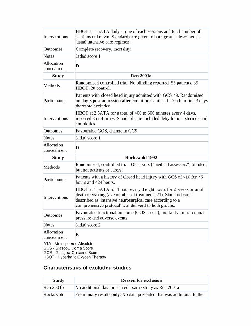
Interventions HBOT at 1.5ATA daily - time of each sessions and total number of sessions unknown. Standard care given to both groups described as 'usual intensive care regimen'.
Outcomes Complete recovery, mortality. Notes Jadad score 1 Allocation concealment D
Study Ren 2001a
Methods Randomised controlled trial. No blinding reported. 55 patients, 35 HBOT, 20 control.
Participants Patients with closed head injury admitted with GCS <9. Randomised on day 3 post-admission after condition stabilised. Death in first 3 days therefore excluded.
Interventions HBOT at 2.5ATA for a total of 400 to 600 minutes every 4 days, repeated 3 or 4 times. Standard care included dehydration, steriods and antibiotics.
Outcomes Favourable GOS, change in GCS Notes Jadad score 1 Allocation concealment D
Study Rockswold 1992
Methods Randomised, controlled trial. Observers ("medical assessors") blinded, but not patients or carers.
Participants Patients with a history of closed head injury with GCS of <10 for >6 hours and <24 hours.
Interventions
HBOT at 1.5ATA for 1 hour every 8 eight hours for 2 weeks or until death or waking (ave number of treatments 21). Standard care described as 'intensive neurosurgical care according to a comprehensive protocol' was deliverd to both groups.
Outcomes Favourable functional outcome (GOS 1 or 2), mortality , intra-cranial pressure and adverse events.
Notes Jadad score 2 Allocation concealment B
ATA - Atmospheres Absolute GCS - Glasgow Coma Score GOS - Glasgow Outcome Score HBOT - Hyperbaric Oxygen Therapy
Characteristics of excluded studies
Study Reason for exclusion Ren 2001b No additional data presented - same study as Ren 2001a Rockswold Preliminary results only. No data presented that was additional to the

1985 full report. Shi 2003 Included only patients at least 3 months after head injury
Additional tables Table 01 Search strategies
EMBASE MEDLINE (OVID)
1. exp head injury/ 2. (head or cerebr$ or crani$ or capitis or brain$ or forebrain$ or skull$ or hemisphere or intracran$ or orbit$).mp. 3. (injur$ or trauma$ or lesion$ or damag$ or wound$ or destruction$ or oedema$ or edema$ or fracture$ or contusion$ or concus$ or commotio$ or pressur$).mp 4. 2 and 3 5. diffuse axonal injur$.mp. 6. 1 or 4 or 5 7. exp coma/ 8. 6 or 7 9. exp hyperbaric oxygen/ 10. (high adj5 (pressur$ or oxygen$)).mp. 11. hyperbaric$.mp. 12. 10 or 11 13. oxygen$.mp. 14. 12 and 13 15. (HBO or HBOT).mp. 16. multiplace chamber$.mp. 17. monoplace chamber$.mp. 18. 9 or 14 or 15 or 16 or 17 19. 8 and 18 20. 19
1. exp head injuries-penetrating 2. exp head injuries-closed 3. exp coma-post head injury 4. exp craniocerebral trauma 5. head or crani$ or capitis or brain$ or forebrain$ or skull$ or hemisphere or intracran$ or orbit$ 6. injur$ or trauma$ or lesion$ or damage$ or wound$ or destruction$ or oedema$ edema$ or fracture$ or contusion$ or concus$ or commotion$ or pressur$ 7. 5 and 6 8. diffuse axonal injur$ 9. 1 or 2 or 3 or 4 or 7 or 8 10. exp hyperbaric oxygenation 11. (high$) adj3 (pressure or tension$) 12. hyperbaric$ 13. oxygen$ 14. 12 or 13 15. 14 and 11 16. HBO or HBOT 17. multiplace chamber$ 18. monoplace chamber$ 19. 10 or 15 or 16 or 17 or 18 20. 9 and 19
References References to studies included in this review
Artru 1976 {published data only}
Artru F, Chacornac R, Deleuze R. Hyperbaric oxygenation for severe head injuries. Preliminary results of a controlled study. European Neurology 1976;14(4):310-18.
Holbach 1974 {published data only}
Holbach KH, Wassmann H, Kolberg T. Improved reversibility of the traumatic midbrain syndrome using hyperbaric oxygen. Acta Neurochirurgica (Wien) 1974;30(3-4):247-56.
Ren 2001a {published data only}

Ren H, Wang W, Ge Z. Glasgow coma scale, brain electrical activity mapping and Glasgow outcome score after hyperbaric oxygen treatment of severe brain injury. Chinese Journal of Traumatology 2001;4(4):239-41.
Rockswold 1992 {published data only}
Rockswold GL, Ford SE, Anderson DC, Bergman TA, ShermanRE. Results of a prospective randomized trial for treatment of severely brain-injured patients with hyperbaric oxygen. Journal of Neurosurgery 1992;76(6):929-34.
* indicates the major publication for the study
References to studies excluded from this review
Ren 2001b
Ren H, Wang W, Ge Z. Clinical, Glasgow coma scale, brain electric earth map, endothelin and transcranial ultrasonic doppler findings after hyperbaric oxygen treatment for severe brain injury. Chinese Medical Journal (English) 2001;114(4):387-90.
Rockswold 1985
Rockswold GL, Ford SE. Preliminary results of a prospective randomized trial of treatment of severely brain-injured patients with hyperbaric oxygen. Minnesota Medical Journal 1985;68(7):533-5.
Shi 2003
Shi XY, Tang ZQ, Xiong B, Bao JX, Sun D, Zhang YQ, Yao Y. Cerebral perfusion SPECT imaging for assessment of the effect of hyperbaric oxygen therapy on patients with postbrain injury neural status. Chinese Journal of Traumatology 2003;6(6):346-9.
References to studies awaiting assessment
Belokurov
Additional references
Alderson 2003
Alderson P, Roberts I. Corticosteroids for acute traumatic brain injury (Cochrane Review). In: The Cochrane Library, Issue 2, 2003. Oxford: Update Software.
Alexander 1992
Alexander E. Global Spine and Head Injury Prevention Project (SHIP). Surgical Neurology 1992;38(6):478-9.
Artru 1976
Artru F, Philippon B, Gau F, Berger M, Deleuze R. Cerebral blood flow, cerebral metabolism and cerebrospinal fluid biochemistry in brain-injured patients after exposure to hyperbaric oxygen. European Neurology 1976;14(5):351-64.
Clark 1982

Clark JM. Oxygen toxicity. In: Bennett PB, Elliott DH, editor(s). The Physiology and Medicine of Diving 3rd Edition. London: Bailliere, Tindall and Cox, 1982:200-38.
Fasano 1964
Fasano VA, Nunno T, Urciolo R, Lombard G. First observation on the use of oxygen under high pressure for the treatment of traumatic coma. In: Boerema, Brummelkamp and Meigne, editor(s). Clinical Application of Hyperbaric Oxygen Amsterdam: Elsevier, 1964:168-73.
Fearnside 1997
Fearnside MR, Gurka JA. The challenge of traumatic brain injury. Medical Journal of Australia 1997;167(6):293-4.
Fiskum 2000
Fiskum G. Mitochondrial participation in ischemic and traumatic neural cell death. Journal of Neurotrauma 2000;17(10):843-55.
Hayakawa 1971
Hayakawa T, Kanai N, Kuroda R. Response of cerebrospinal fluid pressure to hyperbaric oxygenation. Journal of Neurology, Neurosurgery and Psychiatry 1971;34(5):580-6.
Hills 1999
Hills BA. A role of oxygen-induced osmosis in hyperbaric oxygen therapy. Medical Hypotheses 1999;52(3):259-63.
Holbach 1977
Holbach KH, Caroli A, Wassmann H. Cerebral energy metabolism in patients with brain lesions of normo- and hyperbaric oxygen pressures. Journal of Neurology 1977;217(1):17-30.
Ikeda 1990
Ikeda Y, Long DM. The molecular basis of brain injury and brain edema: the role of oxygen free radicals. Neurosurgery 1990;27(1):1-11.
Jadad 1996
Jadad AR, Moore RA, Carroll D, Jenkinson C, Reynolds DJ, Gavaghan DJ, McQuay HJ. Assessing the quality of reports of randomized clinical trials: is blinding necessary?. Controlled Clinical Trials 1996;17(1):1-12.
Jouvet 1960
Jouvet DJ. [Etudes semiologiques des troubles prolonges de la conscience. Ses bases physiopathologiques]. Lyon Medecine 1960;201:1401-20.
Khan 2003
Khan B, Evans AW, Easterbrook M. Refractive changes in patients undergoing hyperbaric oxygen therapy: a prospective study. Undersea and Hyperbaric Medicine 2003;24 (Suppl):9.
Langham 2003

Langham J, Goldfrad C, Teasdale G, Shaw D, Rowan K. Calcium channel blockers for acute traumatic brain injury (Cochrane Review). In: The Cochrane Library, Issue 2, 2003. Oxford: Update Software.
Muizelaar 1989
Muizelaar JP. Cerebral blood flow, cerebral blood volume and cerebral metabolism after severe head injury. In: Becker DP, Gudeman SK, editor(s). Textbook of Head Injury Philadelphia: WB Saunders, 1989:221-40.
Neubauer 1994
Neubauer RA, Gottlieb SF, Pevsner NH. Hyperbaric oxygen for treatment of closed head injury. Southern Medical Journal 1994;87(9):933-6.
Roberts 1995
Roberts I. Letter from Chengdu: China takes to the roads. BMJ 1995;310(6990):1311-13.
Roberts 2003a
Roberts I. Barbiturates for acute traumatic brain injury (Cochrane Review). In: The Cochrane Library, Issue 2, 2003. Oxford: Update Software.
Roberts 2003b
Roberts I, Schierhout G. Hyperventilation therapy for acute traumatic brain injury (Cochrane Review). In: The Cochrane Library, Issue 2, 2003. Oxford: Update Software.
Robertson 1989
Robertson CS, Narayan RK, Gokaslan ZL, Pahwa R, Grossman RG, Caram P Jr, Allen E. Cerebral arteriovenous oxygen difference as an estimate of cerebral blood flow in comatose patients. Journal of Neurosurgery 1989;70(2):222-30.
Schierhout 2003
Schierhout G, Roberts I. Anti-epileptic drugs for preventing seizures following acute traumatic brain injury (Cochrane Review). In: The Cochrane Library, Issue 2, 2003. Oxford: Update Software.
Siesjo 1989
Siesjo BK, Agardh CD, Bengtsson F. Free radicals and brain damage. Cerebrovascular and Brain Metabolism Review 1989;1:165-211.
Suckoff 1982
Suckoff MH, Ragatz RE. Hyperbaric oxygenation for the treatment of acute cerebral edema. Neurosurgery 1982;10(1):29-38.
Thurman 1999
Thurman DJ, Alverson C, Browne D et al. Traumatic brain injury in the United States: a report to congress. US Department of health and Human Services, National Centre for Injury Prevention and Control 1999.

Tymianski 1996
Tymianski M, Tator CH. Normal and abnormal calcium homeostasis in neurons: a basis for the pathophysiology of traumatic and ischemic central nervous system injury. Neurosurgery 1996;38(6):1176-95.
Graphs Graphs and Tables
To view a graph or table, click on the outcome title of the summary table below.
01 Good functional outcome (GOS <3 or similar)
Outcome title No. of studies
No. of participants
Statistical method Effect size
01 Good functional outcome at end of treatment period to one month
2 159 Relative Risk (Random) 95% CI
2.66 [0.73, 9.68]
02 Good functional outcome at six months 1 55 Relative Risk
(Fixed) 95% CI 2.76 [1.39, 5.49]
03 Good functional outcome at 12 months 1 166 Relative Risk
(Fixed) 95% CI 0.98 [0.73, 1.30]
04 Best case scenario (12 months) 1 168 Relative Risk
(Fixed) 95% CI 1.00 [0.75, 1.33]
05 Worst case (12 months) 1 168 Relative Risk (Fixed) 95% CI
0.96 [0.72, 1.27]
06 Good functional outcome at final follow-up 4 380
Relative Risk (Random) 95% CI
1.94 [0.92, 4.08]
07 Best case good outcome at final follow-up 4 382
Odds Ratio (Random) 95% CI
3.25 [0.99, 10.65]
08 Worst case good outcome at final follow-up 4 382
Odds Ratio (Random) 95% CI
3.19 [0.93, 10.97]
09 Good functional outcome - subgroup by treatment pressure
4 380 Relative Risk (Random) 95% CI
1.94 [0.92, 4.08]
02 Death at final follow-up
Outcome title No. of studies
No. of participants Statistical method Effect size
01 Death at final follow-up 3 325 Relative Risk (Fixed)
95% CI 1.46 [1.13, 1.87]
02 Best case death 3 327 Relative Risk (Fixed) 95% CI
1.48 [1.16, 1.90]

03 Worst case death 3 327 Relative Risk (Fixed) 95% CI
1.45 [1.13, 1.86]
03 Intra-cranial pressure
Outcome title No. of studies
No. of participants Statistical method Effect size
01 Mean peak ICP at any time overall 1 156
Weighted Mean Difference (Fixed) 95% CI
3.10 [-3.37, 9.57]
02 Mean peak ICP at any time (subgroups)
Weighted Mean Difference (Fixed) 95% CI
Subtotals only
04 Adverse events of treatment
Outcome title No. of studies
No. of participants Statistical method Effect size
01 Pulmonary 2 228 Relative Risk (Fixed) 95% CI
0.06 [0.01, 0.47]
02 Neurological toxicity 1 168 Relative Risk (Fixed)
95% CI 0.20 [0.01, 4.10]
03 Ear barotauma 1 168 Relative Risk (Fixed) 95% CI
0.20 [0.01, 4.10]
Cover sheet
Hyperbaric oxygen therapy for the adjunctive treatment of traumatic brain injury
Reviewer(s) Bennett MH, Trytko B, Jonker B
Contribution of Reviewer(s)
Bennett: Conception, principal author, search strategy, identification of trials, critical appraisal and data extraction. Content expert on hyperbaric medicine and clinical epidemiology.
Jonker: Co-author, critical appraisal and data extraction. Content expert on brain trauma.
Trytko: Co-author, data extraction. Content expert on hyperbaric medicine and intensive care.
Issue protocol first published
2004 issue 1
Issue review first published
2004 issue 4
Date of last minor amendment
20 November 2003

Date of last substantive amendment
16 June 2004
Most recent changes Information not supplied by reviewer
Date new studies sought but none found
12 December 2003
Date new studies found but not yet included/excluded
Information not supplied by reviewer
Date new studies found and included/excluded
Information not supplied by reviewer
Date reviewers' conclusions section amended
Information not supplied by reviewer
Contact address Dr Michael Bennett FANZCA, DipDHM Barker Street Randwick NSW AUSTRALIA 2031 Telephone: +61 2 9382 3880 Facsimile: +61 2 9382 3882 E-mail: [email protected]
Cochrane Library number
CD004609
Editorial group Cochrane Injuries Group
Editorial group code INJ
Sources of support External sources of support
• No external sources of support AUSTRALIA
Internal sources of support
• Prince of Wales Hospital, Sydney AUSTRALIA
Synopsis Hyperbaric oxygen may reduce death rate after brain injury but does not seem to improve quality of life for survivors

Traumatic brain injury is common and has a high impact on the wellbeing of those affected. Hyperbaric oxygen therapy (HBOT) involves people breathing pure oxygen in a specially designed chamber and it is sometimes used as a treatment to increase the supply of oxygen to the injured brain, in an attempt to reduce the area of brain that will die. We found some evidence that people with traumatic brain injury are less likely to die if they receive hyperbaric oxygen therapy as part of their treatment. However, there is no good evidence that people are more likely to have a good health outcome. It is possible, therefore, that the overall effect of hyperbaric oxygen is only to make it more likely that people will survive severely disabled after such injuries. Our conclusions are based on four randomised trials, with a limited number of patients. Further research is needed.
Keywords Humans; Brain Injuries[*therapy]; *Hyperbaric Oxygenation; Randomized Controlled Trials