hypoestes aristata (vahl) sol. ex roem. & schult var
TRANSCRIPT
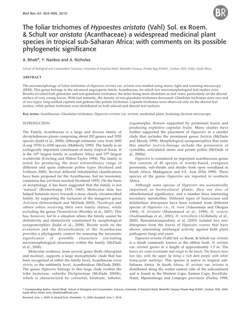
403BHATT ET AL. Biol Res 43, 2010, 403-409Biol Res 43: 403-409, 2010 BRThe foliar trichomes of Hypoestes aristata (Vahl) Sol. ex Roem.& Schult var aristata (Acanthaceae) a widespread medicinal plantspecies in tropical sub-Saharan Africa: with comments on its possiblephylogenetic significance
A. Bhatt*, Y. Naidoo and A. Nicholas
School of Biological and Conservation Sciences, University of KwaZulu-Natal, Westville Campus, Private Bag X54001, Durban, KZN, 4000, South Africa
ABSTRACT
The micromorphology of foliar trichomes of Hypoestes aristata var. aristata was studied using stereo, light and scanning microscopy(SEM). This genus belongs to the advanced angiosperm family Acanthaceae, for which few micromorphological leaf studies exist.Results revealed both glandular and non-glandular trichomes, the latter being more abundant on leaf veins, particularly on the abaxialsurface of very young leaves. With leaf maturity, the density of non-glandular trichomes decreased. Glandular trichomes were rare andof two types: long-stalked capitate and globose-like peltate trichomes. Capitate trichomes were observed only on the abaxial leafsurface, while peltate trichomes were distributed on both adaxial and abaxial leaf surfaces.
Key terms: Acanthaceae, Glandular trichomes, Hypoestes aristata var. aristata, medicinal plant, Scanning electron microscope.
* Corresponding Author: Arvind Bhatt, School of Biological and Conservation Sciences, University of KwaZulu-Natal, Westville Campus Private Bag X54001, Durban, KZN, 4000,South Africa, Email: [email protected]
Received: June 1, 2009. In revised form: November 13, 2009. Accepted: June 7, 2010.
INTRODUCTION
The Family Acanthaceae is a large and diverse family ofdicotyledonous plants comprising about 202 genera and 3520species (Judd et al., 2008); although estimates vary from 2600(Long 1970) to 4300 species (Mabberly 1990). The family is anecologically important constituent of many tropical floras. Itis the 14th largest family in southern Africa and 15th largestworldwide (Cowling and Hilton-Taylor 1994). The family isnoted for producing the most extraordinary range ofdifferent and quite elaborate pollen types (Scotland andVollesen 2000). Several different infrafamilial classificationshave been proposed for the Acanthaceae, but no taxonomicconsensus has yet been reached (Scotland 1995). On the basisof morphology it has been suggested that the family is not‘natural’ (Bremerkamp 1955; 1965). Molecular data hashelped botanists move towards a more clearly circumscribedfamily, by supporting the inclusion of the mangrove genusAvicennia (Schwarzbach and McDade 2002), Thunbergia andothers (often receiving their own family status), whileexcluding the genus Thomandersia (Wortley et al., 2007). Thishas, however, led to a situation where the family cannot bedefinitively and distinctively constrained by morphologicalsynapomorphies (Judd et al., 2008). Recent work on theevolution and the diversification of the Acanthaceaeprovides a phylogenetic context for assessing the taxonomicsignificance of possible characters (includingmicromorphological structures) within the family (McDadeet al., 2008).
Molecular evidence, from several genes (both chloroplastand nuclear), supports a large monophyletic clade that hasbeen recognized at either the family level, Acanthaceae sensustricto, or the subfamily level, Acanthoideae (McDade 2000).The genus Hypoestes belongs to this large clade (within thetribe Justicieae, subtribe Diclipterinae (McDade 2000b),which is characterized by colourful, bilabiate, tubular,
zygomorphic flowers supported by prominent bracts andproducing explosive capsular fruits. Many studies havefurther supported the placement of Hypoestes in a smallerclade that includes the prominent genus Justicia (McDadeand Moody 1999). Morphological synapomorphies that unitethis smaller Justicia-lineage include the possession ofcystoliths, articulated stems and porate pollen (McDade etal., 2000a).
Hypoestes is considered an important acanthaceous genusthat consists of 40 species of woody-based, evergreenperennials, sub-shrubs and shrubs from open woodland inSouth Africa, Madagascar and S.E. Asia (Ellis 1999). Threespecies of the genus Hypoestes are reported in southernAfrica.
Although some species of Hypoestes are economicallyimportant as horticultural plants, they are also ofethnobotanical significance, and a number have interestingsecondary metabolites. Different types of fusicoccane anddolabellane diterpenes have been isolated from differentspecies of Hypoestes i.e., H. rosea (Adesomoju and Okogun1984), H. forskalei (Muhammad et al., 1998), H. serpens(Andriamihaja et al., 2001), H. verticillaris (Al-Rehaily et al.,2002). Rasoamiaranjanahary et al. (2003) isolated two newditerpenes from the leaves of Hypoestes serpens that haveshown interesting antifungal activity against both plantpathogenic fungi and yeast.
Hypoestes aristata (Vahl) Sol. ex Roem. & Schult var. aristatais a shrub commonly known as the ribbon bush. H. aristatavar. aristata grows to a height of approximately 1.5 m. Theleaves are ovate-acuminate and rough to the touch. The flowers havetwo lips, with the upper lip being a rich dark purple with whitehoney-guide markings. This species is native to tropical sub-Saharan Africa. In South Africa, H. aristata var. aristata isdistributed along the wetter eastern side of the subcontinentand is found in the Western Cape, Eastern Cape, KwaZulu-Natal, Mpumalanga and Limpopo provinces (Pooley 1998;
BHATT ET AL. Biol Res 43, 2010, 403-409404
Joffe 2001). A taxonomic revision for the genus in southernAfrica was undertaken by Balkwill and Getliffe Norris (1985).
The amaZulu of South Africa use the crushed leaves of H.aristata var. aristata for the treatment of sore eyes (Hulme,1954). Whole plant infusions are used to drench calvessuffering from a condition referred to as white scours(Hutchings 1996). Plant decoctions are used in the treatmentof breast disease. Roots are chewed for influenza, cough,colds and sore throats in East Africa (Kokwaro 1976). Theroot bark of H. aristata var. aristata is reported to be used forthe treatment of malaria (Iwu 1993).
A unique chemistry (as is seen in some species of thisgenus) may be connected with the medicinal properties of H.aristata var. aristata, and in turn this could relate to theglandular trichomes of the leaves.
Although not a universally useful taxonomic character,the structure of folia trichomes have nonetheless proved tobe taxonomically useful in some families, such as theSolanaceae (Adedeji et al., 2007), and even in some orders,such as the Urticales (Gangadhara and Inamdar, 2005).Although many taxonomic works on the Acanthaceaemention the presence of folio trichomes, few studies explorein detail the micromorphology of these interestingstructures. While the unique hygroscopic hairs of the seed ofsome species is well known and documented (Balkwill andGetliff Norris 1988), to date no detailed survey of generalfolio trichomes has been undertaken for the whole of theAcanthaceae, Such a survey may eventually contribute tofinding possible morphological synapomorphies that mightcorrelate with the clades postulated using molecularevidence; as has been done with the hygroscopic trichomesof the seeds in the Tribe Whitfieldieae (Manktelow 2001).The correlation of phenotypic characters with genomic mayhelp towards a holistic taxonomy for the Acanthaceae andpossibly even aid in a small way to solving familycircumscriptions in the problematic order Lamiales. It is thelack of detailed studies of the folio micromorphology ofmember species of this important family, and Hypoestes inparticular, that motivated this investigation. In consequence,this paper examines the morphology and distribution of leaftrichomes of H. aristata var. aristata at different stages of leafdevelopment. This paper builds on the initial observations ofthese trichomes by Balkwill and Getliffe Norris (1985).
MATERIAL AND METHODS
The leaves of Hypoestes aristata var. aristata were collectedfrom cultivated plants on the University of KwaZulu-Natal,Westville campus. Fresh leaves were used for experimentalwork and a voucher specimen (Nicholas & Bhatt, 2994) wasalso deposited in the Ward herbarium (UDW) at theUniversity of KwaZulu-Natal, Durban campus. Leaves ofthree different developmental stages were selected for thepresent study: Stage I- very young (2.0- 4.0 cm long), StageII- young (4.1-6.0 cm long) and Stage III- fully expanded(above 6.1 cm long). Five plants of H. aristata var. aristatawere sampled and from each plant, five leaves of differentdevelopmental stage were used for study. Fresh leaves wereexamined and photographed with a stereomicroscope (NikonAZ 100).
For light microscopic studies, semi-thin sections of leafmaterial were embedded in low viscosity Spurr’s (1969)
resin. Sections of the resin-embedded tissue, ranging inthickness from 0.5-2 µm, were cut using glass knives andheat fixed onto pre-cleaned glass slides with drops ofdistilled water. These sections were stained with 0.5%Toluidine blue-0 consisting of 0.1% sodium carbonate at apH 11.1 (Feder and O’Brein 1968). Sections were stained forapproximately 1 min. over heat, washed briefly in distilledwater and mounted in immersion oil. Slides were viewedand photographed with a Leitz light microscope.
For scanning electron microscopy, sections of leaves atdifferent developmental stages were prepared by rapidquenching in liquid nitrogen and freeze dried in an EdwardsModulyo freeze dryer at -60°C in a vacuum of 10-2 Torr forapproximately 48 hrs. Leaf segments were secured onto brassstubs with carbon conductive tape and sputter coated withgold for 4 mins. Observations were carried out using a Jeol6100 scanning electron microscope operated at 12 kV, with aworking distance of 15 mm.
RESULTS
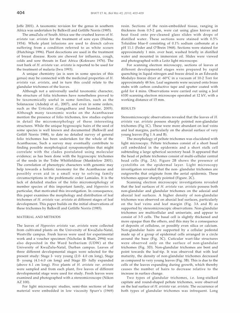
Stereomicroscopic observations revealed that the leaves of H.aristata var. aristata possess sharply pointed non-glandulartrichomes (Fig 1C). These were more abundant on leaf veinsand leaf margins, particularly on the abaxial surface of veryyoung leaves (Fig 1 A and B).
The morphology of peltate trichomes was elucidated withlight microscopy. Peltate trichomes consist of a short basalcell embedded in the epidermis and a short stalk cellsubtending a large spherical secretory head. It appeared thatthe head of peltate trichomes consist of multi-cellular centralhead cells (Fig. 2A). Figure 2B shows the presence ofcystoliths on the epidermal layer. Light microscopyinvestigation revealed that the non- glandular trichomes areoutgrowths that originate from the aerial epidermis. Thesetrichomes appear sharply pointed (Figure. 2C).
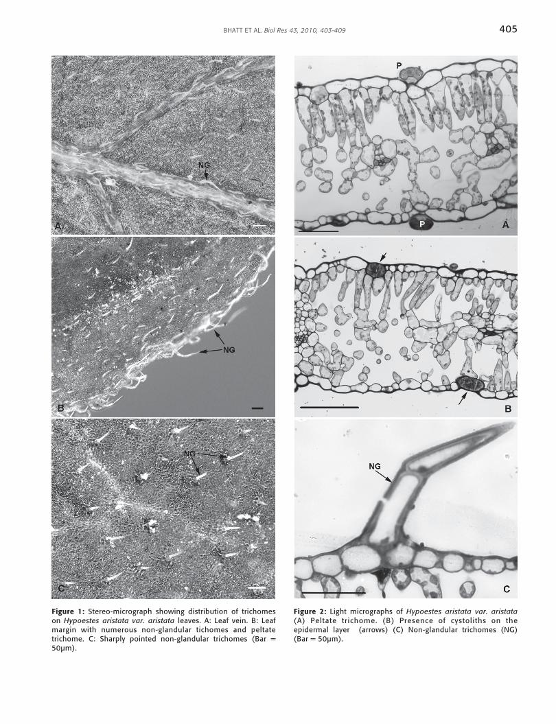
Scanning electron microscopic investigations revealedthat the leaf surfaces of H. aristata var. aristata possess bothnon-glandular and glandular trichomes on the adaxial andabaxial leaf surfaces. A higher density of non-glandulartrichomes was observed on abaxial leaf surfaces, particularlyon the leaf veins and leaf margin (Fig. 3A and B) assupported by stereomicroscopic observations. Non-glandulartrichomes are multicellular and uniseriate, and appear toconsist of 3-5 cells. The basal cell is slightly thickened andmore opaque than the others, and this may be a consequenceof deposits of cellulose, or possibly even silica or calcium.Non-glandular hairs are supported by a cellular pedestalmade up of a group of epidermal cells arranged in a circlearound the base (Fig. 3C). Cuticular wart-like structureswere observed only on the surface of non-glandulartrichomes (Fig. 3D). Non-glandular trichomes are bent andpoint towards the leaf-tip. It was observed that with leafmaturity, the density of non-glandular trichomes decreasedas compared to very young leaves (Fig. 3B). This is due to thecells of the leaves expanding during growth, which therebycauses the number of hairs to decrease relative to theincrease in surface change.
Two types of glandular trichomes, i.e. long-stalkedcapitate and round-shaped peltate trichomes, were observedon the leaf surface of H. aristata var. aristata. The occurrence ofboth types of glandular trichomes was infrequent. Long
405BHATT ET AL. Biol Res 43, 2010, 403-409
Figure 1: Stereo-micrograph showing distribution of trichomeson Hypoestes aristata var. aristata leaves. A: Leaf vein. B: Leafmargin with numerous non-glandular tichomes and peltatetrichome. C: Sharply pointed non-glandular trichomes (Bar =50µm).
Figure 2: Light micrographs of Hypoestes aristata var. aristata(A) Peltate trichome. (B) Presence of cystoliths on theepidermal layer (arrows) (C) Non-glandular trichomes (NG)(Bar = 50µm).
BHATT ET AL. Biol Res 43, 2010, 403-409406
Figure 3: SEM micrographs showing distribution and types of trichomes on Hypoestes aristata var. aristata leaves. A: Distribution ofnon-glandular trichomes (NG) on leaf vein (Bar = 20µm). B: Leaf margin (Bar = 20µm). C: Sparse distribution of non-glandulartrichomes on mature leaf (Bar = 20µm). D: Fully developed non-glandular trichomes (NG) supported by basal cellular pedestal (CP)on leaf surface (Bar = 10µm). E: Distribution of long stalked capitate trichomes (C). Capitate trichomes at different phases ofsecretion (PS- Pre-Secretory phase; SP- Secretory Phase; Ps- Post secretory phase) (Bar = 10µm). F: Peltate trichomes showingsmooth surface (Bar = 1µm)
407BHATT ET AL. Biol Res 43, 2010, 403-409
stalked capitate trichomes were seen only on the abaxial leafsurface (Fig. 3E). This type of trichome was observed only onvery young leaves. For capitates trichomes, different stages ofsecretion were observed through the SEM i.e.: Phase I-Presecretory, Phase II- Secretory and Phase III- Post secretory(Fig. 3F). Capitate trichomes appear to be supported by a oneto two celled stalk. In contrast, the round-shaped peltatetrichomes are distributed on both the abaxial and adaxial leafsurfaces. The surface of these peltate trichomes is smooth (Fig.3F). The different developmental stages of peltate trichomeswere observed throughout leaf maturation.
DISCUSSION
Trichomes are unicellular or multicellular outgrowths thatoriginate from the aerial epidermis and which vary inmorphological features, location and mode of secretion(Werker 2000). A long history of published literatureindicates that the type and density of trichomes differ amongspecies and may vary in organs of the same plant (Uphof1962). It has been suggested that non-glandular trichomesserve various functions in plants i.e., to reduce the heat load,reflectance of UV light, provide protection from insects andherbivores, increase tolerance to freezing and maintain waterbalance in leaves (Werker 2000; Mauricio and Rausher, 1997;Liakoura et al., 1997). Glandular trichomes are associatedwith the production of chemicals that provide defenseagainst herbivores and pathogens.
The density of non-glandular trichomes in H. aristata var.aristata was higher on the leaf veins and leaf margin ofyoung leaves, and decreased with leaf maturity. A higherdensity of trichomes on the leaf veins and apex is a commontrend seen in angiosperms (Oppenheimer 1959). Thisadaptation is possibly used to limit incoming UV light andthus protect vascular tissue. It is assumed that non-glandulartrichomes play an important role in leaf protection,particularly during the early stages of leaf development in H.aristata var. aristata. Similar results have been reported inother species (Corsi and Bottega 1999; Tattini et al., 2000;Werker 2000; Valkama et al., 2004). The non-glandulartrichome is supported by a basal cellular pedestal. It hasbeen reported that the basal cellular pedestal providesmechanical support and serves as a point for the attachmentof trichomes to the epidermis (Payne 1978; Ascensao et al.,1999). The thickened basal cell, also reported by Metcalfe andChalk (1950), may add extra support to this trichome. Thestalk of the non-glandular trichome is densely covered withcuticular warts, which could be indicative of leaf maturity(Werker 2000) and which may be involved in helping thehairs stay free of dust by promoting cleaning during rainfall;the so called ‘Lotus effect’ (Nosonovsky 2007; Nosonovskyand Bhushan 2007). Although Hypoestes produces simplenon-glandular trichomes, other genera of the Acanthaceae,such as Baleria and Ruellia, have been reported to producecompound trichomes that are 2-armed or candelabra-like(Metcalfe and Chalk 1950).
We observed the presence of peltate trichomes on bothleaf surfaces. Our findings are consistent with observationsin several lamiaceous genera: Salvia officinalis (Corsi andBottega 1999); Plectranthus ornatus (Ascensão et al., 1999)and Mentha arvensis (Sharma et al., 2003). In H. aristata var.aristata, peltate trichomes were mostly distributed on the
inter-vein area of the leaves . A similar pattern ofdistr ibution of peltate tr ichomes was reported inPlectranthus medagascariensis (Ascensão et al . , 1998).However, long-stalked capitate trichomes were observedonly on the abaxial leaf surface of H. aristata var. aristata inthe early stages of leaf development. Related literature onTeucrium species (Maleci and Servettaz 1991) showed asimilar type of long stalked capitate trichomes to those weobserved in H. aristata var. aristata. In the Lamiaceae,peltate trichomes are postulated to be the site of productionof essential oils, while the long-stalked capitate trichomesmay produce essential oils and polysaccharides (Maleci andGiuliani 2006). The function of these glandular hairs inAcanthaceae, however, has not yet been determined but,unlike the Lamiaceae and the closely related familyVerbenaceae, this family is not reported to be aromatic.Further research into the histochemical and ultrustructurecharacterization of glandular trichomes in H. aristata var.aristata would be beneficial in understanding theirpotential role in the plant.
Molecular studies have resolved the phylogeneticplacement of some families in the Lamiales. However, theAcanthaceae and Lamiaceae are part of a large nucleus ofphylogenetically unresolved families (Bremer et al., 2002;Olmstead et al . , 2001). More data, includingmicromorphological, are needed to help resolve thesecomplex relationships. Within the Asterid I clade, sometimescalled the Lamiids (Soltis et al., 2005), glandular trichomeshave been reported for the Gesneriaceae, Plantaginaceae,Scrophulariaceae, Orobanchaceae, Lentibulariaceae andVerbenaceae (Judd et al., 2008). Non-glandular or eglandularhairs, similar to those found in the Acanthaceae andLamiacaeae, have also been found in the Strychnaceae andApocynaceae (Naidoo unpublished data). The widespreadsimilarities in structure of both non-glandular and glandulartrichomes within the Asterids may or may not have somephylogenetic significance.
We observed that non glandular and peltate trichomesinitiate and senesce during all stages of leaf development.These findings are in agreement with Wagner et al. (2004)who reported that some trichomes may senesce duringvarious stages of leaf development. Similar results werereported for other species, where trichome developmentcontinues throughout leaf maturity (Oosthuizen and Coetzee1983; Sharma et al., 2003).
The presence of cystoliths is also interesting. Thesestructures are outgrowths or ingrowths of the epidermallayer that become filled with calcium carbonate. They maybe simple or can become quite complex. Cystoliths seem tobe associated with Justicia and allies, but are not present intaxa outside of this acanthaceous lineage, they are also notpresent in the Lamiaceae. They have, however, been reportedfrom the Boraginaceae, Scrophulariaceae and Verbenaceae(Metcalfe and Chalk 1950). Most works onmicromorphological structure are descriptive, unfortunately,few attempts have been made establish the phylogeneticsignificance of these (Cantino 1990). A continued survey offoliar trichomes and other micromorphological structures inthe Lamiales (22 families with about 20 000 species (Judd etal., 2008)) is needed. However, these should aim to establishtheir phylogenetic and taxonomic significance, and not justelucidate their structural manifestation.
BHATT ET AL. Biol Res 43, 2010, 403-409408
ACKNOWLEDGMENTS
This research was carried out with financial support from theNational Research Foundation, South Africa. We thank thestaff of the Electron Microscope Unit, UKZN Westvillecampus for their assistance with the microscopy. Our thanksto A. Rajh for assistance with the photographic plates. Theauthors would also like to thank Dr Hugh Glen of theKwaZulu-Natal Herbarium, of the Southern African NationalBiodiversity Institute, for helping track down some of thenecessary literature.
REFERENCE
ADEDEJI O, AJUWON OY, BABAWALE OO (2007) Foliar epidermalstudies, organographic distribution and taxonomic importance oftrichomes in the family Solanaceae. Internatl J Bot 3(3): 276-282.
ADESOMOJU AA, OKOGUN JI (1984) Roseatoxide and dihypoestoxide:additional new diterpenoids from Hypoestes rosea. J Nat Prod 47:308-311.
AL-REHAILY A, AL-YAHYA MA, MIRZA HH, AHMED B (2002)Verticillarone: a new seco-fusicoccane ditrepenoid ketonepoxidefrom Hypoestes verticillaris. J Asian Nat Prod Res 4: 117-122.
ANDRIAMIHAJA B, MARTIN MT, RASOANAIVO P, FRAPPIER F (2001)A new diterpene from Hypoestes serpens. J Nat Prod 64: 217-218.
ASCENSÃO L, FIGUEIREDO AC, BARROSO JG, PEDRO LG,SCHRIPSEMA J, DEANS SG, SCHEFFER JJC (1998) Plectranthusmadagascariensis: morphology of the glandular trichomes, essential oilcomposition, and its biological activity. Int J Plant Sci 159: 31-38.
ASCENSÃO L, MOTA L, CASTRO, MM (1999) Glandular trichomes onthe leaves and flowers of Plectranthus ornatus : morphology,distribution and histochemistry. Ann Bot 84: 437-447.
BALKWILL K, GETIFFE NORRIS F (1985) Taxonomic studies in theAcanthaceae; the genus Hypoestes in southern Africa. S Afr J Bot51(2):133-144.
BALKWILL K, GETIFFE NORRIS F (1988) Classification of theAcanthaceae: a southern African perspective. Missouri BotanicalGarden Monographs in Systematic Botany 25: 503-516.
BREMEKAMP CEB (1955) Notes on some acanthaceous genera ofcontroversial position. Acta Botanical Neerlandse 4:644-655.
BREMERKAMP CEB (1965) Delimitation and subdivision of theAcanthaceae. Bulletin of the Botanical Survey of India 7:21-30.
BREMER BK, BREMER K, HEIDARI N, ERIXON P, OLMSTEAD RG,ANDERBERG AA, KÄLLERSJ Q M, BARKHORDARIAN EP (2002)Phylogenetics of asterids based on 3 coding and 3 non-codingchloroplast DNA markers and the utility of non-coding DNA athigher taxonomic levels. Mol Phylogenet Evol 24:271-301.
BREMER K, FRIIS EM, BREMER B (2004) Molecular phylogenetic datingof Asterid flowering plants shows early Cretaceous diversification.Syst Biol 53(3): 496-505.
CANTINO PD (1990) The phylogenetic significance of stomata andtrichomes in the Labiatae and Verbenaceae. J Arnold Arboretum 71:323-370.
CORSI Z, BOTTEGA S (1999) Glandular hairs of Salvia officinalis: Newdata on morphology, localization and histochemistry in relation tofunction. Ann Bot 84: 657-664.
COWLING RM, HILTON-TAYLOR C (1994) Patterns of plant diversityand endemism in southern Africa: an overview. Strelitzia 1: 31-52.
Ellis BW (1999) Taylor’s Guide to Annuals: how to select and grow morethan 400 annuals, biennials, and tender perennials. Houghton MifflinCo., Boston. 1-448pp.
FEDER N, O’BRIEN TP (1968) Plant microtechniques: some principlesand new methods. Am J Bot 55: 123-142.
GANGADHARA M, INAMDAR JA (2005) Trichomes and stomata, andtheir taxonomic significance in the Urticales. Plant Systematics andEvolution 127(2-3): 121-137.
HULME MM (1954) Wild Flowers of Natal. Shuter and Shooter,Pietermaritzburg.
HUTCHINGS A (1996) Acanthaceae, in: Zulu Medicinal Plants.University of Natal Press, Pietermaritzburg. 288-291.
IWU MM (1993) Handbook of African medicinal plants. CRC Press,Florida.
JOFFE P (2001) Creative Gardening with Indigenous Plants. A SouthAfrican Guide. Briza Publications, Pretoria. 1-372pp.
JUDD WS, CAMPBELL CS, KELLOGG EA, STEVENS PF, DONOGHUEMJ (2008) Lamiids (Euasterids I) in: Plant Systematics: APhylogenetic Approach. 3rd edition. Sinauer Associates, Sunderland.459-494.
KOKWARO JO (1976) Medicinal plants of East Africa. East AfricanLiterature Bureau.
LIAKOURA V, STEFANOU M, MANETAS Y, CHOLEVAS C,KARABOURNIOTIS G (1997) Trichome density and its UV-Bprotective potential are affected by shading and leaf position on thecanopy. Environ Exp Bot 38:223-229.
LONG RW (1970) The genera of Acanthaceae in the southeastern UnitedState. J Arnold Arbor 51:257-309.
MABBERLEY DJ (1990) The Plant Book. Cambridge University Press,Cambridge. 1-874pp.
MALECI BL, SERVETTAZ O (1991) Morphology and distribution oftrichomes in Italian species of Teucrium sect.Chamaedrys (Labiatae) - ataxonomical evaluation. Plant Syst Evol 174 (1-2):83-91.
MALECI BL, GIULIANI C (2006) The glandular trichomes of the Labiatae:A review. Acta Hort 723: 85-90.
MANKTELOW M, MCDADE LA, OXELMAN B, FURNESS CA,BALKWILL M-J (2001) The enigmatic tribe Whitfieldieae(Acanthaceae): Delimitation and phylogenetic relationships based onmolecular and morphological data. Systematic Botany 26(1): 104-119.
MAURICIO R, RAUSHER MD (1997) Experimental manipulation ofputative selective agents provides evidence for the role of naturalenemies in the evolution of plant defense. Int J Org Evolution 51:1435-1444.
MCDADE LA, MOODY ML (1999) Phylogenetic relationships amongAcanthaceae: Evidence from non coding trnL-trnF chloroplast DNAsequences. Am J Bot 86: 70-80.
MCDADE LA (2000) Hybridization and phylogenetics: Special insightsfrom morphology. In Wiens J (ed.) Morphological Data inPhylogenetic Analysis: Recent Progress and Unresolved Problems.Smithsonian Institution Press.146-164 pp.
MCDADE LA, MASTA SE, MOODY ML, WATERS E (2000a) Phylogeneticrelationships among Acanthaceae: Evidence from two genomes. SystBotany 25(1):106-121.
MCDADE LA, DANIEL, TF MASTA SE, RILEY KM (2000b) Phylogeneticrelationships within the tribe Justicieae (Acanthaceae): Evidence frommolecular sequences, morphology and cytology. Annals of theMissouri Botanical Garden 87(4): 435-458.
MCDADE LA, DANIEL TF, KIEL, C. (2008). Towards a comprehensiveunderstanding of phylogenetic relationships among linages ofAcanthaceae s.l. (Lamiales). Am J Bot 95(9): 1136-1152.
METCALFE CR, CHALK L (1950) Acanthaceae in: Anatomy of theDicotyledons. Vol 2. Claredon Press, Oxford. 1014-1023pp.
MUHAMMAD I, MOSSA JS, RAMADAN AF, EL-FERALY FS, HUFFORDCD (1998) Additional diterpene ketones from Hypoestes forskalei.Phytochemistry 47:1331-1336.
NOSONOVSKY M (2007) Multiscale roughness and stabil ity ofsuperhydrophobic biomimetic interfaces. Langmuir 23:3157-3161.
NOSONOVSKY M, BHUSHAN B (2007) Hierarchical roughnessoptimization for biomimetic superhydrophobic surface.Ultramicroscopy 107: 969-979.
OLMSTEAD RG, DEPAMPHILIS CW, WOLFE AD, YOUNG ND,ELISONS WJ, REEVES PA (2001) Disintegration of theScrophulariaceae. Am J Bot 88(2): 348-361.
OOSTHUIZEN L-M, COETZEE J (1983) Morphogenesis of trichomes ofPelargonium scabrum. S Afr J Bot 2: 305-310.
OPPENHEIMER HR (1959) Adaptation to Drought: Xerophytism. UnitedNations Educational Scientific and Cultural Organisation, UNESCOPublication, Paris. 1-54pp.
PAYNE WW (1978) A glossary of plant hair terminology. Brittonia30(2):239-255.
POOLEY E (1998) Hypoestes aristata in: A Field Guide to Wild FlowersKwaZulu-Natal and the Eastern Region. Natal Flora PublicationsTrust, Durban. 436-437pp.
RASOAMIARANJANAHARY L, MARSTON A, GUILET D, SCHENK K,RANDIMBIVOLOLONA F, HOSTETTMANN K (2003) Antifungalditerpenes from Hypoestes serpens (Acanthaceae). Phytochemistry 62:333-337.
SCHWARZBACH AE, MCDADE L (2002) Phylogenetic relationships ofthe mangrove family Avicenniaceae based on chloroplast andribosomal DNA sequences. Syst Botany 27: 84-98.
409BHATT ET AL. Biol Res 43, 2010, 403-409
SCOTLAND RW, SWEERE JA, REEVES PA, OLMSTEAD RG (1995)Higher-level Systematics of Acanthaceae determined by chloroplastDNA sequences. Am J Bot 82: 266-275.
SCOTLAND RW, VOLLESEN K (2000) Classification of Acanthaceae.Kew Bulletin 55(3): 513-589.
SHARMA S, SANGWAN NS, SANGWAN RS (2003) Development processof essential oil glandular trichome collapsing in menthol mint. CurrSci 84: 544-550.
SOLTIS DE, SOLTIS PS, ENDRESS PK, CHASE MW (2005) Asterids in:Phylogeny and Evolution of Angiosperms. Sinauer Associates,Sunderland. 207-236pp.
SPURR AR (1969) A low viscosity epoxy embedding medium for electronmicroscopy. J Ultrastruct Res 26:31-43.
TATTINI M, GRAVANO E, PINELLI P, MULINACCI N, ROMANI A(2000) Flavonoids accumulate in leaves and glandular trichomes ofPhillyrea latifolia exposed to excess solar radiation. New Phytologist148:69-77.
UPHOF JCT (1962) Plant hairs. Gebriider Borntraeger, Berlin-Nikolassee.VALKAMA E, SALMINEN JP, KORICHEVA J, PIHLAJA K (2004)
Changes in leaf trichomes and epicuticular flavonoids during leafdevelopment in three birch taxa. Ann Bot 94: 233-242.
WAGNER GJ, WANG E, SHEPHERD RW (2004) New approaches forstudying and exploiting an old protuberance, the plant trichome.Ann Bot 93(1):3-11.
WERKER E (2000) Trichome diversity and development. Adv Bot Res31:1-35.
WORTLEY AH, RUDALL PJ, HARRIS DJ, SCOTLAND, RW (2005) Howmuch data are needed to resolve a difficult phylogeny? Case study inLamiales. Syst Biol 54: 697-709.
WORTLEY AH, HARRIS DJ, SCOTLAND RW (2007) On the taxonomyand phylogenetic position of Thomandersia. Syst Bot 32(2):415-444.

BHATT ET AL. Biol Res 43, 2010, 403-409410