hypokalaemia - cicm · objective: to review the metabolism and function of potassium and causes and...
TRANSCRIPT
Hypokalaemia
P. GLOVER Department of Critical Care Medicine, Flinders Medical Centre, Adelaide SOUTH AUSTRALIA
ABSTRACT Objective: To review the metabolism and function of potassium and causes and management of hypokalaemia. Data sources: A review of studies reported from 1966 to 1998 and identified through a MEDLINE search of the English-language literature of hypokalaemia. Summary of review: Potassium is predominantly an intracellular ion that contributes to approximately 50% of the intracellular fluid osmolality and is largely responsible for the resting membrane potential. The latter accounts for its influence on the excitability of muscle and nervous tissue.
Hypokalaemia is defined as a serum potassium of less than 3.5 mmol/L or plasma potassium less than 3.0 mmol/L and may be asymptomatic. Clinical features associated with hypokalaemia include abnormalities of cardiovascular, neurological and metabolic function and may be treated with oral potassium salts, although tachycardia and muscle weakness are the two life threatening disorders which may require rapid intravenous correction. The potassium salts of chloride, phosphate and acetate are often used, although the choice is often guided by the presence of an associated hypochloraemic alkalosis, non-anion gap acidosis or hypophosphataemia, indicating treatment with potassium chloride, potassium acetate, or potassium phosphate, respectively. The infusion rates of intravenous therapy depends upon the salt used. Potassium chloride is usually infused at a rate up to 40 mmol/h, whereas potassium acetate and potassium monohydrogen or dihydrogen phosphate are usually infused up to 5 mmol/h and 2 mmol/h respectively. Conclusions: Hypokalaemia can be asymptomatic or it may cause cardiovascular, neurological or skeletal muscle dysfunction. If intravenous potassium therapy is required, then correction with potassium chloride, acetate, or phosphate salts are usually guided by the presence of a metabolic acidosis, alkalosis or hypophosphataemia. (Critical Care and Resuscitation 1999; 1: 239-251)
Key Words: Potassium, hypokalaemia, potassium deficiency, potassium chloride, potassium acetate, potassium phosphate
Potassium is the principle intracellular cation and is responsible for approximately 50% of the intracellular fluid osmolality. As the cell membrane is approximately 20 times more permeable to potassium ions than sodium ions, potassium is also largely responsible for the resting membrane potential and therefore the function of excitable tissues. Hypokalaemia is defined as a serum potassium of less than 3.5 mmol/L or plasma potassium less than 3.0 mmol/L (serum concentration is 0.7 mmol/l higher than plasma concentration due to in vitro contamination by intracellular red blood cell potassium). Hypokalaemia is
the most frequently seen electrolyte abnormality found in the hospitalised population, occurring in up to 21% of patients1 and is associated with an increased mortality in this group. NORMAL POTASSIUM DISTRIBUTION Total body potassium is 45-50 mmol/kg in males and 35-40 mmol/kg in females, decreasing with increasing age and reduced muscle mass. Potassium, as well as being the most abundant intracellular cation, is predominantly distributed intracellularly, with the intracellular: extracellular ratio being 98%:2%. The
Correspondence to: Dr. P. Glover, Department of Critical Care Medicine, Flinders Medical Centre, Bedford Park, South Australia 5042
239
P. GLOVER Critical Care and Resuscitation 1999; 1: 239-251
intracellular potassium concentration [K+]i ranges from 145 to 155 mmol/L and the extracellular potassium concentration [K+]e ranges from 3.1 to 4.2 mmol/L. About 70% of total body potassium resides in skeletal muscle, 8.5% in erythrocytes and 6.5 % in hepatocytes (Table 1).2,3
Aldosterone. This is secreted by the zona glomerulosa of the adrenal cortex in response to angiotensin II, angiotensin III, ACTH and high plasma potassium concentrations.6 Atrial natriuretic peptide,7 heparin infusions,8 endogenous dopamine9 and hypokalaemia10 can inhibit aldosterone secretion.
Extracellular potassium levels are determined by the balances between extracellular efflux, intracellular influx, gastrointestinal intake, and urinary, gastro-intestinal and tegmental losses. Table 1. Potassium compartments in a 70 kg male
Total (mmol) mmol/kg Total body K
3500
50
Exchangeable K 3300 47 Intracellular K 3200 45 Extracellular K 65 1 Plasma K 12 0.2
The renal site of action of aldosterone is limited to the distal nephron,11 where aldosterone increases the activity of the basolateral cell membrane Na+/K+ pump. This increases intracellular potassium concentration as well as selectively increasing potassium permeability of the luminal cell membrane and sodium permeability of the apical cell membrane.12 This facilitates an intracellular potassium (or H+) exchange with luminal sodium.11 However, the effect of aldosterone on kaliuresis may only be only be significant when supraphysiological doses are administered13 or hyperkalaemia exists.14
The distal filtrate. The important components of this are: POTASSIUM BALANCE
1. sodium delivery to the distal tubule: conditions that increase sodium delivery to the distal tubule (e.g. diuretics, ECF volume expansion) enhance renal potassium excretion;15 conversely a reduction in sodium delivery to the distal nephron reduces renal potassium excretion.16
Gastrointestinal regulation The daily intake in a standard Western society diet varies from 40-150 mmol. Faecal excretion varies from 5-20 mmol/day with only minor losses (<5 mmol/day) occurring through the saliva and skin,3,4 sweat normally has a potassium concentration of 9 mmol/L. 2. presence of unreabsorbable anions: the normal
electrical potential in the distal nephron is lumen negative, and increases throughout the distal nephron from 9-12 mV to 45 mV due to the transport of Na+ from the lumen into the peritubular capillary; as the lumen becomes progressively more electronegative, the movement of anions with sodium reabsorption is encouraged. If anions exist with sodium which cannot be reabsorbed (e.g. sulphate, carbenicillin), then sodium movement will increase the negative potential and increase potassium (or hydrogen ion) excretion.
Gastrointestinal excretion of potassium becomes relatively more important when renal excretion is reduced; the normal 10-15% gastrointestinal loss of the daily ingested potassium load may increase up to 35% by an aldosterone effect enhancing sodium absorption and potassium excretion in the colon.5 While upper gastrointestinal tract secretions have low potassium concentrations (e.g. 5-10 mmol/L), during diarrhoea, potassium concentration in the stool may vary from 10 to 100 mmol/L. Renal Regulation
The normal urinary excretion in a standard Western society diet varies from 30 to 150 mmol/day.3 Glomerular filtration filters 600-900 mmol of potassium daily, all of which is reabsorbed before the filtrate reaches the mineralocorticoid responsive distal segment, irrespective of the body’s potassium status. The distal nephron is therefore responsible for renal potassium excretion, which can vary from 10 – 700 mmol/day.3 Distal nephron potassium excretion is normally influenced by aldosterone, distal delivery of sodium and non-reabsorbable anions.
Metabolic alkalosis enhances renal potassium loss, by encouraging distal nephron Na+/K+, rather than Na+/H+, exchange. Plasma potassium concentration also plays a major regulatory role in potassium excretion.17 When potassium depletion exists, mechanisms to retain potassium are inefficient compared with mechanism for sodium conservation. Even with severe potassium depletion, urinary loss of potassium continues at a rate
240
Critical Care and Resuscitation 1999; 1: 239-251 P. GLOVER
of 10-20 mmol/day. Intracellular fluid (ICF)/extracellular fluid (ECF) potassium balance While plasma potassium concentration normally varies with total body potassium, because the quantity of extracellular potassium is small, minor shifts of potassium into, or out of, cells even in the absence of changes in total body potassium, can produce large and potentially lethal changes in plasma potassium concentrations. Conversely, large changes in total body potassium may occur without major alterations in plasma potassium levels if there is a concomitant intra- or extra-cellular shift in potassium. The negative electrochemical potential difference across the cell membrane generated by membrane bound NaK-ATPase is the major force keeping potassium intracellularly. The two key hormone groups in regulating the transcellular distribution of potassium are the β2–adrenergic agonists and the insulins. Increased activity of cell membrane NaK-ATPase by either hormone increases intracellular potassium concentrations.18 Direct stimulation of the enzyme appears to be the mechanism for β2-adrenergic agonists.19 However, with insulin, the mechanism is complex as the sodium pumped extracellularly had previously entered the cell via the electroneutral Na+/H+ exchanger.20 Potassium is the predominant ion affecting the transmembrane electrical potential, as defined by the Goldmann-Hodgkin-Katz constant field equation:
EM = - 61.5 log PK[K+]i + PNa[Na+]i + PCl[Cl-]i
PK[K+]e + PNa[Na+]e + PCl[Cl-]e where P is the membrane permeability for each ion and [K+]i and [K+]e, etc., are the intracellular and extracellular concentrations of potassium, and the various other ions, respectively. As the resting permeability for potassium is much greater than for sodium or chloride ions, the transmembrane potential approaches that calculated by the Nernst equation for K+:
EM K = -61.5 log [K+]i
[K+]e From this equation it can be seen that the ratio of intracellular to extracellular potassium concentrations is the major determinant of the resting membrane potential and that small changes in Ke can produce large changes in Ki / Ke (and thus, the resting membrane potential). An increase in ECF potassium reduces the resting membrane potential. In excitable tissues, this not only
reduces the ability to generate an impulse but will also slow its conduction velocity. A decrease in ECF potassium increases the resting membrane potential, increasing membrane excitability and conduction velocity, which may lead to supraventricular and ventricular tachycardias in patients with hypokalaemia. Acid-base Acidosis promotes a shift of potassium from the ICF to ECF, whereas alkalosis promotes the reverse shift. It has been reported that for every 0.1 pH unit change, the plasma potassium concentration changes by approximately 0.6 mmol/L,21 but this varies with the type, duration and circumstance of acid-base derangement. For example, during an infusion of hydrochloric acid, for each 0.1 pH unit decrease, the plasma potassium increased by 0.7 mmol/L.22 During an acute metabolic alkalosis, induced by an infusion of sodium bicarbonate, for each 0.1 pH increase, the plasma potassium decreased by 0.5 mmol/L. During acute respiratory acidosis for each 0.1 change in pH, the serum potassium increases by 0.1 mmol/L. Hypokalaemia accompanies acute respiratory alkalosis,23 but an early hyperkalaemic response may be seen.24 Chronic respiratory and metabolic acidosis and alkalosis may be associated with minimal or no change in plasma potassium. However, both chronic metabolic alkalosis and acidosis are often associated with marked total body potassium deficits.25 Tonicity Hypertonicity caused by glucose or mannitol increases plasma potassium by 0.1-0.6 mmol/L for each 10 mosmol/kg increase in osmolality,25 although the increase in insulin release in response to glucose commonly counters the hypertonic hyperkalaemic effect of hyperglycaemia. Insulin Insulin has been shown to decrease the ICF/ECF potassium leak (i.e. hyperpolarising muscle tissue) and stimulate NaK-ATPase, increasing intracellular potassium retention and uptake. The mechanism by which insulin stimulates NaK-ATPase is not well characterised, but is not mediated by cAMP.26 Two isoforms of the α-subunit of NaK-ATPase exist, α1 and α2
27 and insulin increases the affinity of the
α2-subunit for sodium,28 inducing translocation of the α2 –subunit from the intracellular (inactive pool) to the membrane bound (active pool),29 thereby providing a mechanism for increasing NaK-ATPase activity.30 The decrease in plasma potassium begins 10-15 minutes after insulin administration; the peak effect
241
P. GLOVER Critical Care and Resuscitation 1999; 1: 239-251
occurring at 60 minutes and persisting for up to 3 hours. This effect is independent of glucose uptake.3 Initially, the uptake of potassium occurs in the liver; later skeletal muscle plays a major role.31 Potassium also has an effect on insulin release. Hypokalaemia inhibits, and hyperkalaemia stimulates, insulin release, effects which operate to reduce severe alterations in plasma potassium rather than to modulate the normal moment-by-moment plasma potassium levels.10 Sympathomimetic effects Beta2-adrenergic agonists promote cellular uptake of potassium by a cAMP-dependent activation of the Na+/K+ pump,3 an action which can be inhibited by β-adrenergic blockers. Alpha-adrenergic agonists increase the plasma potassium level by increasing hepatic potassium release and decreasing skeletal muscle potassium uptake. Adrenaline, therefore has a biphasic effect,32-34 increasing plasma potassium initially due to an α effect, followed by a decrease in the potassium level due to a β2 effect. During β-blockade, adrenaline causes a dose-dependent increase in plasma potassium.30 HYPOKALAEMIA Causes of hypokalaemia due to inadequate intake, abnormal gastrointestinal and renal losses and transcellular shifts are listed in table 2. Hypokalaemia is a feature of all forms of primary aldosteronism except glucocorticoid-remediable aldosteronism. Spontaneous hypokalaemia with metabolic alkalosis and a serum sodium at the upper end of the normal range, or hypernatraemia, are often observed, and although hypertension is a strong indicator that aldosteronism is present, normokalaemia is present in at least 20% of patients.35 Bartter's syndrome is not a single disease but a set of closely related renal tubular disorders characterised by hypokalaemia, hypochloraemic metabolic alkalosis, increased urinary excretion of potassium, calcium and prostaglandins, normal or low blood pressure despite increased plasma renin activity and high serum aldosterone concentrations. Other features include a relative vascular resistance to the pressor effects of exogenous angiotensin II, and hyperplasia of the juxtaglomerular apparatus.36 Genetic defects result in defective transepithelial transport of sodium chloride in the thick ascending limb of Henle's loop. In Bartter's syndrome, hypomagnesaemia is uncommon and, when present, mild. Gitelman's syndrome is a variant of Bartter's syndrome in which patients have hypomagnesaemia and
hypocalciuria.36,37 Mutations associated with a putative loss of function of the sodium-chloride transporter in the distal convoluted tubule have been identified in patients with Gitelman's syndrome.38 Loss of function of this sodium-chloride transporter causes defective reabsorption of sodium chloride in this segment. This increases solute delivery to the collecting tubule, with consequent mild volume contraction and aldosterone-stimulated secretion of potassium and hydrogen ions, resulting in mild hypokalaemic metabolic alkalosis. In Liddle's syndrome, mutations in the gene encoding the aldosterone-sensitive epithelial sodium channel in the collecting tubule result in persistent unregulated reabsorption of sodium and increased secretion of potassium. Unregulated reabsorption of sodium across the collecting tubule results in volume expansion, inhibition of renin and aldosterone secretion, and hypertension.36 Amphotericin B increases urinary potassium loss by up to 50%. Proposed mechanisms include: a) increased membrane permeability secondary to the interaction of amphotericin and membrane sterols, leading to increased potassium permeability at the luminal membrane39 and, b) concurrent type I renal tubular acidosis. The tubular toxicity of amphotericin is potentiated by concurrent hypokalaemia.40 Concurrent hypomagnesaemia occurs in up to 40% of cases of hypokalaemia,41 particularly in association with loop diuretics, alcohol abuse,42 malabsorption, gentamicin43 and cisplatin.44 Evidence exists that magnesium is essential in the stabilization of the intracellular concentration of potassium and may play a role in the regulation of cellular and subcellular potassium distribution, being a cofactor in the NaK-ATPase pump responsible for active potassium transport into cells. In experimental magnesium depletion, there are increased losses of intracellular potassium, particularly from cardiac and skeletal myocytes.45 Hypomagnesaemia may lead to hypokalaemia by increasing urinary and faecal losses. The mechanisms are unclear but may be associated with, a) increased aldosterone secretion or, b) increased leakage of potassium from luminal cell membranes in the thick ascending loop of Henle46 caused by opening of potassium channels in the presence of hypomagnesaemia. In these instances, correction of hypokalaemia requires initial restoration of serum magnesium; giving potassium alone is ineffective as exogenous potassium is excreted rather than taken up by cells. Hypokalaemia refractory to K+ repletion is highly suggestive of concurrent hypomagnesaemia.
242

Critical Care and Resuscitation 1999; 1: 239-251 P. GLOVER
Table 2. Causes of hypokalaemia Decreased intake (urine K+ < 20 mmol/day)
starvation clay ingestion
Abnormal body losses Gastrointestinal
(urine K+ < 20 mmol/day)
vomiting, nasogastric aspiration, diarrhoea, fistula formation villous adenoma of the colon, laxative abuse
Renal Primary aldosteronism ( urine K+ > 20 mmol/day) Conn’s syndrome
idiopathic aldosteronism unilateral or primary adrenal hyperplasia adrenal carcinoma
Secondary hyperaldosteronism
renal artery stenosis reninoma malignant hypertension volume depletion
Cushing’s syndrome Bartter’s syndrome Gitelman’s syndrome Liddle’s syndrome Congenital adrenal hyperplasia
Ectopic ACTH syndrome oat cell carcinoma of the lung, bronchial carcinoid,
pancreatic carcinoma, thymoma, medullary carcinoma of thyroid, phaechromocytoma
Diuretic phase of acute tubular necrosis Polycystic kidney disease, chronic pyelonephritis Magnesium deficiency Renal tubular acidosis Osmotic diuresis Diabetic ketoacidosis
Drugs: diuretics corticosteroids, carbenicillin, ampicillin, amphotericin B, gentamicin cisplatin, licorice, carbenoloxone
Compartmental shift hypothermia alkalosis insulin NaK-ATPase stimulation β2-adrenergic agonists, methylxanthines (theophylline, caffeine) barium poisoning toluene intoxication chloroquine overdosage verapamil overdosage refeeding syndrome treatment of severe megaloblastic anaemia hyperthyroidism familial hypokalaemic paralysis
243

P. GLOVER Critical Care and Resuscitation 1999; 1: 239-251
reported in Chinese populations. Overt findings of thyrotoxicosis are rarely present with the initial paralytic attack, which usually occurs the second to fourth decades. The mechanism of intracellular potassium shift is unknown, but there are an increased number of cell membrane Na+/K+ pumps present.66
Theophylline-induced hypokalaemia is well estab-lished.47,48 The severity of hypokalaemia has been shown to correlate with the peak serum theophylline concentration.49 Chronic theophylline intoxication is less often associated with hypokalaemia than acute intoxication.50 Mechanisms of hypokalaemia in theophylline overdosage include, a) β2-receptor stimulation (as raised serum adrenaline levels correlate with the hypokalaemia),51 b) adenosine receptor antagonism52 which may facilitate NaK-ATPase activation by increasing intracellular ATP availability, and, c) phosphodiesterase inhibition which will increase intracellular cAMP. Vomiting induced by theophylline toxicity will also induce hypokalaemia. Caffeine has also been associated with hypokalaemia.53
Familial hypokalaemic paralysis has an autosomal mode of inheritance with a male preponderance (3:1). Onset usually occurs during adolescence, with Caucasians most frequently affected. Recurrent episodes of muscle weakness and paralysis may be precipitated by rest following a period of exercise, stress, a carbohydrate meal, and the administration of insulin. Potassium moves intracellularly, the plasma potassium concentration typically falling to 1.5 - 2.5 mmol/L, and returns to normal after 6-48 hours. The membrane defect is not fully understood, but there appears to be a increased cell membrane sodium-potassium pump independent potassium influx.66
Muscular weakness, often accompanied by respiratory failure, is the main clinical feature of barium poisoning, although other features of hypokalaemia, including ventricular arrhythmias occur frequently.54,55 Barium reduces passive membrane permeability to potassium ions, with little effect on either sodium permeability or NaK-ATPase activity, resulting in net movement of potassium intracellularly. As extracellular potassium concentration falls, NaK-ATPase activity declines markedly;56 consequently the resting membrane potential approaches the resting ionic diffusion potential. At this degree of depolarisation, muscle fibers are inexcitable. Reversible blockade of potassium channels also leads to hypokalaemia in chloroquine poisoning, the extent of hypokalaemia correlating with the severity of poisoning.57
Clinical manifestations Clinical manifestations of hypokalaemia vary greatly between patients but features suggestive of hypokalaemia are listed in table 3. Skeletal muscle is affected by hypokalaemia to a greater degree than cardiac muscle. Muscle weakness is usually not seen at plasma levels above 3.0 mmol/L. Below 2 mmol/L, increases in plasma creatine phosphokinase and aspartate transaminase may be seen and, rarely, rhabdomyolysis and myoglobinuria occur.67,68 Rhabdomyolysis occurs as hypokalaemia impairs the augmentation of skeletal blood flow that normally occurs during exercise, leading to ischaemia. This occurs more frequently in individuals with a history of heavy alcohol intake. The pattern of muscle weakness is relatively characteristic. Lower extremities are most commonly involved first, particularly the quadriceps but in more severe cases, muscles of the trunk, upper extremities and eventually of respiration become affected. Muscles innervated by cranial nerves are rarely affected.69
In pseudohypokalaemia, potassium ions are taken up by metabolically active cells after the blood has been drawn, e.g. acute myeloid leukaemia.58 Therefore, blood should be separated and stored at 40C, if there is to be a delay in measuring plasma potassium concentrations. Hypokalaemia is also a feature of many anabolic states. Increased haematopoietic cell production, causing increased potassium uptake by new cells, may be seen within 48 hours of commencing treatment for megaloblastic anaemia,59 and severe hypokalaemia has been reported with granulocyte-macrophage colony stimulating factor.60
Tetany is more frequently associated with hypocalcaemia but occasionally occurs with hypokalaemia;70 however, correction of hypokalaemia in the presence of hypomagnesaemia may also precipitate tetany.71
Activation of NaK-ATPase and hypokalaemia is recognised in a diverse variety of clinical situations including: following head injury,61 during therapy for asthma,62 following massive transfusion,63 and during alcohol withdrawal.64 Use of β2-adrenergic agonists in the management of preterm labour may lead to profound hypokalaemia (2.5mmol/L) and associated cardiac arrhythmias.65
Characteristic ECG changes are seen with hypokalaemia (Table 4), due to delayed repolarisation of myocardial cells, which is normally due to K+ efflux down its electrochemical gradient. With hypokalaemia, membrane potassium permeability is less, leading to reduced potassium efflux. ECG changes do not correlate well with the degree of hypokalaemia in individual
Thyrotoxic periodic paralysis has a marked male preponderance (20:1), and over 90% of cases have been
244
Critical Care and Resuscitation 1999; 1: 239-251 P. GLOVER
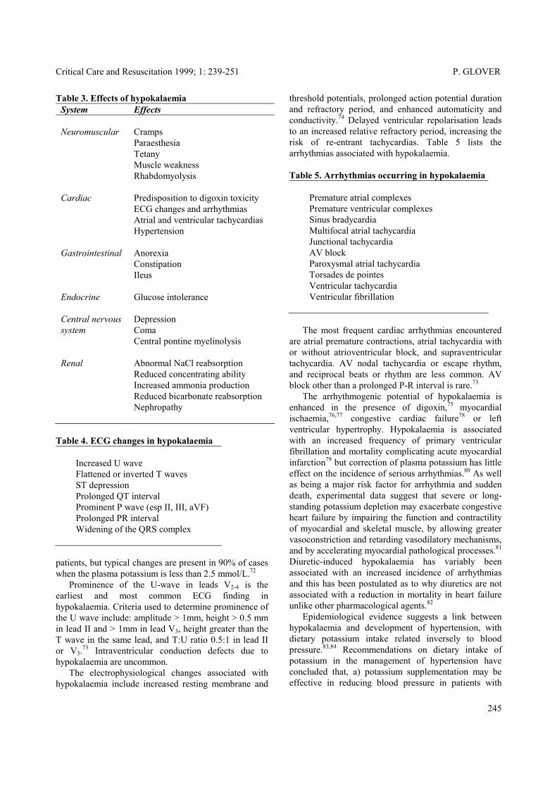
Table 3. Effects of hypokalaemia System Effects Neuromuscular
Cramps Paraesthesia Tetany Muscle weakness Rhabdomyolysis
Cardiac Predisposition to digoxin toxicity ECG changes and arrhythmias Atrial and ventricular tachycardias Hypertension
Gastrointestinal Anorexia Constipation Ileus
Endocrine Glucose intolerance
Central nervous system
Depression Coma Central pontine myelinolysis
Renal Abnormal NaCl reabsorption Reduced concentrating ability Increased ammonia production Reduced bicarbonate reabsorption Nephropathy
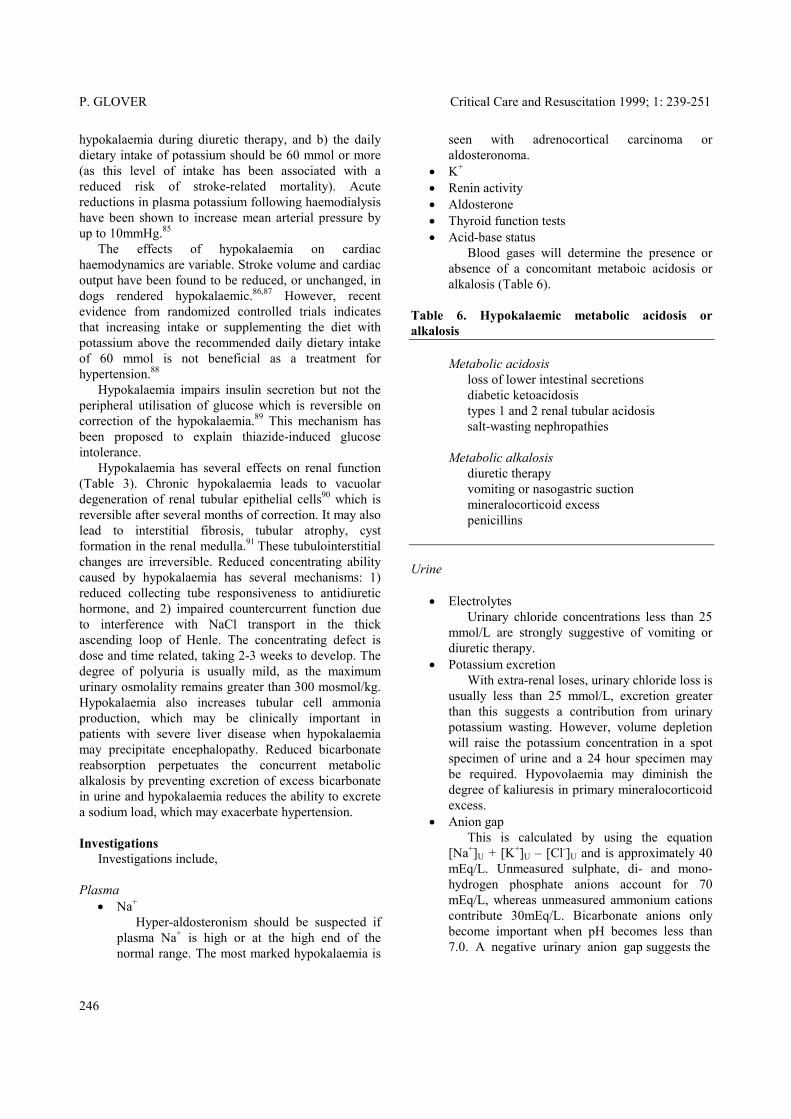
Table 4. ECG changes in hypokalaemia
Increased U wave Flattened or inverted T waves ST depression Prolonged QT interval Prominent P wave (esp II, III, aVF) Prolonged PR interval Widening of the QRS complex
patients, but typical changes are present in 90% of cases when the plasma potassium is less than 2.5 mmol/L.72 Prominence of the U-wave in leads V2-4 is the earliest and most common ECG finding in hypokalaemia. Criteria used to determine prominence of the U wave include: amplitude > 1mm, height > 0.5 mm in lead II and > 1mm in lead V3, height greater than the T wave in the same lead, and T:U ratio 0.5:1 in lead II or V3.73 Intraventricular conduction defects due to hypokalaemia are uncommon. The electrophysiological changes associated with hypokalaemia include increased resting membrane and
threshold potentials, prolonged action potential duration and refractory period, and enhanced automaticity and conductivity.74 Delayed ventricular repolarisation leads to an increased relative refractory period, increasing the risk of re-entrant tachycardias. Table 5 lists the arrhythmias associated with hypokalaemia. Table 5. Arrhythmias occurring in hypokalaemia
Premature atrial complexes Premature ventricular complexes Sinus bradycardia Multifocal atrial tachycardia Junctional tachycardia AV block Paroxysmal atrial tachycardia Torsades de pointes Ventricular tachycardia Ventricular fibrillation
The most frequent cardiac arrhythmias encountered are atrial premature contractions, atrial tachycardia with or without atrioventricular block, and supraventricular tachycardia. AV nodal tachycardia or escape rhythm, and reciprocal beats or rhythm are less common. AV block other than a prolonged P-R interval is rare.73 The arrhythmogenic potential of hypokalaemia is enhanced in the presence of digoxin,75 myocardial ischaemia,76,77 congestive cardiac failure78 or left ventricular hypertrophy. Hypokalaemia is associated with an increased frequency of primary ventricular fibrillation and mortality complicating acute myocardial infarction79 but correction of plasma potassium has little effect on the incidence of serious arrhythmias.80 As well as being a major risk factor for arrhythmia and sudden death, experimental data suggest that severe or long-standing potassium depletion may exacerbate congestive heart failure by impairing the function and contractility of myocardial and skeletal muscle, by allowing greater vasoconstriction and retarding vasodilatory mechanisms, and by accelerating myocardial pathological processes.81 Diuretic-induced hypokalaemia has variably been associated with an increased incidence of arrhythmias and this has been postulated as to why diuretics are not associated with a reduction in mortality in heart failure unlike other pharmacological agents.82 Epidemiological evidence suggests a link between hypokalaemia and development of hypertension, with dietary potassium intake related inversely to blood pressure.83,84 Recommendations on dietary intake of potassium in the management of hypertension have concluded that, a) potassium supplementation may be effective in reducing blood pressure in patients with
245
P. GLOVER Critical Care and Resuscitation 1999; 1: 239-251
hypokalaemia during diuretic therapy, and b) the daily dietary intake of potassium should be 60 mmol or more (as this level of intake has been associated with a reduced risk of stroke-related mortality). Acute reductions in plasma potassium following haemodialysis have been shown to increase mean arterial pressure by up to 10mmHg.85 The effects of hypokalaemia on cardiac haemodynamics are variable. Stroke volume and cardiac output have been found to be reduced, or unchanged, in dogs rendered hypokalaemic.86,87 However, recent evidence from randomized controlled trials indicates that increasing intake or supplementing the diet with potassium above the recommended daily dietary intake of 60 mmol is not beneficial as a treatment for hypertension.88 Hypokalaemia impairs insulin secretion but not the peripheral utilisation of glucose which is reversible on correction of the hypokalaemia.89 This mechanism has been proposed to explain thiazide-induced glucose intolerance. Hypokalaemia has several effects on renal function (Table 3). Chronic hypokalaemia leads to vacuolar degeneration of renal tubular epithelial cells90 which is reversible after several months of correction. It may also lead to interstitial fibrosis, tubular atrophy, cyst formation in the renal medulla.91 These tubulointerstitial changes are irreversible. Reduced concentrating ability caused by hypokalaemia has several mechanisms: 1) reduced collecting tube responsiveness to antidiuretic hormone, and 2) impaired countercurrent function due to interference with NaCl transport in the thick ascending loop of Henle. The concentrating defect is dose and time related, taking 2-3 weeks to develop. The degree of polyuria is usually mild, as the maximum urinary osmolality remains greater than 300 mosmol/kg. Hypokalaemia also increases tubular cell ammonia production, which may be clinically important in patients with severe liver disease when hypokalaemia may precipitate encephalopathy. Reduced bicarbonate reabsorption perpetuates the concurrent metabolic alkalosis by preventing excretion of excess bicarbonate in urine and hypokalaemia reduces the ability to excrete a sodium load, which may exacerbate hypertension. Investigations Investigations include, Plasma
• Na+ Hyper-aldosteronism should be suspected if plasma Na+ is high or at the high end of the normal range. The most marked hypokalaemia is
seen with adrenocortical carcinoma or aldosteronoma.
• K+ • Renin activity • Aldosterone • Thyroid function tests • Acid-base status
Blood gases will determine the presence or absence of a concomitant metaboic acidosis or alkalosis (Table 6).
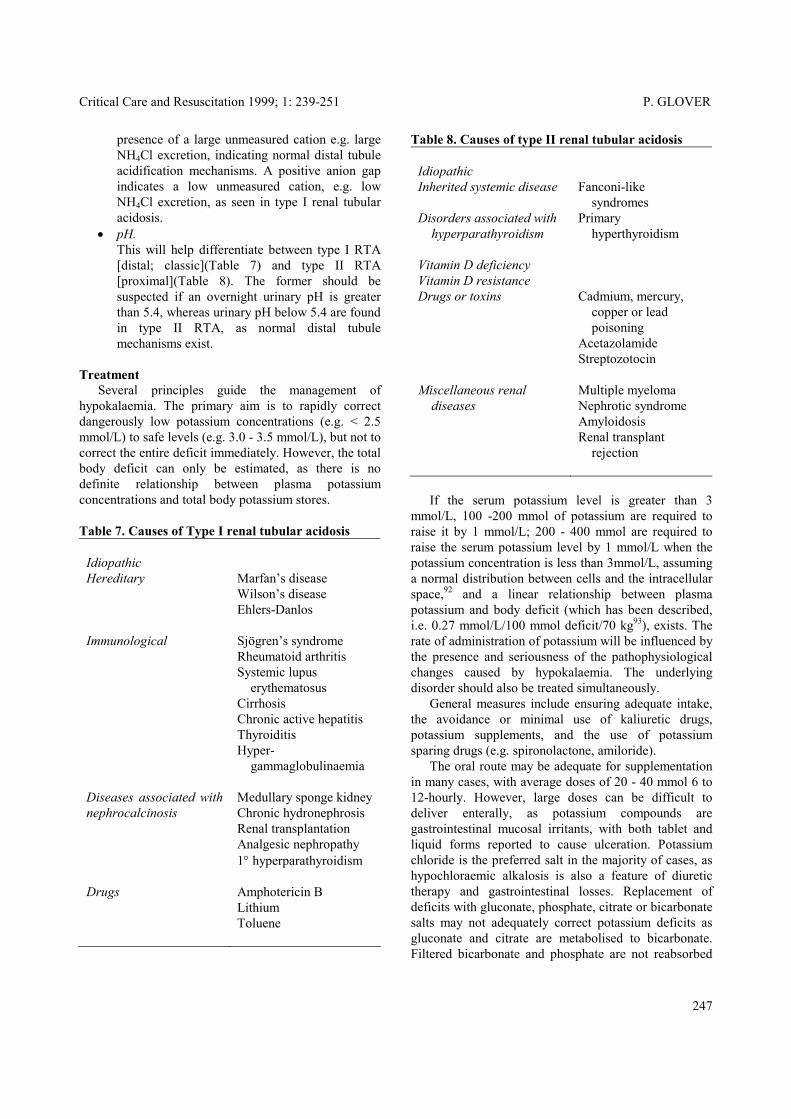
Table 6. Hypokalaemic metabolic acidosis or alkalosis Metabolic acidosis loss of lower intestinal secretions diabetic ketoacidosis types 1 and 2 renal tubular acidosis salt-wasting nephropathies Metabolic alkalosis diuretic therapy vomiting or nasogastric suction mineralocorticoid excess penicillins Urine
• Electrolytes Urinary chloride concentrations less than 25 mmol/L are strongly suggestive of vomiting or diuretic therapy.
• Potassium excretion With extra-renal loses, urinary chloride loss is usually less than 25 mmol/L, excretion greater than this suggests a contribution from urinary potassium wasting. However, volume depletion will raise the potassium concentration in a spot specimen of urine and a 24 hour specimen may be required. Hypovolaemia may diminish the degree of kaliuresis in primary mineralocorticoid excess.
• Anion gap This is calculated by using the equation [Na+]U + [K+]U – [Cl-]U and is approximately 40 mEq/L. Unmeasured sulphate, di- and mono-hydrogen phosphate anions account for 70 mEq/L, whereas unmeasured ammonium cations contribute 30mEq/L. Bicarbonate anions only become important when pH becomes less than 7.0. A negative urinary anion gap suggests the
246
Critical Care and Resuscitation 1999; 1: 239-251 P. GLOVER
Table 8. Causes of type II renal tubular acidosis presence of a large unmeasured cation e.g. large NH4Cl excretion, indicating normal distal tubule acidification mechanisms. A positive anion gap indicates a low unmeasured cation, e.g. low NH4Cl excretion, as seen in type I renal tubular acidosis.
Idiopathic
Inherited systemic disease
Fanconi-like syndromes
Disorders associated with hyperparathyroidism
Primary hyperthyroidism
Vitamin D deficiency Vitamin D resistance
Drugs or toxins
Cadmium, mercury, copper or lead poisoning Acetazolamide Streptozotocin
Miscellaneous renal diseases
Multiple myeloma Nephrotic syndrome Amyloidosis Renal transplant rejection
• pH. This will help differentiate between type I RTA [distal; classic](Table 7) and type II RTA [proximal](Table 8). The former should be suspected if an overnight urinary pH is greater than 5.4, whereas urinary pH below 5.4 are found in type II RTA, as normal distal tubule mechanisms exist.
Treatment Several principles guide the management of hypokalaemia. The primary aim is to rapidly correct dangerously low potassium concentrations (e.g. < 2.5 mmol/L) to safe levels (e.g. 3.0 - 3.5 mmol/L), but not to correct the entire deficit immediately. However, the total body deficit can only be estimated, as there is no definite relationship between plasma potassium concentrations and total body potassium stores.
If the serum potassium level is greater than 3 mmol/L, 100 -200 mmol of potassium are required to raise it by 1 mmol/L; 200 - 400 mmol are required to raise the serum potassium level by 1 mmol/L when the potassium concentration is less than 3mmol/L, assuming a normal distribution between cells and the intracellular space,92 and a linear relationship between plasma potassium and body deficit (which has been described, i.e. 0.27 mmol/L/100 mmol deficit/70 kg93), exists. The rate of administration of potassium will be influenced by the presence and seriousness of the pathophysiological changes caused by hypokalaemia. The underlying disorder should also be treated simultaneously.
Table 7. Causes of Type I renal tubular acidosis
Idiopathic
Hereditary Marfan’s disease Wilson’s disease Ehlers-Danlos
Immunological Sjögren’s syndrome Rheumatoid arthritis Systemic lupus erythematosus Cirrhosis Chronic active hepatitis Thyroiditis Hyper- gammaglobulinaemia
Diseases associated with nephrocalcinosis
Medullary sponge kidney Chronic hydronephrosis Renal transplantation Analgesic nephropathy 1° hyperparathyroidism
Drugs Amphotericin B Lithium Toluene
General measures include ensuring adequate intake, the avoidance or minimal use of kaliuretic drugs, potassium supplements, and the use of potassium sparing drugs (e.g. spironolactone, amiloride). The oral route may be adequate for supplementation in many cases, with average doses of 20 - 40 mmol 6 to 12-hourly. However, large doses can be difficult to deliver enterally, as potassium compounds are gastrointestinal mucosal irritants, with both tablet and liquid forms reported to cause ulceration. Potassium chloride is the preferred salt in the majority of cases, as hypochloraemic alkalosis is also a feature of diuretic therapy and gastrointestinal losses. Replacement of deficits with gluconate, phosphate, citrate or bicarbonate salts may not adequately correct potassium deficits as gluconate and citrate are metabolised to bicarbonate. Filtered bicarbonate and phosphate are not reabsorbed
247
P. GLOVER Critical Care and Resuscitation 1999; 1: 239-251
in the distal tubule, obligating further potassium loss in the distal tubule. Potassium bicarbonate can be used for maintenance therapy for the hypokalaemia associated with type I RTA. When phosphate depletion also exists, potassium phosphate may be used, although phosphate can be infused only slowly (e.g. maximum rate of 2 - 6 mmol/h/70kg).94 Intravenous administration of potassium salts should be in a saline solution, as metabolism of dextrose (if a dextrose solution is used) may initially worsen the hypokalaemia due to stimulation of insulin release.95 Replacement rates of up to 40 mmol/h have been safely administered to critically ill patients independent of renal function96 and up to 100 mmol/h have been replaced in diabetic ketoacidosis when hypokalaemia is present at presentation. Some have advocated an antiarrhythmic dose of 2 mmol as a bolus 97 or 6 mmol over 1 minute (to increase the plasma potassium by 2 mmol/L)98 to manage hypokalaemic tachyarrhythmias; however, even with such a low dose it is conceivable that in patients with low cardiac outputs, a dangerously high level of plasma potassium might occur transiently to cause sudden sinus arrest, AV block or asystole.99 In the presence of acute myocardial infarction, hypokalaemia should be corrected rapidly; intravenous potassium at a rate of 10 mmol/30 min and repeated as necessary until the serum potassium is 4.0-4.5 mmol/L has been recommended.100 When a transcellular shift has led to hypokalaemia, the plasma potassium concentration will normalise when the precipitating factor is alleviated;101 the vigorous administration of potassium supplements during the hypokalaemic period has later been associated with hyperkalaemia,61 which has occasionally been fatal.102 However, potassium administration may be required during the period of hypokalaemia, particularly if ECG changes and arrhythmias are present. Administration of an intravenous non-cardioselective β-adrenergic blocker e.g. propranolol may be of benefit in cases of hypokalaemia when NaK-ATPase activity has produced the trancellular shift, reversing the receptor-mediated activation of the membrane-bound enzyme. This has been successfully used in cases of theophylline and β2-agonist overdoses.55 Acute episodes of weakness in familial hypokalaemic periodic paralysis and thyrotoxic periodic paralysis may be treated with oral potassium supplements, but paralysis requires intravenous supplementation at 20 mmol/h while monitoring the patient clinically. A low carbohydrate diet with potassium supplements and spironolactone have been used as prophylaxis against recurrence in familial hypokalaemic periodic paralysis, although
acetazolamide has been shown to be the most effective agent in this condition, with the induction of a metabolic acidosis blunting the intracellular translocation of serum potassium.66 However, acetazolamide exacerbates thyrotoxic periodic paralysis. Beta-adrenergic blockers can block attacks of thyrotoxic paralysis but control of the hyperthyroid state is the most successful prophylaxis. Received: 2 May 1999 Accepted: 20 August 1999 REFERENCES 1. Paice BJ, Paterson KR, Onyanga-Omara F, Donnelly T,
Gray JM, Lawson DH. Record linkage study of hypokalaemia in hospitalized patients. Postgrad Med J 1986;62:187-191.
2. Stockigt JR. Potassium metabolism. Anaesth Intens Care 1977;5:317-325.
3. Tannen RL. Potassium disorders. In Kokko JP, Tannen RL (eds). Fluids and Electrolytes. WB Saunders Co, Philadelphia, 1986, pp150-228.
4. Kliger AS, Hayslett JP. Disorders of potassium balance. In Brenner BM, Stein JH, eds. Acid-base and potassium homeostasis in Contemporary Issues in Nephrology, Vol 2. New York, Churchill-Livingstone, 1978:168-204.
5. Fisher KA, Binder HJ, HayslettJP. Potassium secretion by colonic mucosal cells after potassium adaptation. Am J Physiolo 1976;231:987-994.
6. Fraser R, Brown JJ, Lever AF, Mason PA, Robertson JIS. Control of aldosterone secretion. Clin Sci 1979; 56:389-399.
7. Anderson JV, Struthers AD, Payne NN et al. Atrial natriuretic peptide inhibits the aldosterone response to angiotensin II in man. Clin Sci 1986;70:507-511.
8. O’Kelly R, Magee F, McKenna TJ. Routine heparin therapy inhibits adrenal aldosterone production. J Clin Endocrinol Metab 1983;56:108-112.
9. Carey RM, Thorner MO, Ortt E. Effects of metoclopramide and bromocriptine on the renin-angiotensin-aldosterone system in man. J Clin Invest 1979;63:727-732.
10. Gennari FJ. Hypokalaemia. N Engl J Med 1998;339: 451-458.
11. Wright FS, Giebisch G. Renal potassium transport: contributions of individual nephron segments and populations. Am J Physiol 1978;235:F515-527.
12. White PC. Disorders of aldosterone biosynthesis and action. N Engl J Med 1994;331:250-258.
13. Young DA, Paulsen AW. Interrelated effects of aldosterone and plasma potassium on potassium excretion. Am J Physiol 1983;244:F28-F34
14. Horisberger JD ,Diezi J. Effects of mineralocorticoids on Na+ and K+ excretion in the adrenalectomised rat. Am J Physiol 1983;245:F89-F99.
15. Berliner RW, Kennedy TJ Jr, Hilton JG. Renal mechanisms for excretion of potassium. Am J Physiol 1950;162:348-367.
248

Critical Care and Resuscitation 1999; 1: 239-251 P. GLOVER
36. Scheinman SJ, Guay-Woodford LM, Thakker RV, Warnock DG. Mechanisms of Disease: Genetic Disorders of Renal Electrolyte Transport. N Engl J Med 1999;340:1177-87.
16. Peterson LN, Wright FS. Effect of sodium intake on renal potassium excretion. Am J Physiol. 1977;233: F225-234.
17. Rabinowitz L. Homeostatic regulation of potassium excretion. J Hypertens 1989 7:433-442. 37. Gitelman HJ, Graham JB, Welt LG. A new familial
disorder characterized by hypokalemia and hypomagnesemia.Trans Assoc Am Physicians 1966;79: 221-235.
18. Clausen T, Everts ME. Regulation of the Na, K pump in skeletal muscle. Kidney Int 1989; 35:1-13.
19. Williams ME, Gervino EV, Rosa RM, Landsberg L, Young JB, Silva P, Epstein FH. Catecholamine modulation of rapid potassium shifts during exercise. N Engl J Med 1985;312:823-827.
38. Simon DB, Nelson-Williams C, Bia MJ, et al. Gitelman's variant of Bartter's syndrome, inherited hypokalaemic alkalosis, is caused by mutations in the thiazide-sensitive Na-Cl cotransporter. Nat Genet 1996;12:24-30.
20. Zierler K. Insulin hyperpolarises rat myotubule primary culture without stimulating glucose uptake. Diabetes 1987;36:1035-1040. 39. Cheng J-T, Witty RT, Robinson RR, Yarger WE.
Amphotericin B nephrotoxicity: increased renal resistance and tubule permeability. Kidney Int 1982;22:626-633.
21. Simmons DH, Avedon M. Acid-base alterations and plasma potassium concentration. Am J Physiol 1959;197:319-326.
22. Swan RC, Pitts RF. Neutralisation of infused acid by nephrectomised dogs. J Clin Invest 1955;96:323-330.
40. Bernardo JF, Murakami S, Branch RA, Sabra R. Potassium depletion potentiates amphotericin-B-induced toxicity to renal tubules. Nephron 1995;70:235-241. 23. Androgue HJ, Madias NE. Changes in the plasma
potassium concentration during acute acid-base disturbances. Am J Med 1981;71:456-467.
41. Boyd JC, Bruns DE, Wills MR. Frequency of hypomagnesemia in hypokalemic states. Clin Chem 1983;29:178-179. 24. Krapf R, Caduff P, Wagdi P, Staubli M, Hulter HN.
Plasma potassium response to acute respiratory alkalosis. Kidney Int 1995;47:217-224.
42. Elisaf M, Merkouropoulos M, Tsianos EV, Siamopoulos KC. Pathogenetic mechanisms of hypomagnesemia in alcoholic patients. J Trace Elem Med Biol 1995;9:210-214.
25. Saxton CR, Seldin QW. Clinical interpretation of laboratory values. In Kokko JP, Tannen RL (eds). Fluids and Electrolytes. WB Saunders Co, Philadelphia, 1986, pp 3-62.
43. Nanji AA, Denegri JF. Hypomagnesemia associated with gentamicin therapy. Drug Intell Clin Pharm 1984;18:596-598. 26. Erlij D, Grinstein S. The number of sodium ion pumping
sites in skeletal muscle and its modification by insulin. J Physiol (Lond) 1976;259:13-31.
44. Rodriguez M, Solanki DL, Whang R. Refractory potassium repletion due to cisplatin-induced magnesium depletion. Arch Intern Med 1989;149:2592-2594. 27. Lytton J, Lin JC, Guidotti G. Identification of two
molecular forms of (Na+,K+)-ATPase in rat adipocytes: Relation to insulin stimulation of the enzyme. J Biol Chem 1985;260:1177-1184.
45. Wills MR. Magnesium and potassium. Inter-relationships in cardiac disorders. Drugs 1986;31 Suppl 4:121-131.
46. Kelepouris E. Cytosolic Mg2+ modulates whole cell K+ and Cl- currents in cortical thick ascending limb (TAL) cells of rabbit kidney. Kidney Int 1990;37:564.
28. Lytton J. Insulin affects the sodium affinity of the rat adipocyte (Na+,K+)-ATPase. J Biol Chem 1985;260: 10075-10080.
29. Hundal HS, Marette A, Mitsumoto Y, Ramlal T, Blostein R, Klip A. Insulin induces translocation of α2 and β1 subunits of the Na+/K+-ATPase from intracellular compartments to the plasma membrane in mammalian skeletal muscle. J Biol Chem 1992;267:5040-5043.
47. Hall KW, Dobson KE, Dalton JG, Ghigone MC, Penner SB. Metabolic abnormalities associated with intentional theophylline overdose. Ann Intern Med 1984; 101:457-462.
48. Sessler CN. Theophylline toxicity: clinical features of 116 consecutive cases. Am J Med 1990; 88:567-576. 30. Allon M. Treatment and prevention of hyperkalaemia in
end-stage renal disease. Kidney Int 1993;43:1197-1209. 49. Amitai Y, Lovejoy FH. Hypokalemia in acute theophylline poisoning. Am J Emerg Med 1988;6:214-218.
31. Decaux G, Soupart A, Cauchie P, Delwiche F. Potassium homeostasis in liver cirrhosis. Arch Intern Med 1988;148:547-548. 50. Shannon M. Predictors of major toxicity after
theophylline overdose. Ann Intern Med 1993;119:1161-1167.
32. D’Silva JL. The action of adrenaline on serum potassium. J Physiol 1934;82:392-398.
51. Shannon M. Hypokalaemia, hyperglycaemia and plasma catecholamine activity after severe theophylline intoxication. J Toxicol Clin Toxicol 1994;32:41-47.
33. Williams ME, Rosa RM, Silva P, Brown RS, Epstein FH. Impairment of extrarenal potassium disposal by alpha-adrenergic stimulation. N Engl J Med 1984;311:145-149. 52. Vassallo R, Lipsky JJ. Theophylline: recent advances in
the understanding of its mode of action and uses in clinical practice. Mayo Proc Clin 1998;73:346-354.
34. Brown MJ, Brown DC, Murphy MB. Hypokalemia from beta2-receptor stimulation by circulating epinephrine. N Engl J Med 1983;309:1414-1419. 53. Leson CL, McGuigan MA, Bryson SM. Caffeine
overdose in an adolescent male. J Toxicol Clin Toxicol 1988;26:407-415.
35. Ganguly A. Primary Aldosteronism. N Engl J Med 1998;339:1828-1844.
249
P. GLOVER Critical Care and Resuscitation 1999; 1: 239-251
74. Helfant RH. Hypokalemia and arrhythmias. Am J Med 1986;80:13-22.
54. Schorn TF, Olbricht C, Schüler A, Franz A, Wittek K, Balks HJ, Hausmann E, Welhoener HH. Barium carbonate intoxication. Intensive Care Med 1991:17:60-62.
75. Gomez-Arnau J, Maseda J, Burgos R, Cordon J, Dominguez R, Criado A, Avello F. Cardiac arrest due to digitalis intoxication with normal serum digoxin levels: effects of hypokalemia. Drug Intell Clin Pharm 1982;16:160-161.
55. Bradberry SM, Vale JA. Disturbances of potassium homeostasis in poisoning. J Toxicol Clin Toxicol 1995;33:295-310.
76. Nordrehaug JE, Johannessen KA, Von der Lippe. Serum potassium concentration as a risk factor of ventricular arrhythmias early in acute myocardial infarction. Circulation 1985;71:645-649.
56. Layzer RB. Periodic paralysis and the sodium-potassium pump. Ann Neurol 1982;11:547-552.
57. Clemessy JL, Favier C, Borron SW, Hantson PE, Vicaut E, Baud FJ. Hypokalaemia related to acute chloroquine ingestion. Lancet 1995;346:877-880. 77. Brezins M, Elyassov S, Elimelech I, Roguin
NComparison of patients with acute myocardial infarction with and without ventricular fibrillation Am J Cardiol 1996;78:948-50.
58. Naparstek Y, Gutman A. Case report: spurious hypokalemia in myeloproliferative disorders. Am J Med Sci 1984;288:175-177.
78. Leier CV, Dei Cas L, Metra M. Clinical Relevance and management of the major electrolyte abnormalities in congestive heart failure: hyponatremia, hypokalemia, and hypomagnesemia. Am Heart J 1994;128:564-574.
59. Lawson DH, Murray RM, Parker JL. Early mortality in the megaloblastic anaemias. Q J Med 1972;41:1-14.
60. Viens P, Thyss A, Garnier G, Ayela P, Lagrange M, Schneider M. GM-CSF treatment and hypokalemia. Ann Intern Med 1989;111:263. 79. Volpi A, Cavalli A, Santoro L, Negri E. Incidence and
prognosis of early primary ventricular fibrillation in acute myocardial infarction--results of the Gruppo Italiano per lo Studio della Sopravvivenza nell'Infarto Miocardico (GISSI-2) database. Am J Cardiol 1998;82:265-271.
61. Schaefer M, Link J, Hannemann L, Rudolph KH. Excessive hypokalemia and hyperkalemia following head injury. Intensive Care Med 1995;21:235-237.
62. Udezue E, D’Souza L, Mahajan M. Hypokalemia after normal doses of nebulized albuterol (salbutamol). Am J Emerg Med 1995;13:168-171. 80. Solomon RJ. Ventricular arrhythmias in patients with
myocardial infarction and ischaemia. The role of serum potassium. Drugs 1986;31 Suppl 4:112-120.
63. Carmichael D, Hosty T, Kastl D, Beckman D. Hypokalemia and massive transfusion. South Med J 1984;77:315-317. 81. Leier CV, Dei Cas L, Metra M. Clinical Relevance and
management of the major electrolyte abnormalities in congestive heart failure: hyponatremia, hypokalemia, and hypomagnesemia. Am Heart J 1994;128:564-574.
64. Manhem P, Nilsson LH, Moberg AL, Wadstein J, Hokfelt B. Hypokalaemia in alcohol withdrawal caused by high circulating adrenaline levels. Lancet. 1984;1: 679. 82. Krishna GG. Hypokalemic states: current clinical issues.
Semin Nephrol 1990;10(6):515-552. 65. Braden GL, von Oeyen PT, Germain MJ, Watson DJ, Haag BL. Ritodrine- and terbutaline-induced hypokalaemia in preterm labor: mechanisms and consequences. Kidney Int 1997;51:1867-1875.
83. Langford HG. Dietary potassium and hypertension: epidemiologic data. Ann Intern Med 1983;98:770-772.
84. Krishan GG. Role of potassium in the pathogenesis of hypertension. Am J Med Sci 1994;307:S21-S25. 66. Stedwell RE, Allen KM, Binder LS. Hypokalemic
paralyses: a review of the etiologies, pathophysiology, presentation and therapy. Am J Emer Med 1992;10:143-148.
85. Dolson GM, Ellis KJ, Bernardo MV, Prakash R, Adrogué HJ. Acute decreases in serum potassium augment blood pressure. Am J Kid Dis 1995;26:321-326. 67. Knochel JP. Neuromuscular manifestations of
electrolyte disorders. Am J Med 1982;72:521-535. 86. Abrecht PH. Cardiovascular effects of chronic potassium deficiency in dogs. Am J Physiol 1972;223: 555-559.
68. Knochel JP. Rhabdomyolysis and effects of potassium deficiency on muscle structure and function. Cardiovasc Med 1978;3:247-261. 87. Bahler RC, Rakita L. Cardiovascular function in
potassium depleted dogs. Am Heart J 1971;81:650-657. 69. Rose BD. Hypokalemia. In Rose BD (ed).Clinical Physiology of Acid-Base and Electrolyte Disorders. McGraw-Hill Inc, 1994, pp776-822.
88. Burgess E, Lewanczuk R, Bolli P, et al. Lifestyle modifications to prevent and control hypertension. 6. Recommendations on potassium, magnesium and calcium. Canadian Hypertension Society, Canadian Coalition for High Blood Pressure Prevention and Control, Laboratory Centre for Disease Control at Health Canada, Heart and Stroke Foundation of Canada. CMAJ 1999;160(9 Suppl):S35-S45.
70. Davies JS, Aguirre G, Cassidy DM, Griffiths BE. Latent tetany associated with the hypokalaemia of Conn's syndrome. Int J Clin Pract 1998;52:347-348.
71. Cohen L. Potassium replacement associated with the development of tetany in a patient with hypomagnesaemia. Magnes Res 1993;6:43-45.
89. Rowe JW, Tobin JD, Rosa RM, Andres R. Effect of experimental potassium deficiency on glucose and insulin metabolism. Metabolism 1980;29:498-502.
72. Fletcher GF, Hurst JW, Schlant RC. Electrocardiographic changes in severe hypokalemia. A reappraisal. Am J Cardiol 1967;20:628-631.
90. Alpern RJ, Toto RD. Hypokalaemic nephropathy: a clue to cystogenesis. N Engl J Med 1990;322:398-399.
73. Chung EK. Electrocardiographic findings in hypokalemia. Postgrad Med. 1972;251:285-287.
250

Critical Care and Resuscitation 1999; 1: 239-251 P. GLOVER
97. Selmonosky CA, Flege JB. The effect of small doses of potassium on postoperative ventricular arrhythmias. J Thorac Cardiovasc Surg 1967;53:349-352.
91. Torres VE, Young WF, Offord KP et al. Association of hypokalaemia, aldosteronism and renal cysts. N Engl J Med 1990;322:345-351.
98. Halperin ML, Kamel KS. Potassium. Lancet 1998;352: 135-140.
92. MacLeod SM. The rational use of potassium supplements. Postgrad Med 1975;57;123-128.
99. Tanaka K, Pettinger WA. Pharmacokinetics of bolus potassium injections for cardiac arrhythmias. Anesthesiology 1973;38:587-589.
93. Sterns RH, Cox M, Feig PU, Singer I. Internal potassium balance and the control of the plasma potassium concentration. Medicine1981;60:339-354.
100. Standards and guidelines for cardiopulmonary resuscitation (CPR) and emergency cardiac care (ECC). JAMA 1992;268:2172-2288.
94. Worthley LIG.Blood biochemical analysis. In Worthley LIG (ed). Handbook of Emergency Laboratory Tests. Churchill Livingstone, 1996;pp9-98.
101. Chua S, Razvi K, Wong MT, Tay R, Arulkumaran S. Is there a need to treat hypokalaemia associated with intravenous salbutamol infusion? J Obstet Gynaecol Res 1997;23:381-387.
95. Agarwal A, Wingo CS. Treatment of hypokalemia. N Engl J Med 1999;340:154-155.
96. Hamill RJ, Robinson LM, Wexler HR, Moote C. Efficacy and safety of potassium infusion therapy in hypokalemic critically ill patients. Crit Care Med 1991;19:694-699.
102. Zydlewski AW, Hasbargen JA. Hypothermia-induced hypokalemia. Mil Med 1998;163:719-721.
251