hypoxia, respiratory failure and altered mental status alicia m. mohr, md surgical fundamentals...
TRANSCRIPT

Hypoxia, Respiratory Failure and Altered
Mental Status
Alicia M. Mohr, MD
Surgical Fundamentals Session 2
July 21, 2006

Objectives
• To learn a logical method for determining the nature of respiratory failure and its treatment
• To determine if a patient requires intubation and ventilation
• To learn the differential diagnosis and treatment of altered mental status
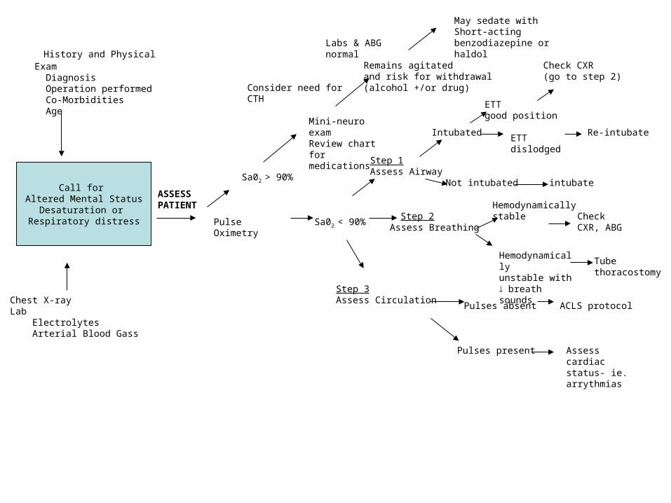
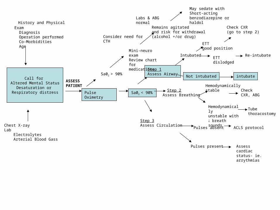
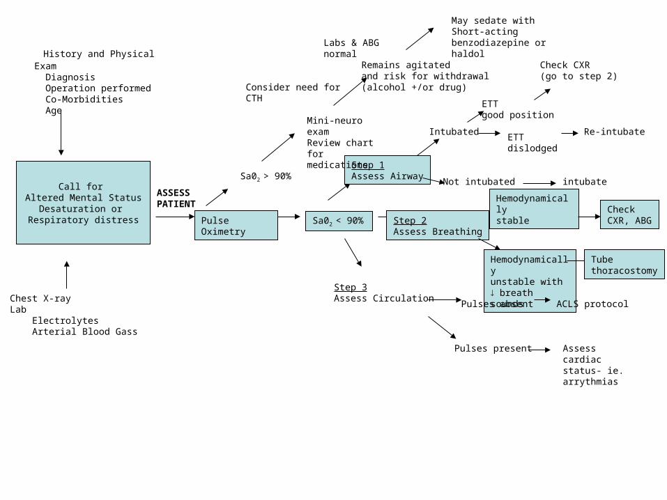
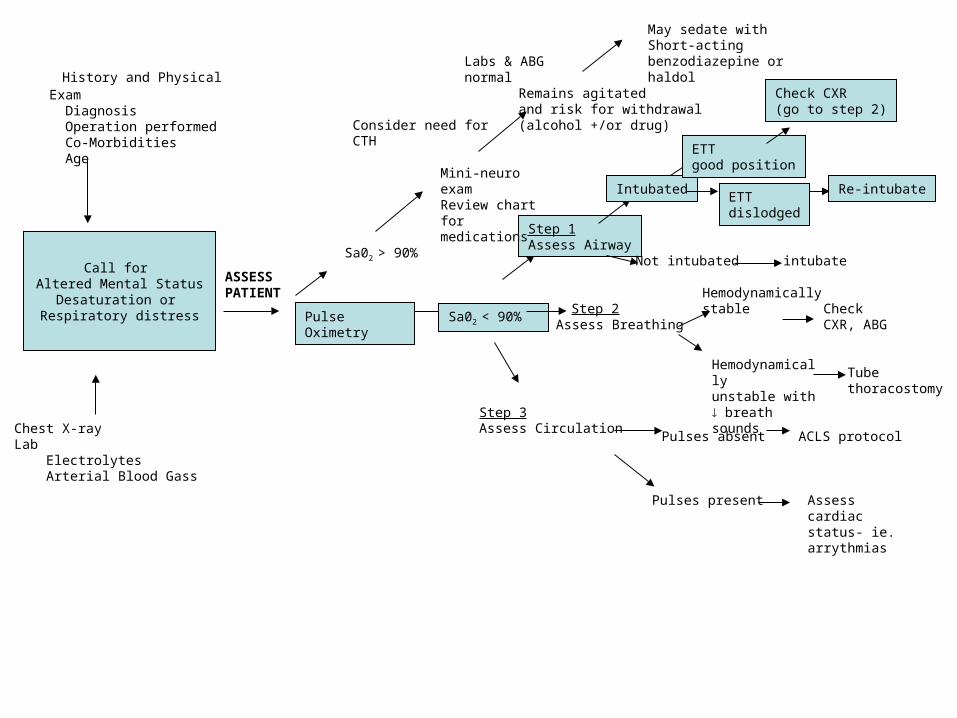
History and Physical Exam Diagnosis Operation performed Co-Morbidities Age
Chest X-rayLab Electrolytes Arterial Blood Gass
Pulse Oximetry
Sa02 > 90%
Sa02 < 90%
Remains agitatedand risk for withdrawal(alcohol +/or drug)
May sedate withShort-acting benzodiazepine or haldol
Step 1Assess Airway
Step 2
Step 3Assess Circulation
Intubated
Not intubated
ETT good position
Check CXR(go to step 2)
Re-intubate
intubate
Hemodynamically stable
Assess Breathing
Hemodynamicallyunstable with breath sounds
Check CXR, ABG
Tubethoracostomy
Pulses absent ACLS protocol
Pulses present Assess cardiacstatus- ie.arrythmias
Labs & ABG normal
ETTdislodged
Mini-neuro examReview chart for medications
Consider need for CTH
Call for Altered Mental Status
Desaturation or Respiratory distress
ASSESS PATIENT

History

History
• Can’t catch my breath• Lightedheadedness• Usually acute onset• Minimal symptoms

Physical Exam Findings

Physical Exam Findings
• Tachypnea• Dyspnea• Retractions• Nasal flaring• Grunting• Diaphoresis• Tachycardia• Hypertension
• Altered mental status Confusion Agitation Restlessness Somnolence
• Cyanosis (need 5mg/dl of unoxygenated blood)
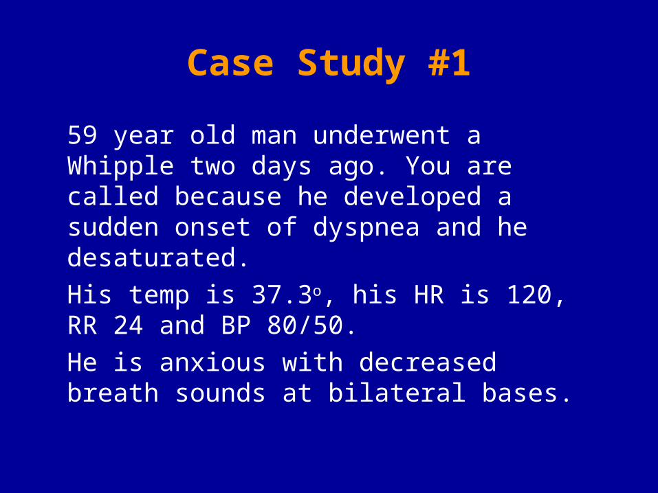
Case Study #1
59 year old man underwent a Whipple two days ago. You are called because he developed a sudden onset of dyspnea and he desaturated.
His temp is 37.3o, his HR is 120, RR 24 and BP 80/50.
He is anxious with decreased breath sounds at bilateral bases.

A - Airway
B - Breathing
C - Circulation
Oxygen delivery to tissues
Carbon dioxide
removal from tissues
Assess, change, reassess
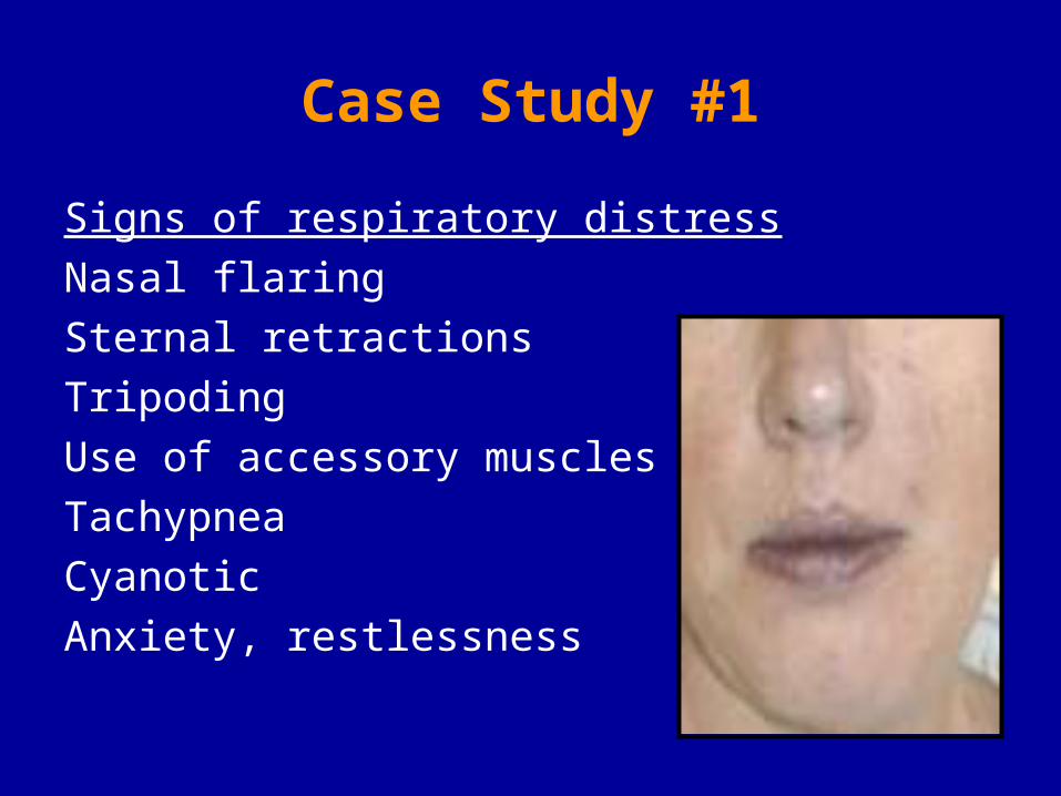
Case Study #1
Signs of respiratory distress
Nasal flaring
Sternal retractions
Tripoding
Use of accessory muscles
Tachypnea
Cyanotic
Anxiety, restlessness

Case Study #1
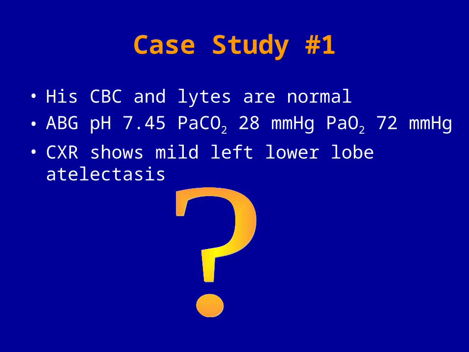
• His CBC and lytes are normal
• ABG pH 7.45 PaCO2 28 mmHg PaO2 72 mmHg
• CXR shows mild left lower lobe atelectasis

Indications for Intubation

Indications for Intubation
1. Airway protection
Loss of gag reflex, GCS <8
Massive facial trauma
2. Failure to ventilate
Increased work of breathing
PaCO2 > 55 mm Hg
3. Failure to oxygenate
Hypoxemia or PaO2 < 60 mm Hg
Severe metabolic acidosis or shock
Need for bronchopulmonary toilet

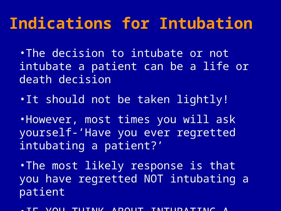
•The decision to intubate or not intubate a patient can be a life or death decision
•It should not be taken lightly!
•However, most times you will ask yourself-’Have you ever regretted intubating a patient?’
•The most likely response is that you have regretted NOT intubating a patient
•IF YOU THINK ABOUT INTUBATING A PATIENT YOU SHOULD PROBABLY DO IT!
Indications for Intubation
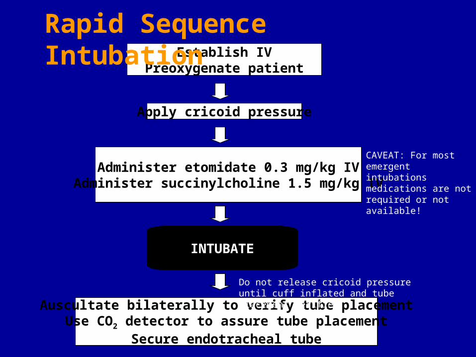
Establish IVPreoxygenate patient
Administer etomidate 0.3 mg/kg IVAdminister succinylcholine 1.5 mg/kg IV
Apply cricoid pressure
INTUBATE
Auscultate bilaterally to verify tube placementUse CO2 detector to assure tube placement
Secure endotracheal tube
Rapid Sequence Intubation
Do not release cricoid pressure until cuff inflated and tube placement verified
CAVEAT: For most emergent intubations medications are not required or not available!
Case Study #1
• His CBC and lytes are normal
• ABG pH 7.45 PaCO2 28 mmHg PaO2 72 mmHg
• CXR shows mild left lower lobe atelectasis

Pathophysiology of
Respiratory Failure
Due to mismatch of ventilation and
perfusion in lung units
History and Physical Exam Diagnosis Operation performed Co-Morbidities Age
Chest X-rayLab Electrolytes Arterial Blood Gass
Pulse Oximetry
Sa02 > 90%
Sa02 < 90%
Remains agitatedand risk for withdrawal(alcohol +/or drug)
May sedate withShort-acting benzodiazepine or haldol
Step 1Assess Airway
Step 2
Step 3Assess Circulation
Intubated
Not intubated
ETT good position
Check CXR(go to step 2)
Re-intubate
intubate
Hemodynamically stable
Assess Breathing
Hemodynamicallyunstable with breath sounds
Check CXR, ABG
Tubethoracostomy
Pulses absent ACLS protocol
Pulses present Assess cardiacstatus- ie.arrythmias
Labs & ABG normal
ETTdislodged
Mini-neuro examReview chart for medications
Consider need for CTH
Call for Altered Mental Status
Desaturation or Respiratory distress
ASSESS PATIENT

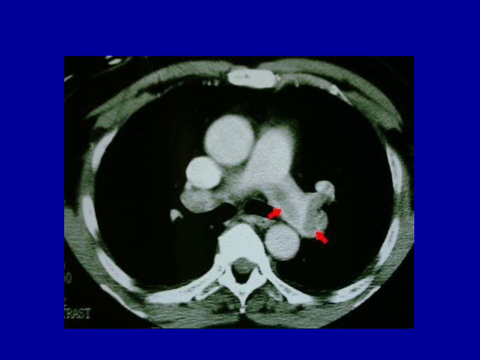
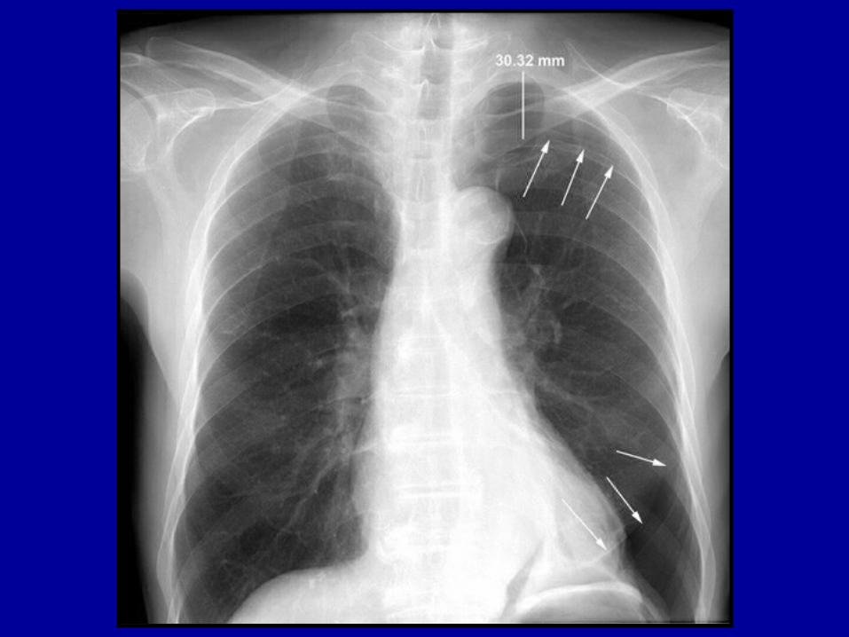
Case Study #2
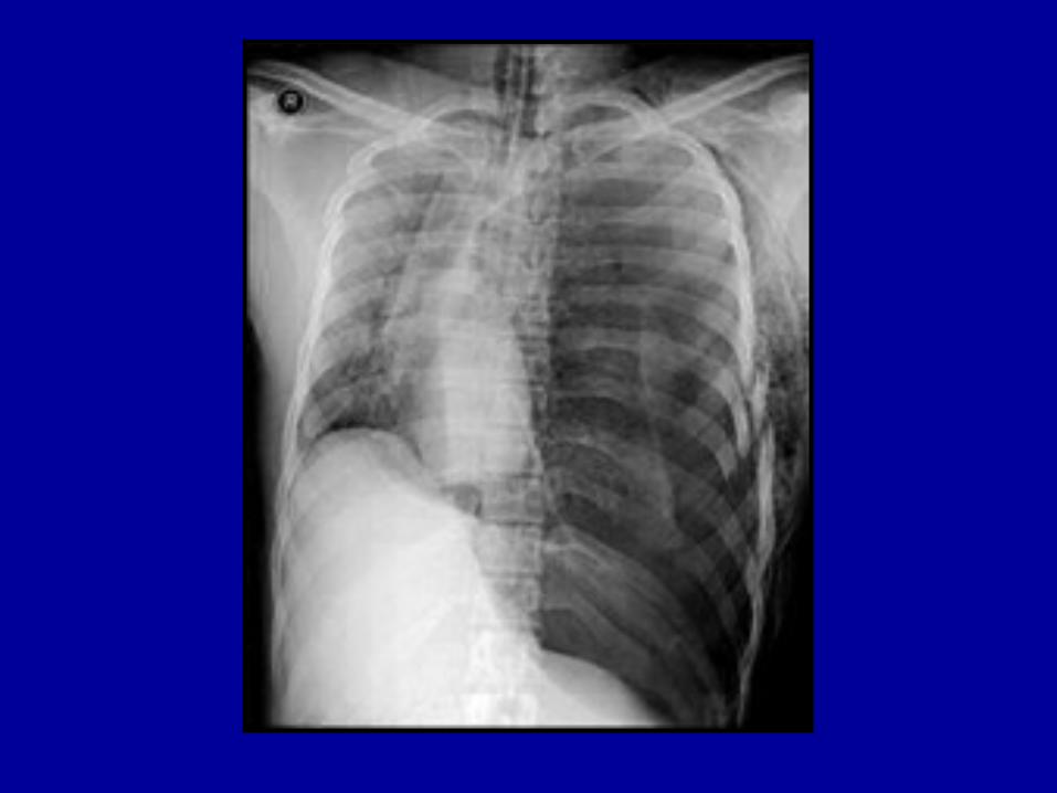
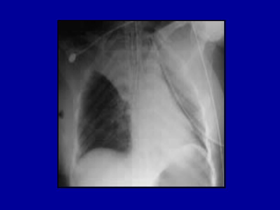
22 year old man was admitted five days ago after an MVC. He sustained a left rib fractures, a left pneumothorax and a left femur fracture. The nurse states the patient is short of breath.
His temp is 37.1o, his HR is 95, RR 30 and BP 120/70.
His saturation on room air is 85%

Differential Diagnosis
Differential Diagnosis
• Pneumothorax• Pneumonia• Lobar collapse• Pulmonary embolus


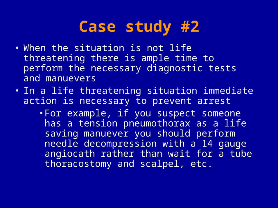
Case study #2• When the situation is not life threatening there is
ample time to perform the necessary diagnostic tests and manuevers
• In a life threatening situation immediate action is necessary to prevent arrest
• For example, if you suspect someone has a tension pneumothorax as a life saving manuever you should perform needle decompression with a 14 gauge angiocath rather than wait for a tube thoracostomy and scalpel, etc.
History and Physical Exam Diagnosis Operation performed Co-Morbidities Age
Chest X-rayLab Electrolytes Arterial Blood Gass
Pulse Oximetry
Sa02 > 90%
Sa02 < 90%
Remains agitatedand risk for withdrawal(alcohol +/or drug)
May sedate withShort-acting benzodiazepine or haldol
Step 1Assess Airway
Step 3Assess Circulation
Intubated
Not intubated
ETT good position
Check CXR(go to step 2)
Re-intubate
intubate
Hemodynamically stable
Step 2Assess Breathing
Hemodynamicallyunstable with breath sounds
Check CXR, ABG
Tubethoracostomy
Pulses absent ACLS protocol
Pulses present Assess cardiacstatus- ie.arrythmias
Labs & ABG normal
ETTdislodged
Mini-neuro examReview chart for medications
Consider need for CTH
Call for Altered Mental Status
Desaturation or Respiratory distress
ASSESS PATIENT
Case Study #3
72 year old man was admitted two days ago after an assault. He sustained an orbital fracture, scalp laceration and a frontal contusion. The nurse states the patient is confused and restless.
Case Study #3
What do you want to know?• Is this a change in his mental status?• Was he just medicated?• Has this happened before?• What are his vital signs?• What is his saturation?
Altered Mental Status
Five major causes:• Metabolic derangement• Drug toxicity/overdose/withdrawal• Infectious• Strutural abnormality• Psychiatric
Altered Mental Status
Metabolic abnormality• Rule out hypoxia
» Check ABG, saturation• Rule out hypoglycemia, DKA
» Assess blood glucose• Rule out uremia
» Assess urine output, BUN, creatinine• Rule out hepatic encephalopathy
» Check ammonia • Rule electrolyte abnormalities
» Send electrolytes
Altered Mental Status
Structural abnormality• Assess GCS• Assess for suspected head injury• Assess for focal neurologic deficits• Assess for possible post-ictal state• Emergent CT head
Altered Mental Status
Infectious cause• Assess for post operative sepsis• Assess risk of meningitis• Assess need for CT

Altered Mental StatusDrug toxicity/overdose/withdrawal
• Assess recent prescribed medications• Assess for potential self prescribed
medications• Check pupils• Check for sweating, agitation, hallucinations• Assess HR and blood pressure• May prescribe narcan or naloxone if OD• May prescribe benzodiazepine if withdrawal

Altered Mental Status
Altered Mental Status
Psychiatric cause• Assess for hallucinations• Assess for delusions• Mini-neuro exam

History and Physical Exam Diagnosis Operation performed Co-Morbidities Age
Chest X-rayLab Electrolytes Arterial Blood Gass
Pulse Oximetry
Sa02 > 90%
Sa02 < 90%
Remains agitatedand risk for withdrawal(alcohol +/or drug)
May sedate withShort-acting benzodiazepine or haldol
Step 1Assess Airway
Step 2
Step 3Assess Circulation
Intubated
Not intubated
ETT good position
Check CXR(go to step 2)
Re-intubate
intubate
Hemodynamically stable
Assess Breathing
Hemodynamicallyunstable with breath sounds
Check CXR, ABG
Tubethoracostomy
Pulses absent ACLS protocol
Pulses present Assess cardiacstatus- ie.arrythmias
Labs & ABG normal
ETTdislodged
Mini-neuro examReview chart for medications
Consider need for CTH
Call for Altered Mental Status
Desaturation or Respiratory distress
ASSESS PATIENT

Case Study #4
70 year old female had a colon resection five days ago. You are called by the nurse because she is dyspneic.
Her temp is 100o, her RR is 30, her HR is 110, and her BP is 140/90.
Her saturation is 95% on a non-rebreather.
Differential Diagnosis
Differential Diagnosis
• Pneumonia• Lobar collapse• Pulmonary embolus• Aspiration• Sepsis• Pulmonary edema• Congestive heart failure• Myocardial infarction
Case Study #4Causes of post-operative dyspnea
• Rule out pneumonia, atelectasis, collapse, aspiration» Check ABG, saturation, CXR» Assess abdomen, need for NGT
• Rule out sepsis» Assess for fever, abdominal exam, CTA/P
• Rule out pulmonary embolus» Assess leg swelling, duplex, CT chest» Can heparin be started empirically?
• Rule out myocardial infarction» Check EKG, troponin, myocardial enzymes» Can aspirin be given?
• Rule out fluid overload, CHF» Listen to lungs, assess fluid balance» Check home medications» Give diuretic
Case Study #4Does this patient need to be moved to monitored bed or
ICU?• Does this patient require intubation now?• May this patient need to be intubated in the next
few hours?• How likely is it that the patient is having an MI?• Is the patient having an arrythmia?• Does the patient need invasive monitoring?• How likely is it that the patient is going to
decompensate?• How likely is it that I am going to be presenting
this at M&M?
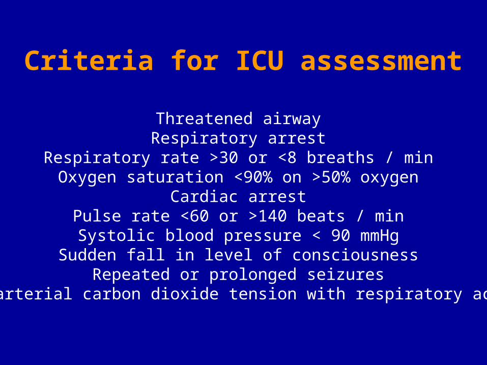
Criteria for ICU assessment
Threatened airway Respiratory arrest
Respiratory rate >30 or <8 breaths / min Oxygen saturation <90% on >50% oxygen
Cardiac arrest Pulse rate <60 or >140 beats / min Systolic blood pressure < 90 mmHg Sudden fall in level of consciousness
Repeated or prolonged seizures Rising arterial carbon dioxide tension with respiratory acidosis

Case Study #545 year old male in the ICU admitted four days ago with necrotizing pancreatitis. He was intubated on admission. His current ventilator settings are IMV rate of 14, tidal volume 600 mL, PEEP 5 and FiO2 50%.
The nurse calls you because after the patient was turned and washed he desaturated to 70%.
She has already turned the FiO2 up to 100% and his saturation has not responded.

Differential Diagnosis

Differential Diagnosis• Pneumonia• Lobar collapse• Pneumothorax• Pulmonary embolus• Aspiration• Sepsis• Pulmonary edema• Mucous plugging• Bronchospasm• ETT is dislodged

What do you do?• Take patient off the ventilator and hand bag
» Rule out ventilator problem» Assess degree of airway resistance
• Listen to the lungs» Rule out pneumothorax, fluid overload, bronchospasm
• Order a CXR, ABG» ABG will be bad, but will assess acidosis, and ventilation» CXR will assess ETT placement, lobar collapse, effusion,
pneumonia, etc.» Does patient require bronchoscopy?
• Pass a suction catheter» Rule out an occluded, dislodged ETT and assess secretions
• Give a bronchodilator» Can’t hurt! May loosen secretions
• If chest tubes in place, make sure on suction and assess for air leak• Adjust ventilator to compensate worsening respiratory failure
History and Physical Exam Diagnosis Operation performed Co-Morbidities Age
Chest X-rayLab Electrolytes Arterial Blood Gass
Pulse Oximetry
Sa02 > 90%
Sa02 < 90%
Remains agitatedand risk for withdrawal(alcohol +/or drug)
May sedate withShort-acting benzodiazepine or haldol
Step 1Assess Airway
Step 2
Step 3Assess Circulation
Intubated
Not intubated
ETT good position
Check CXR(go to step 2)
Re-intubate
intubate
Hemodynamically stable
Assess Breathing
Hemodynamicallyunstable with breath sounds
Check CXR, ABG
Tubethoracostomy
Pulses absent ACLS protocol
Pulses present Assess cardiacstatus- ie.arrythmias
Labs & ABG normal
ETTdislodged
Mini-neuro examReview chart for medications
Consider need for CTH
Call for Altered Mental Status
Desaturation or Respiratory distress
ASSESS PATIENT
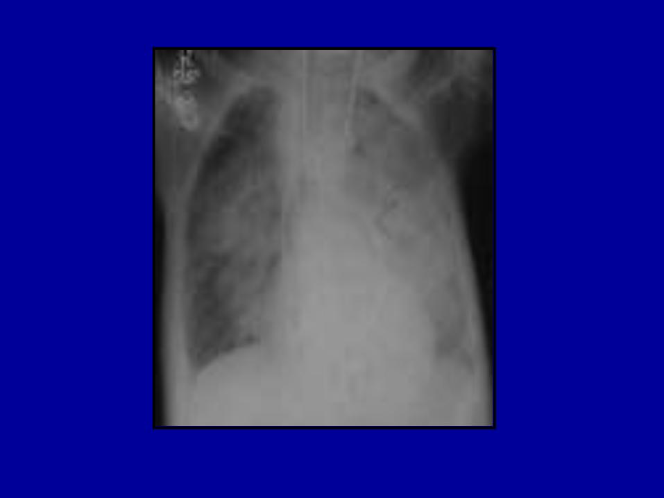

ARDS• A patient must meet all of the following:
– Acute onset of respiratory symptoms– CXR with bilateral infiltrates– No evidence of left heart failure
– PaO2/FiO2 < 200mm Hg (regardless of PEEP)– American-European Consensus Conference on ARDS (Am J Resp Crit
Care Med 149:818, 1994)
• The following are implied:– Previously normal lungs– Decreased lung compliance– Increased shunting– Hypoxemic respiratory failure

